HAL Id: inserm-00090465
https://www.hal.inserm.fr/inserm-00090465
Submitted on 31 Aug 2006
HAL is a multi-disciplinary open access
archive for the deposit and dissemination of
sci-entific research documents, whether they are
pub-lished or not. The documents may come from
teaching and research institutions in France or
abroad, or from public or private research centers.
L’archive ouverte pluridisciplinaire HAL, est
destinée au dépôt et à la diffusion de documents
scientifiques de niveau recherche, publiés ou non,
émanant des établissements d’enseignement et de
recherche français ou étrangers, des laboratoires
publics ou privés.
Impact of HFE genetic testing on clinical presentation of
hereditary hemochromatosis: new epidemiological data.
Virginie Scotet, Gérald Le Gac, Marie-Christine Mérour, Anne-Yvonne
Mercier, Brigitte Chanu, Chandran Ka, Catherine Mura, Jean-Baptiste
Nousbaum, Claude Férec
To cite this version:
Virginie Scotet, Gérald Le Gac, Marie-Christine Mérour, Anne-Yvonne Mercier, Brigitte Chanu, et
al.. Impact of HFE genetic testing on clinical presentation of hereditary hemochromatosis: new
epidemiological data.. BMC Medical Genetics, BioMed Central, 2005, 6, pp.24.
�10.1186/1471-2350-6-24�. �inserm-00090465�
Open Access
Research article
Impact of HFE genetic testing on clinical presentation of hereditary
hemochromatosis: new epidemiological data
Virginie Scotet
1, Gérald Le Gac
2, Marie-Christine Mérour
2,
Anne-Yvonne Mercier
2, Brigitte Chanu
2, Chandran Ka
2,3, Catherine Mura
3,
Jean-Baptiste Nousbaum
4and Claude Férec*
1,2Address: 1INSERM U 613 "Génétique moléculaire et génétique épidémiologique", Brest, France, 2Etablissement Français du Sang, Site de Brest,
Brest, France, 3Université de Bretagne Occidentale, Brest, France and 4Service d'Hépato-Gastroentérologie, Centre Hospitalier Universitaire La
Cavale Blanche, Brest, France
Email: Virginie Scotet - virginie.scotet@univ-brest.fr; Gérald Le Gac - gerald.legac@univ-brest.fr; Marie-Christine Mérour - secretariat-labos.brest@efs.sante.fr; Anne-Yvonne Mercier - anne-yvonne.mercier@efs.sante.fr; Brigitte Chanu - brigitte.chanu@efs.sante.fr; Chandran Ka - chandran.ka@univ-brest.fr; Catherine Mura - catherine.mura@univ-brest.fr; Jean-Baptiste Nousbaum - jean-baptiste.nousbaum@chu-brest.fr; Claude Férec* - claude.ferec@univ-brest.fr
* Corresponding author
Abstract
Background: Hereditary hemochromatosis (HH) is a common inherited disorder of iron
metabolism in Northern European populations. The discovery of a candidate gene in 1996 (HFE), and of its main mutation (C282Y), has radically altered the way to diagnose this disease. The aim of this study was to assess the impact of the HFE gene discovery on the clinical presentation and epidemiology of HH.
Methods: We studied our cohort of 415 patients homozygous for the C282Y allele and included
in a phlebotomy program in a blood centre in western Brittany, France.
Results: In this cohort, 56.9% of the patients were male and 21.9% began their phlebotomy
program before the implementation of the genetic test. A significant decrease in the sex ratio was noticed following implementation of this DNA test, from 3.79 to 1.03 (p < 10-5), meaning that the
proportion of diagnosed females relatives to males greatly increased. The profile of HH patients at diagnosis changed after the DNA test became available. Serum ferritin and iron values were lower and there was a reduced frequency of clinical signs displayed at diagnosis, particularly skin pigmentation (20.1 vs. 40.4%, OR = 0.37, p < 0.001) and hepatomegaly (11.0 vs. 22.7%, OR = 0.42, p = 0.006). In contrast, fatigue became a more common symptom at diagnosis (68.0 vs. 51.2%, OR = 2.03, p = 0.004).
Conclusion: This study highlights the importance of the HFE gene discovery, which has simplified
the diagnosis of HH and modified its clinical presentation and epidemiology. This study precisely measures these changes. Enhanced diagnosis of HFE-related HH at an early stage and implementation of phlebotomy treatment are anticipated to maintain normal life expectancy for these patients.
Published: 01 June 2005
BMC Medical Genetics 2005, 6:24 doi:10.1186/1471-2350-6-24
Received: 24 September 2004 Accepted: 01 June 2005 This article is available from: http://www.biomedcentral.com/1471-2350/6/24
© 2005 Scotet et al; licensee BioMed Central Ltd.
This is an Open Access article distributed under the terms of the Creative Commons Attribution License (http://creativecommons.org/licenses/by/2.0), which permits unrestricted use, distribution, and reproduction in any medium, provided the original work is properly cited.
BMC Medical Genetics 2005, 6:24 http://www.biomedcentral.com/1471-2350/6/24
Background
Hereditary hemochromatosis (HH) is a common genetic disorder of iron metabolism that is usually inherited in an autosomal recessive pattern and associated with missense mutations in the HFE gene. This pathology displays a large genetic heterogeneity because several other types of hemochromatosis, associated with different genes and patterns of inheritance, have been reported [1]: HH type 2B is a juvenile form linked to the HAMP gene (encoding for hepcidin) [2-4], HH type 3 is linked to the TfR2 gene (encoding for transferrin receptor 2) [5-7], HH type 4 is linked to the SLC11A3 gene (encoding for ferroportin, an intestinal iron transporter) [8,9]. and HH type 5 is linked to a gene encoding subunit H of ferritin [10]. Moreover, the gene responsible for juvenile hemochromatosis (HH type 2A), and whose protein product is called hemojuve-lin, has recently been cloned [11].
The main form of HH (i.e. type I which is linked to the
HFE gene) occurs predominantly in Northern European
populations, with a prevalence of approximately 3 to 8 in 1000 [12-16]. It is characterised by excessive iron absorp-tion, which progressively leads to the destruction of tis-sues in different organs of the body. After a phase of latency, the first signs of biochemical expression appear, generally around the age of 20. This is characterised by increases in serum iron parameters (transferrin saturation, ferritin). The clinical expression manifests later during adulthood, generally around the age of 40 in males and later in females, around the age of 50, because of the pro-tective effects of menstrual blood loss and pregnancies [17-19]. The clinical picture may include at an early stage, non-specific symptoms such as persistent fatigue and arthralgias, and at a later stage, clinical signs such as skin pigmentation, hepatomegaly, arthropathy, cardiomyopa-thy, diabetes and cirrhosis [20-22]. Classically, this clini-cal expression occurs more frequently in males than in females (sex ratio of 3:1) [23].
HH can be treated or prevented by periodic phlebotomies. This simple and efficient treatment prevents iron accumu-lation and clinical complications. Without this early treat-ment, the disease may progress towards irreversible damage such as cirrhosis and hepatocellular carcinoma [17-19].
A candidate gene for HH type 1, HFE, was identified in 1996 on chromosome 6 and encodes the HFE protein, a transmembrane glycoprotein that is implicated in modu-lation of iron uptake [24,25]. Currently, about twenty dif-ferent mutations have been identified in this gene worldwide and, one of them, termed C282Y, is present at homozygous state in 80 to 95% of HH patients. This molecular anomaly corresponds to the substitution of a tyrosine for a cysteine at amino acid 282, which prevents
formation of a disulfide bond [24,26]. The two other most common mutations of the HFE gene are associated with milder forms of HH (H63D and S65C) [1,21,27-29]. The discovery of the HFE gene in 1996 and the fact that one of its mutations (C282Y) is responsible for the large majority of HH cases enabled the implementation of effi-cient strategies for molecular diagnosis [30], what has altered the way in which HH is diagnosed. Initially, the diagnosis relied on a high index of suspicion associated with evidence of elevated iron parameter values [18,20,22]. Following discovery of the HFE gene, a DNA test was proposed to confirm the diagnosis of HH. Such a test, which allows the detection of at least the C282Y mutation, is now widely available. This discovery has sim-plified the diagnostic strategy and enabled pre-sympto-matic or earlier diagnosis in some patients. If phlebotomy treatment is implemented before the appearance of irre-versible damage, the excess iron can be removed and patients have a normal life expectancy [20,31].
In this study, we assessed the impact of HFE genetic test-ing on the clinical presentation and epidemiology of HH in a cohort of 415 patients homozygous for the C282Y mutation who were followed in a blood centre in western Brittany, France. This report contains objective data to measure this impact.
Methods
Study population
The present study was conducted in Brittany, a region of nearly three million inhabitants located in the north-west-ern part of France, where HH is particularly frequent (car-rier rate: 1 in 7) [32]. This disease presents a large genetic and allelic heterogeneity, but as the majority of patients are homozygous for the C282Y mutation, we decided to include in this study only the patients homozygous for this mutation. This study included all the C282Y homozygous patients who are or were included in a phle-botomy program in a blood centre of western Brittany and who presented with transferrin saturations of greater than or equal to 45%. The first patients began their treatment in the early eighties and the last date of patient entry for inclusion in this study was December 31st 2003. The
pro-portion of homozygous C282Y patients before and after the implementation of the genetic test did not change significantly.
Clinical questionnaire
A detailed clinical questionnaire was completed during the clinical exam performed at the first visit of patients to the blood centre (i.e. at entry in the phlebotomy pro-gram). Information contained in this questionnaire was previously described in detail [33]. Briefly, it provided information regarding socio-demographic characteristics
of patients, their age at diagnosis, the circumstances of HH discovery, the biochemical parameters and the clini-cal signs associated at the time of diagnosis. This question-naire also included data related to the treatment, such as the number and quantity of phlebotomies needed to reach depletion and the quantity of iron extracted. The date of the beginning of the treatment (i.e. entry into the phlebotomy program) was also documented. This date was used to determine if the treatment of patients began before or after the availability of the genetic test (i.e. 1996). The intake of alcohol was assessed by a detailed item included in the questionnaire, which measured the number of glasses of alcohol drunk each day (including glasses of wine, beer and liquor). These data enabled the quantity of ethanol (in grams) consumed each day, by each patient of the cohort, to be determined. Excessive alcohol consumption was defined as a daily consumption greater than or equal to 60 grams of ethanol [33]. Determination of biochemical parameter levels and of
HFE genotype
Serum iron parameters (i.e. serum iron, serum ferritin and serum transferrin saturation) were determined by stand-ard biochemical methods (including collection of serum after a 12-hour fast, confirmation by at least two measurements).
Analysis of the C282Y mutation relies on amplification of the specific gene region by the polymerase chain reaction, followed by mutation detection using restriction enzymes [29,32]. Recently, another method was adopted for this analysis: the denaturing high-performance liquid chroma-tography method [34]. If the DNA test confirms the pres-ence of mutations in a patient, family testing is offered to the relatives of this newly diagnosed patient. Family test-ing combines the collection of biochemical and clinical evidence of iron overload for the patients relatives with analysis of the main HFE mutations. Before 1996, family testing was already possible by analysis of HLA haplotypes in families [35] and this was commonly practised in our region where the disease incidence is high. The genotype of the patients diagnosed before 1996 was retrospectively determined when the genetic test became available. Statistical analysis
Data were analysed using Epi-Info software (version 6.04; Centers for Disease Control and Prevention, Atlanta, Georgia) and SAS statistical package (version 8.2; SAS Institute). Quantitative variables, expressed as means and standard deviations, were compared with the Student's t test or ANOVA, whereas qualitative variables, expressed as percentages, were compared with the Chi square test or the Fisher's exact test (in case of small sample size). A log-arithmic transformation was performed for the serum fer-ritin variable which had a skewed distribution. A
significance level of 5% was used for all of the analyses which were performed two-sided.
The analyses consisted of determining the influence of the implementation of the genetic test on the sex ratio, the age at diagnosis, the circumstances of HH discovery, the levels of biochemical parameters and the frequency of clinical signs associated at the time of diagnosis. The comparisons of biochemical and clinical data, which were made sepa-rately for males and females, were adjusted for age at diag-nosis and for alcohol consumption, because we showed previously that excessive alcohol consumption (>60 g/ day) increased HH expressivity [33]. The relation between the time of diagnosis (i.e. before or after availability of
HFE genotyping) and each of the different clinical
symp-toms was assessed by calculating the odds-ratio (OR) and its 95% confidence interval (CI).
This study complied with French bioethical regulations. Informed consent of patients was obtained before blood samples were taken.
Results
Description of the study population
This study included 415 C282Y homozygous patients of whom 56.9% were male, resulting in a sex ratio of 1.3:1 (236/179). The age at diagnosis, ranging from 13 to 76 years, was significantly higher in females than in males
Evolution of the sex ratio: number of males and females diag-nosed before and after the implementation of the genetic test
Figure 1
Evolution of the sex ratio: number of males and females diag-nosed before and after the implementation of the genetic test. 72 19 164 160 0 50 100 150 200 250 300 350 No Yes
Male Fem ale
Gender Number
of patients
BMC Medical Genetics 2005, 6:24 http://www.biomedcentral.com/1471-2350/6/24
(48.5 yrs (σ = 14.3) vs. 46.1 yrs (σ = 12.5), p = 0.037). Overall, the diagnosis was mainly made on the basis of clinical features (61.4%) or through family testing (30.9%). The circumstances of diagnosis remained unknown for one patient. Among these 415 patients, 21.9% began their phlebotomy program before the implementation of the molecular testing (n = 91, 72 males and 19 females) and 78.1% after this date (n = 324, 164 males and 160 females).
Characteristics of patients before and after the implementation of the genetic test
Socio-demographic characteristics of patients in relation to the type of diagnosis (based on HFE genotyping or not)
A significant decrease in the sex ratio was noted following development of the genetic test, as illustrated in figure 1. The sex ratio was 3.79 (72/19) before discovery of the gene and 1.03 (164/160) after this date (p < 10-5),
mean-ing that the proportion of females diagnosed since 1996 increased greatly: from 20.9% (19/91) to 49.4% (160/ 324). These females seemed to be diagnosed earlier. Their age at diagnosis tended to decrease from 52.9 yrs (σ = 9.8) before the implementation of the genetic test to 47.9 yrs (σ = 14.7) after this date. Nevertheless, this difference was not significant because a small number of women was diagnosed before 1996 (n = 19, p = 0.210). This pattern was not observed in males, their age at diagnosis increased from 42.7 yrs (σ = 10.4) to 47.6 yrs (σ = 13.1) after 1996 (p = 0.007).
After introduction of molecular testing, the symptom of unexplained and persistent fatigue was more commonly
present at diagnosis of HH. The frequency of this symp-tom increased from 51.2 to 68.0% following introduction of the DNA-based testing (OR = 2.03, 95% CI: 1.21, 3.41; p = 0.004), notably in males (from 47.1 to 58.4%). On the other hand, the proportion of patients detected by family testing was not significantly changed after 1996 (31.3 vs. 29.7%).
Biochemical parameters in relation to the type of diagnosis (based on HFE genotyping or not)
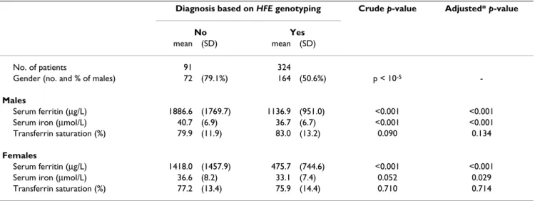
Differences were also observed in biochemical parameter values. The results of the comparison of the biochemical data of patients before and after the development of the genetic test are presented in table 1. Serum ferritin and serum iron values were significantly lower in patients diagnosed after implementation of the genetic test (serum ferritin: 810.4 vs. 1788.8 µg/liter, p < 0.001; serum iron: 34.9 vs. 39.8 µmol/liter, p < 0.001). These results were observed in males (serum ferritin: 1136.9 vs. 1886.6 µg/ liter, p < 0.001; serum iron: 36.7 vs. 40.7 µmol/liter, p < 0.001) and in females (serum ferritin: 475.7 vs. 1418.0 µg/liter, p < 0.001; serum iron: 33.1 vs. 36.6 µmol/liter, p = 0.052). As shown in table 1, the results remained unchanged after adjustment for age at diagnosis and alco-hol consumption. In this cohort, 8.0% of the patients declared having excessive alcohol consumption (≥ 60 g/ day; n = 33) comprising 9.9% of the patients diagnosed before 1996 (n = 9) and 7.4% of those diagnosed after 1996 (n = 24). A decrease was not observed for the third iron parameter, transferrin saturation (79.5 vs. 79.4%, p = 0.930), for which an elevated value (>45%) was used as a selection criterion for this study.
Table 1: Biochemical characteristics of the C282Y homozygous patients before and after the implementation of the genetic test. Diagnosis based on HFE genotyping Crude p-value Adjusted* p-value
No Yes
mean (SD) mean (SD)
No. of patients 91 324
Gender (no. and % of males) 72 (79.1%) 164 (50.6%) p < 10-5
-Males
Serum ferritin (µg/L) 1886.6 (1769.7) 1136.9 (951.0) <0.001 <0.001 Serum iron (µmol/L) 40.7 (6.9) 36.7 (6.7) <0.001 <0.001 Transferrin saturation (%) 79.9 (11.9) 83.0 (13.2) 0.090 0.134
Females
Serum ferritin (µg/L) 1418.0 (1457.9) 475.7 (744.6) <0.001 <0.001 Serum iron (µmol/L) 36.6 (8.2) 33.1 (7.4) 0.052 0.029 Transferrin saturation (%) 77.2 (13.4) 75.9 (14.4) 0.710 0.714 * Adjusted for age at diagnosis and alcohol consumption
Frequency of the main clinical signs and symptoms at the time of diagnosis in males, before and after the implementation of the genetic test
Figure 2
(A) Frequency of the main clinical signs and symptoms at the time of diagnosis in males, before and after the implementation of
the genetic test. (B) Frequency of the main clinical signs and symptoms at the time of diagnosis in females, before and after the implementation of the genetic test
A
0
20
40
60
80
100
Fatigue
Skin pigmentation
Arthritis
Hepatomegaly
Diabetes
Frequency (%)
B0
20
40
60
80
100
Fatigue
Skin pigmentation
Arthritis
Hepatomegaly
Diabetes
Diagnosis based
on
HFE genotyping
Frequency (%)
BMC Medical Genetics 2005, 6:24 http://www.biomedcentral.com/1471-2350/6/24
Clinical signs in relation to type of diagnosis (based on HFE genotyping or not)
The frequency of the main clinical signs observed at the time of diagnosis before and after the availability of the molecular testing is shown in figures 2A and 2B. The clin-ical signs were less frequent in the subjects diagnosed after the development of the genetic test, particularly skin pig-mentation (20.1 vs. 40.4%; OR = 0.37, 95% CI = 0.22, 0.63; p < 0.001) and hepatomegaly (11.0 vs. 22.7%; OR = 0.42, 95% CI = 0.21, 0.83; p = 0.006). This change was more significant in women. Indeed, only the two signs mentioned above tended to be less frequently observed in men following the introduction of genetic testing although these changes did not reached statistical signifi-cance (skin pigmentation: 32.1 vs. 43.7%; OR = 0.61, 95% CI = 0.33, 1.13; p = 0.089 – hepatomegaly: 14.7 vs. 22.5%; OR = 0.59, 95% CI = 0.27, 1.32; p = 0.160), whereas in women, four symptoms were significantly less frequent following implementation of molecular testing: skin pigmentation (7.6 vs. 27.8%; OR = 0.22, 95% CI = 0.06, 0.84; p = 0.006), hepatomegaly (7.3 vs. 23.5%; OR = 0.26, 95% CI = 0.06, 0.86; p = 0.028), arthritis (47.5 vs. 76.5%; OR = 0.28, 95% CI = 0.07, 0.98; p = 0.023) and diabetes (2.0 vs. 16.7%; OR = 0.10, 95% CI = 0.01, 0.71; p = 0.001). These results were similar in the sub-group of patients having no excessive alcohol consumption (data not shown).
Discussion
The discovery of the HFE gene in 1996 constituted a con-siderable advance in the medical and scientific field. This discovery concerned one of the most common inherited disorders in white populations, HH – a disorder that was complex to diagnose but for which a treatment existed – and identified one of the few undiscovered genes that has an important impact on public health.
The symptomatology of HH has evolved over the past years and it is now rare to diagnose severe forms of the dis-ease, associated with diabetes, cirrhosis and darkened skin [36,37]. Through a survival analysis based on a cohort of 251 patients diagnosed between 1947 and 1991, Nied-erau et al. showed that the percentage of patients with early diagnoses increased 3-fold during the period of 1970–1981 compared to the period of 1947–1969, and that there was a further 20–25% increase in the early diag-nosis rate during the period of 1981–1991 [37]. These changes occurred before the discovery of the HFE gene, and were probably the consequences of improved educa-tion of physicians and the implementaeduca-tion of HLA testing for family members of probands.
The current study highlights the importance of the discov-ery of the HFE gene in 1996 and demonstrates how the clinical presentation and epidemiology of HH have
changed since the availability of the DNA test. Our results objectively measure these changes, and show that the sex ratio of this disease has altered: the proportion of females currently diagnosed has increased and has reached that of males. This study also highlights that the profile of HH patients has changed: the patients have lower iron param-eter values (serum ferritin and iron) and a lower frequency of clinical signs at the time of diagnosis, notably skin pig-mentation and hepatomegaly. This change is more pro-nounced in females in whom clinical manifestations of HH appears later than in males (around the age of 50 ver-sus around the age of 40 in males). This study included all the C282Y homozygous patients who are or were included in a phlebotomy program in a blood centre of western Brittany. For the patients diagnosed before the implementation of the genetic test, the genotype was ret-rospectively determined in 1996 if they were still alive at this date. Consequently, the patients who died before 1996 were not genotyped and not included in this study. With this bias, some severe cases of the disease have been missed and the difference between the two groups should therefore be even greater than reported here.
Identification of the HFE gene and of its main mutation (C282Y) has greatly simplified diagnosis of, and family testing for, HH [20,31]. The fact that homozygosity for the C282Y mutation is responsible for the majority of HH cases has enabled use of the molecular test for this muta-tion as a diagnostic criterion for HH. Before the genetic test was available, diagnosis of HH required a high index of suspicion (as the clinical signs are non-specific) and evidence of elevated iron parameters [18,20,22]. Tradi-tionally, diagnosis was based on the measurement of transferrin saturation. A liver biopsy then enabled confirmation of iron overload by detection of elevated hepatic iron levels [27]. The discovery of the HFE gene enabled molecular analysis to be included in the diagnos-tic strategy and thus genediagnos-tic testing for confirmation of the diagnosis was proposed. In this way, HH could easily be differentiated from all other types of iron overload. Cur-rently, the diagnosis combines molecular testing with tra-ditional biochemical methods. The diagnostic strategy is as follows: 1) To suspect the diagnosis from non-specific symptoms (such as persistent fatigue, arthralgias), and not only when presented with classical signs of HH (such as skin pigmentation, diabetes and cirrhosis); 2) Once the disease is suspected, the second step is to determine the serum transferrin saturation; 3) If the value of this iron parameter is elevated, molecular analysis of the main HFE mutations (C282Y +/- H63D) must be done to confirm the diagnosis of HH [20]. The diagnostic strategy has changed, and as a consequence, patients with increased iron parameter values and a genotype of HH are now diag-nosed as having HH. The molecular basis of the disease has been evidenced and inclusion of genetics in the
diagnostic strategy has enabled detection of iron overload that is expressed only at a biochemical level.
The HFE gene discovery has improved our knowledge of this complex disease. It has enabled the genotypes of patients to be determined, and by considering this infor-mation in relation to other factors such as age, gender and environment, elucidation of genotype/phenotype correla-tions has begun [33,38,39]. The HFE gene discovery also raised the complex issue of the penetrance, which is clearly incomplete [22,40-44]. It is probable that some of the patients who exhibit biochemical evidence of iron overload and a genotype of HH would never progress towards the clinical manifestations of HH. Two studies reported that less than 1% of the C282Y homozygous subjects develops clinical hemochromatosis [40,45]. Unfortunately, studies on penetrance generally suffer from bias that results in under or over-estimation of the frequency of the disease [22,46]. Until more data are available on the penetrance of the C282Y homozygous state, screening using HFE genotyping remains controver-sial [27,47].
Nevertheless, looking beyond this complex issue of pene-trance, the gene discovery has led to a better understand-ing of some of the phenotypic variability observed in HH. This improved knowledge has been conducive to better medical education of physicians, such that they now may more often suspect a diagnosis of HH when presented with non-specific symptoms (such as unexplained and persistent fatigue) than they did previously. This educa-tion is certainly not perfect at this time but we can observe in the present study that the proportion of patients, partic-ularly males, diagnosed with the symptom of fatigue has already increased since the availability of HFE genotyping. With astute clinical assessment and HFE genetic testing, patients can be diagnosed and treated before the appear-ance of irreversible damage, and this therefore avoids development of severe forms of HH. In our study, an increase in the age at diagnosis after the introduction of the DNA test was observed in men. This could be explained by the fact that a diagnosis of HH has been done in some men older than 65 presenting with fatigue, arthralgia and a discrete ferritin elevation. Those C282Y homozygous patients would probably never have been diagnosed ten years ago. The inclusion of these old diag-nosed C282Y homozygous men in our cohort significantly increased the age at diagnosis of HH during the last years.
Family testing performed among the relatives of a newly diagnosed patient also enables detection of subjects in the pre-symptomatic phase. The discovery of the HFE gene has not significantly altered family testing for HH because, prior to 1996 it was already possible to analyze
the transmission of HLA haplotypes in families (the HFE gene is located near the HLA complex). Such testing was commonly practiced in our region where HH is common. In our study, the proportion of patients detected by family testing before and after the introduction of HFE genotyp-ing was similar (29.7% versus 31.3%). Consequently, the inclusion, in the present study, of patients identified through family testing did not alter our findings. The impact of the HFE gene test on the identification of HH through family testing is expected to be higher in other regions where family testing was not practiced as system-atically as in our region prior to 1996.
This pre-symptomatic or early diagnosis of HH and fol-low-up phlebotomy treatment should prove efficacious in preventing organ damage and therefore aid in achieving normal life expectancy for patients [48]. Early detection can completely prevent premature death caused by HH. Illustrating this point, Milman et al. found, through anal-ysis of a cohort of patients diagnosed in Denmark between 1945 and 1985, that the survival of HH patients without cirrhosis or diabetes mellitus was similar to that in the general population [48].
Conclusion
In conclusion, HFE mutation testing has supplemented the determination of serum iron parameters as a criterion for the diagnosis of HH. This has increased the proportion of women diagnosed with HH and has decreased the fre-quency of certain clinical signs at diagnosis. The method of diagnosis of HH has changed and this has contributed to modify the epidemiology of this disease, with the sex ratio reduced to close to 1.0 and a weaker clinical expres-sion than observed previously. This study highlights an example of the progress enabled by Genomic Medicine [49] and shows that knowledge of the molecular basis of a disease (following identification of its gene and muta-tions involved) can lead to a change in the epidemiology of that disease.
Competing interests
The author(s) declare that they have no competing interests.
Authors' contributions
VS contributed to the conception and design of the work, analysed the data and wrote the paper. GLG and CM were involved in genetic analysis and revised the paper. MCM, AYM, BC and JBN helped in the acquisition of data. CF contributed to the conception and design of the work and supervised the study. All authors read and approved the final manuscript.
Acknowledgements
The authors thank the reviewer Robert Britton for his helpful comments that improve the manuscript. This work was supported by grants from the
BMC Medical Genetics 2005, 6:24 http://www.biomedcentral.com/1471-2350/6/24
Projet Hospitalier de Recherche Clinique "Mise en place d'un diagnostic phénotypique des surcharges en fer primaire" and the Etablissement Français du Sang.
References
1. Bomford A: Genetics of haemochromatosis. Lancet 2002, 360:1673-1681.
2. Nicolas G, Viatte L, Lou DQ, Bennoun M, Beaumont C, Kahn A, Andrews NC, Vaulont S: Constitutive hepcidin expression pre-vents iron overload in a mouse model of hemochromatosis.
Nat Genet 2003, 34:97-101.
3. Muckenthaler M, Roy CN, Custodio AO, Minana B, deGraaf J, Mon-tross LK, Andrews NC, Hentze MW: Regulatory defects in liver and intestine implicate abnormal hepcidin and Cybrd1 expression in mouse hemochromatosis. Nat Genet 2003, 34:102-107.
4. Bridle KR, Frazer DM, Wilkins SJ, Dixon JL, Purdie DM, Crawford DH, Subramaniam VN, Powell LW, Anderson GJ, Ramm GA: Dis-rupted hepcidin regulation in HFE-associated haemochro-matosis and the liver as a regulator of body iron homoeostasis. Lancet 2003, 361:669-673.
5. Camaschella C, Roetto A, Cali A, De Gobbi M, Garozzo G, Carella M, Majorano N, Totaro A, Gasparini P: The gene TfR2 is mutated in a new type of haemochromatosis mapping to 7q22. Nat
Genet 2000, 25:14-15.
6. Roetto A, Daraio F, Alberti F, Porporato P, Cali A, De Gobbi M, Camaschella C: Hemochromatosis due to mutations in trans-ferrin receptor 2. Blood Cells Mol Dis 2002, 29:465-470.
7. Roetto A, Totaro A, Piperno A, Piga A, Longo F, Garozzo G, Cali A, De Gobbi M, Gasparini P, Camaschella C: New mutations inacti-vating transferrin receptor 2 in hemochromatosis type 3.
Blood 2001, 97:2555-2560.
8. Njajou OT, Vaessen N, Joosse M, Berghuis B, van Dongen JW, Breun-ing MH, Snijders PJ, Rutten WP, Sandkuijl LA, Oostra BA, van Duijn CM, Heutink P: A mutation in SLC11A3 is associated with auto-somal dominant hemochromatosis. Nat Genet 2001, 28:213-214.
9. Montosi G, Donovan A, Totaro A, Garuti C, Pignatti E, Cassanelli S, Trenor CC, Gasparini P, Andrews NC, Pietrangelo A: Autosomal-dominant hemochromatosis is associated with a mutation in the ferroportin (SLC11A3) gene. J Clin Invest 2001, 108:619-623. 10. Kato J, Fujikawa K, Kanda M, Fukuda N, Sasaki K, Takayama T, Kob-une M, Takada K, Takimoto R, Hamada H, Ikeda T, Niitsu Y: A muta-tion, in the iron-responsive element of H ferritin mRNA, causing autosomal dominant iron overload. Am J Hum Genet 2001, 69:191-197.
11. Papanikolaou G, Samuels ME, Ludwig EH, MacDonald ML, Franchini PL, Dube MP, Andres L, MacFarlane J, Sakellaropoulos N, Politou M, Nemeth E, Thompson J, Risler JK, Zaborowska C, Babakaiff R, Radomski CC, Pape TD, Davidas O, Christakis J, Brissot P, Lockitch G, Ganz T, Hayden MR, Goldberg YP: Mutations in HFE2 cause iron overload in chromosome 1q-linked juvenile hemochromatosis. Nat Genet 2004, 36:77-82.
12. Bradley LA, Haddow JE, Palomaki GE: Population screening for haemochromatosis: a unifying analysis of published interven-tion trials. J Med Screen 1996, 3:178-184.
13. Edwards CQ, Griffen LM, Goldgar D, Drummond C, Skolnick MH, Kushner JP: Prevalence of hemochromatosis among 11,065 presumably healthy blood donors. N Engl J Med 1988, 318:1355-1362.
14. McDonnell SM, Phatak PD, Felitti V, Hover A, McLaren GD: Screen-ing for hemochromatosis in primary care settScreen-ings. Ann Intern
Med 1998, 129:962-970.
15. Niederau C, Niederau CM, Lange S, Littauer A, Abdel-Jalil N, Maurer M, Haussinger D, Strohmeyer G: Screening for hemochromato-sis and iron deficiency in employees and primary care patients in Western Germany. Ann Intern Med 1998, 128:337-345.
16. Phatak PD, Sham RL, Raubertas RF, Dunnigan K, O'Leary MT, Brag-gins C, Cappuccio JD: Prevalence of hereditary hemochroma-tosis in 16031 primary care patients. Ann Intern Med 1998, 129:954-961.
17. Niederau C, Strohmeyer G, Stremmel W: Epidemiology, clinical spectrum and prognosis of hemochromatosis. Adv Exp Med
Biol 1994, 356:293-302.
18. Powell LW, George DK, McDonnell SM, Kowdley KV: Diagnosis of hemochromatosis. Ann Intern Med 1998, 129:925-931.
19. Piperno A: Classification and diagnosis of iron overload.
Hema-tologica 1998, 83:447-455.
20. Lyon E, Frank EL: Hereditary hemochromatosis since discov-ery of the HFE gene. Clin Chem 2001, 47:1147-1156.
21. Hanson EH, Imperatore G, Burke W: HFE gene and hereditary hemochromatosis: a HuGE review. Am J Epidemiol 2001, 154:193-206.
22. McCullen MA, Crawford DH, Hickman PE: Screening for hemochromatosis. Clin Chim Acta 2002, 315:169-186.
23. Moirand R, Adams PC, Bicheler V, Brissot P, Deugnier Y: Clinical features of genetic hemochromatosis in women compared with men. Ann Intern Med 1997, 127:105-110.
24. Feder JN, Gnirke A, Thomas W, Tsuchihashi Z, Ruddy DA, Basava A, Dormishian F, Domingo R Jr, Ellis MC, Fullan A, Hinton LM, Jones NL, Kimmel BE, Kronmal GS, Lauer P, Lee VK, Loeb DB, Mapa FA, McClelland E, Meyer NC, Mintier GA, Moeller N, Moore T, Morikang E, Prass CE, Quintana L, Starnes SM, Schatzman RC, Brunke KJ, Drayna DT, Risch NJ, Bacon BR, Wolff RK: A novel MHC class I-like gene is mutated in patients with hereditary haemochromatosis. Nat Genet 1996, 13:399-408.
25. Feder JN, Tsuchihashi Z, Irrinki A, Lee VK, Mapa FA, Morikang E, Prass CE, Starnes SM, Wolff RK, Parkkila S, Sly WS, Schatzman RC: The hemochromatosis founder mutation in HLA-H disrupts beta2-microglobulin interaction and cell surface expression.
J Biol Chem 1997, 272:14025-14028.
26. Waheed A, Parkkila S, Zhou XY, Tomatsu S, Tsuchihashi Z, Feder JN, Schatzman RC, Britton RS, Bacon BR, Sly WS: Hereditary hemo-chromatosis: effects of C282Y and H63D mutations on asso-ciation with beta2-microglobulin, intracellular processing, and cell surface expression of the HFE protein in COS-7 cells. Proc Natl Acad Sci U S A 1997, 94:12384-12389.
27. Burke W, Thomson E, Khoury MJ, McDonnell SM, Press N, Adams PC, Barton JC, Beutler E, Brittenham G, Buchanan A, Clayton EW, Cogswell ME, Meslin EM, Motulsky AG, Powell LW, Sigal E, Wilfond BS, Collins FS: Hereditary hemochromatosis: gene discovery and its implications for population-based screening. JAMA 1998, 280:172-178.
28. Barton JC, Sawada-Hirai R, Rothenberg BE, Acton RT: Two novel missense mutations of the HFE gene (I105T and G93R) and identification of the S65C mutation in Alabama hemochro-matosis probands. Blood Cells Mol Dis 1997, 25:147-155. 29. Mura C, Raguenes O, Ferec C: HFE mutations analysis in 711
hemochromatosis probands: evidence for S65C implication in mild form of hemochromatosis. Blood 1999, 93:2502-2505. 30. Trent RJ, Le H, Yu B, Young G, Bowden DK: DNA testing for
haemochromatosis: diagnostic, predictive and screening implications. Pathology 2000, 32:274-279.
31. Press RD: Hereditary hemochromatosis: impact of molecular and iron-based testing on the diagnosis, treatment, and pre-vention of a common, chronic disease. Arch Pathol Lab Med 1999, 123:1053-1059.
32. Mura C, Nousbaum JB, Verger P, Moalic MT, Raguenes O, Mercier AY, Ferec C: Phenotype-genotype correlation in haemochro-matosis subjects. Hum Genet 1997, 101:271-276.
33. Scotet V, Merour MC, Mercier AY, Chanu B, Le Faou T, Raguenes O, Le Gac G, Mura C, Nousbaum JB, Ferec C: Hereditary hemochro-matosis: effect of excessive alcohol consumption on disease expression in patients homozygous for the C282Y mutation.
Am J Epidemiol 2003, 158:129-134.
34. Le Gac G, Mura C, Ferec C: Complete scanning of the heredi-tary hemochromatosis gene (HFE) by use of denaturing HPLC. Clin Chem 2001, 47:1633-1640.
35. Simon M, Le Mignon L, Fauchet R, Yaouanq J, David V, Edan G, Bourel M: A study of 609 HLA haplotypes marking for the hemo-chromatosis gene: (1) mapping of the gene near the HLA-A locus and characters required to define a heterozygous pop-ulation and (2) hypothesis concerning the underlying cause of hemochromatosis-HLA association. Am J Hum Genet 1987, 41:89-105.
36. Adams PC, Kertesz AE, Valberg LS: Clinical presentation of hemochromatosis: a changing scene. Am J Med 1991, 90:445-449.
Publish with BioMed Central and every scientist can read your work free of charge "BioMed Central will be the most significant development for disseminating the results of biomedical researc h in our lifetime."
Sir Paul Nurse, Cancer Research UK Your research papers will be:
available free of charge to the entire biomedical community peer reviewed and published immediately upon acceptance cited in PubMed and archived on PubMed Central yours — you keep the copyright
Submit your manuscript here:
http://www.biomedcentral.com/info/publishing_adv.asp
BioMedcentral
37. Niederau C, Fischer R, Purschel A, Stremmel W, Haussinger D, Stro-hmeyer G: Long-term survival in patients with hereditary hemochromatosis. Gastroenterology 1996, 110:1107-1119. 38. Jacolot S, Le Gac G, Scotet V, Quere I, Mura C, Ferec C: HAMP as
a modifier gene that increases the phenotypic expression of the HFE p.C282Y homozygous genotype. Blood 2004, 103:2835-2840.
39. Le Gac G, Scotet V, Ka C, Gourlaouen I, Bryckaert L, Jacolot S, Mura C, Ferec C: The recently identified type 2A juvenile haemo-chromatosis gene (HJV), a second candidate modifier of the C282Y homozygous phenotype. Hum Mol Genet 2004, 13:1913-1918.
40. Beutler E, Felitti VJ, Koziol JA, Ho NJ, Gelbart T: Penetrance of 845G>A (C282Y) HFE hereditary haemochromatosis muta-tion in the USA. Lancet 2002, 359:211-218.
41. Olynyk JK, Cullen DJ, Aquilia S, Rossi E, Summerville L, Powell LW: A population-based study of the clinical expression of the hemochromatosis gene. N Engl J Med 1999, 341:718-724. 42. Bulaj ZJ, Ajioka RS, Phillips JD, LaSalle BA, Jorde LB, Griffen LM,
Edwards CQ, Kushner JP: Disease-related conditions in rela-tives of patients with hemochromatosis. N Engl J Med 2000, 343:1529-1535.
43. McCune A, Worwood M: Penetrance in hereditary hemochromatosis. Blood 2003, 102:2696-2697.
44. Waalen J, Nordestgaard BG, Beutler E: The penetrance of hered-itary hemochromatosis. Best Pract Res Clin Haematol 2005, 18:203-220.
45. McCune CA, Al Jader LN, May A, Hayes SL, Jackson HA, Worwood M: Hereditary haemochromatosis: only 1% of adult HFE C282Y homozygotes in South Wales have a clinical diagnosis of iron overload. Hum Genet 2002, 111:538-543.
46. Adams P, Brissot P, Powell LW: EASL International Consensus Conference on Haemochromatosis. J Hepatol 2000, 33:485-504.
47. Njajou OT, Alizadeh BZ, van Duijn CM: Is genetic screening for hemochromatosis worthwhile? Eur J Epidemiol 2004, 19:101-108. 48. Milman N, Pedersen P, Steig T, Byg KE, Graudal N, Fenger K: Clini-cally overt hereditary hemochromatosis in Denmark 1948– 1985: epidemiology, factors of significance for long-term sur-vival, and causes of death in 179 patients. Ann Hematol 2001, 80:737-744.
49. Guttmacher AE, Collins FS: Genomic medicine – a primer. N
Engl J Med 2002, 347:1512-1520.
Pre-publication history
The pre-publication history for this paper can be accessed here: