HAL Id: inserm-00484177
https://www.hal.inserm.fr/inserm-00484177
Submitted on 18 May 2010HAL is a multi-disciplinary open access archive for the deposit and dissemination of sci-entific research documents, whether they are pub-lished or not. The documents may come from teaching and research institutions in France or abroad, or from public or private research centers.
L’archive ouverte pluridisciplinaire HAL, est destinée au dépôt et à la diffusion de documents scientifiques de niveau recherche, publiés ou non, émanant des établissements d’enseignement et de recherche français ou étrangers, des laboratoires publics ou privés.
Coordinated maintenance of muscle cell size control by
AMP-activated protein kinase.
Louise Lantier, Rémi Mounier, Jocelyne Leclerc, Mario Pende, Marc Foretz,
Benoit Viollet
To cite this version:
Louise Lantier, Rémi Mounier, Jocelyne Leclerc, Mario Pende, Marc Foretz, et al.. Coordinated maintenance of muscle cell size control by AMP-activated protein kinase.. FASEB Journal, Federa-tion of American Society of Experimental Biology, 2010, 24 (9), pp.3555-61. �10.1096/fj.10-155994�. �inserm-00484177�
Coordinated maintenance of muscle cell size control by
AMP-activated protein kinase
Louise Lantiera,b*, Rémi Mouniera,b*, Jocelyne Leclerca,b, Mario Pendec,d, Marc Foretza,b, and Benoit Violleta,b,1
Affiliations: a
Institut Cochin, Université Paris Descartes, CNRS (UMR 8104), Paris, France; bINSERM
U1016, Paris, France, cINSERM, U845, Paris, France; dUniversité Paris Descartes,
UMRS-845, Paris, France.
*These authors have equally contributed to this work
Running Title: AMPK controls muscle cell size
1To whom correspondence may be addressed:
Benoit Viollet, INSERM U1016, Institut Cochin, Département Endocrinologie, Métabolisme et Cancer, 24, rue du Faubourg Saint-Jacques, 75014 Paris, France.
Abstract
Skeletal muscle mass is regulated by signaling pathways that govern protein synthesis and cell proliferation, the mammalian target of rapamycin (mTOR) playing a key role in these processes. Recent studies suggested the crucial role of AMP-activated protein kinase (AMPK) in the inhibition of protein synthesis and cell growth. Here, we address the role of AMPK in the regulation of muscle cell size in vitro and in vivo. The size of AMPK-deficient myotubes
was 1.5-fold higher than for controls. A marked increase in p70S6K Thr389 and rpS6 Ser235/236
phosphorylation was observed concomitantly with an up-regulation ofprotein synthesis rate.
Treatment with rapamycin prevented both p70S6K phosphorylation and rescued cell size control in AMPK-deficient cells. Furthermore, myotubes lacking AMPK were resistant to further cell size increase beyond AMPK deletion alone, as MyrAkt-induced hypertrophy was
absent in these cells. Moreover, in skeletal muscle-specific deficient AMPKα1/α2 KO mice,
soleus muscle showed a higher mass with myofibers of larger size and was associated with
increased p70S6K and rpS6 phosphorylation. Our results uncover the role of AMPK in the maintenance of muscle cell size control and highlight the crosstalk between AMPK and mTOR/p70S6K signaling pathways coordinating a metabolic checkpoint on cell growth.
Introduction
Regulation of muscle mass depends on a thin balance between growth promoting and growth suppressing factors. Alterations in skeletal muscle mass are controlled by the regulation of complex cell signaling pathways that govern muscle protein synthesis, breakdown and cell proliferation.
Signaling through the mammalian target of rapamycin (mTOR) playsa key role in cell growth
regulation and particularly in the control of proteinsynthesis (1). The activation of mTOR by
the GTPases rheb and rag and the sequential activation of its downstream targets enhances mRNA translation, initiation and elongation, resulting in an increase of muscle protein synthesis and growth (2). mTOR is part of two distinct multiprotein complexes: mTORC1
and mTORC2. The mTORC1 complex (containing mTOR, mLST8/GβL, PRAS40 and
raptor) is responsible for cell growth. It is rapamycin sensitive and isactivated by amino
acids, hormones/growth factors and energy signals (2). In contrast, the mTORC2 complex
(containing mTOR, mLST8 and rictor) is important in the organization of actin cytoskeleton (3). There is clear evidence that signaling through mTOR is required for muscle hypertrophy (4, 5) and this process is mediated in part via activation of skeletal muscle p70S6K (6, 7). Phosphorylation of p70S6K is associated with increased muscle mass following hypertrophic stimuli, whereas muscle atrophy is associated with reduced p70S6K phosphorylation and an overall decrease in protein abundance (4).
A number of recent studies have revealed the key role of AMP-activated protein kinase (AMPK) in the inhibition of protein synthesis by suppressing the function of multiple translation regulators of the mTORC1 signaling complex in response to cellular energy depletion and low metabolic conditions (8, 9). AMPK is an evolutionary conserved serine/threonine kinase that regulates energy homeostasis and metabolic stress (10). AMPK is
a heterotrimeric complex, consisting of a catalytic α-subunit and the regulatory β- and
γ-subunits, which functions as a fuel sensor to coordinate the balance between energy consuming and energy producing processes. When the cellular AMP/ATP ratio is high, AMPK is activated, switching off consuming anabolic pathways and switching on ATP-producing catabolic pathways. This would typically occur when AMPK is activated as a result of energy deprivation, the net result being suppression of protein synthesis and cell growth. AMPK could therefore modulate skeletal muscle mass and limit overloading-induced muscle hypertrophy (11-13). Several lines of evidence suggest that AMPK reduces both the initiation and the elongation of ribosomal peptide synthesis (14-16). One mechanism is linked to the ability of AMPK to effectively suppress mTORC1 signaling (17). AMPK directly
phosphorylates the TSC2 tumour suppressor (9), mTOR (18) as well as the critical mTORC1 binding subunit raptor to induce rapid suppression of mTORC1 activity.
While AMPK activation is emerging as a powerful way to suppress protein synthesis through the downregulation of mTOR signaling in skeletal muscle (17, 19, 20), the regulation of these signaling pathways has not been studied in muscle cells lacking AMPK. Therefore, the aims of the present study were to define whether the deletion of AMPK catalytic subunits affects muscle cell size control in unstimulated as well as in response to hypertrophic stimuli. We
tested the hypothesis that AMPK deletion would result in enhancedactivation of signaling
pathways that promote protein synthesis and cell hypertrophy. Herein, we found that
AMPK-deficient myotubes have a greater cell size, a marked increase in p70S6K Thr389 and rpS6
Ser235/236 phosphorylation, and an enhanced protein synthesis rate. Following transfection
with an adenovirus expressing MyrAkt, an effector known to enhance protein synthesis through activation of the mTOR pathway, we have observed an increase in Wild Type (WT) myotube size while there was no difference in AMPK-deficient myotubes. Furthermore, we report that soleus muscle from skeletal muscle AMPK-deficient mice showed a shift of fiber
size distribution toward higher values correlated with increased p70S6K Thr389 and rpS6
Ser235/236 phosphorylation. Our findings highlight the crosstalk between AMPK and
mTOR/p70S6K signaling pathway in the adaptative response of skeletal muscle for the control of muscle cell size.
Material and methods
Animals
To obtain skeletal muscle AMPK-deficient mice (AMPKα1-/-α2fl/fl HSA-Cre+ mice),
AMPKα1-/-α2fl/fl mice (21, 22) were interbred with transgenic mice expressing the Cre
recombinase under the control of the human skeletal actin (HSA) promoter, which is expressed in differentiated multinucleated skeletal fibers (23). Animals were maintained on a 12h:12h light-dark cycle and received standard rodent chow and water ad libitum. All procedures were performed in accordance with the principles and guidelines established by the European Convention for the Protection of Laboratory Animals. Animal studies described herein were reviewed and approved (agreement no. 75-886) by the Directeur Départemental des Services Vétérinaires of the Préfecture de Police de Paris.
Cell cultures, adenovirus infection and immunostaining
Primary muscle cell cultures were derived from mice as described previously (13). WT and
multiplicity of infection (m.o.i.) of 50. After 24h, myoblasts were submitted to a second round of infection in the same conditions. Diameter of myotubes was measured after four days of differentiation using Metamorph software (Molecular Devices) as previously described (13). Diameter of myotubes was measured in a region where myonuclei were absent and diameter was constant. For immunostaining, myotubes were fixed with 4% formaldehyde,
permeabilized with Triton X-100 and exposed to P-rpS6 (Ser235/236) antibody (Cell Signaling
Technology).
Tissue collection
Animals were anaesthetized with xylazine-ketamine and soleus muscles of both legs were removed, cleaned and precisely weighed. Muscles were then frozen in liquid nitrogen for protein extraction or in liquid nitrogen-chilled isopentane for preservation of fiber morphology and stored at -80°C until processed. The fiber cross-sectional area of soleus
muscle was determined by staining serial transverse sections (12-16 μm thick) with mouse
anti-dystrophin Dys2 antibody (Novocastra) as previously described (13).
Protein isolation and immunoblotting
Total protein from soleus muscles and myotubes were extracted as described previously (13)
and subjected to immunoblot. Blots were probed with antibodies against total AMPKα1,
AMPKα2 (a kind gift from G. Hardie), b-actin (Sigma), phosphorylated and total forms of
AMPK (Thr172), ACC (Ser79), raptor (Ser792), rpS6 (Ser235/236) and p70S6K (Thr389) (Cell
Signaling Technology).
Protein synthesis measurements
After 4 days of differentiation, myotubes were serum-starved for two hours and then exposed to rapamycin (30mM, 1h). Protein synthesis rate was then measured as previously described by incorporation of 35S-Methionine and 35S-Cysteine (EasyTag Express Protein Labelling mix, Perkin Elmer, Waltham, MA, USA). (16). Identical incubations were conducted on parallel plates that contained no cells for blanks measurement.
Statistical analysis
Results are expressed as means ± SEM. We used Student’s t test for unpaired data. Differences were considered significant if P < 0.05.
Results
Generation of myotubes lacking both AMPKα1 and α2 catalytic subunits
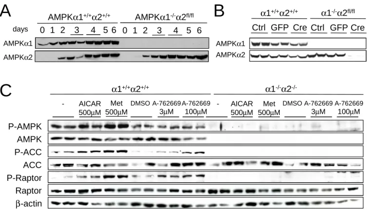
In undifferentiated WT myoblasts, only a low AMPKα1 subunit expression was noticeable
progresses, levels of both AMPKα1 and α2 catalytic subunits gradually increased, reaching a
maximum by day 4 (Figure 1A). In AMPKα1-/-AMPKα2fl/fl myotubes, AMPKα1 subunit was
absent in both undifferentiated myoblasts and differentiated myotubes, whereas AMPKα2
subunit expression levels showedan increase during differentiation without any significant
difference with WT myotubes (Figure 1A). To obtain myotubes lacking both AMPKα1 and
AMPKα2 catalytic subunits, we infected primary AMPKα1-/-AMPKα2fl/fl myoblasts with Cre-expressing adenovirus (Ad-Cre). Myoblasts were infected with GFP-expressing
adenovirus as a control. Expression of Cre resulted in the loss of AMPKα2 protein during the
differentiation process and AMPKα2 protein was absent after 4 days of differentiation
(Figure 1B). Additionally, AMPKα1 protein expression was undetectable in Ad-Cre-infected
myoblasts and 4 days after initiation of differentiation (Figure 1B). AMPK-deficient myoblasts efficiently initiate the differentiation program, as evidenced by the typical up-regulation of myogenin expression after 1 day of differentiation to about equal levels in both WT and AMPK-deleted cells (data not shown). AMPK-deficient myotubes were fully differentiated at day 4 as assessed by morphology (Figure 2A).
Lack of ACC and raptor phosphorylation following AICAR, metformin and A-762669 treatment in AMPK-deficient myotubes
Myotubes lacking both expression of AMPKα1 and α2 catalytic subunits (referred to as
AMPKα1-/-α2-/-) were treated with the AMPK activators AICAR
(5-aminoimidazole-4-carboxamide-1-β-D-ribofuranoside) (24), metformin (25) and A-762669 (26).
Phosphorylation of AMPK at Thr172 was increased in response to AMPK activators in WT
myotubes but was undetectable in AMPK-deficient myotubes (Figure 1C). The effect of
AMPK activators on ACC phosphorylation was abolished in AMPKα1-/-α2-/- myotubes
confirming that induction of ACC phosphorylation by AICAR, metformin and A-762669 is completety dependent on AMPK activation in muscle cells. We also examined phosphorylation of raptor, a recently identified AMPK target in the regulation of protein
synthesis pathway (8). Raptor is phosphorylated at Ser792 following AICAR, metformin and
A-762669 treatment in WT but not in AMPKα1-/-α2-/- myotubes, precisely paralleling ACC
phosphorylation pattern (Figure 1C).
Regulation of muscle cell size in the absence of AMPK
To determine whether AMPK contributes to the control of muscle cell size, we measured the effect of AMPK deletion on myotube size at differentiation day 4 (Figure 2A). The size of
Accordingly, the distribution of AMPKα1-/-α2-/- myotubes size showed higher number of large cells and a smaller proportion of small cells (Figure 2C). Myoblasts fusion was similar
in WT and AMPKα1-/-α2-/- muscle cultures indicating that cell hypertrophy promoted in the
absence of AMPK is due to enhanced cytoplasmic growth rather than increased myoblasts fusion (data not shown).
To address whether inactivation of AMPK signaling was sufficient to upregulate the mTOR pathway, we measured the phosphorylation of p70S6K as surrogate marker of mTOR kinase
activity. Interestingly, deletion of AMPK results in a rise in p70S6K phosphorylation at Thr389
and of its downstream target ribosomal protein S6 at Ser235/236 as assessed by Western blot
and immunofluorescence analysis (Figures 3A and 3B). Interestingly, the protein quantity
recovered per well was twice as high in AMPKα1-/-α2-/- myotubes compared to WT myotubes
(Figure 3C). The rate of protein synthesis measured by 35S-methionine incorporation was
increased 2-fold in AMPKα1-/-α2-/- myotubes (Figure 3D) compared to WT myotubes. The
protein synthesis rate was sensitive to rapamycin in both WT and AMPKα1-/-α2-/- myotubes,
as it was decreased by half after rapamycin treatment (30μM, 1h), decreasing the protein
synthesis rate in AMPK-deficient myotubes to the level of WT myotubes in basal conditions (Figure 3D). Accordingly, treatment with rapamycin abolished phosphorylation of p70S6K at
Thr389 in both WT and AMPKα1-/-α2-/- myotubes (Figure 3E). We therefore wished to
investigate the effect of rapamycin on cell size and mTOR downstream componentsleading to
the stimulation of skeletal muscle protein synthesisin AMPKα1-/-α2-/- myotubes. This mTOR
pathway blunting significantly affected the size of both WT and AMPKα1-/-α2-/- myotubes,
decreasing the myotube diameter of AMPK-deficient cells to the level of WT cells (Figures 2A, B and C).
MyrAkt-induced cell hypertrophy in WT but not in AMPKα1-/-α2-/- myotubes
To investigate the functional consequence of AMPK deletion on mTOR-mediated
hypertrophy, WT and AMPKα1-/-α2-/- muscle cells were infected with an adenovirus
expressing constitutive active Akt (MyrAkt). The diameter of myotubes increased by 20% in
WT cells whereas it was not affected in AMPKα1-/-α2-/- myotubes (Figures 4A and B). In
addition, we measured the phosphorylation of p70S6K as surrogate marker of mTOR kinase activity. We observed a significant increase in p70S6K phosphorylation only in WT myotubes introduced with MyrAkt, consistent with an absence of negative action of AMPK on
mTOR/p70S6K signaling pathway in response to hypertrophy in AMPKα1-/-α2-/- myotubes
Increased muscle fiber size in skeletal muscle AMPK-deficient mice
To extend our findings in vivo, AMPKα1-/-α2fl/fl mice were interbred with transgenic mice
expressing the Cre recombinase under the control of the human skeletal actin (HSA) promoter, which is exclusively expressed in differentiated multinucleated skeletal fibers (23). The resulting skeletal muscle AMPK-deficient mice were viable and appeared normal in all
respects as compared to HSA-Cre transgenic controls. The body weight of AMPKα1-/-α2fl/fl
HSA-Cre+ mice was similar with that of control mice (data not shown). Skeletal
muscle-specific deletion of AMPKα2 was examined by western blot analysis. AMPKα2 expression
was undetectable in soleus muscle from muscle AMPK-deficient mice (Figure 5A), with no
sign of deletion in liver and heart (data not shown). Additionally, AMPKα1 was also absent
in soleus muscle from muscle AMPK-deficient mice (Figure 5A). To determine whether AMPK deletion affects skeletal muscle mass, we compared the soleus muscle weight from
AMPKα1-/-α2fl/fl HSA-Cre+ and HSA-Cre- mice. Soleus muscle mass was significantly higher
by 42% in muscle AMPK-deficient mice compared to control mice (Figure 5B). Histological analysis of AMPK-deficient soleus muscle revealed increased cross-sectional area of the fibers (Figure 5C). Furthermore, myofiber size distribution of AMPK-deficient soleus muscle exhibits increased number of large myofibers and fewer small myofibers compared to control
soleus muscle (Figures 5D). Finally, fiber hypertrophy in AMPK-deficient soleus muscle was
associated with increased phosphorylation of p70S6K at Thr389 (Figure 5A) and of its
downstream target ribosomal protein S6 at Ser235/236 (Figure 5A).
Discussion
The mTORC1 signaling pathway has recently emerged as a crucial point of convergence for signaling by nutrients (glucose, amino acids), growth factors and cellular energy to promote skeletal muscle growth and fiber size (2, 4, 5, 7). As opposed to the mTORC1 signaling pathway, AMPK is activated under conditions of energy stress leading to high intracellular AMP/ATP ratio and restores cellular energy balance by promoting ATP-generating pathways, while simultaneously inhibiting ATP-utilizing pathways (10). Cell growth is energetically demanding and its inhibition is therefore an important mechanism to save energy under metabolic stress. To suppress mTOR signaling, AMPK utilizes both direct and indirect targets
via phosphorylation and activation of TSC2 on Thr1227 andSer1345 (9), and/or phosphorylation
and inactivation of mTORon Thr2446 (18). AMPK has also recently been shown to inhibit
mTORC1 directly, via phosphorylation of raptor on Ser722/Ser792 (8). Here, we confirmed that
of its phosphorylation was lost in muscle cells deleted for both AMPK catalytic subunits (Figure 1C), indicating an AMPK-dependent effect.
Recent evidence indicates that AMPK acts as a cellular energy checkpoint to prevent cell growth when cellular energy reserves are insufficient (8, 27). Hence, AMPK inhibits protein synthesis when nutrient conditions are limited by interfering with anabolic signaling mediated by the mTOR pathway. Our results provide strong evidence for the crucial role of AMPK in the maintenance of cell size through the control of mTOR/p70S6K signaling pathway. Deletion of AMPK in muscle cells enhanced p70S6K phosphorylation and in turn, protein synthesis rate, contributing to the shift in muscle cell and fiber size distribution toward high values (Figure 2C, 3A and 3D). Interestingly, treatment of AMPK-deficient cells with rapamycin completely blocked the up-regulation of p70S6K phosphorylation and rescued
AMPKα1-/-α2-/- muscle cell size (Figure 2C and 3E). Consistent with our results, analysis of
signaling downstream of mTORC1 has revealed the contribution of p70S6K phosphorylation in the control of muscle cell size and muscle hypertrophy (7). Muscle cells lacking p70S6K exhibited reduced cell size and failed to adapt their size to the inhibitory effect of rapamycin or amino acid starvation (7). Suppression of p70S6K signaling triggers an energy stress response defined by an increase in the AMP/ATP ratio leading to the activation of AMPK (6). This increase in AMPK activity blunts the growth responses to nutrient availability and inhibits signals that affect the regulation of muscle size. In this context, AMPK inhibition
restores muscle cell growth and sensitivityto nutrient signals (6). Of note, inactivation of
mTORC1 has previously been demonstrated to be critical for the ability of AMPK to integrate energy sensing with cell growth and proliferation requirement (9, 28). In contrast, loss of AMPK induces an absence of p70S6K inhibition, leading to higher protein synthesis rate and ultimately to increased muscle cell size. Interestingly, AMPKα1-/-α2-/- myotubes are resistant to MyrAkt hypertrophic action (Figure 4A and 4B) and p70S6K phosphorylation was not further increased in the absence of AMPK suggesting a ceiling effect (Figure 4C). One possible explanation is that mTOR signaling pathway is already activated at its maximal level in the absence of AMPK and no further increase is possible in response to Akt stimulation. Furthermore, it is possible that AMPK deletion also limits energy production for cell growth in response to MyrAkt-induced hypertrophy. Impaired mitochondrial function has been
demonstrated in skeletal muscle of mice expressing a kinase-dead AMPKα2 subunit in
skeletal muscle (29). Accordingly, impaired ATP generation was observed in skeletal muscle of these mice following chronic energy deprivation (30) and exercise (29). In addition, a marked energy disturbances was also reported during exercise in skeletal muscle of AMPK
2-/- mice (31) supporting the role of AMPKα2 in metabolic adaptations of skeletal muscle (32). These observations are completely consistent with the model that AMPK plays an essential role in the coordination between cell growth and cellular energy levels.
The inability of AMPK-deficient muscle cells to sense the cellular energy status triggers a signal equivalent to high energy content and a nutrient-rich environment, leading to the
hypertrophy of AMPKα1-/-α2-/- myotubes. These results show the major impact of AMPK
deletion on muscle cell size in our model. The physiological relevance of these data has been investigated in our skeletal muscle AMPK-deficient mice model. Interestingly, the increase in muscle mass and fiber size found in the soleus muscle is associated with an activation of the p70S6K/mTOR pathway (Figure 5). These findings demonstrate that in vivo as well as in
vitro, AMPK is highly implicated in the maintenance of muscle cell size. Thus, a crosstalk
between p70S6K and AMPK establishes a thin balance integrating cellular energy and nutrition content, and leading to changes in growth and metabolic rates. Evidence is accumulating that the molecular interplay between AMPK and mTOR signaling also limit hypertrophy in the heart to various stimuli. Treatment of neonatal rat cardiomyocytes with metformin, AICAR or resveratrol activated AMPK and inhibited the development of hypertrophy during phenylephrine treatment (33, 34). Notably, increased heart hypertrophy
was observed in AMPKα2-deficient mice following isoproterenol treatment (35) or transverse
aortic constriction (36), which correlated with dramatic increases in mTORC1 signaling. Recent studies have also linked AMPK and protein degradation in the muscle. Indeed, AICAR and metformin treatments decreased protein synthesis and increased protein degradation in an AMPK-dependant manner in C2C12 myotubes (37). Another study, also in C2C12 myotubes, has shown that AMPK activation stimulates myofibrillar protein degradation by increasing FOXO transcription factors (38). In our AMPK-deficient models, protein degradation may well be downregulated, thus emphasizing the observed phenotype. The increased protein quantity and associated hypertrophy observed may result from both an increase in protein synthesis and a decrease in protein degradation.
AMPK has emerged over the last decade as a central integrator of signals that control energy balance. Our results extend this notion by showing that AMPK controls muscle cell size and is involved in the cell size maintenance through the regulation of mTOR/p70S6K pathway. The recent findings that mTORC1 regulates muscle insulin signaling (39) and metabolism (40) in addition to protein synthesis suggest that AMPK metabolic effects might be mediated,
crosstalk between of AMPK and mTOR/p70S6K defines a metabolic program coordinating muscle plasticity.
Acknowledgements
We thank Judith Melki for kindly supplying the HSA-Cre transgenic mice for breeding, Kei
Sakamoto for the kind gift of A-769662 and Grahame Hardie for providing anti-AMPKα1
and α2 antibodies. We are indebted to Dr Luc Bertrand for protein synthesis protocol. We also thank Athanassia Sotiropoulos for helpful and fruitful discussions.
This work was supported by the European Commission integrated project (LSHM-CT-2004-005272), ANR (PHYSIO 2006 R06428KS) and AFM (grant 14138).
References
1. Glass, D. J. (2005) Skeletal muscle hypertrophy and atrophy signaling pathways. The
international journal of biochemistry & cell biology 37, 1974-1984
2. Wullschleger, S., Loewith, R., and Hall, M. N. (2006) TOR signaling in growth and
metabolism. Cell 124, 471-484
3. Sarbassov, D. D., Ali, S. M., Kim, D. H., Guertin, D. A., Latek, R. R.,
Erdjument-Bromage, H., Tempst, P., and Sabatini, D. M. (2004) Rictor, a novel binding partner of mTOR, defines a rapamycin-insensitive and raptor-independent pathway that regulates the cytoskeleton. Curr Biol 14, 1296-1302
4. Bodine, S. C., Stitt, T. N., Gonzalez, M., Kline, W. O., Stover, G. L., Bauerlein, R.,
Zlotchenko, E., Scrimgeour, A., Lawrence, J. C., Glass, D. J., and Yancopoulos, G. D. (2001) Akt/mTOR pathway is a crucial regulator of skeletal muscle hypertrophy and can prevent muscle atrophy in vivo. Nature cell biology 3, 1014-1019
5. Pallafacchina, G., Calabria, E., Serrano, A. L., Kalhovde, J. M., and Schiaffino, S.
(2002) A protein kinase B-dependent and rapamycin-sensitive pathway controls skeletal muscle growth but not fiber type specification. Proceedings of the National
Academy of Sciences of the United States of America 99, 9213-9218
6. Aguilar, V., Alliouachene, S., Sotiropoulos, A., Sobering, A., Athea, Y., Djouadi, F.,
Miraux, S., Thiaudiere, E., Foretz, M., Viollet, B., Diolez, P., Bastin, J., Benit, P., Rustin, P., Carling, D., Sandri, M., Ventura-Clapier, R., and Pende, M. (2007) S6 kinase deletion suppresses muscle growth adaptations to nutrient availability by activating AMP kinase. Cell Metab 5, 476-487
7. Ohanna, M., Sobering, A. K., Lapointe, T., Lorenzo, L., Praud, C., Petroulakis, E.,
Sonenberg, N., Kelly, P. A., Sotiropoulos, A., and Pende, M. (2005) Atrophy of S6K1(-/-) skeletal muscle cells reveals distinct mTOR effectors for cell cycle and size control. Nature cell biology 7, 286-294
8. Gwinn, D. M., Shackelford, D. B., Egan, D. F., Mihaylova, M. M., Mery, A.,
Vasquez, D. S., Turk, B. E., and Shaw, R. J. (2008) AMPK phosphorylation of raptor mediates a metabolic checkpoint. Molecular cell 30, 214-226
9. Inoki, K., Zhu, T., and Guan, K. L. (2003) TSC2 mediates cellular energy response to
control cell growth and survival. Cell 115, 577-590
10. Hardie, D. G. (2007) AMP-activated/SNF1 protein kinases: conserved guardians of
cellular energy. Nature reviews 8, 774-785
11. Gordon, S. E., Lake, J. A., Westerkamp, C. M., and Thomson, D. M. (2008) Does
AMP-activated protein kinase negatively mediate aged fast-twitch skeletal muscle mass? Exercise and sport sciences reviews 36, 179-186
12. Thomson, D. M., and Gordon, S. E. (2005) Diminished overload-induced hypertrophy
in aged fast-twitch skeletal muscle is associated with AMPK hyperphosphorylation. J
Appl Physiol 98, 557-564
13. Mounier, R., Lantier, L., Leclerc, J., Sotiropoulos, A., Pende, M., Daegelen, D.,
Sakamoto, K., Foretz, M., and Viollet, B. (2009) Important role for AMPKalpha1 in limiting skeletal muscle cell hypertrophy. Faseb J 23, 2264-2273
14. Kimura, N., Tokunaga, C., Dalal, S., Richardson, C., Yoshino, K., Hara, K., Kemp, B.
E., Witters, L. A., Mimura, O., and Yonezawa, K. (2003) A possible linkage between AMP-activated protein kinase (AMPK) and mammalian target of rapamycin (mTOR) signalling pathway. Genes Cells 8, 65-79
15. Krause, U., Bertrand, L., and Hue, L. (2002) Control of p70 ribosomal protein S6
kinase and acetyl-CoA carboxylase by AMP-activated protein kinase and protein phosphatases in isolated hepatocytes. Eur J Biochem 269, 3751-3759
16. Horman, S., Browne, G., Krause, U., Patel, J., Vertommen, D., Bertrand, L., Lavoinne, A., Hue, L., Proud, C., and Rider, M. (2002) Activation of AMP-activated protein kinase leads to the phosphorylation of elongation factor 2 and an inhibition of protein synthesis. Curr Biol 12, 1419-1423
17. Bolster, D. R., Crozier, S. J., Kimball, S. R., and Jefferson, L. S. (2002)
AMP-activated protein kinase suppresses protein synthesis in rat skeletal muscle through down-regulated mammalian target of rapamycin (mTOR) signaling. The Journal of
biological chemistry 277, 23977-23980
18. Cheng, S. W., Fryer, L. G., Carling, D., and Shepherd, P. R. (2004) Thr2446 is a novel
mammalian target of rapamycin (mTOR) phosphorylation site regulated by nutrient status. The Journal of biological chemistry 279, 15719-15722
19. Thomson, D. M., Fick, C. A., and Gordon, S. E. (2008) AMPK activation attenuates
S6K1, 4E-BP1, and eEF2 signaling responses to high-frequency electrically stimulated skeletal muscle contractions. J Appl Physiol 104, 625-632
20. Dreyer, H. C., Fujita, S., Cadenas, J. G., Chinkes, D. L., Volpi, E., and Rasmussen, B.
B. (2006) Resistance exercise increases AMPK activity and reduces 4E-BP1 phosphorylation and protein synthesis in human skeletal muscle. The Journal of
physiology 576, 613-624
21. Jorgensen, S. B., Viollet, B., Andreelli, F., Frosig, C., Birk, J. B., Schjerling, P.,
Vaulont, S., Richter, E. A., and Wojtaszewski, J. F. (2004) Knockout of the alpha2 but not alpha1 5'-AMP-activated protein kinase isoform abolishes 5-aminoimidazole-4-carboxamide-1-beta-4-ribofuranoside but not contraction-induced glucose uptake in skeletal muscle. The Journal of biological chemistry 279, 1070-1079
22. Viollet, B., Andreelli, F., Jorgensen, S. B., Perrin, C., Geloen, A., Flamez, D., Mu, J.,
Lenzner, C., Baud, O., Bennoun, M., Gomas, E., Nicolas, G., Wojtaszewski, J. F., Kahn, A., Carling, D., Schuit, F. C., Birnbaum, M. J., Richter, E. A., Burcelin, R., and Vaulont, S. (2003) The AMP-activated protein kinase alpha2 catalytic subunit controls whole-body insulin sensitivity. J Clin Invest 111, 91-98
23. Miniou, P., Tiziano, D., Frugier, T., Roblot, N., Le Meur, M., and Melki, J. (1999)
Gene targeting restricted to mouse striated muscle lineage. Nucleic acids research 27, e27
24. Corton, J. M., Gillespie, J. G., Hawley, S. A., and Hardie, D. G. (1995)
5-aminoimidazole-4-carboxamide ribonucleoside. A specific method for activating AMP-activated protein kinase in intact cells? Eur J Biochem 229, 558-565
25. Zhou, G., Myers, R., Li, Y., Chen, Y., Shen, X., Fenyk-Melody, J., Wu, M., Ventre, J.,
Doebber, T., Fujii, N., Musi, N., Hirshman, M. F., Goodyear, L. J., and Moller, D. E. (2001) Role of AMP-activated protein kinase in mechanism of metformin action. J
Clin Invest 108, 1167-1174
26. Cool, B., Zinker, B., Chiou, W., Kifle, L., Cao, N., Perham, M., Dickinson, R., Adler,
A., Gagne, G., Iyengar, R., Zhao, G., Marsh, K., Kym, P., Jung, P., Camp, H. S., and Frevert, E. (2006) Identification and characterization of a small molecule AMPK activator that treats key components of type 2 diabetes and the metabolic syndrome.
Cell Metab 3, 403-416
27. Hardie, D. G. (2008) AMPK and Raptor: matching cell growth to energy supply.
Molecular cell 30, 263-265
28. Shaw, R. J., Bardeesy, N., Manning, B. D., Lopez, L., Kosmatka, M., DePinho, R. A.,
and Cantley, L. C. (2004) The LKB1 tumor suppressor negatively regulates mTOR signaling. Cancer cell 6, 91-99
29. Lee-Young, R. S., Griffee, S. R., Lynes, S. E., Bracy, D. P., Ayala, J. E., McGuinness,
essential for the metabolic response to exercise in vivo. The Journal of biological
chemistry 284, 23925-23934
30. Zong, H., Ren, J. M., Young, L. H., Pypaert, M., Mu, J., Birnbaum, M. J., and
Shulman, G. I. (2002) AMP kinase is required for mitochondrial biogenesis in skeletal muscle in response to chronic energy deprivation. Proceedings of the National
Academy of Sciences of the United States of America 99, 15983-15987
31. Klein, D. K., Pilegaard, H., Treebak, J. T., Jensen, T. E., Viollet, B., Schjerling, P.,
and Wojtaszewski, J. F. (2007) Lack of AMPKalpha2 enhances pyruvate dehydrogenase activity during exercise. American journal of physiology 293, E1242-1249
32. McGee, S. L., Mustard, K. J., Hardie, D. G., and Baar, K. (2008) Normal hypertrophy
accompanied by phosphoryation and activation of AMP-activated protein kinase alpha1 following overload in LKB1 knockout mice. The Journal of physiology 586, 1731-1741
33. Chan, A. Y., Dolinsky, V. W., Soltys, C. L., Viollet, B., Baksh, S., Light, P. E., and
Dyck, J. R. (2008) Resveratrol inhibits cardiac hypertrophy via AMP-activated protein kinase and Akt. The Journal of biological chemistry 283, 24194-24201
34. Chan, A. Y., Soltys, C. L., Young, M. E., Proud, C. G., and Dyck, J. R. (2004)
Activation of AMP-activated protein kinase inhibits protein synthesis associated with hypertrophy in the cardiac myocyte. The Journal of biological chemistry 279, 32771-32779
35. Zarrinpashneh, E., Carjaval, K., Beauloye, C., Ginion, A., Mateo, P., Pouleur, A. C.,
Horman, S., Vaulont, S., Hoerter, J., Viollet, B., Hue, L., Vanoverschelde, J. L., and Bertrand, L. (2006) Role of the alpha2-isoform of AMP-activated protein kinase in the metabolic response of the heart to no-flow ischemia. Am J Physiol Heart Circ Physiol
291, H2875-2883
36. Zhang, P., Hu, X., Xu, X., Fassett, J., Zhu, G., Viollet, B., Xu, W., Wiczer, B.,
Bernlohr, D. A., Bache, R. J., and Chen, Y. (2008) AMP activated protein kinase-alpha2 deficiency exacerbates pressure-overload-induced left ventricular hypertrophy and dysfunction in mice. Hypertension 52, 918-924
37. Nystrom, G. J., and Lang, C. H. (2008) Sepsis and AMPK Activation by AICAR
Differentially Regulate FoxO-1, -3 and -4 mRNA in Striated Muscle. International
journal of clinical and experimental medicine 1, 50-63
38. Nakashima, K., and Yakabe, Y. (2007) AMPK activation stimulates myofibrillar
protein degradation and expression of atrophy-related ubiquitin ligases by increasing FOXO transcription factors in C2C12 myotubes. Bioscience, biotechnology, and
biochemistry 71, 1650-1656
39. Harrington, L. S., Findlay, G. M., Gray, A., Tolkacheva, T., Wigfield, S., Rebholz, H.,
Barnett, J., Leslie, N. R., Cheng, S., Shepherd, P. R., Gout, I., Downes, C. P., and Lamb, R. F. (2004) The TSC1-2 tumor suppressor controls insulin-PI3K signaling via regulation of IRS proteins. The Journal of cell biology 166, 213-223
40. Bentzinger, C. F., Romanino, K., Cloetta, D., Lin, S., Mascarenhas, J. B., Oliveri, F.,
Xia, J., Casanova, E., Costa, C. F., Brink, M., Zorzato, F., Hall, M. N., and Ruegg, M. A. (2008) Skeletal muscle-specific ablation of raptor, but not of rictor, causes metabolic changes and results in muscle dystrophy. Cell Metab 8, 411-424
Figure Legends
Figure 1: Generation of AMPKα1-/-α2-/- myotubes
AICAR, metformin and A-769662-induced AMPK and ACC phosphorylation in muscle cells. (A) AMPKα1 and α2 expression in WT and AMPKα1-/-α2fl/fl myotubes during differentiation. Myoblasts were differentiated and proteins were harvested every 24 hours during 6 days of differentiation. (B) Myoblasts were twice subjected to AdGFP or AdCre infection (50 m.o.i.). Twenty four hours after the second infection, myoblasts were split and differentiated in myotubes. Cells were harvested on day 4 of differentiation and proteins were
subjected to an α1 or α2 immunoblotting. (C) Western blot of P-AMPK, AMPK, P-ACC,
ACC, P-raptor, raptor and β-actin on myotubes incubated with AICAR (500μM, 5 hours),
metformin (500μM, 5 hours) or A-769662 (10 or 50μM, 2h).
Figure 2: Cell size control in muscle cells lacking AMPK. (A and B) Cells on day 2 of
differentiation were treated with 20 nM rapamycin or vehicle. After another 2 days, bright-field images were taken and myotube size was measured and expressed as percentage of WT
muscle cells. (C) Frequency distribution of size of WT and AMPKα1-/-α2-/- myotubes.
Results are represented as means ± SEM. Different from untreated cells of the same genotype:
#P<0.05; different for the same conditions of treatment: **
P<0.01.
Figure 3: Upregulation of mTOR signaling pathway in AMPKα1-/-α2-/- myotubes. (A)
Western blot of AMPKα1, AMPKα2, P-p70S6K, p70S6K, P-rpS6 and rpS6 in myotubes at
day 4 of differentiation. (B) Immunostaining for P-rpS6 in WT and AMPKα1-/-α2
-/-myotubes. (C) Protein quantity was measured in WT and α1-/-α2-/- myotubes after 4 days of
differentiation from wells seeded at the same density. (D) Protein synthesis rate as measured
before and after treatment with rapamycin (30μM, 1h) (E) Western blot of P-p70S6K and
p70S6K in myotubes treated with rapamycin (30μM, 1h) or vehicle. Different from untreated
cells of the same genotype: ##P<0.01; different for the same conditions of treatment: **P<0.01; ***P<0.001.
Figure 4: MyrAkt induced cell hypertrophy in WT but not in AMPKα1-/-α2-/- myotubes (A) Size of WT and AMPKα1-/-α2-/- myotubes transduced with AdGFP and AdMyrAkt (75 m.o.i.). Results are presented as a percentage of WT muscle cells transduced with AdGFP.
(B) Frequency distribution of size of WT and AMPKα1-/-α2-/- myotubes transduced with AdGFP and AdMyrAkt. (C) Western blot of P-p70S6K and total p70S6K. Results are
represented as means ± SEM. Different from WT cells infected with AdGFP : #P<0.05; ##
P<0.01.
Figure 5: Increased fiber size in soleus muscle lacking AMPKα1 and α2. (A) AMPKα1,
AMPKα2, p70S6K Thr389, p70S6K, rpS6 Ser235/236 and rpS6 expression in soleus muscle from
AMPKα1+/+α2+/+Cre+ and AMPKα1-/-α2fl/flCre+ mice. (B) Mass of soleus muscle from
AMPKα1+/+α2+/+Cre+ and AMPKα1-/-α2fl/flCre+ mice. Results are represented as means ±
SEM (n=6). (C) Representative bright-field images of soleus muscle fibers from AMPKα1+/+α2+/+Cre+ and AMPKα1-/-α2fl/flCre+ mice. (D). Frequency distribution of cross sectional area fibers in soleus muscle from AMPKα1+/+α2+/+Cre+ and AMPKα1-/-α2fl/flCre+ mice (n=6). The fiber cross-sectional area of fibers (n=417 in mean) was determined from different muscle areas of 6 muscles in each group. Different from WT: *P<0.05; **P<0.01.
Figure 1
A
B
DMSO A-762669 3μM A-762669 100μM α1+/+α2+/+ α1-/-α2 -/-Met 500μM AICAR 500μM - DMSO A-762669 3μM A-762669 100μM Met 500μM AICAR 500μM -P-AMPK P-ACC AMPK ACC β-actin P-Raptor RaptorC
AMPKα1 AMPKα1 0 1 2 3 4 5 6 AMPKα1-/-α2fl/fl AMPKα1+/+α2+/+ days 0 1 2 3 4 5 6 Ctrl GFP Cre α1+/+α2+/+ α1-/-α2fl/fl Ctrl GFP Cre AMPKα2 AMPKα2α1-/-α2 -/-α1+/+α2+/+ unt reat ed rapam yc in
A
C
B
0 20 40 60 80 100 120 140 160 M y ot ube di am et er (% W T )**
α1+/+α2+/+ α1-/-α2 -/-rapamycin-
+
-
+
# # α1+/+α2+/+ α1+/+α2+/++ rapamycin α1-/-α2 -/-α1-/-α2-/-+ rapacyminMyotube size (a.u.)
0 20 40 60 80 100 120 0-2 >2 >4 >6 >8 >10>12>14>16>18>20>22>24>26>28>30>32 M y ot ube s ize di s tri but ion (% )
Figure 2
A
P-p70S6K p70S6K P-rpS6 rpS6 AMPKα1 AMPKα2 0 2 4 6 8 10 12 P rot ei n (ug/ 3. 10 5c e lls )***
P-rpS6 P-p70S6K p70S6K AMPKα1-/-α2 -/-AMPKα1+/+α2+/+ rapamycin rapamycinD
rapamycin 35S -M et (10 3 dpm -bl k )**
##-
+
-
+
α1-/-α2 -/-α1+/+α2+/+ ## 0 2 4 6 8 10E
Figure 3
B
C
α1-/-α2 -/-α1+/+α2+/+ α1-/-α2 -/-α1+/+α2+/+ P-rpS6P-p70S6K GFP MyrAkt GFP MyrAkt p70S6K
C
A
M y ot ube di am et e r (% W T GF P ) α1+/+α2+/+ α1-/-α2 -/-MyrAkt GFP MyrAkt GFP α1+/+α2+/+ α1-/-α2-/-Figure 4
B
0 20 40 60 80 100 120 0-2>2>4>6>8>10>12>14>16>18>20>22>24>26>28>30>32 α1+/+α2+/++ GFP α1+/+α2+/++ MyrAkt α1-/-α2-/-+ GFP α1-/-α2-/-+ MyrAktMyotube size (a.u.)
M y ot ube s ize di s tri but ion (% ) 0 30 60 90 120 150 180 210 ## # ##
A
C
AMPKα1 AMPKα2 P-p70S6K p70S6K P-rpS6 rpS6 P-AMPK AMPK S o leus m a s s (m g)**
0 20 40 60 80 100 120 140Figure 5
α1-/-α2fl/flCre+ α1+/+α2+/+Cre+B
α1-/-α2fl/flCre+ α1+/+α2+/+Cre+ 0 5 10 15 20 25 30 M u s c le fi ber s ize (% )Fiber size (a.u.)