HAL Id: inserm-00698628
https://www.hal.inserm.fr/inserm-00698628
Submitted on 17 May 2012
HAL is a multi-disciplinary open access
archive for the deposit and dissemination of
sci-entific research documents, whether they are
pub-lished or not. The documents may come from
teaching and research institutions in France or
abroad, or from public or private research centers.
L’archive ouverte pluridisciplinaire HAL, est
destinée au dépôt et à la diffusion de documents
scientifiques de niveau recherche, publiés ou non,
émanant des établissements d’enseignement et de
recherche français ou étrangers, des laboratoires
publics ou privés.
Cysteinyl leukotriene signaling through perinuclear
CysLT(1) receptors on vascular smooth muscle cells
transduces nuclear calcium signaling and alterations of
gene expression.
Alison Eaton, Edit Nagy, Mathilde Pacault, J. Fauconnier, Magnus Bäck
To cite this version:
Alison Eaton, Edit Nagy, Mathilde Pacault, J. Fauconnier, Magnus Bäck. Cysteinyl leukotriene
signaling through perinuclear CysLT(1) receptors on vascular smooth muscle cells transduces nuclear
calcium signaling and alterations of gene expression.. Journal of Molecular Medicine, Springer Verlag,
2012, epub ahead of print. �10.1007/s00109-012-0904-1�. �inserm-00698628�
ORIGINAL ARTICLE
Cysteinyl leukotriene signaling through perinuclear CysLT
1
receptors on vascular smooth muscle cells transduces nuclear
calcium signaling and alterations of gene expression
Alison Eaton&Edit Nagy&Mathilde Pacault& Jérémy Fauconnier&Magnus Bäck
Received: 11 February 2012 / Revised: 22 March 2012 / Accepted: 12 April 2012 # The Author(s) 2012. This article is published with open access at Springerlink.com
Abstract Leukotrienes are pro-inflammatory mediators that are locally produced in coronary atherosclerotic plaques. The response induced by cysteinyl leukotrienes (CysLT) in human coronary arteries may be altered under pathological condi-tions, such as atherosclerosis. The aim of the present study was to elucidate cysteinyl leukotriene signaling in vascular smooth muscle cells (SMCs) and the effects of inflammation on this process. Immunohistochemical analysis of human carotid endarterectomy samples revealed that the CysLT1
leukotriene receptor was expressed in areas that also stained positive for α-smooth muscle actin. In human coronary artery smooth muscle cells, lipopolysaccharide significantly upregu-lated the CysLT1 receptor and significantly enhanced the
changes in intracellular calcium induced by leukotriene C4
(LTC4). In these cells, the CysLT1receptor exhibited a
peri-nuclear expression, and LTC4 stimulation predominantly
enhanced nuclear calcium increase, which was significantly inhibited by the CysLT1receptor antagonist MK-571.
Micro-array analysis revealed, among a number of significantly upregulated genes after 24 h stimulation of human coronary artery smooth muscle cells with LTC4, a 5-fold increase in
mRNA levels for plasminogen activator inhibitor (PAI)-2. The LTC4-induced increase in PAI-2 expression was confirmed by
real-time quantitative PCR and ELISA and was inhibited by the CysLT1receptor antagonist MK-571 and by calcium
che-lators. In summary, pro-inflammatory stimulation of vascular SMCs upregulated a perinuclear CysLT1receptor expression
coupled to nuclear calcium signaling and changes in gene expression, such as upregulation of PAI-2. Taken together, these findings suggest a role of nuclear CysLT1 receptor
signaling in vascular SMCs inducing gene expression patterns associated with atherosclerosis.
Keywords Atherosclerosis . Eicosanoids . Inflammation . Lipoxygenase . PAI-2
Introduction
Although initially identified as targets in the treatment of asthma, recent findings have brought attention to leuko-trienes (LTs) as potential mediators of cardiovascular dis-ease [1], such as atherosclerosis [2], abdominal aortic aneurysms [3], and aortic stenosis [4]. Human coronary artery atherosclerotic lesions are a source of cysteinyl LTs (i.e., LTC4, D4, and E4) [5], and urinary levels of LTE4are
increased in acute coronary syndromes [6], implicating these mediators in coronary atherosclerosis and plaque in-stability. The notion of cysteinyl LTs as potential effectors of atherosclerosis has also received support from animal models showing beneficial effects on atherosclerosis burden [7,8] and
Electronic supplementary material The online version of this article (doi:10.1007/s00109-012-0904-1) contains supplementary material, which is available to authorized users.
A. Eaton
:
E. Nagy:
M. Pacault:
M. BäckDepartment of Medicine, Karolinska Institutet, Stockholm, Sweden
J. Fauconnier
Department of Physiology & Pharmacology, Karolinska Institutet, Stockholm, Sweden
J. Fauconnier
INSERM U1046, Université Montpellier 1, Université Montpellier 2,
Montpellier, France M. Bäck (*)
Center for Molecular Medicine L8:03, Karolinska University Hospital, 171 76 Stockholm, Sweden e-mail: Magnus.Back@ki.se DOI 10.1007/s00109-012-0904-1
intimal hyperplasia [9] by specific antagonists of the leukotri-ene CysLT1receptor.
The cysteinyl LTs induce their action through G-protein-coupled receptors referred to as CysLT1and CysLT2, and the
existence of further subclasses of CysLT receptors also has been suggested [10]. In human carotid atherosclerotic lesions, a 3-fold higher CysLT1receptor expression compared with the
CysLT2receptor has been reported [11], although the cellular
localization of these receptors has not yet been resolved. The signaling through the CysLT1 receptor subtypes have been
widely studied in the context of bronchoconstriction and asthma [10]. Recently, the CysLT1receptor antagonist
mon-telukast was shown to be associated with a decreased risk of ischemic stroke and a decreased risk of myocardial infarction in males [12]. Although the latter report provides a first indication of beneficial effects of clinically used anti-leukotriene drugs, the exact role of CysLT receptor signaling in atherosclerosis remains to be established.
In addition to being bronchoconstrictors, cysteinyl LTs are also potent vasoconstrictors in the human lung [13]. Their role in the coronary vasculature, however, can be said to be contextually antithetical, as healthy human coronary arteries are unresponsive to cysteinyl LTs, but a contractile response to either LTC4or LTD4is observed in
atheroscle-rotic coronaries [14,15]. Although no previous study has addressed the mechanism for this differential sensitivity between healthy and atherosclerotic vessels, it is interesting to note that these leukotriene-induced contractions are inhibited by CysLT1 receptor antagonists [14]. The latter
observation is, however, in contrast to the dominant CysLT2
receptor expression in human coronary artery smooth mus-cle cells (SMCs) [16].
With the notion in mind that a local production of cys-teinyl LTs within the atherosclerotic lesion [5] could poten-tially activate CysLT receptors within the vascular wall, we engaged in this study with the hypothesis that the inflam-matory environment of atherosclerosis could lead to an upregulation of CysLT1receptors on vascular SMCs. Here,
we have investigated this idea, with the aim to quantify and describe the nature of the increased CysLT1signaling.
Fur-ther, we have explored the potential downstream effects of this phenomenon on both intracellular signaling and gene expression in vascular SMCs in an effort to link CysLT1
receptor signaling to atherogenic properties of SMCs in the context of inflammation and leukotriene stimulation.
Methods Cell culture
Human coronary artery SMC purchased from Clonetics (Cambrex Bio Science, Walkersville, MD, USA) were
cultured in SmGM2 kit medium as previously described [17] and harvested for experiments between passages 5 and 8. Cells were seeded in six-well plates (105cells/well) containing SmGM2 48 h before the respective treatment, which was replaced with Dulbecco’s modified Eagle’s me-dium supplemented with 2 % fetal calf serum (starvation medium) 24 h before experiments. Subsequently, cells were incubated in the absence or presence of lipopolysaccharide (LPS; from Sigma, 10 μg/mL), interleukin-6 (IL-6; from Peprotech, 20 ng/mL), interferon-γ (IFN-γ; from Peprotech, 20 ng/mL), or tumor necrosis factor-α (TNF-α; from Pepro-tech, 10 ng/mL). Duration of treatment was experiment-dependent, as described in detail below. Cells were treated with various substances across experiments, including LTC4, LTD4, and LTE4(from Cayman Chem, Ann Arbor,
MI, USA; 1 μM), the CysLT1receptor antagonist MK571
(from Cayman; 1 μM), ethylene glycol tetraacetic acid (EGTA; from Sigma, 5 mM), and 1,2-bis(2-aminophe-noxy)ethane-N,N,N′,N′-tetraacetic acid tetrakis(acetoxy-methyl ester) (BAPTA-AM; from Invitrogen, 10 μM). Immunostainings
Atherosclerotic vascular tissue was collected from eight patients undergoing carotid endarterectomy. All experi-ments were performed in accordance with the ethical stand-ards laid down in the 1964 Declaration of Helsinki and were approved by the local ethics committee (reference number 02/147). All persons gave their informed consent prior to their inclusion in the study. Immunofluorescent stainings were performed on acetone-fixed frozen sections of carotid endarterectomies and on human coronary artery SMCs cul-tured in LabTek slides after fixation and permeabilization with acetone–methanol. Rabbit anti-human CysLT1receptor
(from Cayman Chem) and mouse anti-human α-smooth muscle actin (from DAKO) were used as primary antibod-ies. Isotype-specific either DyLight 594 or DyLight 488-conjugated secondary antibodies (from Vector) were used, and the nuclei were counterstained with 4′, 6-diamino-2-phenylindol (DAPI; Vector). Images were captured with confocal microscope Leica DMI.
Calcium signaling experiments
SMCs incubated for 48 h in the absence or presence of LPS (10 μg/mL) were washed and loaded with the fluorescent Ca2+indicator fluo-3. The cells were subsequently stimulat-ed with LTC4(1 μM, 30 min at room temperature) in either
Tyrode’s solution (composition, millimolars: NaCl 121, KCl 5.0, NaHCO3 24, CaCl2 0.5, MgCl2 0.4, NaH2PO4 0.4,
EDTA0.1, and glucose 5.5) gassed with 5 % CO2in O2or
Tyrode’s solution containing the CysLT1receptor antagonist
MK571 (1 μM). Changes in [Ca2+] were recorded using a
BioRad MRC 1024 confocal microscopy unit attached to a Nikon Diaphot 200 inverted microscope with a Nikon Plan Apo ×20 or ×60 oil immersion objective (N.A.1.3), as previously described [18].
RNA extraction, cDNA synthesis, and TaqMan real-time PCR
Total RNA was extracted using RNeasy Mini kit (from Qiagen, Hilden, Germany) with an on-column DNase diges-tion step. RNA quantity was assessed using a Nanodrop ND-1000 microvolume spectrophotometer (Thermo Fisher Scientific), and RNA quality was assessed by a Bioanalyzer capillary electrophoresis system (Agilent Technologies, Palo Alto, CA, USA). First-strand cDNA was synthesized using Superscript II (Invitrogen, Carlsbad, CA, USA) with random hexamers according to the manufacturer’s instruc-tions. Quantitative TaqMan PCR was performed on a 7900HT Fast Real-Time PCR system (Applied Biosystems) with primer/probe pairs that were obtained using Assay-on-demand™ from Applied Biosystems for human CysLT1
(Hs00272624_s1) and Plasminogen Activator Inhibitor 2 (PAI-2/SERPIN B2; Hs01010736_m1). Levels of mRNA were normalized to expression levels of cyclophilin A (Hs99999904_m1), which previously has been determined as an appropriate housekeeping gene in these cells [17] Microarray analysis
Microarray analysis experiments were performed on RNA derived from three separate SMC culture experiments, using either Agilent one-color whole human genome (44 K) kit (Agilent Technologies, Redwood City, CA, USA; n02) or the Affymetrix Human Genome U133 Plus 2.0 array (n01). Microarrays were analyzed with the Agilent high-resolution microarray scanner. Data were subsequently uploaded to GeneSpring GX10 (Agilent technologies) and analyzed us-ing advanced analysis workflow for the Agilent one-color arrays. The set of data was normalized according to recom-mendations by GeneSpring for one-color arrays. (http:// www.chem.agilent.com/cag/bsp/products/gsgx/manuals/
GeneSpring-manual.pdf). Variability between chips was
accounted for by applying a shift to the 75th percentile (dividing all measured signals by a 75th percentile value). Per-gene normalization was performed by bringing the base-line to the median of all samples. Probe sets were firstly filtered by confidence of detection, where genes that were not confidently detected in any sample were excluded from further analysis. Further filtering based on expression dis-carded any genes where less than 100 % of samples in either the relapse or diagnosis condition had expression values below the 20th percentile. The most differentially expressed genes, defined as those with an uncorrected P value of <0.05
and demonstrating a fold change in expression of 2.0 or greater, were selected for analysis. The list of 90 genes generated was subsequently compared to data from the Affymetrix arrays, and the genes of interest were verified in terms of direction of regulation. Genes meeting all these criteria are presented in Table1. The 45 genes listed were submitted to Ingenuity® pathway analysis for prediction of canonical pathways and functional gene networks affected by the significant differential expression of these genes. ELISA
PAI-2 ELISA was carried out on supernatants from untreat-ed SMCs and SMCs treatuntreat-ed LTC4(1 μM) for 24 h using
IMUBIND® PAI-2 ELISA kit (from American Diagnostica GmBH, Pfungstadt, Germany) according to manufacturer’s protocol.
Data analysis
All results are expressed as mean±SE. Statistically significant differences were determined by either a Student’s t test (for pair-wise comparisons) or a one-way analysis of variances, followed by Holm–Sidak post hoc test, for multiple compar-isons, using Sigma Stat software. A P value of less than 0.05 was considered significant.
Results
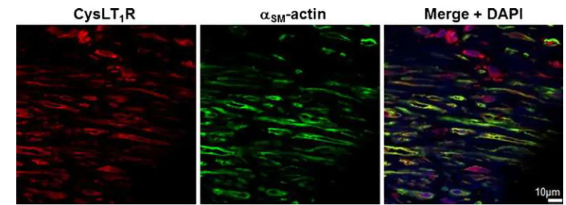
CysLT1receptor expression on vascular SMC
Immunohistochemical staining showed colocalization of the CysLT1 receptor protein with markers for SMC (α-smooth
muscle actin) in human atherosclerotic lesions (Fig. 1). In human coronary artery SMCs, the transcriptional levels of the CysLT1receptor were time-dependently increased by LPS,
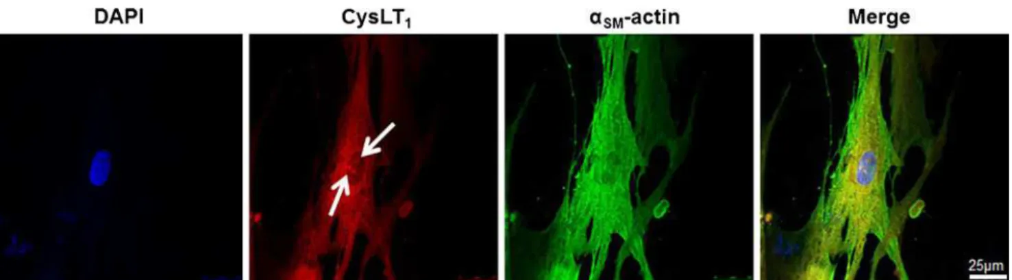
IL-6, and IFN-γ (Fig.2). Fluorescent immunostainings revealed a predominantly perinuclear localization of the CysLT1receptor
in human coronary artery SMCs compared with α-smooth muscle actin, which stained positive in the whole cytoplasm (Fig. 3). The CysLT1 receptor in some cases demonstrated
nuclear inclusions, as indicated by arrows in Fig.3.
LTC4-induced nuclear calcium signaling in vascular SMCs
To evaluate whether CysLT1receptors expressed on
vascu-lar SMC were functional, calcium changes in human coro-nary artery SMC were studied using the fluorescent Ca2+ indicator fluo-3 (Fig. 4a). LTC4induced a dose-dependent
increase in intracellular calcium, which was predominantly located in the nucleus (Fig.4b). The LTC4-induced calcium
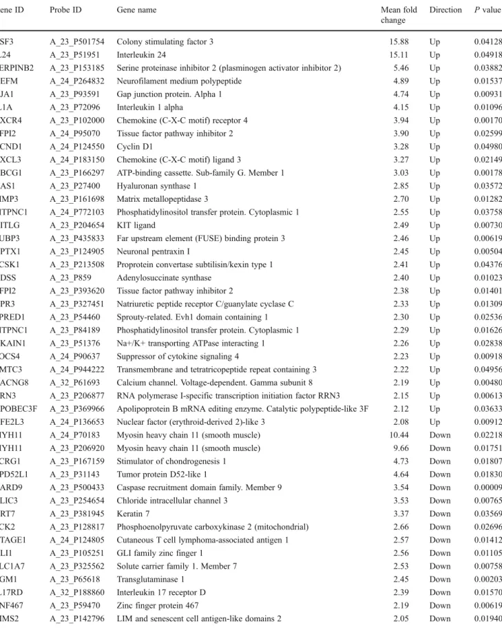
Table 1 Most significantly differentially expressed genes in response to LTC4(1 μM) in LPS-primed human coronary artery SMCs (sorted by fold change)
Gene ID Probe ID Gene name Mean fold
change
Direction P value
CSF3 A_23_P501754 Colony stimulating factor 3 15.88 Up 0.04128
IL24 A_23_P51951 Interleukin 24 15.11 Up 0.04918
SERPINB2 A_23_P153185 Serine proteinase inhibitor 2 (plasminogen activator inhibitor 2) 5.46 Up 0.03882 NEFM A_24_P264832 Neurofilament medium polypeptide 4.89 Up 0.01537 GJA1 A_23_P93591 Gap junction protein. Alpha 1 4.74 Up 0.00931
IL1A A_23_P72096 Interleukin 1 alpha 4.15 Up 0.01096
CXCR4 A_23_P102000 Chemokine (C-X-C motif) receptor 4 3.94 Up 0.00170 TFPI2 A_24_P95070 Tissue factor pathway inhibitor 2 3.90 Up 0.02599
CCND1 A_24_P124550 Cyclin D1 3.28 Up 0.04980
CXCL3 A_24_P183150 Chemokine (C-X-C motif) ligand 3 3.27 Up 0.02149 ABCG1 A_23_P166297 ATP-binding cassette. Sub-family G. Member 1 3.03 Up 0.00178
HAS1 A_23_P27400 Hyaluronan synthase 1 2.85 Up 0.03572
MMP3 A_23_P161698 Matrix metallopeptidase 3 2.70 Up 0.01282 PITPNC1 A_24_P772103 Phosphatidylinositol transfer protein. Cytoplasmic 1 2.55 Up 0.03758
KITLG A_23_P204654 KIT ligand 2.49 Up 0.00730
FUBP3 A_23_P435833 Far upstream element (FUSE) binding protein 3 2.46 Up 0.00619
NPTX1 A_23_P124905 Neuronal pentraxin I 2.45 Up 0.00504
PCSK1 A_23_P213508 Proprotein convertase subtilisin/kexin type 1 2.41 Up 0.04376
ADSS A_23_P859 Adenylosuccinate synthase 2.40 Up 0.01023
TFPI2 A_23_P393620 Tissue factor pathway inhibitor 2 2.38 Up 0.01401 NPR3 A_23_P327451 Natriuretic peptide receptor C/guanylate cyclase C 2.33 Up 0.01309 SPRED1 A_23_P54460 Sprouty-related. Evh1 domain containing 1 2.30 Up 0.02536 PITPNC1 A_23_P84189 Phosphatidylinositol transfer protein. Cytoplasmic 1 2.29 Up 0.01626 NKAIN1 A_23_P51376 Na+/K+ transporting ATPase interacting 1 2.26 Up 0.02838 SOCS4 A_24_P90637 Suppressor of cytokine signaling 4 2.23 Up 0.00918 TMTC3 A_24_P944222 Transmembrane and tetratricopeptide repeat containing 3 2.22 Up 0.04956 CACNG8 A_32_P61693 Calcium channel. Voltage-dependent. Gamma subunit 8 2.19 Up 0.00480 RRN3 A_23_P206877 RNA polymerase I-specific transcription initiation factor RRN3 2.15 Up 0.00613 APOBEC3F A_23_P369966 Apolipoprotein B mRNA editing enzyme. Catalytic polypeptide-like 3F 2.12 Up 0.03633 NFE2L3 A_24_P136653 Nuclear factor (erythroid-derived 2)-like 3 2.08 Up 0.00912 MYH11 A_24_P70183 Myosin heavy chain 11 (smooth muscle) 10.44 Down 0.02218 MYH11 A_23_P206920 Myosin heavy chain 11 (smooth muscle) 9.66 Down 0.01751 SCRG1 A_23_P167159 Stimulator of chondrogenesis 1 4.73 Down 0.01807 TPD52L1 A_23_P31143 Tumor protein D52-like 1 4.64 Down 0.01830 CARD9 A_23_P500433 Caspase recruitment domain family. Member 9 3.54 Down 0.00009 CLIC3 A_23_P254654 Chloride intracellular channel 3 3.53 Down 0.00765
KRT7 A_23_P381945 Keratin 7 3.37 Down 0.03569
PCK2 A_23_P128817 Phosphoenolpyruvate carboxykinase 2 (mitochondrial) 2.66 Down 0.02696 CTAGE1 A_24_P124805 Cutaneous T cell lymphoma-associated antigen 1 2.57 Down 0.01412 GLI1 A_23_P105251 GLI family zinc finger 1 2.56 Down 0.01105 SLC1A7 A_23_P325562 Solute carrier family 1. Member 7 2.53 Down 0.00758
TGM1 A_23_P65618 Transglutaminase 1 2.45 Down 0.00203
IL17RD A_32_P188860 Interleukin 17 receptor D 2.39 Down 0.01570 ZNF467 A_23_P59470 Zinc finger protein 467 2.19 Down 0.00619 LIMS2 A_23_P142796 LIM and senescent cell antigen-like domains 2 2.05 Down 0.01940 Note that some genes may be listed twice due to significant differences detected by independent probes
compared with untreated cells (Fig. 4c). In LPS-treated cells, the LTC4-induced increase in nuclear calcium was
significantly inhibited by the CysLT1 receptor antagonist
MK571 (Fig. 4c). The time course of the LTC4-induced
calcium increase in the nuclear and cytosolic compartments is shown in Fig.4d. The increase in nuclear calcium pre-ceded the increase in cytosolic calcium (Fig.4d).
LTC4-induced gene expression in vascular SMCs
The genes most significantly differentially expressed in response to LTC4(1 μM) in LPS-primed human coronary
artery SMCs are presented in Table1. PAI-2 (SERPIN B2), a member of the serine protease inhibitor superfamily, pre-sented as one of the most significantly upregulated genes in the microarray analysis. This finding was confirmed by quantitative PCR (Fig.5a), and in addition, increased PAI-2 protein levels were detected in the supernatant derived from LTC4-stimulated human coronary artery SMCs,
com-pared with unstimulated cells (Fig. 5b). The increased mRNA levels of PAI-2 induced by LTC4 (1 μM) were
mimicked by LTD4 (1 μM) but not LTE4 (1 μM) and
significantly inhibited by the CysLT1 receptor antagonist
MK 571 (1 μM; Fig. 5a). In addition, the LTC4-induced
increase in PAI-2 mRNA was abolished by the removal of intra- and extracellular calcium, through experiments per-formed in the presence of BAPTA-AM and EGTA (Fig.5c).
Ingenuity pathway analysis identified several functional gene networks predicted to be significantly affected by LTC4
stimulation as based on the 45 genes determined to be differ-entially expressed through our microarray analysis (Table1). The highest-scoring network (with a score of 35, equating to a fishers’ exact test score of 1×10⁻³⁵) containing 15 genes from
Table 1is suggested to be implicated in cellular movement and hematopoietic system function and development. An outline of the network is shown in Supplementary Fig.1.
Discussion
The results of the present study showed an upregulation of predominantly perinuclear CysLT1 receptors in vascular
SMCs under inflammatory conditions which was associat-ed with increasassociat-ed nuclear calcium signaling and changes in gene expression. Taken together, these findings suggest a role of nuclear CysLT1 receptor signaling in vascular
Fig. 1 CysLT1receptor expression in human atherosclerotic lesions. Representative
immunofluorescent staining of human atherosclerotic plaques from carotid artery showing colocalization of the CysLT1with α-smooth muscle actin-positive vascular smooth muscle cells. Original magnification, ×40
Fig. 2 CysLT1receptor expression in human coronary artery smooth muscle cells is upregulated by pro-inflammatory stimuli. Real-time quan-titative TaqMan RT-PCR for CysLT receptor mRNA in SMCs incubated in the absence and presence of LPS (10 μg/mL) for 1, 4, and 8 h (a) and IL-6
(20 ng/mL), TNF-α (10 ng/mL), or IFN-γ (20 ng/mL) for either 8 h (b) or 24 h (c). Results are expressed as fold increase compared with untreated cells (n03–5). *P<0.05 vs. time-matched control
SMCs inducing gene expression patterns associated with atherosclerosis.
Previous studies have suggested a dominant expression of the leukotriene CysLT2 receptor subtype in human
Fig. 3 Perinuclear CysLT1receptor expression in human coronary artery smooth muscle cells. Fluorescent labeling of CysLT1receptor protein (DyLight 594 red chromogen) and αSM-actin (DyLight 488 green
chromogen) in SMCs. Nuclei were stained with DAPI. Arrows indicate nuclear inclusions. Original magnification, ×63
Fig. 4 LTC4-induced calcium signaling in human coronary artery SMC. a Representative micrographs of Ca2+
fluorescence in the absence and presence of LTC4(1 μM). b Concentration–response curves for Ca2+fluorescence in nuclei (blue symbols) and cytosol (black symbols) of SMCs incubated for 48 h in the presence of LPS (10 μg/ml). c Ca2+fluorescence in nuclei (blue bars) and cytosol (black bars) of SMCs incubated for 48 h in the absence (control) or presence of LPS (10 μg/ml) prior to stimulation with LTC4 (1 μM, 30 min). d The time course of the LTC4-induced calcium increase shows that the increase in nuclear calcium (blue symbols) preceded the increase in cytosolic calcium (black symbols). *P<0.05 vs. controls,#P<0.05 vs Nuclear Ca2+
coronary arteries [16]. In the present study, priming of vascular SMCs with LPS upregulated CysLT1 receptor
mRNA and enhanced LTC4-induced effects. Similar
find-ings have been reported in endothelial cells, which under resting conditions exhibit a dominant CysLT2receptor, but
in which prolonged exposure to LPS or pro-inflammatory cytokines upregulate CysLT1 receptor expression [19]. In
the present study, CysLT1 receptor expression was also
upregulated in SMCs by IL-6 and by prolonged exposure to IFN-γ. Taken together, these observations suggest that a pro-inflammatory environment, such as atherosclerosis, may induce CysLT1receptor expression within the vascular wall.
In support of the latter notion, it has been shown that LTC4
induces contractions of atherosclerotic but not healthy cor-onary arteries [14,15] and that CysLT1receptor signaling,
but not CysLT2receptor signaling, is coupled to
vasocon-striction in isolated systemic vessels [20]. LTC4 has also
been associated with SMC proliferation and the shift of SMCs into a synthetic phenotype [21], which is in line with findings that CysLT1 receptor antagonism inhibits intimal
hyperplasia after vascular injury in mice [9].
The present study is the first demonstration of a perinu-clear localization of functional CysLT1receptors in vascular
SMCs and a leukotriene-induced nuclear calcium signaling in these cells. These findings are nevertheless consistent with other G-protein-coupled receptors in vascular SMCs.
For example, the ETA endothelin receptor and the AT1
angiotensin II receptor exhibit a perinuclear localization in vascular SMCs coupled to nuclear calcium signaling [22–24]. In addition, we have recently demonstrated a peri-nuclear localization of the CysLT1receptor in valvular
in-terstitial cells derived from human aortic valves [4], corroborating prior observations of CysLT1receptor
expres-sion at the outer nuclear membrane in intestinal epithelial cells [25]. In both of these cell types, leukotriene stimulation induces an increase in nuclear calcium [4,25]. The present study extends those findings by demonstrating an enhanced nuclear calcium increase in response to LTC4after priming
of cells with LPS and that the increase in nuclear calcium preceded the increase in cytosolic calcium. Whereas the present study cannot definitely conclude whether the sub-cellular CysLT1receptor localization represents an
internal-ization process, previous studies support a translocation of CysLT1receptors between different cellular compartments,
including nuclear inclusions [26].
Nuclear calcium is a key regulator of gene expression [27], and in line with this notion, LTC4induced significant changes
in expression of several genes in the present study. Of partic-ular interest was the appearance of PAI-2 as one of the most significantly upregulated genes in the microarray analysis. This was confirmed with qPCR analysis, and ELISA measures in addition showed that PAI-2 protein secretion from human
Fig. 5 LTC4-induced upregulation of PAI-2 in human coronary artery SMC. a Real-time quantitative RT-PCR for PAI-2 mRNA in human coronary artery SMC incubated in the ab-sence and preab-sence of LTC4, LTD4or LTE4(1 μM) for 24 h. In some experiments, cells were pretreated with the CysLT1 re-ceptor antagonist MK571 (1 μM) for 1 h before addition of LTC4. *P<0.05 vs. non-LTC4 stimulat-ed Contr (n03–6). b PAI-2 concentrations in supernatants from human coronary artery SMC incubated 24 h in the absence (Contr) or presence of LTC4(1 μM). *P<0.05 vs. Contr (n07). c Increase in PAI-2 mRNA levels induced by LTC4 (1 μM) in human coronary artery SMC incubated in the absence (Contr) or presence of either BAPTA-AM or EGTA. *P<0.05 vs. Contr (n03–6)
coronary artery SMCs was increased by LTC4stimulation.
PAI-1 and PAI-2 are members of the serine proteinase inhibitor family and act as important inhibitors of fibrinolysis by inter-fering with the plasminogen system. PAI-1 is induced by atherogenic stimuli in vascular SMCs and may participate in cell growth and matrix degradation associated with atheroscle-rosis [28]. In addition, using cDNA representational difference analysis, PAI-2 has previously been identified as one of the most differentially expressed genes in atherosclerotic lesions compared with normal vessels, with elevated PAI-2 expression preferentially observed in unstable carotid plaques [29]. In addition, another study using serial analysis of gene expression in human vascular SMCs also identified PAI-2 as one of the most upregulated genes in response to conditioned media derived from macrophages activated by oxidized low-density lipoprotein [30]. Finally, immunohistochemical analysis of human atherosclerotic lesions has also confirmed that vascular SMCs stain positive for PAI-2 [29,30]. In addition to acting as a plasminogen activator inhibitor, PAI-2 may serve as a regu-lator of Th1 immune responses through the modulation of cytokine-induced responses [31]. Furthermore, PAI-2 may be associated with the process of wound healing post-plaque rupture [29].
The LTC4-induced increase in PAI-2 mRNA was
abol-ished when experiments were performed in the presence of calcium chelators, suggesting a calcium dependent upregu-lation of PAI-2. The latter notion is supported by previous studies showing that angiotensin II is a potent inducer of PAI-2 in vascular SMCs through the AT1 receptor [32],
which is in line with a perinuclear AT1receptor localization
and an EGTA-sensitive nuclear calcium signaling induced by angiotensin II [33].
Ingenuity pathway analysis revealed a significant number of LTC4-upregulated genes to be implicated in a functional
gene network linked to hematopoietic system function and cellular movement. Of note in this network was the involve-ment of cAMP response eleinvolve-ment binding protein (CREB), a transcription factor and member of the leucine zipper family of DNA binding proteins, which is known to be activated by nuclear calcium [27]. Furthermore, the PAI-2 gene promoter region contains a binding site for CREB (−1,319 bp), and this transcription factor has been shown to be associated with the induction of PAI-2 expression [34]. Taken together, these findings suggest that LTC4-induced changes in gene
expression may be induced through an increase in nuclear calcium leading to CREB activation. However, the pathway analysis also revealed other pathways that may be involved in LTC4-induced gene expression, such as the NF-κB signaling
pathway which has been previously shown to be activated after CysLT1receptor ligation in leukocytes [10].
In summary, we have shown that pro-inflammatory stim-ulation of vascular SMCs enhances perinuclear CysLT1
receptor expression coupled to nuclear calcium signaling
and results in changes in gene expression, such as upregu-lation of PAI-2. Since cysteinyl-LT production is increased in atherosclerosis [5] and acute coronary syndromes [6], an altered vascular sensitivity to leukotriene-induced SMC gene expression secretion may further enhance the inflam-matory response. As such, targeting CysLT1receptors could
potentially be of therapeutic interest in atherosclerosis.
Acknowledgments The authors would like to Professor Håkan West-erblad for helpful advice on the study. This work was supported by the Swedish Heart and Lung Foundation, the Swedish Research Council (grant numbers 06816 and 2011-2988), and the French–Swedish Foundation. AE was supported by a fellowship from Mach-Gaensslen Foundation of Canada.
Conflict of interest None declared.
Open Access This article is distributed under the terms of the Crea-tive Commons Attribution License which permits any use, distribution, and reproduction in any medium, provided the original author(s) and the source are credited.
References
1. Poeckel D, Funk CD (2010) The 5-lipoxygenase/leukotriene path-way in preclinical models of cardiovascular disease. Cardiovasc Res 86:243–253. doi:10.1093/cvr/cvq016
2. Bäck M, Hansson GK (2006) Leukotriene receptors in atherosclero-sis. Ann Med 38:493–502
3. Houard X, Ollivier V, Louedec L, Michel JB, Bäck M (2009) Differential inflammatory activity across human abdominal aortic aneurysms reveals neutrophil-derived leukotriene B4 as a major chemotactic factor released from the intraluminal thrombus. FASEB J 23:1376–1383. doi:10.1096/fj.08-116202
4. Nagy E, Andersson DC, Caidahl K, Eriksson MJ, Eriksson P, Franco-Cereceda A, Hansson GK, Bäck M (2011) Upregulation of the 5-lipoxygenase pathway in human aortic valves correlates with severity of stenosis and leads to leukotriene-induced effects on valvular myofibroblasts. Circulation 123:1316–1325. doi:10.1161/CIRCULATIONAHA.110.966846
5. Piomelli D, Feinmark SJ, Cannon PJ (1987) Leukotriene biosynthe-sis by canine and human coronary arteries. J Pharmacol Exp Ther 241:763–770
6. Carry M, Korley V, Willerson JT, Weigelt L, Ford-Hutchinson AW, Tagari P (1992) Increased urinary leukotriene excretion in patients with cardiac ischemia. In vivo evidence for 5-lipoxygenase activa-tion. Circulation 85:230–236
7. Mueller CF, Wassmann K, Widder JD, Wassmann S, Chen CH, Keuler B, Kudin A, Kunz WS, Nickenig G (2008) Multidrug resistance protein-1 affects oxidative stress, endothelial dysfunc-tion, and atherogenesis via leukotriene C4 export. Circulation 117:2912–2918. doi:CIRCULATIONAHA.107.747667
8. Jawien J, Gajda M, Wolkow P, Zuranska J, Olszanecki R, Korbut R (2008) The effect of montelukast on atherogenesis in apoE/ LDLR-double knockout mice. J Physiol Pharmacol 59:633–639 9. Kaetsu Y, Yamamoto Y, Sugihara S, Matsuura T, Igawa G, Matsubara
K, Igawa O, Shigemasa C, Hisatome I (2007) Role of cysteinyl leukotrienes in the proliferation and the migration of murine vascular smooth muscle cells in vivo and in vitro. Cardiovasc Res 76:160– 166. doi:10.1016/j.cardiores.2007.05.018
10. Bäck M, Dahlen SE, Drazen JM, Evans JF, Serhan CN, Shimizu T, Yokomizo T, Rovati GE (2011) International union of basic and clinical pharmacology. LXXXIV: leukotriene receptor nomenclature, distribution, and pathophysiological functions. Pharmacol Rev 63:539–584. doi:10.1124/pr.110.004184
11. Lotzer K, Spanbroek R, Hildner M, Urbach A, Heller R, Bretschneider E, Galczenski H, Evans JF, Habenicht AJ (2003) Differential leukotriene receptor expression and calcium responses in en-dothelial cells and macrophages indicate 5-lipoxygenase-dependent circuits of inflammation and atherogenesis. Arte-rioscler Thromb Vasc Biol 23:e32–e36. doi:10.1161/01.ATV. 0000082690.23131.CB
12. Ingelsson E, Yin L, Bäck M (2012) Nationwide cohort study of the leukotriene receptor antagonist montelukast and incident or recur-rent cardiovascular disease. J Allergy Clin Immunol. doi:10.1016/ j.jaci.2011.11.052
13. Bäck M (2002) Functional characteristics of cysteinyl-leukotriene receptor subtypes. Life Sci 71:611–622
14. Allen SP, Dashwood MR, Chester AH, Tadjkarimi S, Collins M, Piper PJ, Yacoub MH (1993) Influence of atherosclerosis on the vascular reactivity of isolated human epicardial coronary arteries to leukotriene C4. Cardioscience 4:47–54
15. Allen S, Dashwood M, Morrison K, Yacoub M (1998) Differential leukotriene constrictor responses in human atherosclerotic coronary arteries. Circulation 97:2406–2413
16. Kamohara M, Takasaki J, Matsumoto M, Matsumoto S, Saito T, Soga T, Matsushime H, Furuichi K (2001) Functional character-ization of cysteinyl leukotriene CysLT(2) receptor on human cor-onary artery smooth muscle cells. Biochem Biophys Res Commun 287:1088–1092
17. Bäck M, Bu DX, Branström R, Sheikine Y, Yan ZQ, Hansson GK (2005) Leukotriene B4 signaling through NF-kappaB-dependent BLT1 receptors on vascular smooth muscle cells in atherosclerosis and intimal hyperplasia. Proc Natl Acad Sci U S A 102:17501– 17506
18. Tavi P, Hansson A, Zhang SJ, Larsson NG, Westerblad H (2005) Abnormal Ca(2+) release and catecholamine-induced arrhythmias in mitochondrial cardiomyopathy. Hum Mol Genet 14:1069–1076. doi:10.1093/hmg/ddi119
19. Gronert K, Martinsson-Niskanen T, Ravasi S, Chiang N, Serhan CN (2001) Selectivity of recombinant human leukotriene D(4), leukotriene B(4), and lipoxin A(4) receptors with aspirin-triggered 15-epi-LXA(4) and regulation of vascular and inflamma-tory responses. Am J Pathol 158:3–9. doi:10.1016/S0002-9440 (10)63937-5
20. Mechiche H, Candenas L, Pinto FM, Nazeyrollas P, Clément C, Devillier P (2004) Characterization of cysteinyl leukotriene receptors on human saphenous veins: antagonist activity of montelukast and its metabolites. J Cardiovasc Pharmacol 43:113–120
21. Palmberg L, Claesson HE, Thyberg J (1989) Effects of leuko-trienes on phenotypic properties and growth of arterial smooth muscle cells in primary culture. J Cell Sci 93(Pt 3):403–408
22. Bkaily G, Choufani S, Hassan G, El-Bizri N, Jacques D, D'Orleans-Juste P (2000) Presence of functional endothelin-1 receptors in nuclear membranes of human aortic vascular smooth muscle cells. J Cardiovasc Pharmacol 36:S414–S417
23. Bkaily G, Sleiman S, Stephan J, Asselin C, Choufani S, Kamal M, Jacques D, Gobeil F Jr, D'Orleans-Juste P (2003) Angiotensin II AT1 receptor internalization, translocation and de novo synthesis modulate cytosolic and nuclear calcium in human vascular smooth muscle cells. Can J Physiol Pharmacol 81:274–287
24. Haller H, Lindschau C, Erdmann B, Quass P, Luft FC (1996) Effects of intracellular angiotensin II in vascular smooth muscle cells. Circ Res 79:765–772
25. Nielsen CK, Campbell JI, Ohd JF, Morgelin M, Riesbeck K, Landberg G, Sjolander A (2005) A novel localization of the G-protein-coupled CysLT1 receptor in the nucleus of colorectal ade-nocarcinoma cells. Cancer Res 65:732–742
26. Jiang Y, Borrelli LA, Kanaoka Y, Bacskai BJ, Boyce JA (2007) CysLT2 receptors interact with CysLT1 receptors and down-modulate cysteinyl leukotriene dependent mitogenic responses of mast cells. Blood 110:3263–3270. doi:10.1182/blood-2007-07-100453 27. Hardingham GE, Bading H (1998) Nuclear calcium: a key regulator
of gene expression. Biometals 11:345–358
28. Dichtl W, Stiko A, Eriksson P, Goncalves I, Calara F, Banfi C, Ares MP, Hamsten A, Nilsson J (1999) Oxidized LDL and lyso-phosphatidylcholine stimulate plasminogen activator inhibitor-1 expression in vascular smooth muscle cells. Arterioscler Thromb Vasc Biol 19:3025–3032
29. Tyson KL, Weissberg PL, Shanahan CM (2002) Heterogeneity of gene expression in human atheroma unmasked using cDNA rep-resentational difference analysis. Physiol Genomics 9:121–130. doi:10.1152/physiolgenomics.00116.2001
30. Beauchamp NJ, van Achterberg TA, Engelse MA, Pannekoek H, de Vries CJ (2003) Gene expression profiling of resting and activated vascular smooth muscle cells by serial analysis of gene expression and clustering analysis. Genomics 82:288–299 31. Schroder WA, Le TT, Major L, Street S, Gardner J, Lambley E,
Markey K, MacDonald KP, Fish RJ, Thomas R et al (2010) A physiological function of inflammation-associated SerpinB2 is regulation of adaptive immunity. J Immunol 184:2663–2670. doi:10.4049/jimmunol.0902187
32. Feener EP, Northrup JM, Aiello LP, King GL (1995) Angiotensin II induces plasminogen activator inhibitor-1 and -2 expression in vascular endothelial and smooth muscle cells. J Clin Invest 95:1353–1362. doi:10.1172/JCI117786
33. Haller H, Lindschau C, Quass P, Distler A, Luft FC (1994) Nuclear calcium signaling is initiated by cytosolic calcium surges in vascular smooth muscle cells. Kidney Int 46:1653–1662
34. Park JM, Greten FR, Wong A, Westrick RJ, Arthur JS, Otsu K, Hoffmann A, Montminy M, Karin M (2005) Signaling pathways and genes that inhibit pathogen-induced macrophage apoptosis— CREB and NF-kappaB as key regulators. Immunity 23:319–329. doi:10.1016/j.immuni.2005.08.010