HAL Id: hal-01540356
https://hal.sorbonne-universite.fr/hal-01540356
Submitted on 16 Jun 2017HAL is a multi-disciplinary open access archive for the deposit and dissemination of sci-entific research documents, whether they are pub-lished or not. The documents may come from teaching and research institutions in France or abroad, or from public or private research centers.
L’archive ouverte pluridisciplinaire HAL, est destinée au dépôt et à la diffusion de documents scientifiques de niveau recherche, publiés ou non, émanant des établissements d’enseignement et de recherche français ou étrangers, des laboratoires publics ou privés.
Effectiveness of heart rate control on hemodynamics in
critically ill patients with atrial tachyarrhythmias
managed by amiodarone
Joe-Elie Salem, Pauline Dureau, Christian Funck-Brentano, Jean-Sébastien
Hulot, Maria Aissaoui, Nadia Aissaoui, Saik Urien, Christophe Faisy
To cite this version:
Joe-Elie Salem, Pauline Dureau, Christian Funck-Brentano, Jean-Sébastien Hulot, Maria Aissaoui, et al.. Effectiveness of heart rate control on hemodynamics in critically ill patients with atrial tach-yarrhythmias managed by amiodarone. Pharmacological Research, Elsevier, 2017, 122, pp.118 - 126. �10.1016/j.phrs.2017.06.004�. �hal-01540356�
Effectiveness of heart rate control on hemodynamics in
critically ill patients with atrial tachyarrhythmias managed
by amiodarone
Running title: AT, heart rate, amiodarone and hemodynamics in ICU
Joe-Elie Salem, MD-PhD †#, Pauline Dureau, MD †, Christian Funck-Brentano, MD-PhD †, Jean-Sébastien Hulot MD-MD-PhD †, Maria El-Aissaoui, MD ‡, Nadia Aissaoui, MD-PhD ‡, Saik Urien*, MD-PhD ‖, Christophe Faisy* MD-PhD †‖
*Saik Urien and Christophe Faisy contributed equally as last authors
† AP-HP, Pitié-Salpêtrière Hospital, Department of Pharmacology and CIC-1421, F-75013 Paris, France; INSERM, CIC-1421 and UMR ICAN 1166, F-75013 Paris, France; Sorbonne Universités, UPMC Univ Paris 06, Faculty of Medicine, Department of Pharmacology and UMR ICAN 1166, F-75013 Paris, France; Institute of Cardiometabolism and Nutrition (ICAN) ‡ Critical Care Unit, Hôpital Européen Georges Pompidou, Assistance Publique – Hôpitaux de Paris, University Paris Descartes Sorbonne Paris Cité, Paris, France.
‖ CIC-1419 INSERM, EAU-08 University Paris Descartes Sorbonne Paris Cité, Paris, France. # Cardiology - Rythmology Unit, Pitié-Salpêtrière Hospital, Assistance Publique – Hôpitaux de Paris, F-75013 Paris, France
Request for reprints and correspondence: Joe-Elie Salem, joeelie.salem@gmail.com, Centre d'Investigation Clinique Paris-Est, Hôpital La Pitié-Salpêtrière, 47-83 Bld de l'hôpital, 75651
Graphical abstract
ABSTRACT
Atrial tachyarrhythmias (AT) are common in intensive care unit (ICU) patients and might contribute to hemodynamic instability if heart rate (HR) is persistently too rapid. We aimed to assess if HR control below 115 or 130 bpm with amiodarone improves hemodynamics in ICU patients with AT.
This observational study included 73 ICU patients with disabling AT receiving amiodarone for HR control. A total of 525 changes (mainly within 4-8 h) in mean arterial pressure (MAP) and 167 changes in plasma lactate in response to HR variations above 115 or 130 bpm were analyzed. Epinephrine, sedative drugs, fluid loading, use of diuretics, continuous renal replacement therapy and amiodarone dosing were among covariables assessed.
Univariable analysis showed that HR variations above 115 bpm were poorly correlated to change in MAP (r=0.11, p<0.01). Multivariable analysis showed that changes in MAP were still positively associated to HR variation (p<0.05) and to initiation or termination of epinephrine (p<0.05) or sedatives infusions (p<0.05). Changes in plasma lactate did not correlate to HR variations above 115 bpm. When considering 130 bpm as a threshold, HR variations were not associated to changes in MAP or to changes in
plasma lactate. Amiodarone dose was associated to HR decrease but not to MAP or plasma lactate increase.
In ICU patients with AT, strict HR control below 115 bpm or 130 bpm with amiodarone does not improve hemodynamics. A prospective randomized trial assessing strict versus lenient HR control in this setting is needed.
ABBREVIATIONS LIST AT: atrial tachyarrhythmias
bpm: beats per minute
CRRT: continuous renal replacement therapy
ECG: electrocardiogram
ECV: electrical cardioversion
HR: heart rate
ICU: intensive care unit
IV: intravenous
MAP: mean arterial pressure
mg: milligram
Key words: amiodarone, atrial tachyarrhythmia, pharmacology, shock, heart rate.
INTRODUCTION
Atrial tachyarrhythmias (AT) (atrial fibrillation, atrial flutter, atrial tachycardia or atrioventricular arrhythmias) are highly prevalent and are associated with a poor outcome in critically ill patients admitted in intensive care units (ICU) [1,2]. AT are often
associated with a ventricular heart rate (HR) above 120–130 beats per minute (bpm) which contributes to baroreflex impairment,[3] diastolic dysfunction and may lead to
cardiac systolic dysfunction. Such induced ventricular tachycardiomyopathy is associated with hemodynamic instability and increased morbidity or mortality [2,4,5]. In
severely ill patients with AT, a rate or rhythm control strategy with amiodarone is recommended for the treatment or prevention of ventricular tachycardiomyopathy [6-7].
Attempts to restore sinus rhythm are frequently unsuccessful in the setting of critically ill patients [8-10], particularly when using electrical cardioversion without antiarrhythmic
pretreatment. HR control is thus the preferred strategy in this situation [10-12]. In
non-ICU patients, guidelines recommend aiming at a HR control between 90–115 bpm during stress situations [6,7,13]. There are no specific guidelines for patients in severely
ill condition but it is commonly advocated that reaching a HR below 115 bpm or a maximum of 130 bpm is advisable [6,7,14]. Thus, it is unknown whether exceeding these
HR cut-offs, even for short periods of time, is associated with alterations of hemodynamics, i.e. fall of mean arterial pressure (MAP) [15,16] or increase of plasma
lactate levels [17].
In a former study [18], we modelled the relationship between amiodarone dose and
heart rate variation but did not assess the influence of heart rate control on restoration of hemodynamics. The objective of this study was to determine if magnitude of HR variations above 115 or 130 bpm are associated to variations in MAP or plasma lactate. The analyses took into account variables such as paroxysmal vs persistent AT and others confounding factors relevant to ICU patients [19] such as epinephrine treatment,
sedative or curare drugs infusion, fluid loading, amiodarone dose, use of diuretics or continuous renal replacement therapy (CRRT).
MATERIALANDMETHODS
Patients and Study Design
This observational, cohort study was performed from January 2007 to April 2012 in the 18-bed medical ICU of a tertiary teaching hospital. In accordance with the French legislation on observational studies, approval by an investigational review board was not required. The use of confidential electronically processed patient data was approved by the French National for Data Protection Commission (Commission
Nationale de l'Informatique et des Libertés; reference: 1922081). Data were extracted
from the files of 73 consecutive critically ill patients who had received at least one dose of amiodarone (Mylan laboratory, Saint-Priest, FRANCE) to treat a disabling ATduring their ICU stay (Figure 1). HR and MAP data (average of at least 3 measures over 5 minutes) were collected at the time of the first amiodarone administration in intensive
care unit (ICU) and then 4 to 6 times daily over 6 days or until death or ICU discharge. MAP values were obtained through an arterial catheter attached to a fluid-filled pressure transducer system incorporating a flush system, which continuously infused a solution under pressure to maintain patency of the catheter. An attached transducer senses arterial pressure and converted the pressure signal to a waveform on a Phillips® bedside monitor. The waveform reflected pressure generated by the left ventricle during systole. The monitor also displayed automatically numerical MAP values. HR were also displayed on the monitor and computed by automatic QRS detection from continuous acquisition of a digital electrocardiographic signal. Plasma lactate levels were drawn at the discretion of the treating physician without any predefined protocol and were determined by an arterial blood gas analyser (ABL 825, Radiometer, Copenhagen, Denmark). Details concerning modalities of amiodarone administration in this cohort and estimation of pharmacodynamically active amiodarone dose at each time point were estimated as previously described in detail
[18]. Briefly, amiodarone could be administered either intravenously (IV) as amiodarone
hydrochloride 150mg/3mL or orally as 200 mg tablet(s). Route, frequency of administrations and doses were left at the discretion of the treating physician. Other treatments were administered according to standard guidelines [14].
Variables collection
The following covariables were recorded at the time of first HR and MAP collection: age, gender, body weight, severity score at ICU admission, diabetes, hypertension, AT history, bilirubinemia, previous amiodarone treatment and type of AT. Paroxysmal and persistent AT were defined as an arrhythmia occurring less or more than one week before the first amiodarone administration in ICU, respectively. Prior chronic and sub-acute amiodarone treatment before ICU were defined as continuous amiodarone treatment since more than one month or a cumulated amiodarone dose of less than 4 grams in the last month, respectively.
Time-dependent covariables potentially interacting with hemodynamic conditions were recorded at the time of each HR, MAP, and plasma lactate collection. These were: body temperature, arterial pH or PaO2, hemoglobin, electrical cardioversion, fluid
loading,pharmacodynamically active amiodarone dose or use of other antiarrhythmic drugs, loop diuretics, catecholamine, curare or sedative drugs and need for CRRT. Fluid loading was defined as administration of more than 0.5 L of saline solution in 30
min. Electrocardiographic acquisition allowing for accurate evaluation of type of rhythm was left at the discretion of the treating physician.
As previously reported [18], pharmacodynamically active amiodarone dose for each
subject was determined at the time of each HR, MAP, and plasma lactate collection. In brief, amiodarone pharmacokinetics was ascribed to a virtual compartment model including zero or first order input rates. This virtual compartment, A(t), represents the biophase in which amiodarone amount is in equilibrium with the observed effect on heart rate. Pharmacodynamic half-life (KDE) was determined to be 3.33 days. G(t) represents the amiodarone amount in the gut at a given time and ka, the first-order absorption rate, which was fixed to 8. Amiodarone bioavailability (F) was fixed to 0.33.
dA(t)/dt = input – KDE × A(t) (1)
input = ka × G(t) × F if administered orally (2)
Statistics
Results are expressed as numbers (%), means ± standard deviation, or medians (interquartile ranges) as appropriate. Normality was assessed by the D’Agostino-Pearson omnibus normality test. Comparison of continuous variables were analyzed by Mann-Whitney test for non-paired and non-parametric distribution. Comparison of categorical variables were analyzed by Chi-2 test. The correlation (r) between linear variables was assessed by calculating Pearson’s for parametric distribution or Spearman’s coefficient for non-parametric distribution (Prism 6, GraphPad software®, San Diego, USA). Of note, we had a power of at least 80% to detect a modest correlation (r>0.27) between variation of HR and variation of hemodynamic surrogates (MAP and plasma lactate). Multivariable analysis was performed by using linear mixed effect modeling that takes into account repeated measures (package nlme, Linear and Nonlinear Mixed Effects Models, R statistical software, https://www.r-project.org). The general equation is:
Y = intercept + β1*X1 + β2*X2 +…+ βn*Xn + (3)
Where Y is the dependent variable, X1,..., Xn and β1,..., βn stand for the
explanatory variables and the corresponding regression coefficient
respectively. denotes the random effect (it models the differences between the patients). Estimation of pharmacodynamically active amiodarone doses [18] at each
time-point were derived using R statistical software (https://www.r-project.org). Statistical significance was accepted for P<.05.
RESULTS
Study Population and Time-Dependent Observations
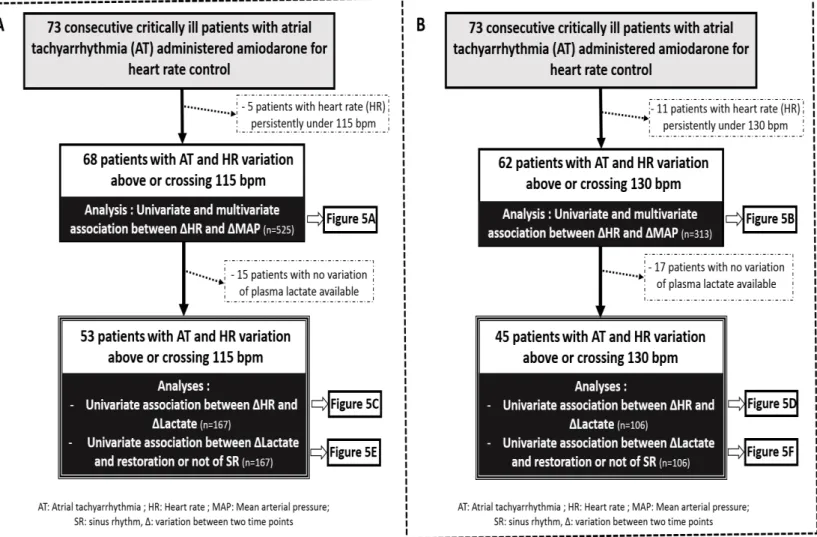
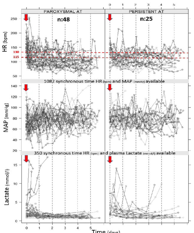
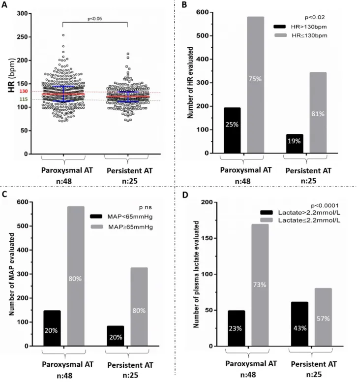
Seventy three consecutive patients admitted to ICU received at least one amiodarone dose for AT HR control and were eligible for analysis [Figures 1-2]. Of these 73 patients, 68 and 62 patients had at least one HR above 115 bpm or 130 bpm, respectively, thereby allowing for analysis of association between concomitant variations of MAP (ΔMAP) and HR (ΔHR), above 115 bpm (n = 525) or 130 bpm (n = 313) (Figure 1). Most of these variations were evaluated during a period of time of 4 to 8 hours (Table 1). Fifty three and 45 patients had at least one HR above 115 bpm and 130 bpm and at least two plasma lactate levels available, respectively, allowing for analysis of association between concomitant variations of plasma lactate (Δlactate) and HR (ΔHR), above 115 bpm (n = 167) or 130 bpm (n = 106) (Figures 1-2). Most of these variations were evaluated during a period of time of 6 to 24 h (Table 1). Patients included were severely ill. They were mainly admitted for cardiogenic shock or sepsis, had cardiovascular comorbidities and initially received amiodarone during their ICU stay for heart rate control either IV or mostly orally (Table 2). Dobutamine (11%) or epinephrine (61%) were the only IV catecholamine used. CRRT (25%), loop diuretics (49%), sedative drugs (60%) and fluid loading (70%) were frequently used. The main clinical, demographic and biological patient’s characteristics and other treatments that could interfere with hemodynamic status are summarized in Table 2. Forty eight patients (66%) had paroxysmal AT and 25 patients (34%) had persistent AT. Despite a higher proportion of uncontrolled HR collected above 130 bpm in patients with paroxysmal than persistent AT (25% vs 19%, p=0.01), proportion of MAP below 65 mmHg was not different (20% vs 20%, p=ns) and proportion of plasma lactate >2.2mmol/L was lower in patients with paroxysmal AT as compared to patients with persistent AT (23% vs 43%, p<0.0001) [Figure 3]. Variations in estimated pharmacodynamically active dose of amiodarone were correlated to variations in HR (r = 0.31, P <.0001) [Figure 4].
Heart rate variation and hemodynamics
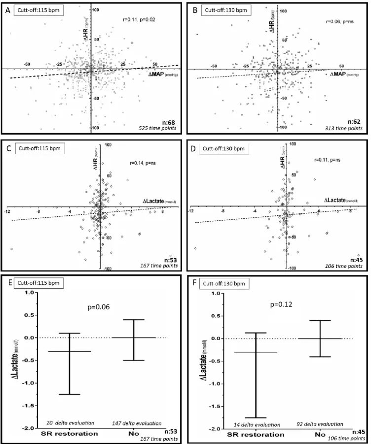
Univariable analysis showed that HR variations above 115 bpm were poorly associated to changes in MAP (r= 0.11±0.04, P.02; Figure 5A). The results of multivariable analysis integrated catecholamine, curare or sedative drugs infusion, fluid loading, persistent versus paroxysmal AT, use of diuretics or CRRT and estimated
pharmacodynamically active amiodarone dose or its variation. It showed that variations in MAP were associated to initiation or termination/weaning of epinephrine (β= 14.5±6.5 for initiation vs. termination/weaning, P=0.02; β= 10.6±4.9 for no change vs. termination/weaning, P=0.03) and sedative drugs (β= 18.3±8.4 for termination vs. initiation, P=0.03). Variation in HR was still positively associated to variation in MAP (β =0.06±0.03, P=0.03) but contribution of amiodarone pharmacodynamically active amount or other covariables were insignificant to explain variation in MAP.
When considering 130 bpm as threshold, HR variations were not associated to changes in MAP in univariable analysis (Figure 5B). Multivariable analysis showed that variations in MAP were also associated to initiation or termination/weaning of epinephrine (β= 27±9.4 for initiation vs. termination/weaning, P=0.005; β= 16±7.8 for no change vs. termination/weaning, P=0.04) and sedative drugs (β= 27.2±11.9 for termination vs. initiation, P=0.02). Variation of amiodarone pharmacodynamically active amount was also positively associated with variation in MAP (β= 0.01± 0.005, P=0.03). Other co-variables, including variation in HR, were not associated to variation in MAP.
We did not test other HR threshold because too few HR were available above 150 or 200 bpm in a limited number of patients (Table 1). Restoration of sinus rhythm could not be examined in our multivariable analysis because we had too few accurate characterizations of rhythm type evaluated by electrocardiogram (Table 1). However, univariable analysis showed that changes in plasma lactate were not associated to HR variations above 115 bpm (Figure 5C) but tended to be associated to restoration of sinus rhythm (-0.3 vs 0 mmol/L, P= 0.06, Figure 5E). Univariable analysis showed that changes in plasma lactate were not associated to HR variations above 130 bpm (ns; Figure 5D) or to restoration of sinus rhythm (-0.3 vs 0 mmol/L, P= 0.12, Figure 5F). DISCUSSION
The present study indicates that, in critically ill patients with AT, acute HR increases above 115 bpm or 130 bpm are not associated with acute deterioration of hemodynamic surrogates, i.e. increase of plasma lactate and decreases of MAP. In this setting, use of epinephrine or absence of sedative drugs are major determinants of MAP increase while restoration of sinus rhythm might improve plasma lactate levels. Patients with paroxysmal AT had a higher proportion of HR above 130bpm than patients with persistent AT. Thus, patients with paroxysmal AT had a lower proportion of plasma lactate above 2.2mmol/L, as compared to patients with
persistent AT. This finding further enhance absence of negative association between increased HR and deleterious hemodynamic response. We also found that amiodarone slows HR without altering hemodynamic markers.
Avoiding Ventricular Tachycardiomyopathy
AT, if persistent and rapid, can lead to tachycardia-induced cardiomyopathy with heart failure [3]. This condition is reversible, either by HR or rhythm control. Amiodarone
is the gold standard medication to prevent or cure this condition in critically ill patients with AT, particularly if kidney failure, ionic disturbances and heart failure are present
[6,7]. Whether brief episodes of rapid AT or sinus rhythm exceeding thresholds of 115
or 130 bpm carry a significant risk of tachycardia-induced cardiomyopathy in ICU patients remains unknown. Also, the best target HR and molecule to use are still unknown [5,6,20,21].
Amiodarone and Hemodynamics
In this observational study, amiodarone was mainly administered by oral route. In the past, the IV route of administration was known to be associated with transient hypotension and hemodynamic impairment [22-24]. However, the aqueous form of IV
amiodarone used in our patients was not expected to be associated with this specific side effect [25]. Our results did not show any negative association between the
estimated pharmacodynamically active amiodarone dose in organism and surrogates of global hemodynamic status. These data further support the safety of newer IV amiodarone hydrochloride formulation. Beta-blockers and verapamil were exceptionally used in this study because of their negative inotropic effects [20]. Digoxin
was also very rarely used because of its poor efficacy to rapidly slow HR, important dependency to renal clearance, and significant interaction with amiodarone or ionic disorder that may lead to adverse events [6,7].
Epinephrine, Sedative Drugs and Hemodynamics
In our study, we found that epinephrine use was associated with an increase of MAP while sedative drugs had an opposite effect. Epinephrine has alpha-1 and beta adrenergic agonist properties, determining its inotropic and vasopressive effects. In contrast, dobutamine, an inodilator selective beta adrenergic agonist [25], was not found
to be a major determinant of MAP variation. This might be explained by vasodilatation compensating its inotropic effect. Sedative drugs used in these patients were systematically relying on an association of a morphine derivate (sufentanil) with benzodiazepine (midazolam), which association is well known to decrease MAP [26].
Limitations
In our multivariable analysis, there was no significant influence of fluid loading. Its effect on MAP is expected to occur in the short term and not necessarily after a few hours of administration. Since the delay between two MAP collections was of a few hours, the effect of fluid therapy on MAP was possibly not sustained enough to be detected. Neither furosemide administration nor CRRT were identified as significant covariables. We hypothesize that patients treated with these depletive strategies had high pre-therapeutic filling pressures and did not experience hypovolemia and reactional MAP decrease.
In this observational study in critically ill patients, other covariables of interest such as left ventricular ejection fraction, plasma catecholamine levels, oxidative stress measurement, baroreflex and autonomic function analysis, which may influence hemodynamic response were not taken into consideration. However, this study shows for the first time the lack of effectiveness of aggressive acute heart rate control strategy to restore hemodynamics in critically ill patients.
Plasma levels of amiodarone and its metabolite N-desethylamiodarone, were not tested in our analysis because amiodarone therapeutic drug monitoring is not recommendedin clinical practice [6,7]. It should be emphasized that individual plasma
concentrations are poorly predictive of amiodarone pharmacodynamics response [27,28]. Perspectives
Several studies have evaluated the potential benefit of lenient versus strict heart rate control or rhythm control in the context of persistent AT in non-critically ill patients
[12,13,29]. These studies did not show any difference in outcome among these strategies.
Recently, Gillinov et al. further showed that strategies of rate or rhythm control to treat postoperative atrial fibrillation were not significantly different in terms of numbers of days of hospitalization, complication rates and rates of persistent atrial fibrillation 60 days after onset. [30]
Our study is the first to address this issue in critically ill patients. Its findings emphasize the importance of conducting a prospective trial of amiodarone dosing in critically ill patients with mostly “reactional” AT due to severe conditions comparing a lenient versus a strict rate control or even a rhythm control strategy. We recently proposed a tailored amiodarone dosing strategy according to duration of AT (paroxysmal versus persistent) and amiodarone pretreatment or not, which might help achieve a heart rate of 115bpm more conveniently (less systemic side effects,
bradycardia and hypotension) than a highly aggressive strategy relying on physician’s decisions without any standardized protocol [18]. Paroxysmal AT needed lower doses
of amiodarone to achieve the same level of rate control, as compared to persistent AT. Furthermore, addition of intravenous magnesium and vascular filling improved amiodarone efficacy to slow HR, as opposed to concomitant use of dobutamine [18].
CONCLUSION
In critically ill patients with AT, strict heart rate control below 115 bpm or even 130 bpm is not associated with hemodynamic improvement. Our data suggest that lenient heart rate control around 130 bpm might not be deleterious for these patients. A prospective randomized trial assessing strict versus lenient heart rate control or even rhythm control in critically ill patients with AT is needed to further address this issue.
DISCLOSURES
JES, PD, CFB, MEA, NA, SU, CF declare that they have no conflict of interest.
Conflict of interest: None
ACKNOWLEDGMENTS
Dr Salem had full access to all of the data in the study and takes responsibility for the integrity of the data and the accuracy of the data analysis. Dr Salem, Dr Aissaoui and Pr Faisy contributed to the conception and design of the study. Dr Salem, Dr El-Aissaoui and Dr Dureau contributed to data collection. Dr Salem, Dr Urien, Pr Faisy, Pr Funck-brentano and Pr Hulot contributed to analysis and/or interpretation of the data. Dr Salem, Pr Faisy, and Dr Urien contributed to drafting the manuscript. All authors contributed to revision of the manuscript and accepted the submitted version. We thank Margaux Alazard for help in data collection.
REFERENCES
1. Artucio H, Pereira M. Cardiac arrhythmias in critically ill patients: epidemiologic study. Crit Care Med. 1990;18(12):1383-8.
2. Walkey AJ, Hogarth DK, Lip GY. Optimizing atrial fibrillation management: from ICU and beyond. Chest. 2015;148(4):859-64.
3. Field ME, Wasmund SL, Page RL, Hamdan MH. Restoring Sinus Rhythm Improves Baroreflex Function in Patients With Persistent Atrial Fibrillation. J Am Heart Assoc. 2016; 23(2):5.
4. Nerheim P, Birger-Botkin S, Piracha L, Olshansky B. Heart failure and sudden death in patients with tachycardia-induced cardiomyopathy and recurrent tachycardia. Circulation. 2004;110(3):247‑52.
5. Tseng YH, Ko HK, Tseng YC, Lin YH, Kou YR. Atrial Fibrillation on Intensive Care Unit Admission Independently Increases the Risk of Weaning Failure in Non heart Failure Mechanically Ventilated Patients in a Medical Intensive Care Unit: A Retrospective Case-Control Study. Medicine (Baltimore). 2016;95(20):e3744.
6. European Heart Rhythm Association, European Association for Cardio-Thoracic Surgery, Camm AJ, Kirchhof P, Lip GY et al. Guidelines for the management of atrial fibrillation: the Task Force for the Management of Atrial Fibrillation of the European Society of Cardiology (ESC). Eur Heart J. 2010(19);31:2369‑429.
7. January CT, Wann LS, Alpert JS. 2014 AHA/ACC/HRS guideline for the management of patients with atrial fibrillation: a report of the American College of Cardiology/American Heart Association Task Force on Practice Guidelines and the Heart Rhythm Society. Circulation. 2014(23);130:2071-104.
8. Arrigo M, Jaeger N, Seifert B, Spahn DR, Bettex D, Rudiger A. Disappointing Success of Electrical Cardioversion for New-Onset Atrial Fibrillation in Cardiosurgical ICU Patients. Crit Care Med. 2015;43(11):2354-9.
9. Mayr A, Knotzer H, Mutz N, Hasibeder W. Atrial tachyarrhythmia after cardiac surgery. Intensive Care Med. 1999;25(2):242‑3.
10. Mitrić G, Udy A, Bandeshe H, Clement P, Boots R. Variable use of amiodarone is associated with a greater risk of recurrence of atrial fibrillation in the critically ill. Crit Care. 2016;20(1):90.
11. Delle Karth G, Geppert A, Neunteufl T et al. Amiodarone versus diltiazem for rate control in critically ill patients with atrial tachyarrhythmias. Crit Care Med. 2001; 29(6):1149‑53. 12. Carlsson J, Miketic S, Windeler J et al; STAF Investigators. Randomized trial of rate-control versus rhythm-rate-control in persistent atrial fibrillation: the Strategies of Treatment of Atrial Fibrillation (STAF) study. J Am Coll Cardiol. 2003;41(10):1690‑6.
13. Van Gelder IC, Hagens VE, Bosker HA et al; Rate Control versus Electrical Cardioversion for Persistent Atrial Fibrillation Study Group. A comparison of rate control and rhythm control in patients with recurrent persistent atrial fibrillation. N Engl J Med. 2002; 347(23):1834‑40. 14. Khoo CW, Lip GY. Acute management of atrial fibrillation. Chest. 2009;135(3):849-59. 15. Rhodes A, Evans LE, Alhazzani W, et al. Surviving Sepsis Campaign: International Guidelines for Management of Sepsis and Septic Shock: 2016. Intensive Care Med. 2017 Jan 18.
16. Thiele H, Ohman EM, Desch S, Eitel I, de Waha S. Management of cardiogenic shock. Eur Heart J. 2015 May 21;36(20):1223-30.
17. Kraut JA, Madias NE. Lactic acidosis. N Engl J Med. 2014;371(24):2309-19.
18. Salem JE, El-Aissaoui M, Alazard M et al. Modeling of Amiodarone Effect on Heart Rate Control in Critically Ill Patients with Atrial Tachyarrhythmias. Clin Pharmacokinet. 2016 Aug;55(8):991-1002
19. Lafont E, Urien S, Salem JE, Heming N, Faisy C. Modeling for critically ill patients: An introduction for beginners. J Crit Care. 2015;30(6):1287-94.
20. Walkey AJ, Evans SR, Winter MR, Benjamin EJ. Practice Patterns and Outcomes of Treatments for Atrial Fibrillation During Sepsis: A Propensity-Matched Cohort Study. Chest. 2016;149(1):74-83.
21. Patel J. Don't Rush to "Block" Atrial Fibrillation in Sepsis. Chest. 2016;149(5):1348. 22. Schwartz A, Shen E, Morady F et al. Hemodynamic effects of intravenous amiodarone in patients with depressed left ventricular function and recurrent ventricular tachycardia. Am Heart J. 1983;106(4Pt2):848‑56.
23. Remme WJ, Kruyssen HA, Look MP et al. Hemodynamic effects and tolerability of intravenous amiodarone in patients with impaired left ventricular function. Am Heart J. 1991; 122(1Pt1):96‑103.
24. Souney PF, Cooper WD, Cushing DJ. PM101: intravenous amiodarone formulation changes can improve medication safety. Expert Opin Drug Saf. 2010;9(2):319-33.
25. Salem JE, Aissaoui N, Paluszkiewicz L et al. Impact of inodilator drugs on echocardiographic assessments of left ventricular filling pressure in patients with decompensated end-stage heart failure. Crit Care Med. 2014;42(12):2508-17.
26. Crozier TA, Langenbeck M, Müller J, Kietzmann D, Sydow M, Kettler D. Total intravenous anaesthesia with sufentanil-midazolam for major abdominal surgery. Eur J Anaesthesiol. 1994;11(6):449-59.
27. Greenberg ML, Lerman BB, Shipe JR, Kaiser DL, DiMarco JP. Relation between amiodarone and desethylamiodarone plasma concentrations and electrophysiologic effects, efficacy and toxicity. J Am Coll Cardiol. 1987;9(5):1148–55.
28. Deharo JC, Durand A, Macaluso G, et al. Clinical electrophysiologic effects of a single high oral dose of amiodarone. Fundam Clin Pharmacol. 1997;11(3):275–80.
29. Van Gelder IC, Groenveld HF, Crijns HJ et al; RACE II Investigators. Lenient versus strict rate control in patients with atrial fibrillation. N Engl J Med. 2010;362(15):1363-73.
30. Gillinov AM, Bagiella E, Moskowitz AJ et al; CTSN. Rate Control versus Rhythm Control for Atrial Fibrillation after Cardiac Surgery. N Engl J Med. 2016;374(20):1911-21.
FIGURE 1. Flow chart of patients included and analyses performed; when considering 115 bpm (A), or 130 bpm (B) as threshold.
FIGURE 2. Representation of HR (bpm), MAP (mmHg), and plasma lactate (mmol/l)
collected over time (days) in the 73 studied patients with AT treated by
amiodarone in ICU (arrow representing time of first dosing). Each gray-scale line linking the different circles represents a single patient.
Figure 3. HR (bpm) in paroxysmal and persistent AT groups, represented as
median and interquartile range (A). HR (bpm), MAP (mmHg) and plasma lactate
(mmol/l) in paroxysmal and persistent AT groups, represented as proportion of HR>130bpm (B), MAP<65mmHg (C) and plasma lactate>2.2mmol/L (D). Statistics were performed by Mann-Whitney or Chi-2 tests, as appropriate.
FIGURE 4. Correlation between variation in heart rate (ΔHR, bpm) and variation in
FIGURE 5. Correlation between variations of heart rate (ΔHR, bpm) and mean arterial pressure
(ΔMAP, mmHg) when considering 115bpm (A) or 130bpm (B) as thresholds. Correlation between
concomitant variations of heart rate (ΔHR, bpm) and plasma lactate (ΔLactate, mmol/l) when considering 115bpm (C) or 130bpm (D) as thresholds. Variation of plasma lactate (ΔLactate, mmol/l)
as a function of restoration of sinus rhythm (SR restoration) or not (No), when considering 115bpm (E) or 130bpm (F) as thresholds.
Table 1. Heart rate, rhythm and hemodynamic data of the 73 patients
Characteristic Value
Heart rate data
Total number of HR collected 1193
Median time between consecutive HR collected, hours 5.04(4.08–7.44) Median time between first and last HR collected, days 4.85(2.2–5.09) Total number of HR collected ≥ 200 bpm / Number of patients concerned 10 / 5
Total number of HR collected ≥ 150 bpm / Number of patients concerned 106 / 40 Total number of HR collected ≥ 130 bpm / Number of patients concerned 289 / 62 Total number of HR collected ≥ 115 bpm / Number of patients concerned 492 / 68 Total number of HR collected ≤ 75 bpm / Number of patients concerned 85 / 26 Total number of HR collected ≤ 60 bpm / Number of patients concerned 6 / 5 Mean arterial Pressure data
Total number of MAP collected 1138
Median time between consecutive MAP collected, hours 5.52(4.08–7.68) Total number of synchronous MAP and HR collected 1082
Proportion of patients with at least one MAP≤65 mmHg 55 (75%) Plasma lactate data
Total number of plasma lactate collected 359
Median time between consecutive plasma lactate collected, hours 12(7.92–23.76) Total number of synchronous plasma lactate and HR collected 350
Proportion of patients with at least one plasma lactate≥2.2 mmol/l 37 (51%) Rhythm
Total number of rhythm evaluation collected 280
Abbreviations: bpm, beat per minute; HR, Heart rate; MAP, Mean arterial pressure. Statistics: Values are given as the median (interquartile range 25-75) or number.
Table 2. Main Characteristics of the 73 patients studied
Characteristic Value
Demographic status and comorbidities
Age, year 7513
Male sex 41(56%)
Body weight, kilogram 7116
Diabetes/Hypertension 17(23%)/36(49%)
Ischemic heart disease 30(41%)
Previous history of cardiac insufficiency 28(38%)
Previous history of supra-ventricular tachyarrhythmia 21(29%)
Prior amiodarone treatment before ICU: Chronic/Sub-acute 8(11%)/13(18%)
Chronic obstructive pulmonary disease 18(25%)
Chronic renal insufficiency (Clearance <60ml/min/m²) 17(23%)
APACHEII/SAPSII at admission in ICU 278/6119
ICU mortality 38(52%)
Bio-clinical values at first HR/MAP collection (first amiodarone dosing in ICU)
Mean arterial pressure <65mmHg / Need for catecholamine 28(38%) /39(53%)
Body Temperature, (°Celsius, eardrum) 37.1(36.4-38)
Non-invasive/invasive mechanical ventilation 8(11%) / 41(56%) Cardiac systolic dysfunction (Ejection fraction<45%) 27(37%)
Sepsis 34(47%)
Paroxysmal/persistent atrial tachyarrhythmia 48(66%)/25(34%) Atrial fibrillation / Flutter / Other atrial tachyarrhythmias 63(86%)/4(5%)/6(8%)
pH 7.36(7.26–7.44)
PaO2 (mmHg) 87(70-104)
Hemoglobin (g/dL) 10.6(9.1–12.2)
Creatinine clearance (ml/min/m²) 39(21-73)
Total bilirubin (mmol/l) 15(9-28)
Treatments administered during HR, MAP and plasma lactate data collection
Attempted/Successful electrical cardioversion 9(12%) / 4(5%) At least one amiodarone IV dose / oral dose 14(19%) / 69(95%) Median amiodarone dose (IV route), mg/dose 300 (150–300)
Median amiodarone dose (oral), mg/dose 400 (200–1200)
Other anti-arrhythmic (digoxin, beta-blocker, verapamil) 2(3%), 1(1%), 1(1%)
Catecholamine (-agonist): dobutamine 8(11%)
Catecholamine (α-and -agonist): epinephrine 45(61%)
Fluid loading 51(70%)
Renal replacement therapy/Diuretics 18(25%) / 36(49%)
Sedative drugs/Curare 44(60%) / 20(27%)
Estimated amiodarone dose at each time point (HR collection)
Median estimated pharmacodynamically active amiodarone dose (mg) 426 (281-713) Mean estimated pharmacodynamically active amiodarone dose (mg) 516±361 Min/Maximal estimated pharmacodynamically active amiodarone dose (mg) 0/1893
Abbreviations: APACHEII, Acute Physiology And Chronic Health Evaluation at intensive care unit admission; HR, heart rate; ICU, intensive care unit; MAP, mean arterial pressure; SAPSII, simplified acute physiology score at intensive care unit admission; IV, intravenous; mg, milligram
Statistics: Values are given as the mean standard deviation, median (IQ, interquartile range 25-75) or number (%).