HAL Id: hal-01471980
https://hal.sorbonne-universite.fr/hal-01471980
Submitted on 20 Feb 2017HAL is a multi-disciplinary open access archive for the deposit and dissemination of sci-entific research documents, whether they are pub-lished or not. The documents may come from teaching and research institutions in France or abroad, or from public or private research centers.
L’archive ouverte pluridisciplinaire HAL, est destinée au dépôt et à la diffusion de documents scientifiques de niveau recherche, publiés ou non, émanant des établissements d’enseignement et de recherche français ou étrangers, des laboratoires publics ou privés.
Thrombus composition in sudden cardiac death from
acute myocardial infarction
Johanne Silvain, Jean-Philippe Collet, Paul Guedeney, Olivier Varenne,
Chandrasekaran Nagaswami, Carole Maupain, Jean-Philippe Empana,
Chantal Boulanger, Muriel Tafflet, Stephane Manzo-Silberman, et al.
To cite this version:
Johanne Silvain, Jean-Philippe Collet, Paul Guedeney, Olivier Varenne, Chandrasekaran Nagaswami, et al.. Thrombus composition in sudden cardiac death from acute myocardial infarction. Resuscitation, Elsevier, 2017, �10.1016/j.resuscitation.2017.01.030�. �hal-01471980�
Thrombus Composition in Sudden Cardiac Death from Acute Myocardial Infarction.
Johanne Silvain1, Jean-Philippe Collet1, Paul Guedeney1, Olivier Varenne2, Chandrasekaran
Nagaswami3, Carole Maupain1, Jean-Philippe Empana4, Chantal Boulanger4, Muriel Tafflet4 ,
Stephane Manzo-Silberman5, Mathieu Kerneis1, Delphine Brugier1, Nicolas Vignolles1, John W.
Weisel3 , Xavier Jouven4, Gilles Montalescot1, Christian Spaulding4
1 Sorbonne Université - Univ Paris 06 (UPMC), ACTION Study Group, INSERM UMRS 1166, , Institut de Cardiologie, Hôpital Pitié-Salpêtrière (AP-HP), Paris, France.
2 Cardiology Department, Cochin Hospital, Paris 5 School of Medicine, Rene Descartes University, Paris, France.
3 Department of Cell and Developmental Biology, University of Pennsylvania School of Medicine, Philadelphia, Pennsylvania.
4 Département de cardiologie, Hôpital Européen Georges Pompidou, Université Paris
Descartes, Paris Cardiovascular Research Centre (PARCC), INSERM UMRS 970, Paris Sudden Death Expertise Centre, Paris, France.
5 Cardiology department, Inserm U942, Lariboisiere Hospital, Paris Diderot University, Paris, France.
Corresponding author:
Dr Johanne Silvain, Institut de Cardiologie, Bureau 278, Pitié-Salpêtrière University Hospital, 47 blvd de l’Hôpital, 75013 Paris, France. Tel: +33 01 42 16 29 61; Fax: +33 01 42 16 29 31. Email:
johanne.silvain@aphp.fr
Abstract
Background and aim: It was hypothesized that the pattern of coronary occlusion (thrombus
composition) might contribute to the onset of ventricular arrhythmia and sudden cardiac death (SCD) in myocardial infarction (MI).
Methods: The TIDE (thrombus and inflammation in sudden death) study included patients with angiographically-proven acute coronary occlusion as the cause of a ST elevation MI (STEMI)
thrombo-aspiration before primary percutaneous coronary stenting and analyzed with a quantitative method using scanning electron microscopy. We compared the composition of the thrombi
responsible for the coronary occlusion between the two groups and evaluated factors influencing its composition.
Results: We included 121 patients and found that thrombus composition was not different between the SCD group (n=23) and the STEMI group (n=98) regarding content of fibrin fibers (60.3±18.4 % vs. 62.4±18.4% respectively, p=0.68), platelets (16.3±19.2% vs. 15.616.7±%, p=0.76), erythrocytes (14.6±12.5% vs. 13±12.1%, p=0.73) and leukocytes (0.6±0.9% vs. 0.8±1.5%, p=0.93). Thrombus composition did not differ between patients receiving upstream-use of glycoprotein IIb/IIIa platelet receptor inhibitors (GPI) and patients free of GPI. The only factor found to influence thrombus composition was the ischemic time from symptom onset to primary PCI, with a decreased content in fibrin fibers (57.8±18.5% vs. 71.9±10.1%, p=0.0008) and a higher platelet content (19.2±19.1% vs. 7.9±5.7% p=0.014) in early presenters (< 3 hours of ischemic time) vs. late presenters (>6 hours of ischemic time).
Conclusion: Composition of intracoronary thrombi in STEMI patients does not differ between those presenting with and without SCD. Time from symptom onset to coronary reperfusion seems to be the strongest factor influencing thrombus composition in MI.
Abbreviation list:
STEMI: ST-elevated myocardial infarction SCD: sudden cardiac death
TIDE study: Thrombus and Inflammation in sudden Death study GPI: glycoprotein IIb/IIIa inhibitors
IQR: interquartile range
Key words: Sudden cardiac death; ST elevated myocardial infarction; coronary thrombus
INTRODUCTION
Sudden cardiac death (SCD) accounts for 4 million deaths every year worldwide.[1]A frequent cause of SCDs is ventricular fibrillation or fast ventricular tachycardia in the setting of an acute coronary artery occlusion. [2,3]Several risk factors of SCD have been identified in previous studies, however
the relationship between an acute coronary artery occlusion and the onset of ventricular arrhythmia is unknown.[4–9]
In ST-elevation myocardial infarction (STEMI) patients, a hypothesis is that differences in patterns of STEMI development leading to more rapid coronary occlusion could trigger SCD. In the TIDE
(Thrombus and Inflammation in sudden Death) microparticle study, we reported higher
concentrations of intracoronary endothelial microparticles in STEMI with SCD at presentation versus those without SCD, suggesting that ventricular arrhythmia in the setting of acute myocardial ischemia is not entirely explained by rhythmic vulnerability.[10] A vascular vulnerability related to a specific pattern of abrupt coronary occlusion may also be involved. The TIDE Thrombus study (NCT00748111) was designed to investigate this hypothesis. Our aim was to assess whether thrombus architecture per se could affect the occurrence of SCD in myocardial infarction and characterize the independent correlates of thrombus composition.
METHODS Study design
We prospectively screened all the STEMI and SCD patients referred to the catheterization laboratory of the Pitié-Salpêtrière and Cochin Hospital in Paris France for primary percutaneous coronary intervention (PCI). In the thrombus analysis of the TIDE study, we included SCD patients with documented STEMI (SCD group) and STEMI patients without ventricular arrhythmias (STEMI group) meeting the following inclusion criteria: age > 18 years; documented acute coronary artery occlusion with thrombolysis in myocardial infarction (TIMI) flow of 0, 1, or 2; coronary blood sampling using an aspiration catheter available; a time delay from symptom onset to ventricular arrhythmia or
pulseless condition of less than 1 h; successful out-of-hospital resuscitation with Return Of
Spontaneous Circulation (ROSC) after SCD. Due to logistical constraints, we evaluated only patients presenting during ‘on hours’. Our aim was to include 4 STEMI for each SCD. Based on our previous experience in the analysis of thrombus composition in STEMI presenters, we estimated a sample size of 100 STEMI and 25 SCD to be reasonable although no sample size calculation could be performed due to the lack of available data.[11] Recruitment of the TIDE study was slower than expected and patients with STEMI from the thrombus registry of the Pitié-Salpêtrière hospital who matched the inclusion criteria of the TIDE study were included in the present analysis as explained in the figure 1. Thrombus collection
Thrombo-aspiration during primary PCI was performed by a low-profile catheter (Export 6F,
Medtronic, Santa Rosa, California). Collected thrombi were immediately washed with saline and fixed with 2% glutaraldehyde in 50 mmol/l Na Cacodylate buffer (pH 7.3). Patients received 250 mg of aspirin intravenously; 600 mg of clopidogrel (crushed and administrated in the nasogastric tube if necessary for SCD patients) and the use of glycoprotein IIb/IIIa platelet receptor inhibitors (GPI) were
administered before and/or during the procedure at the discretion of the physicians. Study oversight
The study was reviewed and accepted by the ethics committee of the Institutional Review Board of the Cochin Hospital, Paris, France and the thrombus registry of the Pitié-Salpêtrière is declared as part of the ePARIS STEMI registry which was reviewed and accepted by the ethics committee of the Institutional Review Board of the Pitié-Salpêtrière Hospital. Informed consent was obtained before the procedure in all STEMI patients without SCD. The ethics committee allowed blood sampling and thrombus collection without consent during PCI procedures in case of SCD. However, the data could be used only if informed consent was obtained from the next of kin before or after the procedure and from the patient if s/he survived.
Scanning electron microscope analysis
Sample fixation, dehydration, and preparation were performed according to a previously published method.[12]High-definition photographs (x3,000 magnification) were obtained using a Philips/FEI XL20 scanning electron microscope 4-nm resolution (FEI, Hillsboro, Oregon). To control for
composition heterogeneity in the analysis of the surface of thrombus, we covered several areas (at least 12 according to a grid and the size of the thrombus) for each thrombus and used a validated analysis approach to determine the proportion of each component of thrombus composition (platelet, fibrin, erythrocytes, leukocytes, cholesterol crystal) of images for a single thrombus.[11] Each thrombus analysis comprised for 10 to 15 high magnification scanning electron microscopy (SEM) pictures at randomly chosen locations to eliminate selection bias. All the structures visualized were easy to identify on the basis of SEM images described previously. SEM and image analysis were performed by two independent trained technicians blinded to the clinical data. Very low inter-individual variability was found in the thrombus composition analysis (<5%).
Microparticle analysis
Specific microparticle sub-populations were identified by flow cytometry, as previously reported in our previous publication [10]. They were measured in the blood from the culprit coronary lesion, the systemic blood before the coronary intervention (Pre-PCI) and in the systemic blood after the coronary intervention (Post PCI). We report in this manuscript the micropaticle results in patients having both thrombus and micropaticle data available within the TIDE study (subanalysis). AnnexinV+ micro-particles, endothelium-derived microparticles (EMPs; CD144+), platelet-derived microparticles (CD41+), leukocyte-derived microparticles (CD11a+), erythrocyte-derived microparticles (CD235a+) were expressed as nb/μl and compared with two-by-two comparisons of median (interquartile range).
Clinical data collection
Clinical data were prospectively collected at admission and during the hospital stay. A certified core laboratory reviewed all angiographic data. Clinical and biological data were analyzed to identify
predictors of thrombus composition. For SCD patients, the location of SCD, initial cardiac rhythm, intervals between the onset of SCD and basic life support (‘no-flow’), between basic life support and ROSC (‘low-flow’) were collected from the emergency medical system physicians according to the Utstein recommendations. [13]
Statistical analyses
Descriptive statistics are reported as mean ± SD, median and interquartile range (IQR) (25th to 75th)
percentiles and percentages when appropriate. Differences in baseline characteristics between two groups were compared using Chi Square (Fisher exact test if expected count <5). Potential
associations between clinical and biological parameters such as thrombus composition were
compared by Student’s t-test and ANOVA test. As performed in a previous publication we divided the data in 3 groups on the basis of ischemic time and we used patients with ischemic time <3hours as the reference group.[11] A Kruskal–Wallis test was used for the trend between the three groups. Statistical analysis was performed with Graphpad Prism (GraphPad Software, Inc., USA).
RESULTS
Between March 2007 and December 2009, 77 patients were prospectively recruited in the TIDE study, and 44 patients of the Pitié-Salpêtrière Thrombus registry matching the inclusion criteria of the TIDE study were added to complete the final analysis (Figure 1). Of the 121 STEMI patients with thrombus analyzed, 23 were in the SCD group and 98 in the STEMI group. Patients in the STEMI group had longer median ischemic times than patients included in the SCD group (120 min [IQR at 60-179]) vs. 207min [IQR at 145-325]), p=0.0008) (Table 1). The rate of glycoprotein IIb/IIIa inhibitors (GPI) use was higher in the STEMI group than the SCD group due to upstream prehospital use. In the SCD group, the first recorded rhythm was ventricular tachycardia or ventricular fibrillation in the vast majority of patients (Table 2). The median values before any resuscitation (no-flow) was 0 min (IQR 0-5) reflecting a group of SCD patients with immediate or fast care, and the median values before the return of spontaneous circulation (low-flow) was 18.5 min (IQR 10-26). As expected, in-hospital mortality rate was much lower in the STEMI group as compared with the SCD group.
Thrombus analysis
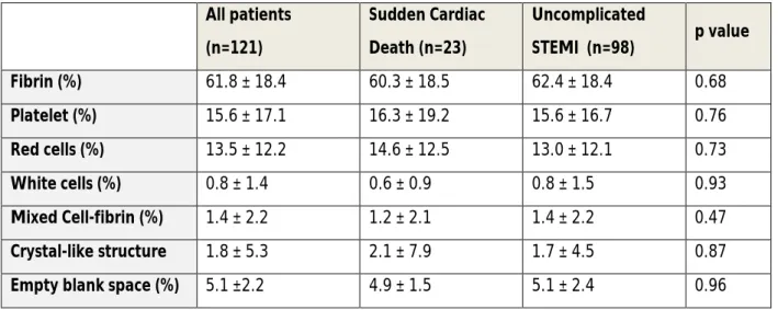
Fibrin fibers were the major component of the thrombus representing more than 60% of its
composition. Platelets, erythrocytes, cholesterol crystals and leukocytes all together represented the remaining 40% (Figure 2). Components of thrombus could be easily identified in our SEM high definition pictures (supplemental figure S1). Examples of the 1210 SEM pictures analysed in this study are shown in supplemental figure S2A with an analysis of a thrombus from the SCD group and in supplemental figure S2B with an analysis of a thrombus from the STEMI group. We did not find any difference in thrombus composition between the two groups of patients (Table 3).
Impact of GPI and ischemic time on thrombus composition
in fibrin content (respectively 61.0±21.6%, 63.6±16.4% and 61.3±18.7%, p =0.897) and platelet content (respectively 12.1±13.4%, 12.5±13.9% and 18.1±19.1%, p=0.197) of the thrombus between the GPI-free group, upstream-use and per-procedural-use of GPI groups, respectively. It is
noteworthy that ischemic time did not differ according to GPI use (median of 210min [IQR at 80-360], 185min [IQR at 125-245] and 175min [IQR at 132-356] for GPI-free, upstream-use and
per-procedural-use respectively, p=0.83).
The total ischemic time, defined as the time delay from symptom onset to thrombus retrieval, was the only factor impacting thrombus composition. Platelet content decreased from 9.2±19.1% to 7.9±5.1% among patients with an ischemic time of <3 hours and >6h, respectively (p=0.014 for trend) while fibrin content increased from 57.8±18.5% to 71.9±10.1% (p =0.0008 for trend) (Figure 3). Content in red cells and white cells was not affected by ischemic time.
Microparticle analysis
We presented in the supplemental table S1 the results of the sub-analysis of microparticle
measurement in the SCD and STEMI groups, in patients having both thrombus and microparticle data available. Data were consistent with our previous finding with higher concentration of MP in the SCD group, especially the platelet-derived microparticles (CD41+) in the systemic blood prePCI.
DISCUSSION
The TIDE thrombus study was designed to demonstrate whether the pattern of coronary occlusion (thrombus composition) could contribute to the onset of ventricular arrhythmia and SCD in
myocardial infarction and to characterize the independent correlates of thrombus composition. The main results of our study can be summarized as follows. First, intra-coronary thrombi retrieved during percutaneous myocardial revascularization are mainly made of fibrin. Second, there is no difference in thrombus composition between STEMI presenters complicated by SCD and STEMI without SCD. Third, the use of GPI had no impact on thrombus composition even with early prehospital treatment initiation. Fourth, ischemic time is the only independent predictor of the thrombus content.
Manual thrombectomy was a common adjunctive technique to PCI in STEMI patients, allowing better coronary stent implantation and a lower rate of stent thrombosis, until randomized trials demonstrated no mortality benefit and an increased risk of stroke. [14–16] This device offers a unique opportunity to study intracoronary thrombus in STEMI patients using various approaches, histopathology, immunohistochemistry, angioscopy or scanning electron microscopy. [11,17–19] The results of the TIDE study are in line with our previous findings on thrombus composition in
myocardial infarction and confirm the crucial role of fibrin fibers as the main component of the architecture of intra-coronary thrombus, whereas platelets and erythrocytes are second in line in
terms of composition.[11] We also found that there was a large variation in the proportion of each component of thrombus composition, with percentages of fiber or platelet content in a thrombus both varying from 0% to 80%, indicating a large variation in terms of thrombus composition in an individual patient. Some thrombi are mainly composed of platelets and some mainly of fibrin fibers.
TIDE also demonstrates that ischemic time is the main factor influencing thrombus
composition, with a decrease of platelet content and an increase in fibrin content over time. This is also in line with our previous work, although confirmed here in a much larger sample of patients.[11] These results are consistent with recent studies from other groups and with the generally accepted pathogenesis of clot formation where platelets trigger thrombus formation and are more present in “fresh” thrombi, while fibrin strengthens the overall clot structure and comes into play later on.[20– 22] The absence of difference in thrombus composition according to the use of GPI was unexpected and cannot be interpreted outside the results of previous randomized trials showing a benefit in early GPI administration in the prehospital setting.[23]
The hypothesis that STEMI patients with ventricular arrhythmia leading to SCD have a different pattern of coronary thrombus composition (more platelet-rich) as compared to STEMI patients without SCD was not confirmed by the present work. Indeed, thrombus architecture analyzed in both groups were similar despite the fact that SCD patients had reduced ischemic time and less frequent upstream use of GPI as compared to STEMI patients without cardiac arrest. In the TIDE inflammatory study published previously, intracoronary concentration of endothelial
microparticles (EMP) were significantly higher among SCD patients compared to STEMI patients supporting the hypothesis that occurrence of fast ventricular tachycardia or ventricular fibrillation in the setting of myocardial infarction is not explained only by a rhythmic vulnerability but could also involve a vascular vulnerability.[10] In the present work, we did not manage to find any specific pattern with SCD, as we found no significant difference between our two populations. Although we found differences in platelet microparticles (CD41+) level which were increased in the SCD group, and especially in the systemic blood samples measured before the coronary intervention, our results were most likely affected by the difference in the upstream use of GPI which was higher in the STEMI group.
We acknowledged several limitations. First, STEMI patients screened in this trial were selected and might not be representative of the entire population. Second, the analyses were performed on the main aspirated piece of thrombus and we may have missed the most informative pieces, which may have been damaged during the process of aspiration. This may account for the lack of effect of GPI on thrombus composition. However, the number of thrombi retrieved and the random localization of the pictures that were analyzed should guarantee an unbiased measurement and control for composition heterogeneity in the pieces of thrombi that were obtained. In addition, intra-coronary thrombi are stiff structures and much more stable than in vitro clots, and the
preserved appearance of cells and fibrin fibers in the microscopy images confirmed that distortion was minimal. [24] Third, although the fixation of thrombus sample in the glutaraldehyde solution gave the advantages to be conserved almost eternally for additional imaging analysis, such method does not allow additional immunological analysis and may limit the extent of the comprehension of this analysis. Finally, the aspirated thrombus may be older than expected from the duration of the ischemic time and younger thrombus could be superimposed with an older thrombus, thereby potentially confounding our observations.
CONCLUSION
The composition of intracoronary thrombi that is responsible for acute myocardial infarction does not differ according to the occurrence of SCD at presentation. Ischemic time remains the main factor impacting thrombus composition in patients with myocardial infarction.
Conflicts of interest
Johanne Silvain reports the following disclosures during the past 2 years : Research Grants to Institution from the Fondation de France and the Institute of Cardiolometabolism (ICAN); Consulting fees from Actelion, Amed, Astra-Zeneca , Bayer, Daiichi-Sankyo, Eli Lilly, Gilead Science and Sanofi-Aventis; Speaker honorariums from AstraZeneca, Amgen, Algorythm , Daiichi Sankyo, Eli Lilly, Iroko Cardio and Travels Support from Amgen , Astra-Zeneca and Saint-Jude Medical.
Jean-Philippe Collet reports the following disclosures: Research Grants to Institution or honorarium from AstraZeneca, Bayer, Bristol-Myers Squibb, Daiichi-Sankyo, Eli-Lilly, Fédération Française de Cardiologie, Lead-Up, Medtronic, MSD, Sanofi-Aventis, WebMD.
Paul Guedeneydoes not report any conflict of interest. Olivier Varenne does not report any conflict of interest.
Chandrasekaran Nagaswami does not report any conflict of interest. Carole Maupain does not report any conflict of interest.
Jean-Philippe Empana does not report any conflict of interest. Chantal Boulanger does not report any conflict of interest. Muriel Tafflet does not report any conflict of interest.
Stephane Manzo-Silberman does not report any conflict of interest. Delphine Brugier does not report any conflict of interest.
Nicolas Vignolles does not report any conflict of interest. Xavier Jouven does not report any conflict of interest.
John W. Weisel reports the following disclosures during the past 2 years: Research grant to Institution from Bayer Healthcare; expert witness for Jenner and Block LLP.
Gilles Montalescot reports the following disclosures : Research Grants to Institution or honorarium from ADIR, Amgen, AstraZeneca, Bayer, Berlin Chimie AG, Boehringer Ingelheim, Bristol-Myers Squibb, Beth Israel Deaconess Medical, Brigham Women’s Hospital, Cardiovascular Research
Foundation, Celladon, CME Resources, Daiichi-Sankyo, Eli-Lilly, Europa, Elsevier, Fédération Française de Cardiologie, Fondazione Anna Maria Sechi per il Cuore, Gilead, ICAN, Janssen, Lead-Up,
Menarini, Medtronic, MSD, Pfizer, Sanofi-Aventis, Servier, The Medicines Company, TIMI Study Group, WebMD.
Christian Spaulding reports the following disclosures in the past two years: research grants from the French Ministry of Health, consulting fees from Abiomed, Zoll, Medtronic, Medpass, speaker fees from Astra-Zeneca, Cordis, Servier, Lead-Up, Bayer, the Medicines Company, Eli Lilly, WebMD.
Funding:
ANR, ACTION study group www.actioncoeur.fr
References
[1] Priori SG, Blomström-Lundqvist C, Mazzanti A, et al. 2015 ESC Guidelines for the
management of patients with ventricular arrhythmias and the prevention of sudden
cardiac death: The Task Force for the Management of Patients with Ventricular
Arrhythmias and the Prevention of Sudden Cardiac Death of the European Society of
Cardiology (ESC). Endorsed by: Association for European Paediatric and Congenital
Cardiology (AEPC). Eur Heart J 2015;36:2793–867. doi:10.1093/eurheartj/ehv316.
[2] Spaulding CM, Joly LM, Rosenberg A, et al. Immediate coronary angiography in
survivors of out-of-hospital cardiac arrest. N Engl J Med 1997;336:1629–33.
doi:10.1056/NEJM199706053362302.
[3] Eisenberg MS, Mengert TJ. Cardiac resuscitation. N Engl J Med 2001;344:1304–13.
doi:10.1056/NEJM200104263441707.
[4] Siscovick DS, Weiss NS, Fletcher RH, Lasky T. The incidence of primary cardiac arrest
during vigorous exercise. N Engl J Med 1984;311:874–7.
doi:10.1056/NEJM198410043111402.
[5] Jouven X, Desnos M, Guerot C, Ducimetière P. Predicting sudden death in the
population: the Paris Prospective Study I. Circulation 1999;99:1978–83.
[6] Albert CM, Chae CU, Grodstein F, et al. Prospective study of sudden cardiac death
among women in the United States. Circulation 2003;107:2096–101.
[7] Albert CM, Oh K, Whang W, Manson JE, et al. Dietary alpha-linolenic acid intake and
risk of sudden cardiac death and coronary heart disease. Circulation 2005;112:3232–8.
doi:10.1161/CIRCULATIONAHA.105.572008.
[8] Jouven X, Empana J-P, Schwartz PJ, Desnos M, Courbon D, Ducimetière P. Heart-rate
profile during exercise as a predictor of sudden death. N Engl J Med 2005;352:1951–8.
doi:10.1056/NEJMoa043012.
[9] Dekker LRC, Bezzina CR, Henriques JPS, et al. Familial sudden death is an important risk
factor for primary ventricular fibrillation: a case-control study in acute myocardial
infarction patients. Circulation 2006;114:1140–5.
doi:10.1161/CIRCULATIONAHA.105.606145.
[10] Empana J-P, Boulanger CM, Tafflet M, et al. Microparticles and sudden cardiac death
due to coronary occlusion. The TIDE (Thrombus and Inflammation in sudden DEath)
study. Eur Heart J Acute Cardiovasc Care 2015;4:28–36.
doi:10.1177/2048872614538404.
[11] Silvain J, Collet J-P, Nagaswami C, et al. Composition of coronary thrombus in acute
myocardial infarction. J Am Coll Cardiol 2011;57:1359–67.
doi:10.1016/j.jacc.2010.09.077.
[12] Weisel JW, Nagaswami C. Computer modeling of fibrin polymerization kinetics
correlated with electron microscope and turbidity observations: clot structure and
assembly are kinetically controlled. Biophys J 1992;63:111–28.
doi:10.1016/S0006-3495(92)81594-1.
[13] Perkins GD, Jacobs IG, Nadkarni VM, et al. Cardiac arrest and cardiopulmonary
resuscitation outcome reports: update of the Utstein Resuscitation Registry Templates
for Out-of-Hospital Cardiac Arrest: a statement for healthcare professionals from a task
force of the International Liaison Committee on Resuscitation (American Heart
Association, European Resuscitation Council, Australian and New Zealand Council on
Resuscitation, Heart and Stroke Foundation of Canada, InterAmerican Heart
Foundation, Resuscitation Council of Southern Africa, Resuscitation Council of Asia);
and the American Heart Association Emergency Cardiovascular Care Committee and
the Council on Cardiopulmonary, Critical Care, Perioperative and Resuscitation.
Circulation 2015;132:1286–300. doi:10.1161/CIR.0000000000000144.
[14] Windecker S, Kolh P, Alfonso F, et al. 2014 ESC/EACTS Guidelines on myocardial
revascularization: The Task Force on Myocardial Revascularization of the European
Society of Cardiology (ESC) and the European Association for Cardio-Thoracic Surgery
(EACTS)Developed with the special contribution of the European Association of
Percutaneous Cardiovascular Interventions (EAPCI). Eur Heart J 2014;35:2541–619.
doi:10.1093/eurheartj/ehu278.
[15] Fröbert O, Lagerqvist B, Olivecrona GK, et al. Thrombus aspiration during ST-segment
elevation myocardial infarction. N Engl J Med 2013;369:1587–97.
doi:10.1056/NEJMoa1308789.
[16] Jolly SS, Cairns JA, Yusuf S, et al. Randomized trial of primary PCI with or without
routine manual thrombectomy. N Engl J Med 2015;372:1389–98.
doi:10.1056/NEJMoa1415098.
[17] Sato Y, Hatakeyama K, Yamashita A, Marutsuka K, Sumiyoshi A, Asada Y. Proportion of
fibrin and platelets differs in thrombi on ruptured and eroded coronary atherosclerotic
plaques in humans. Heart Br Card Soc 2005;91:526–30. doi:10.1136/hrt.2004.034058.
[18] Kramer MCA, van der Wal AC, Koch KT, et al. Presence of older thrombus is an
independent predictor of long-term mortality in patients with ST-elevation myocardial
infarction treated with thrombus aspiration during primary percutaneous coronary
intervention. Circulation 2008;118:1810–6. doi:10.1161/CIRCULATIONAHA.108.780734.
[19] Uchida Y, Uchida Y, Sakurai T, Kanai M, Shirai S, Morita T. Characterization of coronary
fibrin thrombus in patients with acute coronary syndrome using dye-staining
angioscopy. Arterioscler Thromb Vasc Biol 2011;31:1452–60.
doi:10.1161/ATVBAHA.110.221671.
[20] Iwata H, Sata M, Ando J, et al. Impact of primitive cells in intracoronary thrombi on
lesion prognosis: temporal analysis of cellular constituents of thrombotic material
obtained from patients with acute coronary syndrome. Heart Br Card Soc 2010;96:748–
55. doi:10.1136/hrt.2009.181040.
[21] Zalewski J, Bogaert J, Sadowski M, et al. Plasma fibrin clot phenotype independently
affects intracoronary thrombus ultrastructure in patients with acute myocardial
infarction. Thromb Haemost 2015;113:1258–69. doi:10.1160/TH14-09-0801.
[22] Furie B, Furie BC. Mechanisms of thrombus formation. N Engl J Med 2008;359:938–49.
doi:10.1056/NEJMra0801082.
[23] Van’t Hof AWJ, Ten Berg J, Heestermans T, et al. Prehospital initiation of tirofiban in
patients with ST-elevation myocardial infarction undergoing primary angioplasty
(On-TIME 2): a multicentre, double-blind, randomised controlled trial. Lancet Lond Engl
2008;372:537–46. doi:10.1016/S0140-6736(08)61235-0.
[24] Collet J-P, Shuman H, Ledger RE, Lee S, Weisel JW. The elasticity of an individual fibrin
fiber in a clot. Proc Natl Acad Sci U S A 2005;102:9133–7.
Figure 1: Flow chart of the study
STEMI: ST-segment elevated myocardial infarction; SCD: sudden cardiac death; PCI: percutaneous coronary intervention
Figure 2: Thrombus composition (percentages of each element) in the study population of n=121 Thrombus (Sudden cardiac death group on the right and STEMI group on the left). Red Lines indicate median and interquartile range (25% to 75%).
Figure 3: Relation between ischemic time (duration of thrombus formation) and thrombus
composition. P-values are given for comparison with the group < 3 hours as a reference with multiple student t-test. * p value <0.05. The Kruskal Wallis test for comparison of the three groups resulted in p value =0.048 for the fibrin fibers and p=0.047 for platelets.
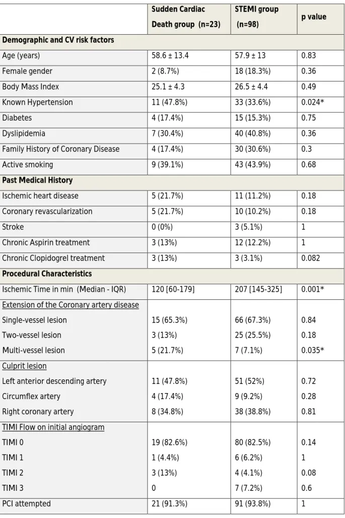
Table 1: Demographic and procedural characteristics description. * indicates a significant results with a p value <0.05. Values are mean ± SD, n (%) or median [interquartile range].
Sudden Cardiac Death group (n=23)
STEMI group
(n=98) p value Demographic and CV risk factors
Age (years) 58.6 ± 13.4 57.9 ± 13 0.83
Female gender 2 (8.7%) 18 (18.3%) 0.36
Body Mass Index 25.1 ± 4.3 26.5 ± 4.4 0.49
Known Hypertension 11 (47.8%) 33 (33.6%) 0.024*
Diabetes 4 (17.4%) 15 (15.3%) 0.75
Dyslipidemia 7 (30.4%) 40 (40.8%) 0.36
Family History of Coronary Disease 4 (17.4%) 30 (30.6%) 0.3
Active smoking 9 (39.1%) 43 (43.9%) 0.68
Past Medical History
Ischemic heart disease 5 (21.7%) 11 (11.2%) 0.18
Coronary revascularization 5 (21.7%) 10 (10.2%) 0.18
Stroke 0 (0%) 3 (5.1%) 1
Chronic Aspirin treatment 3 (13%) 12 (12.2%) 1
Chronic Clopidogrel treatment 3 (13%) 3 (3.1%) 0.082 Procedural Characteristics
Ischemic Time in min (Median - IQR) 120 [60-179] 207 [145-325] 0.001* Extension of the Coronary artery disease
Single-vessel lesion Two-vessel lesion Multi-vessel lesion 15 (65.3%) 3 (13%) 5 (21.7%) 66 (67.3%) 25 (25.5%) 7 (7.1%) 0.84 0.18 0.035* Culprit lesion
Left anterior descending artery Circumflex artery
Right coronary artery
11 (47.8%) 4 (17.4%) 8 (34.8%) 51 (52%) 9 (9.2%) 38 (38.8%) 0.72 0.28 0.81 TIMI Flow on initial angiogram
TIMI 0 TIMI 1 TIMI 2 TIMI 3 19 (82.6%) 1 (4.4%) 3 (13%) 0 80 (82.5%) 6 (6.2%) 4 (4.1%) 7 (7.2%) 0.14 1 0.08 0.6 PCI attempted 21 (91.3%) 91 (93.8%) 1
Successful PCI 21 (100%) 86 (92.8%) 0.58
Number of stent implanted 1.2 ± 0.7 1.3 ± 0.6 0.55
% of Drug eluted stent 2 (9.5%) 15 (16.8%) 0.52
Overall Stent Length (mm) 24.1 ± 14.1 25.5 ± 10.9 0.28
Maximal length stent (mm) 3 ± 0.43 3.8 ± 3.7 0.068
Use of GpIIbIIIa inhibitors 15 (65.2%) 89 (90.8%) 0.0015*
- Upstream use 2 (8.7%) 34 (34.7%) 0.02*
- Cath lab use 13 (56.5%) 55 (56.1%) 0.97
Post- PCI
Cardiogenic shock 15 (65.3%) 10 (10.2%) <0.001*
Intra-aortic balloon pump use 9 (47.4%) 1 (1%) <0.001*
Ventricular assistance 3 (15.8%) 2 (2%) 0.02*
Ejection Fraction (%) 40.7 ± 15.2 49 ± 11.7 0.042*
Peak of Troponin (µg/L) 60.7 ± 102.8 96.3 ± 100.8 0.05* Peak of Creatine Phosphokinase (mmol/L) 3826 ± 3588 2916 ± 2412 0.62
Table 2: Specificity of Sudden Cardiac Death Patients Values are n (%), median [interquartile range] or mean±SD.
Sudden Cardiac Death group (n=23)
Ventricular Tachycardia or Ventricular Fibrillation 18 (78.3 %)
Asystole 5 (21.7%)
No Flow (min) 0 [0-5]
Low Flow (min) 18.5 [10-25.7]
Table 3: Thrombus composition according to clinical presentation Values are mean ± SD. STEMI: ST-segment elevated myocardial infarction.
All patients (n=121) Sudden Cardiac Death (n=23) Uncomplicated STEMI (n=98) p value Fibrin (%) 61.8 ± 18.4 60.3 ± 18.5 62.4 ± 18.4 0.68 Platelet (%) 15.6 ± 17.1 16.3 ± 19.2 15.6 ± 16.7 0.76 Red cells (%) 13.5 ± 12.2 14.6 ± 12.5 13.0 ± 12.1 0.73 White cells (%) 0.8 ± 1.4 0.6 ± 0.9 0.8 ± 1.5 0.93 Mixed Cell-fibrin (%) 1.4 ± 2.2 1.2 ± 2.1 1.4 ± 2.2 0.47 Crystal-like structure 1.8 ± 5.3 2.1 ± 7.9 1.7 ± 4.5 0.87
![Table 2: Specificity of Sudden Cardiac Death Patients Values are n (%), median [interquartile range] or mean±SD](https://thumb-eu.123doks.com/thumbv2/123doknet/14663335.554852/20.892.197.759.128.344/table-specificity-sudden-cardiac-death-patients-values-interquartile.webp)