Dual functions of the Retinal Determination Gene Network member
EYES ABSENT as a transcription factor and protein phosphatase
by
Serena Jean Silver Brown
B.A. Biology
B.A. English and American Literature
New York University, 1999
SUBMITTED TO THE DEPARTMENT OF BIOLOGY IN PARTIAL FULFILLMENT OF THE REQUIREMENTS FOR THE DEGREE OF
DOCTOR OF PHILOSOPHY AT THE
MASSACHUSETTS INSTITUTE OF TECHNOLOGY
Ctef kreer 004-1
© 2004 Serena Jean Silver Brown. All rights reserved. The author hereby grants to MIT permission to reproduce and to distribute publicly paper and electronic copies of this thesis document in
whole or in part.
Signature of Author:
__
(J
Serena J. Silver BrownCertified by:
Ilaria Rebay Associate Professor of Biology
Thesis Supervisor
Accepted by:
Stephen Bell Professor of Biology
Chairman of the Graduate Committee
IMASSACHUSETTS INSRTrE OF TECHNOLOGY JUN Is 2004 *AiCHlVES LIBRARIES 1 _ ~ ~ ~ ~ ~ ~ ~ ~ ~ ~ ~ ~ ~ - I- - - -- - - - I- - - - -
-Dual functions of the Retinal Determination Gene Network member EYES ABSENT as a transcription factor and protein phosphatase
Serena J. Silver Brown
submitted to the Biology Department on June 10, 2004 in partial fulfillment of the
Requirements for the degree of Doctor of Philosophy in Biology
Proper development of cell types and tissues requires the integration of extracellular signals to provide context specific information that insures appropriate differentiation. The Drosophila eye is an excellent model for the study of this signal integration, as its development is orchestrated by the interactions of common signal transduction pathways in conjunction with organ specific gene expression. Signaling through these pathways sets the stage for appropriate deployment of the Retinal Determination (RD) gene network members to direct formation of the eye and other organs.
Our studies have focused on the RD gene network member EYES ABSENT as a point of signal integration necessary for the formation of the Drosophila eye. We have examined two functions for EYA, the first as a transcriptional co-activator, and the second, more novel function as a protein tyrosine phosphatase.
Previous work suggested that EYA functions as a transcriptional co-activator, particularly in a complex with the DNA binding domain containing RD network member SINE OCULIS (SO). In order to better understand RD network regulation, we performed a structure-function analysis of the EYA protein, which defined the P/S/T rich region of EYA as crucial for EYA transactivation potential. This region is also necessary for EYA mediated ectopic eye induction and rescue of the eya2mutant phenotype. We showed that RAS/MAPK signaling potentiates EYA transactivation, providing a mechanism for previously described in vivo activation of EYA by MAPK. We have also demonstrated roles for GROUCHO and DACHSHUND in negative and positive regulation of the EYA-SO transcription factor, respectively.
Recently we have begun to study a novel function of EYA suggested by the homology of the highly conserved EYA domain (ED) to the Haloacid dehalogenase (HAD) family. Using the substrate analog para-nitrophenyl phosphate (pNPP), we showed that recombinant ED possesses phosphatase activity, which is affected by tyrosine phosphatase inhibitors but not
serine/threonine phosphatase inhibitors. To determine whether this activity is important for EYA function in vivo, mutants that reduce or abrogate phosphatase activity, as shown by lower
specific activity or higher Km in pNPP assays, were tested for their ability to induce ectopic eyes or rescue the EYA mutant phenotype. These mutants, which we refer to collectively as EYA AD,
are unable to induce ectopic eyes or rescue the eya2 phenotype to the degree of wildtype EYA. As the EYAHADmutants are all within the ED, which is known to bind to SO, we tested whether these mutants are competent transcriptional coactivators with SO, and found that they retain this activity. Thus the phosphatase and transactivation functions of EYA may represent two distinct essential functions of EYA.
As EYA represents one of the first transcription factors found to possess phosphatase activity, and modulation of phosphorylation state represents a common mode of transcriptional regulation, it will be of particular interest to elucidate the role of EYA phosphatase function in vi'vo, studies which will require identification of transcriptional targets and phosphatase
substrates.
Thesis Advisor: Ilaria Rebay
Table of Contents
CHAPTER 1:
THE RETINAL DETERMINATION GENE NETWORK: A POINT OF SIGNAL INTEGRATION FOR
SPECIFICATION AND PROLIFERATION ... 9
ABSTRACT ... 10
A CONSERVED REGULATORY NETWORK FOR EYE DEVELOPMENT ... I I A Master Regulator? - EYELESS/PAX6 ... 11
EYEGONE, a PAX6-like gene in Drosophila ... 19
EYES ABSENT. a novelprotein with two conservedfunctions ... 21
SO/SIXfamily members, crucial mediators between EYA, DAC, & DNA ... 26
DA CH. A novel DNA binding protein ... 32
EYE DEVELOPMENT REQUIRES THE INTEGRATION OF SIGNALING PATHWAYS WITH RD GENE NETWORK CO M PO N EN TS ... 34
Specification of Eye and Head primordia in the Embryo ... 35
Differentiation of the EYE-ANTENNAL disc ... 36
DPP and HH regulate morphogeneticfurrow progression ... 38
EGFR signaling: Cell survival and differentiation ... 45
Antagonistic signals determine EYE versus CUTICLE ... ... 48
THE RD GENE NETWORK IS DEPLOYED IN THE DEVELOPMENT OF MULTIPLE ORGANS ... 5 1 RD genes can direct muscle specification ... 52
DAC is negatively regulated by EGFR signaling in the leg ... 53
Sex-specific regulation of RD gene network members ... 53
EYA plays a SO independent role in oogenesis ... ... ... ... 57
CONC LUDING REM ARKS ... 59
REFERENCES ... 60
A FUNCTIONAL DISSECTION OF EYES ABSENT REVEALS NEW MODES OF REGULATION WITHIN THE RETINAL DETERMINATION GENE NETWORK ... 77
INTRODUCTION ... 79
MATERIALS AN) METHODS ... ... 82
RESULTS ... ... ... 90
Eya finctions as a transactivator ... ... 90
A Proline, Serine, Threonine rich region is criticalbr Eya transactivation ... 91
RAS/MAPK signaling increases EYA transactivation potential ... 92
ARE-luciferase is responsive to the EYA-SO transcription factor ... 1... 101
The P/S/T rich region of Eya is necessary for transactivation but notJbr E YA-SO interactions .... 102
DAC does not interact with EYA or SO in S2-2H as.says ... ... ... 103
EYA-EYA interactions are mediated via the N-terminus ... 107
Phovsphorylation increases EYA transactivation potential in the context of the EYA-SO transcription fi cto r ... ... 10 8 GROUCHO is a repressor of the EYA-SO transcription fi ctor... ... 113
D ISCUSSION ... 114
REFERENCES ... 122
NOVEL FUNCTION FOR THE TRANSCRIPTION FACTOR EYES ABSENT AS A PROTEIN TYROSINE PHOSPHATASE ... 129
INTRODUCTION ... 131
RESULTS AND DISCUSSION ... 131
METHODS ... 150
SUPPLEMENTARY INFORMATION ... 155
THE LOZENGE MINIMAL EYE ENHANCER CAN BE USED TO EXAMINE THE FUNCTION OF THE
EYA-SO TRANSCRIPTION FACTOR ON A NATIVE PROMOTER ... 173
INTRODUCTION ... 175
RESULTS AND D ISCUSSION ... 175... 7
LMEE-luciferase can be used as readout of EYA -SO activity in S2 cell culture ... 175
DAC is a co-activator of the EYA-SO transcription factor, independent of EYA-mediated phosphatase activity ... 175
RAS-MAPK signaling activates EYA-SO mediated transcription ... 181
Chromatin IP experiments show SO directly hinds to the LMEE in vivo ... 187
MATERIALS AND METHODS ... 175
REFERENCES ... ... 175
DISCUSSION AND FUTURE DIRECTIONS ... 197
REFERENCES ... ... 204
CHARACTERIZATION OF A NEW CLASS OF DROSOPHILA SON-OF-SEVENLESS (SOS) ALLELES HIGHLIGHTS THE COMPLEXITIES OF SOS REGULATION AND FUNCTION IN HIGHER
EUKARYOTES
...
207
ABSTRACT ... ... 208
INTRODUCTION ... ... 209
RESULTS AND DISCUSSION ... 217
Isolation new Sos alleles... 217of Molecular Characterization of SoSEY2-3 alleles ... 221
Analysis of Sos mutant phenotypes in the eye and wing ... 229
A role for SOS in early embryonic patterning... 233
Overexpression of SOSM98 produces dominant negative phenotypes ... 237
CONCLUDING REMARKS ... 245
SUPPLEMENTARY FIGURE ... 246
MATERIALS AND METHODS ... 249
REFERENCES ... 252
GENERATION OF MOUSE AND GUINEA PIG ANTISERA FOR SINE OCULIS ... 259
REFERENCES ... 265
ANALYSIS OF PHOSPHATASE ACTIVITY OF HUMAN BOR AND FLY LOSS OF FUNCTION EYA DOMAIN MUTATIONS ... 267
RESULTS ... 268
REFERENCES ... 271
EYA PROTEIN PROTOCOLS ... 273
PURIFICATION OF EYA DOMAIN GST FUSION PROTEINS WITH FRENCH PRESS ... 274
PROTOCOL FOR EYA DOMAIN PHOSPHATASE ASSAYS ... 276
Assays with pN PP ... ... ... ... ... 276
Assays with Peptide substrate: I( p Y) GEF ... ... 277
Acknowledgements
I was first introduced to the powerful and beautiful genetic tool that is Drosophila by my undergraduate advisor at NYU, Chris Rushlow. When I joined her lab, Chris introduced me to
fly crosses, in situ hybridizations, and the world of developmental biology, all with her infectious love of science. She was an amazing first mentor, and I feel privileged to have had her
friendship and guidance.
I think I knew I would join Ilaria's lab almost as soon as she finished her talk during IAP, and after spending the past four years in her lab, I am still sure that I made the right choice. Ilaria's passion for science is contagious; when labwork seems dismal, a pep talk from her is sure to encourage students to return to the bench full of new fire. In Ilaria's lab I have learned
molecular biology, genetics, and even biochemistry, but most importantly I have learned to trust my instincts and to be unafraid of new challenges (or at least not to be paralyzed by my fear).
She is a wonderful graduate advisor and I am so happy to have been part of her lab. I would like to thank Matthew Voas and Andrina Williams Zink for all of their help and guidance when I first joined the lab. Everyone in our lab has made it a wonderful place to be a graduate student, but I would like to particularly thank Mousumi Mutsuddi, David Doroquez, and Jennifer Jemc for their supportive and insightful discussions over the past few years. I have also been lucky enough have the help of a very talented undergraduate for the past few summers, Laura Doyon. My perspective outside of the world of flies was greatly enriched by discussions with my classmates Maria Courel and Jessica Tytell. I would also like to thank my committee members, Paul Garrity Hazel Sive, and Terry Orr-Weaver, for their helpful advice on many occasions.
I would especially like to thank my classmate, labmate, and very good friend, Tina Tootle. We joined the lab together and worked on very separate projects for three years. Over that time we developed an amazing friendship and support system for each other, a relationship that was strengthened rather than tested by working together for the past year on the EYA phosphatase project. I know that I will always be able to count on her for sound scientific advice as well as a good laugh.
Of course, none of this would be possible without the love and encouragement of my family: first of all, my husband Adam, who has been by my side through the ups and downs of graduate school, keeping me smiling and sane. I would also like to thank my fantastic support network in New York; my sister Jaki, and my parents, Mark & Shelley Silver.
Chapter 1
The Retinal Determination Gene Network:
A Point of Signal Integration for
Specification and Proliferation
Abstract
Proper development of cell types and tissues requires the integration of extracellular signals to provide context specific information that ensures appropriate differentiation. The Drosophila eye is an excellent model for the study of this signal integration, as its development is orchestrated by the interactions of common signal transduction pathways in conjunction with organ specific gene expression. Signaling through the NOTCH, DECAPENTAPLEGIC/ TRANSFORMING GROWTH FACTOR-P, WINGLESS, HEDGEHOG, and EPIDERMAL GROWTH FACTOR RECEPTOR pathways sets the stage for appropriate deployment of the Retinal Determination (RD) gene network members to direct formation of the eye and other organs.
The RD gene network encodes a group of evolutionarily conserved transcription factors and co-factors that are crucial for the formation of many organs including the eye. These nuclear factors, which include proteins in the PAX6, EYA, SIX, and DACH families, are regulated by their interactions with each other and by effectors of the signaling pathways mentioned above. The mechanistic links between the RD gene network and signaling pathways are just beginning to be understood, particularly at the level of phosphorylation and regulation of transcriptional targets. One crucial role for the crosstalk between signaling pathways and this network of transcription factors is to coordinate the processes of cell proliferation and cell differentiation so that appropriate organ size and structure can be achieved.
A CONSERVED REGULATORY NETWORK FOR EYE DEVELOPMENT
Eye development in different organisms produces strikingly different structures: the primitive eye of planaria, the compound eye of insects, and the camera-like eye of vertebrates.
While these visual organs are morphologically distinct, the molecular mechanisms that lead to these different eyes are remarkably conserved. Specification of the eye field in these diverse organisms requires expression of homologous members of the Retinal Determination (RD) gene network, a group of transcription factors and co-factors crucial for the development of the eye as well as other organs. Much has been learned about these transcription factors, which include members of the PAX6, EYA, SIX, and DACH families (Figure 1), from studies in Drosophila as well as vertebrates.
A Master Regulator? - EYELESS/PAX6
The first RD gene network member to be identified molecularly was Drosophila eyeless
(ey), the homolog of vertebrate Pax6 (Quiring et al., 1994). The PAX6 family is a subgroup of
the large family of PAX proteins, each containing two DNA binding motifs; a PAIRED box I)NA binding domain, and a HOMEOBOX type DNA binding motif (Figure 1; Quiring et al.,
1 994).
ey was first named due to eye specific alleles which result in loss of eye tissue (Quiring et
al., 1994). Subsequent cloning of this gene and isolation of null alleles, which are homozygous lethal, revealed a broader role for EY in the embryo and brain (Kammermeier et al., 2001). This pattern of discovery, where isolation of eye specific alleles is followed by recognition of broader roles in development, would be repeated for many of the RD gene network members.
Heterozygosity at the Pax6 locus is associated with aniridia and Peters' anomaly in humans and the small eye phenotype in mouse (Hanson et al., 1994; Quiring et al., 1994), indicating conservation of PAX6 family roles in eye and head development in addition to the striking sequence conservation. Consistent with studies in Drosophila, homozygous mutant vertebrates, including humans, do not survive due to broader requirements for PAX6 during
development (Hanson, 2001).
In addition to ey, the Drosophila melanogaster genome contains a second closely linked PAX6 homolog, twin-of-eyeless (toy), thought to be the result of a duplication during insect evolution, as two PAX6 homologs are also found in the closely related Drosophila virilis and the more distantly related silkmoth Bombyx mori, but not in the grasshopper Schistocerca nitans nor a primitive insect, the springtail Folsomia candida (Czerny et al., 1999). TOY and EY are independently required for proper eye development (Kronhamn et al., 2002; Quiring et al., 1994) and display distinct as well as overlapping embryonic and larval expression patterns
(Kammermeier et al., 2001; Kronhamn et al., 2002). Therefore these homologous genes are not merely redundant, and insects have taken advantage of the presence of two PAX6 genes by adapting them for different uses.
Both TOY and EY are highly homologous to human PAX6, with over 90% identity in the PAIRED domain and 90% identity in the HOMEOBOX domain. TOY is more similar to PAX6 in length and in sequence outside of the two DNA binding domains, and also shows more similar DNA binding specificity (Czerny et al., 1999). This distinction may play a role in the binding specificities of TOY and EY in vivo, as the PAIRED domain of TOY binds to five sites in the eye specific enhancer of their target gene sine oculis, while the PAIRED domain of EY binds
544
Homeobox
Bipartite DNA binding motif
KI
DNA binding motif
409
Homeobox
Bipartite DNA binding motif
1
DNA binding motif
349
EYG
DNA binding motif
1
Homeobox
DNA binding motif
223
Necessary for transactivation potential Stars indicate MAPK phosphorylation sites
1 224
Interacts with SO and DAC Active phosphatase domain
416
Homeobox
Interactions with EYA and with GRO
DNA binding motif
DAC
Ski/Sno like motif Interaction with EYA Structural resemblance to the winged Interaction with Sin3A
helix/ forkhead DNA binding motif Interactions with HDAC3
Interaction with N-CoR Interaction with SIX6
Figure 1: Domain Structures of the RD gene network members
TOY
[
1EY
839 67'1 ** 486EYA
SO
760 1074 1 266 i .In the eye, TOY and EY are expressed in a regulatory hierarchy, whereby TOY acts upstream to activate ey through binding to PAX6 consensus motifs in the ey intron (Czerny et al.,
1999; Hauck et al., 1999), and both are required for proper development of the head and eyes (Kronhamn et al., 2002). Perhaps one of the most striking aspects of PAX6 family members is their ability to act as "master regulators" of eye formation, by directing the formation of ectopic eyes upon overexpression (Halder et al., 1998). Expression of either TOY or EY is sufficient to induce ectopic eyes (Czerny et al., 1999; Halder et al., 1995), and even human PAX6 can induce ectopic eyes when expressed in flies (Halder et al., 1995).
Although PAX6 proteins contain two DNA binding motifs, studies of Drosophila EY suggest they may be used independently. For example, analyses of truncated EY proteins reveal a requirement for the PAIRED domain but not the HOMEOBOX to rescue the eye specific ey2
mutant phenotype and also to induce ectopic eyes (Punzo et al., 2001). The PAIRED domain may also be involved in cooperative interactions with other transcription factors, as is observed for the PAIRED domain of a related protein, PAX5, in its interactions with the transcription factor ETS-1 (Garvie et al., 2001). In contrast, the HOMEOBOX is required to downregulate genes important for the development of appendages, such as distalless, suggesting a role for this I)NA binding domain in repression (Punzo et al., 2001). It is interesting to note that these two DNA binding domains appear to mediate opposite effects, activation versus repression, and function in the development of discrete organs. It is likely that co-factor interactions, such as those observed between PAX5 and ETS-1, may favor the use of one or the other DNA binding domain, co-recruiting it to target sequences and giving PAX6-DNA complexes context
specificity.
TOY and EY function at the top of a transcriptional hierarchy, where they are required for the expression of downstream members of the RD gene network (Figure 2; Halder et al., 1998). Like TOY and EY, members of the RD gene network have largely been characterized as transcription factors, and include the PAX6-like gene eyegone (eyg), which is thought to act in parallel to ey, and downstream components eyes absent (eya), sine oculis (so), and dachschund
(dac). EYA, SO, and DAC are all the founding members of families of proteins important for organ development in both invertebrates and vertebrates, and are homologous to members of the EYA, SIX, and DACH families, respectively. SO contains a HOMEOBOX DNA binding domain (Cheyette et al., 1994; Serikaku and O'Tousa, 1994), while EYA and DAC are novel nuclear proteins (Bonini et al., 1993; Mardon et al., 1994). However, the transcriptional hierarchy observed in the eye is not linear, and EYA, SO, and DAC contribute to positive feedback loops that ensure continued expression of EY (Figure 2; Halder et al., 1998). Another SIX family member, optix, also appears to act in parallel to TOY and EY to direct eye growth (Seimiya et al, 2000).
Despite study of the function of the PAX6 family in many organisms, few direct targets of these transcription factors have been identified. Recently, microarray analysis of ectopic eyes induced by eyeless was performed in order to understand the developmental program initiated upon eye formation, identifying 371 genes induced by EY (Michaut et al., 2003). Of these, many are already known to be involved in eye formation, others have been previously
characterized for roles in other developmental processes, while over half remain uncharacterized, including 100 novel genes (Michaut et al., 2003). It still must be determined which of these
TOY/PAX6
¶1'
EYG/PAX6(5A
SO/SIX'
'YA/EYA1-4
I-OPTIX/SIX3/6
EYE GROWTH AND SPECIFICATION
Figure 2 - Retinal Determination Gene Network
The RD gene network is expressed in a transcriptional hierarch whereby TOY leads to EY expression leading to SO, EYA, and DAC expression. Lower tier members of the network such as EYA and DAC operate in reiterative feedback loops to turn on EY. Other RD members EYG and OPTIX are required independently for proper eye development.
genes are direct targets of EY and which play roles further downstream in eye formation.
Performing similar microarray analysis of tissue expressing the closely related TOY might reveal overlapping and distinct targets for these two PAX6 proteins.
It is of particular note that greater than one-fourth of the total enriched gene class encode novel proteins, especially in light of two novel but now well studied members of the RD gene network, EYA and DAC. EYA and DAC when cloned could not be categorized immediately as transcription factors, enzymes, or DNA binding proteins based on amino acid sequence, but
indeed each perform some of these functions (Ikeda et al., 2002; Ohto et al., 1999; Silver et al., 2003; Tootle et al., 2003). It will require more detailed study to the identify functions of these
100 novel proteins, through analysis of localization, mutant phenotype, structure, and
biochemistry. The study of the novel genes identified by this microarray screen may reveal new paradigms for functional motifs as well as the mechanistic roles of individual genes in organ development.
EYEGONE, a PAX6-like gene in Drosophila
In addition to the PAX6 orthologs TOY and EY, there are two Pax6-like genes in
Drosphila, eyegone (eyg), and its related gene, twin-of-eyegone (toe). Like most RD gene
network members, loss of function alleles of eyg result in loss of eye tissue, while
overexpression of EYG promotes ectopic eye formation (Jang et al., 2003). eyg and toe encode Pax proteins which contain a truncated PAIRED domain and a complete HOMEOBOX (Figure
1; Jang et al., 2003; Jun et al., 1998). The PAIRED DNA binding domain contains two separable motifs, PAI and RED, which can each bind DNA independently (Jun and Desplan, 1996); eyg and toe contain only the RED motif (Jun et al., 1998). eyg expression does not
require the presence of ey, nor does ey expression require eyg function (Jang et al., 2003). Furthermore, ectopic expression of neither ey nor eyg can induce the other, indicating that they may act in parallel during eye formation (Jang et al., 2003).
While there are no homologs of eyg in mammals, it has been suggested that the functional ortholog of this gene is the 5a splice isoform of Pax6 (Pax6(5a)) (Dominguez et al., 2004; Jun et al., 1998), which also contains only the RED motif portion of the PD. In support of this theory, the RED domain of EYG and the RED domain of PAX6(5A) can bind to similar sequences (Jun et al., 1998), and overexpression of PAX6(5A) in Drosophila produces the same overgrowth effects as overexpression of EYG (Dominguez et al., 2004). Therefore the functions of EYG are likely to represent conserved processes in vertebrates.
In Drosophila, EY and EYG can function synergistically to induce larger ectopic eyes, for which a mechanism has recently been suggested. Specifically, EY and EYG may play discrete roles in eye development, where EY is important for eye specification while EYG is crucial for eye growth (Dominguez et al., 2004). It is crucial to the development of a functional organ that these two processes be coordinated during tissue formation, and EY and EYG may be important in this coordination through their parallel roles in differentiation and growth.
In the animal, the discrete roles of EY and EYG can be observed by analysis of marker gene expression in developing eye tissue mutant for either ey or eyg. ey mutant tissue, which can be examined in eye specific ey alleles, develops into the larval eye primordia, the eye disc, but lacks expression of retinal determination markers such as EYA and SO (Halder et al., 1998). Due to the absence of differentiation, this tissue then undergoes programmed cell death, leading to the "eyeless" phenotype (Halder et al., 1998).
In contrast, growth defects in eyg mutant tissue can be observed using the FLP-FRT system (Xu and Rubin, 1993) to induce specific, marked clonal patches of homozygous mutant tissue wherever FLP is expressed. When this is done in the eye disc, eyg mutant clones display clear growth disadvantages compared to wildtype tissue, but express the normal complement of retinal determination markers such as ELAV and EYA (Dominguez et al., 2004). Thus the "eyegone" phenotype is due to growth defects in the developing eye.
Thus two distinct proteins, encoded by different genes in Drosophila but a single gene in vertebrates, separate the control of two essential processes for the development of an organ; growth and differentiation. These processes must be coordinated precisely to generate
appropriate adult structures, coordination that relies upon members of the RD gene network to communicate between multiple signaling pathways.
EYES ABSENT: a novel protein with two conserved functions
eyes absent (eya) was first identified in Drosophila as a gene important for cell survival
and differentiation in the Drosophila eye (Bonini et al., 1993). Further studies of Drosophila EYA and of vertebrate homologs EYA 1-4 have revealed a wider role for EYA in organogenesis (Bonini et al., 1998; Xu et al., 1999). In Drosophila, strong alleles of eya are lethal and affect cell proliferation and apoptosis, most clearly resulting in defects in head morphology, but also defects in gonad formation and body wall musculature (Bonini et al., 1998; Boyle et al., 1997). Weaker alleles survive to adulthood but display defects in eye and ocelli as well as male and female sterility (Bonini et al., 1998; Leiserson et al., 1998).
In humans, heterozygosity for mutations in the Eyal gene are associated with Branchio-oto-renal (BOR) syndrome, as well as Branchio-oto (BO) syndrome and Ocular Defects (OD)
(Abdelhak et al., 1997; Ozaki et al., 2002), while homozygotes display more severe defects and do not survive. Eyal heterozygous mice display similar defects in ears and kidneys, as well as more pleiotropic defects in organogenesis in homozogyotes (Xu et al., 1999; Xu et al., 2002). The four vertebrate EYA homologs have both discrete and overlapping expression patterns, suggesting that their functions may not be wholly redundant (Xu et al., 1997a; Zimmerman et al.,
1997). While knockout mice of Eya2, Eya3, and Eya4 have yet to be reported, in humans mutations in the Eya4 locus are associated with familial hearing impairment (Pfister et al., 2002; Wayne et al., 2001).
The EYA family is characterized by a highly conserved -250 amino acid C-terminal domain called the EYA DOMAIN (ED), while the N-terminus of different EYA orthologs shows little conservation aside from the tyrosine rich EYA DOMAIN2 (ED2; Figure 1; Xu et al.,
1997b; Zimmerman et al., 1997). The amount of EYA must be carefully titrated to maintain viability, as broad overexpression in Drosophila is lethal (Hsiao et al., 2001), while in cell culture overexpression of EYA triggers the programmed cell death pathway (Clark et al., 2002).
The ED was initially characterized as a protein-protein interaction domain, a point of contact between EYA and other RD gene members SO (Pignoni et al., 1997), and DAC (Chen et al., 1997), an observation that has been extended to interactions between vertebrate EYA and SIX and DACH families (Ohto et al., 1999; Heanue et al., 1999).
EYA has been best characterized as a transcriptional co-activator which is recruited to the DNA of target genes via interaction with the SO/SIX family. SIX proteins bind DNA through their HOMEOBOX domain, recruiting the transactivation potential of EYA to the promoter of target genes (Ohto et al., 1999). This transcriptional co-activator function of EYA requires the
Proline/Serine/ Threonine (PST) rich region of EYA's N-terminus including the ED2, which is also required for EYA function in vivo (Figure 1; Silver et al., 2003).
Recently a second function has been described for EYA, through the identification of the ED as a catalytic motif belonging to the Haloacid dehalogenase family of enzymes (Tootle et al., 2003). Recombinant EYA has been shown to dephosphorylate tyrosyl phosphorylated peptides (Rayapureddi et al., 2003; Tootle et al., 2003) and serine/threonine phosphorylated peptides (Li et al., 2003) suggesting it may be a dual-specificity protein phosphatase. Thus far there are only two suggested substrates, both of which can be dephosphorylated by EYA in vitro: RNA polymerase II (Li et al., 2003), and EYA itself (Tootle et al., 2003). The phosphatase function of EYA is required for rescue of the eye specific eya2 allele (Tootle et al., 2003), indicating that this role is utilized in vivo during eye development.
EYA is the first identified transcription factor to posses intrinsic phosphatase function. As phosphorylation plays a key role in modulation of transcription factor activity, and EYA is a nuclear protein associated with DNA, it is likely that the targets of the EYA phosphatase may play important roles in transcriptional regulation. The identification of in vivo substrates of the
EYA phosphatase, and targets of EYA transcriptional regulation, will help determine how the phosphatase and transcription factor functions of EYA are coordinated to establish the
appropriate developmental program.
More insight as to EYA function may come from identification of co-factors other than SO and DAC. Heterotrimeric G proteins are important components of cellular communication, responding to extracellular cues through activation of the Gc subunit. They are known to
regulate the function of numerous transcription factors indirectly through second messengers, but in the case of EYA may directly affect transcription factor function by competition for
factors. Activated Goc and Goq2 proteins have been shown to interact directly with the ED using Yeast and Mammalian-two hybrid analysis as well as glutathione S-transferase (GST)
fusion protein pull-down assays, and may compete with SIX family members for EYA, thus preventing target gene activation (Fan et al., 2000). This represents a novel mode of
transcriptional regulation through Goc subunits, and it is intriguing to ask if this interaction might serve to direct EYA's phosphatase activity towards particular substrates, allowing EYA to regulate other transcription factors or perhaps a different aspect of the cellular machinery.
The ED also contains a potential interaction motif for the GROUCHO (GRO) family of corepressors, although GRO-EYA interactions are not observed by co-immunoprecipitation in
Drosophila cell culture (Silver et al., 2003). Recent genome wide yeast-two-hybrid screens of
Drosophila and C. elegans proteins have identified other potential EYA binding partners, 8 in Drosophila and 51 in C. elegans, whose relevance remains to be determined through more
detailed experimental analysis (Giot et al., 2003; Li et al., 2004). Unfortunately, other than the isolation of SO and its C. elegans homologs ceh-33 and ceh-35, there is no overlap between the two screens, and more analysis will be needed to identify true binding partners.
Although it is best known for its role in eye development, EYA is found in organisms which do not have eyes, including as mentioned above the nematode C. elegans and perhaps more strikingly, plants such as Arabidopsis thaliana. Despite the absence of eyes, C. elegans have many sensory neurons which interact with the environment to sense modalities such as temperature and odor (for review see Mori and Ohshima, 1997), and homologs of RD gene network members EY, SO, and DAC are expressed in the worm head in discrete and overlapping domains (Chisholm and Horvitz, 1995; Dozier et al., 2001; Duncan et al., 1997; Zhang and
Emmons, 1995; A. Brown, personal communication), while the EYA expression pattern has yet to be reported. The presence of the RD gene network in these animals underscores their use in early development of sensory organs, and ultimately the adaptation of this network to organize multiple tissue and organ types in vertebrates (Wawersik and Maas, 2000).
EYA is thus far the only member of the RD gene network to be found in plants, in both monocots and dicots (Takeda et al., 1999). EYA homologs in Oryza sativa (rice) and
,4rabidopsis consist solely of an EYA DOMAIN preceded by a short 18 amino acid peptide, and
Oryza EYA is highly expressed during embryogenesis (Takeda et al., 1999). As the Arabidopsis
genome is now complete and annotated, and searches do not reveal homologs of EY, SO, or DAC, analysis of EYA function in plants may reveal independent and evolutionarily older functions for this family of proteins.
Absence of transcriptional cofactors in addition to the absence of the N-terminal transactivation domain suggests that EYA may not function as a transcription factor in plants. However plant EYA contains all conserved residues necessary for HAD family phosphatase activity, and indeed is the most active EYA of all orthologs tested (Rayapureddi et al., 2003). Thus plant EYA may represent EYA's original role as a protein phosphatase which was later in evolution brought into transcriptional regulation through interactions with "new" proteins such as SO and DAC. This would then bring EYA's phosphatase activity to promoter complexes, where it might act in the regulation of transcription factors through control of phosphorylation state, a common mechanism of transcriptional regulation. It will be interesting to ask whether plant EYA is localized to the nucleus, as constructs of the Drosophila EYA domain alone are both nuclear and cytoplasmic (T. Tootle, personal communication), and whether plant and animal EYA phosphatases will display similar substrate preferences.
SO/SIX family members; crucial mediators between EYA, DAC, & DNA
The SIX family can be grouped into three subgroups, one member each in Drosophila and two members each in vertebrates thought to result from the duplication of a "SIX cluster" (Kawakami et al., 2000; Seo et al., 1999). All family members are marked by the presence of two conserved domains, the SIX domain, which mediates protein-protein interactions, and a HOMEOBOX DNA binding domain (Figure 1; Pignoni et al., 1997). sine oculis (so), the founding member of this family, is crucial for proper proliferation and patterning during eye development (Pignoni et al., 1997), and also plays roles in brain (Daniel et al., 1999) and gonad development (Fabrizio et al., 2003). Strikingly, SO is the only RD gene network member that cannot induce ectopic eyes when expressed alone, although it can synergize with EYA to increase the frequency of ectopic eyes (Pignoni et al., 1997).
so falls into a SIX family subgroup with vertebrate SIXI and SIX2. SIX1 mutant mice
display defects in ear, kidney, thymus, skeletal muscle and nose (Ozaki et al., 2004; Xu et al., 2003; Zheng et al., 2003). Recently SIXI was shown to be upregulated in a mouse model of metastatic skeletal muscle cancer (Yu et al., 2004), and increased levels of SIX1 were associated with greater ability to form metastases (Yu et al., 2004), perhaps by overcoming mitotic
checkpoints in G2 (Ford et al., 1998).
Another subgroup of the SIX family contains Drosophila d-SIX4, which plays an
important role in muscle and gonad formation (Kirby et al., 2001), and vertebrate homologs Six4 and Six5. Six4 mutant mice have no apparent defects (Ozaki et al., 2001), despite its strong expression throughout the nervous system and in many sensory organs, and Six5 knockout mice
2000). The effect of simultaneous knockout of these closely related genes with overlapping expression patterns remains to be examined.
In humans, the Six5 locus is associated with myotonic dystrophy (DM 1), and patients with that disease show lower levels of Six5 expression (Wansink and Wieringa, 2003). Although the loss of Six5 in mice does not mimic the entire phenotype of patients with DM1, it is thought that loss of Six5 may contribute to the cataracts observed in these patients (Wansink and
Wieringa, 2003), while downregulation of the associated gene DM protein kinase (DMPK) is responsible for the myopathy phenotype (Sarkar et al., 2000).
As described earlier, the members of the SIX subfamilies SIX1/2 and SIX4/5 are thought to generally function in a complex with EYA proteins as bipartite transcription factors. The SIX Domain interacts with the EYA domain, forming a transcription factor with the SIX
HOMEOBOX as the DNA binding domain and the N-terminus of EYA as the transactivation domain that together direct transcription of target genes (Kawakami et al., 2000; Silver et al., 2003). This also brings the phosphatase activity of EYA to the promoter region, where it may play a role in regulation of itself or other factors. In another example of independent functions of members of the RD gene network, vertebrate SIX1/2 and SIX4/5 genes have some activation ability independent of EYA (Kawakami et al., 2000).
The most divergent branch of the SIX family includes Drosophila optix and the vertebrate genes Six3 and Six6 (Kawakami et al., 2000). All members of this subfamily show strong expression in the developing eye (Kawakami et al., 1996c; Seimiya and Gehring, 2000), and are necessary for proper eye formation (Carl et al., 2002; Li et al., 2002; Seimiya and G(ehring, 2000). Similar to its vertebrate counterparts, which when overexpressed induce enlarged forebrains (Kobayashi et al., 1998), overexpression of optix leads to ectopic eyes
(Seimiya and Gehring, 2000). OPTIX, like EYG, does not require EY for the induction of ectopic eyes, and may act in a parallel pathway to direct eye formation (Figure 2; Seimiya and Gehring, 2000). However the lack of mutants that affect optix makes it difficult to place this protein more definitively within the RD gene network hierarchy.
Mutations in the human Six3 gene are associated with holoprosencephaly (Pasquier et al., 2000; Wallis et al., 1999), stemming from defects in neural plate formation. Loss of the other human member of that subfamily, Six6, is associated with bilateral anophthalmia, a condition where babies are born with no eyes, as well as pituitary defects (Gallardo et al., 1999). Similar to the human phenotype, mice homozygous for knockout of Six6 survive but have retinal and pituitary hypoplasia (Li et al., 2002). Overexpression of SIX3/6 causes overproliferation, and mutations in these genes cause phenotypes associated with proliferation defects, suggesting that they may play a direct role in cell cycle regulation. Recent work suggests that the SIX3/6 subfamily performs this role through transcriptional regulation and protein-protein interactions.
Unlike the other SIX family members, which are largely thought to coordinate
transcriptional activation through interactions with EYA, the SIX3/6 subfamily do not interact with EYA family members (Kawakami et al., 2000). Instead, they are proposed to act as transcriptional repressors, one mechanism for which may be their interactions with the GROUCHO (GRO) family of corepressors, called GRG in vertebrates, through an engrailed homology 1 (ehl) motif in the SIX domain (Kobayashi et al., 2001; Lopez-Rios et al., 2003; Zhu et al., 2002).
The interaction between SIX3/6 and GRG family members has been demonstrated to be crucial for proper eye and brain formation in zebrafish and medaka (Kobayashi et al., 2001;
homolog TLE can act synergistically with SIX3 and SIX6 to expand the eye field, while a dominant negative form has the opposite effect (Lopez-Rios et al., 2003).
At least one direct target of SIX6 mediated repression is the cyclin-dependent kinase inhibitor p27Kip l, which must be repressed to allow early proliferation of the presumptive eye (Li et al., 2002), providing a direct link between SIX and cell cycle regulation. In this way, SIX family members may serve to coordinate growth with other targets important for appropriate differentiation of the organ.
A transcription independent role of SIX3 in control of cell proliferation has recently been identified in vertebrate eye development. SIX3 and SIX6 were shown to interact with the DNA replication inhibitor GEMININ (Del Bene et al., 2004). GEMININ inhibits cell proliferation through sequestration of CDTI, an important component of the replication machinery, such that
SIX3 can compete with CDTI for GEMININ, thus releasing CDT1 and with it cell cycle
inhibition (Del Bene et al., 2004). This combined with the more direct transcriptional regulation of cell cycle components, illustrates how cell proliferation and cellular differentiation may be knitted together by deployment of common proteins to interact with distinct elements that insure the appropriate development of a complex organ.
Transcriptional repression may not be limited to the SIX3/6 subfamily, as the GRG interaction motif is found in all SIX proteins within the SIX domain (Kobayashi et al., 2001), indicating interactions with this corepressor family may be a common feature of the entire SIX family. Recently we showed that Drosophila GRO can interact with SO, and can repress SO mediated transcription of a reporter gene, likely by competing with EYA for SO binding (Silver
et al., 2003). As studies in mice have shown that SIXI can also have transcriptional repressor function (Li et al., 2003) it is likely that the members of the SIX family may play more elastic
roles in transcriptional regulation dependent upon their specific cofactor or context. As EYA and SO expression patterns are not wholly coincident, this dual function may provide a clear distinction between cells which express genes necessary for differentiation into eye tissue,
expressing EYA and SO, and cells where those genes are actively repressed, expressing only SO. This model may be better understood once more targets of the SO/SIX family are identified.
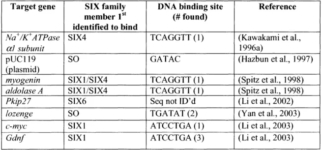
Thus far only a handful of direct transcriptional targets have been identified for the SIX family (Table 3), most of them in mice. The SIX 1I/2 and SIX4/5 subfamilies have the most similar homeobox domains and are likely to bind the same or similar target sequences (Kawakami et al., 2000). The promoters of the myogenin and aldolase A genes contain SIX binding sites known as MEF3 (TCAGGTT), which is necessary for the embryonic expression of
myogenin (Spitz et al., 1998). Another known target from mice is the housekeeping gene
Na+/K+ATPase alpha I subunit which contains a SIX family target sequence in the enhancer element AREC3 (core sequence GGNGNCNGGTTGC, includes TCAGGTT in bold;
Kawakami et al., 1996b).
SIX and SIX4 have been shown to activate transcription via binding to the MEF3 site (Spitz et al., 1998), while SO, SIX2, SIX4 and SIX5 can all bind the AREC3 site and with EYA activate transcription of a reporter gene (Kawakami et al., 1996c; Silver et al., 2003). More recently, microarray analysis of mouse cells expressing dominant active or dominant negative SIX1 suggested c-Myc and Gdnf as direct targets. This result was confirmed by chromatin IP experiments, which showed SIX and EYA localization to the c-Myc and Gdnfpromoters (Li et al., 2003). The single target of this family identified thus far in Drosophila is the lozenge gene, whose expression is activated by SO binding to an imprecise repeat in the lozenge minimal eye
Table 3 - SIX family target genes have been identified in vertebrates and Drosophila
31
Target gene SIX family DNA binding site Reference
member Ist (# found)
identified to bind
Na+/K+ATPase SIX4 TCAGGTT(1) (Kawakami et al.,
acl subunit 1996a)
pUC119 SO GATAC (Hazbun et al., 1997)
(plasmid)
myogenin SIX1/SIX4 TCAGGTT (1) (Spitz et al., 1998)
aldolase A SIX1/SIX4 TCAGGTT(1) (Spitz et al., 1998)
Pkip27 SIX6 Seq not ID'd (Li et al., 2002)
lozenge SO TGATAT (2) (Yan et al., 2003)
c-myc SIXI ATCCTGA (1) (Li et al., 2003)
DACH: A novel DNA binding protein
Dacschund (dac) in Drosophila, and its vertebrate homologs, Dachl and Dach2, encode a family of novel nuclear proteins characterized by two conserved domains, the DachBox-N and the DachBox-C (Figure 1; Davis et al., 2001b; Kozmik et al., 1999). Analysis of the amino acid
sequence of DACH family members revealed some similarity to the ski proto-oncogene (Hammond et al., 1998). Recent crystallization of the human DachBox-N revealed a striking
structural resemblance to the winged helix/forkhead subgroup of the helix-turn-helix family of DNA binding proteins (Kim et al., 2002). While no specific DNA binding sites for DACH have been identified, it has been shown to bind naked DNA (Ikeda et al., 2002). The DachBox-C is thought to be a protein-protein interaction motif, and has been demonstrated to interact with EYA family members via the EYA DOMAIN (Chen et al., 1997). Thus DAC with its DNA binding ability may like SO be a co-factor responsible for bringing the transcriptional activator and phosphatase activity of EYA to the promoter of target genes.
dac homozygous mutants lack eye tissue, and have striking leg and wing defects; those
that survive to adulthood die within a few days (Mardon et al., 1994). In mice lacking Dachl, which like Drosophila dac is expressed in the eye and limbs (Kozmik et al., 1999), there are no gross defects in development but mice die soon after birth (Backman et al., 2003; Davis et al., 2001 a). As the expression patterns of Dachl and Dach2 overlap greatly during development, this relatively mild phenotype may be due to partial redundancy of these genes, and/or may reflect a greater postnatal need for Dach I.
Like other members of the RD gene network, ectopic expression of DAC leads to induction of EY expression and the formation of ectopic eyes (Chen et al., 1997; Shen and
eyes when the two are expressed together (Chen et al., 1997), support for the model that these two proteins act in a complex to direct eye development.
However, there are some discrepancies in the literature regarding the ability of DAC and EYA to interact directly. For example, Heanue et al. has shown that GST-DACH2 can pull down radiolabeled EYA2 (Heanue et al., 1999), while Ikeda et al. found no interaction between GST-DACHI and EYA1, EYA2, or EYA4 in a similar assay. However, Ikeda and colleagues report that a DACH/EYA complex is formed in the presence of Creb binding protein (CBP) using co-immunoprecipitation assays and that the DACH/EYA complex can activate transcription of a synthetic promoter (Ikeda et al., 2002). These results may reflect differences between the 1DACH1 and DACH2, or between cofactors available in different cell types.
Directed yeast-two-hybrid analysis has twice suggested interactions between the DachBox-C and the EYA domain (Bui et al., 2000; Chen et al., 1997), but genome wide yeast-two-hybrid did not observe this interaction (Giot et al., 2003). In Drosophila cell culture, we found that we could not observe interactions between EYA and DAC, nor did DAC affect EYA-SO mediated transcription of the reporter ARE-luciferase (Silver et al., 2003). However, we do observe co-activation properties of DAC with the EYA-SO transcription factor in the context of a more native reporter (Chapter 4). Taken together, these results suggest that interactions between EYA and DAC are likely to be dynamic, highly context-dependent, and may be
influenced by extrinsic factors that strengthen or stabilize the complex.
The DACH protein is a novel nuclear factor that has the potential to promote
transactivation of targets (Ikeda et al., 2002; Chapter 4) and to repress them (Li et al., 2002). The DachBoxN from mouse DACH 1I has been shown to interact with co-repressors such as Histone Deacetylase 3 (HDAC3) and nuclear receptor corepressor (N-CoR), and this repressor
complex can be recruited to DNA through DACHI interactions with SIX6 (Li et al., 2002). One target for repression via this complex is the p27Kip 1 promoter, which must be repressed to allow mitotic cell division (Li et al., 2002). A similar repressor complex between SIX1 and DACH has been described, and it has recently been suggested that this complex can switch from repressor to activator through the function of the EYA protein phosphatase (Li et al., 2003). Our work has
shown a role for DAC in co-activation of EYA-SO mediated regulation of the lozenge eye enhancer independent of EYA phosphatase function (Chapter 4). It is interesting that we do not observe DAC co-activation in the same cells when using a reporter gene of seven tandem SO binding sites, ARE-luciferase (Silver et al., 2003). All of the reported experiments which observe a DAC/DACH dependent effect on transcription use endogenous promoter sequences ranging in size from 250 bp- 2.2 kb, raising the intriguing possibility that the DachBox-N must bind directly to the DNA to play its role in co-regulation of target genes.
EYE DEVELOPMENT REQUIRES THE INTEGRATION OF SIGNALING PATHWAYS WITH RD GENE NETWORK COMPONENTS
The RD gene network is a group of nuclear transcription factors and co-factors that are required for eye development and can even induce ectopic eyes when overexpressed in the fly. Study of these genes and proteins has revealed new paradigms for transcriptional regulation and a valuable model for organ formation. However these nuclear factors do not act alone, but are employed coordinately by and with components of conserved signaling pathways to achieve the
Specification of Eye and Head primordia in the Embryo
The beautifully ordered Drosophila adult eye is the end result of many coordinated signals and processes that begin with the specification of the initial eye primordium in the embryo. The eye primordium is contained within a dorsal region termed the anterior brain/eye anlage, where it can be identified by early expression of the RD gene network member SO (Cheyette et al., 1994; Daniel et al., 1999). This single domain expands as the cells divide until in Stage 11 I when it is split bilaterally and two presumptive eye fields are observed. This split requires that RD gene network expression is repressed in the dorsomedial cells to insure the formation of two discrete eyes, and requires high levels of DPP/TGF[3 signaling (Chang et al., 2001).
DPP is a secreted molecule that is thought to act in a gradient, with the highest levels of protein close to where it is expressed and lower levels as the signal travels across cells. Though high levels of DPP signaling specify non-eye, medium levels of DPP signal are required for eye formation and induce expression of EYA and SO (Chang et al., 2001). In strong or null dpp alleles, the head and eye are not formed, while in weak alleles, only the most dorsal structures, including the head cuticle, are affected, resulting in a cyclopic phenotype (Chang et al., 2001). Thus two discrete levels of DPP signaling are required for appropriate eye and head formation, similar to the high and low levels of DPP signaling required in the wing (Lecuit and Cohen,
1998). Through this graded interpretation of the DPP signal, cells can determine their precise physical location and develop into the appropriate structure, a system that will be redeployed
later in eye development.
Early eye formation also requires signal through the HEDGEHOG (HH) pathway, as hh is found in a transverse stripe along the posterior of the eye field, and is necessary for expression
of the RD gene network member ey, but not eya or so (Chang et al., 2001; Suzuki and Saigo, 2000). Consequently, loss of HH signaling is associated with small head structures and absence of visual structures (Chang et al., 2001; Suzuki and Saigo, 2000), and conversely, ectopic HH induces cyclopia (Chang et al., 2001). Thus, independent activation of both DPP and HH signaling must occur together to achieve appropriate expression of multiple RD gene network members to ensure formation of the eye; EYA and SO induction by DPP, and EY induction by HH.
Differentiation of the EYE-ANTENNAL disc
After embryogenesis, Drosophila melanogaster embryos hatch into a larval stage, which is broken up into three instars and lasts approximately six days, after which the larva forms a pupal case and undergoes metamorphosis before eclosing as an adult fly. During the larval stages, tissues that will give rise to the adult grow and differentiate in epidermal sacs known as imaginal discs. One of these discs is derived from the eye anlage described above and is known as the eye-antennal disc.
Little is known about the steps taken by the eye-antennal discs between embryogenesis and second instar larval stage beyond cell proliferation. However during the third instar larval stage, the larval eye-antennal disc begins its final differentiation program. Proper formation of the adult eye requires a delicate balance of signaling and communication in order to ensure appropriate size, shape, and place.
One of the earliest decisions to be made in the eye-antennal disc is the separation of this single epithelium into two discrete primordia; the eye region that will give rise to the eye and the
both eye and antennal primordia contribute to formation of specific regions of head cuticle. Initially, eyeless is expressed throughout the eye-antennal disc; as cell fates are restricted, these primordia are associated with specific expression of EY in the eye and the marker CUT in the antenna (Kenyon et al., 2003).
One proposed mechanism for this restriction is a balance of antagonism between EGFR and NOTCH signaling, where EGFR induces antennal fate by repression of ey, and NOTCH induces eye fate by repression of the antennal gene dll and activation of ey (Kumar and Moses, 2001). However these studies were performed using eye specific overexpression of dominant active or negative regulators of the NOTCH and EGFR pathways. Dependence on
overexpression analysis to determine epistatic relationships can be misleading, as high levels of protein may behave differently than endogenous levels, and appropriate timing may be crucial for endogenous function.
More recent work using clonal analysis of mutant tissue, as well as temperature sensitive mutations, reveals that NOTCH is not required for expression of RD gene network members; in NOTCH mutant tissue ey, eya, and dac expression are unchanged, and the antennal marker dl is
not ectopically expressed (Kenyon et al., 2003). Thus loss of NOTCH signaling does not result in a fate change from eye to antennal primordia, and it remains to be understood how these primordia truly are restricted. It remains unclear whether EGFR actually plays a homeotic role in antennal specification, however it is certain that in both the eye and antennal primordia EGFR signaling is crucial for cell survival and differentiation, as described below.
Although NOTCH signaling is not required to specify eye fate, it is linked to the maintenance of that fate. If NOTCH signaling is blocked through expression of dominant negative ligands, eye tissue is specified and can express EYA normally (Kenyon et al., 2003).
However this fate is not maintained and EYA expression is soon lost. Tissue without NOTCH signaling displays clear defects in cell proliferation, leading Kenyon et al. to investigate whether the loss of EYA expression might be a secondary defect. They found that EYA expression is tied to cell proliferation, as it can be restored independently of NOTCH signaling via an increase
in cell division (Kenyon et al., 2003). At this stage, a link between EYA expression to cell proliferation may be a mechanism for checks and balances system between cell number and cell differentiation.
Thus NOTCH signal is required for maintenance of eye fate through control of proliferation, and one means by which it signals to the cell proliferation machinery may be activation of the PAX6-like protein EYG. Evidence for the relationship between NOTCH and EYG comes from genetic interactions between eyg and members of the NOTCH signaling pathwayfng, Dl, and NOTCH itself, which suggest a role for eyg as a positive transducer of NOTCH signaling in the eye (Dominguez et al., 2004). In support of this model, eyg expression is induced in regions with high levels of NOTCH signaling, and absent in cells that cannot transduce the NOTCH signal (Dominguez et al., 2004). As discussed earlier, high levels of EYG can induce cell proliferation (Dominguez et al., 2004), and though it is still unclear exactly how EYG performs this function, it is likely to act through transcriptional activation or repression of key cell-cycle regulators.
DPP and HH regulate morphogenetic furrow progression
The eye develops in a wave of differentiation that moves from posterior to anterior and can be visualized by progression of the morphogenetic furrow. For a review of the
be on the establishment and regulation of the furrow itself, which is formed initially at the most posterior part of the eye disc, and requires the expression of dpp and hh (Figure 3; Curtiss and Mlodzik, 2000).
It has been suggested that DPP and HH signaling act somewhat redundantly to initiate and drive morphogenetic furrow progression, as cells that cannot signal through either pathway never differentiate into photoreceptors, while cells defective for only one signal have defects which are less penetrant (Curtiss and Mlodzik, 2000). DPP and HH are both secreted factors, and thus their signal is thought to be received by cells outside the boundaries of their expression pattern in addition to the cells indicated in Figures 3 and 4, and DPP signaling in particular is important for establishment of the preproneural region (PPN; Bessa et al., 2002). One
mechanism for both DPP and HH effects on eye specification is through their regulation of RD gene expression.
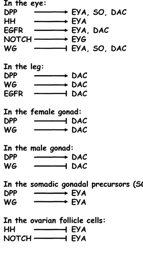
dpp transheterozygotes lose expression of RD gene network members eya, so, and dac in
the eye disc (Chen et al., 1999b), although they do not affect eyeless expression (Kenyon et al., 2003), suggesting that DPP acts downstream of or in parallel to EY to activate expression of RD genes. More detailed work using somatic clonal analysis revealed that DPP signaling is required for EYA, SO, and DAC expression only prior to morphogenetic furrow initiation (Curtiss and Mlodzik, 2000), and that once they are turned on by DPP in the furrow, their expression can be maintained independently of DPP signaling. The expression patterns of the RD gene network members in the eye disc is not wholly coincident (Figure 4), suggesting that even those which can physically interact also play independent roles. EY is expressed in tissue prior to the furrow (Figure 4D; Bessa et al., 2002)), and EYA is expressed just anterior to the morphogenetic furrow and in all eye tissue posterior to the furrow (Figure 4A,D), while DAC is expressed in a
wide stripe centered on the morphogenetic furrow (Figure 4C,D). SO is expressed in all cells in the eye disc (Figure 4B,D), while another SIX family member, OPTIX, is only expressed in undifferentiated tissue anterior to the furrow (Figure 4D; Seimiya and Gehring, 2000).
HH signaling plays a key role in formation of the Drosophila eye, and high levels of HH are expressed just posterior to the DPP signal in the morphogenetic furrow (Figure 4), and are required for proper furrow progression (Pappu et al., 2003), which occurs through mutual activation of HH and DPP. Unlike in the embryo, in the eye disc HH is crucial for EYA expression (Pappu et al., 2003), through what seems to be a permissive mechanism. HH signaling inside the cell is effected by changes in the transcription factor CUBITUS
INTERRUPTUS (CI); in the absence of signal, CI is cleaved to a shorter repressor form (CIr), which enters the nucleus and downregulates target genes, while in the presence of signal,
phosphorylation of Cl is blocked, preventing cleavage thus allowing it to remain an activator and direct transcription of target genes (Chen et al., 1999a). HH's role in regulation of EYA
expression is through elimination of a block to transcription, rather than activation; removal of CIr is sufficient to promote EYA expression, while CIact is not necessary for eye formation (Pappu et al., 2003).
The eye field undergoes a wave of differentiation directed by the expression of DPP and HH at the morphogenetic furrow, which together activate or allow transcription of the second tier of the RD gene network, EYA, SO, and DAC. These three genes are coincident only at, and just adjacent to, the morphogenetic furrow (Figure 4), where they and the DPP signal come together to direct terminal differentiation in the eye. The discrete domains of the eye disc which express each complement of RD gene network members (Figure 4) undergo quite different
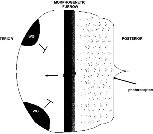
MORPHOGENETIC FURROW POSTERIOR I J-I---= photoreceptors
Figure 3: Morphogenetic furrow progression is driven by DPP and HH signals The eye disc undergoes waves of differentiation as the morphogenetic furrow, driven by
cooperative action of the HH and DPP signaling pathways, moves from the posterior to the anterior of the eye disc. The most posterior cells have differentiated into the photoreceptor cells, while anterior cells still proliferate. WG expression in the dorsal and ventral most anterior regions of the disc prevents eye tissue formation in that region leading to head cuticle formation.
ANTERIOR
y
/ i. 1 I -.pl-· I, ';-· r. ''··i. I·. ;* '·· I· 3 -i.· ·. :.( i Ir I·· ;sl·· I·. I:--· .t'·· "·· rl-· I·· I - :*. Z-4L -;x ·· - I .: .-1-.I
. .~~~~I I: II; I ." r I I. I-I-, I ,I·· *· .I'· ';· i. I; i. I 'r I-. JAnterior
dpp
hh
EY
EYA
SO
DAC
optix
iMF
Posterior
*LL] :i 11 PPNi MF iIV . . . V . . .Figure 4: RD gene network members are expressed in overlapping and distinct domains
in the eye disc
EYA is expressed just before the furrow in the preproneural (PPN) region and in all
differentiated cells after the morphogenetic furrow (MF; A), while SO is expressed in all cells anterior to the furrow and differentiated cells posterior to the furrow (B). DAC is expressed in a broad stripe encompassing the furrow and cells on each side (C). The expression pattern of RD gene network members is depicted schematically in (D), where the eye disc is broken
up into six regions based on the level of differentiation. Lowercase indicates RNA expression, while uppercase indicates protein localization. EYG is not depicted on this schematic as it is expressed in a stripe along the Dorsal/Ventral boundary.
D
I
I I
t
where EY, SO, EYA, and DAC overlap, the neural fate of photoreceptors begins to be specified, along with other accessory cells. Finally just EYA and SO remain expressed in the differentiated cells, perhaps playing a role in their survival or function. Further studies of EYA, SO, and DAC, alone and in combination, should reveal their targets both independently and as a group, yielding insight as to how this precise program of eye development is orchestrated.
EGFR signaling: Cell survival and differentiation
The EGFR/RAS/MAPK pathway is an important growth and differentiation cue, which also plays a key role in control of cell survival (Bergmann et al., 2002). In the Drosophila eye, EGFR signaling is required at a low level in all cells to prevent apoptosis (Bergmann et al., 2002), but is also used selectively in the process of cellular differentiation. EGFR is required for morphogenetic furrow initiation but not its propagation (Kumar and Moses, 2001), and is used reiteratively to specify the fate of every eye cell in waves of differentiation following the recruitment of the first photoreceptor, R8 (Voas and Rebay, 2004).
Signaling through EGFR leads to activation of the small GTPase RAS, which then activates the MAPK cascade, which regulates downstream transcription factors and thus effects transcriptional change in response to receptor activation (Freeman, 1998). The EGFR pathway provides one of the few direct links between a signaling pathway and regulation of the RD gene network, where at least some part of the mechanism linking EGFR signaling and the network is understood. The first hint that EGFR signaling might directly affect RD gene function was the isolation of mutant alleles of eya in a screen for modifiers of the phenotype produced by eye specific expression of an activated negative regulator of EGFR signaling, yan"'ct (Rebay et al.,