HAL Id: hal-03155991
https://hal.sorbonne-universite.fr/hal-03155991
Submitted on 2 Mar 2021HAL is a multi-disciplinary open access archive for the deposit and dissemination of sci-entific research documents, whether they are pub-lished or not. The documents may come from teaching and research institutions in France or
L’archive ouverte pluridisciplinaire HAL, est destinée au dépôt et à la diffusion de documents scientifiques de niveau recherche, publiés ou non, émanant des établissements d’enseignement et de recherche français ou étrangers, des laboratoires
NK cells in the tumor microenvironment: Prognostic
and theranostic impact. Recent advances and trends
Jules Russick, Carine Torset, Edouard Hemery, Isabelle Cremer
To cite this version:
Jules Russick, Carine Torset, Edouard Hemery, Isabelle Cremer. NK cells in the tumor microenvi-ronment: Prognostic and theranostic impact. Recent advances and trends. Seminars in Immunology, Elsevier, 2020, 48, pp.101407. �10.1016/j.smim.2020.101407�. �hal-03155991�
NK cells in The Tumor Microenvironment: prognostic and theranostic impact. Recent advances and trends
Jules Russick1, Carine Torset1, Edouard Hemery1, Isabelle Cremer1
1Centre de Recherche des Cordeliers, INSERM, Sorbonne Université, Université de Paris, Team
Inflammation, complement and cancer, F-75006 Paris, France.
# Correspondence to: Isabelle Cremer, Ph.D.
Cordeliers Research Center, INSERM UMRS 1138;
15 rue de l'Ecole de Medecine; 75006 Paris, France
Phone: 33-1-44-27-90-83/ Fax: 33-1-40-51-04-20,
Abstract
NK cells orchestrate the tumor destruction and control metastasis in a coordinated way with other immune cells of the tumor microenvironment. However, NK cell infiltration in the tumor microenvironment is limited, and tumor cells have developed numerous mechanisms to escape NK cell attack. As a result, NK cells that have been able to infiltrate the tumors are exhausted, and metabolically and functionally impaired. Depending this impairment the prognostic and theranostic values of NK cells differ depending on the studies, the type of cancer, the stage of tumor and the nature of the tumor microenvironment. Extensive studies have been done to investigate different strategies to improve the NK cell function, and nowadays, a battery of therapeutic tools are being tested, with promising results.
Keywords
NK cells, tumor microenvironment, prognostic, theranostic
Abbreviations
A2AR: A2a Adenosine receptor
ADCC: Antibody-Dependent Cell-mediated Cytotoxicity AML: Acute Myeloid Leukemia
APC: Antigen Presenting Cells CAR: Chimeric Antigen Receptor CCL: CC Chemiokine Ligands
CTLA-4: Cytotoxic T-lymphocyte antigen-4 CXCR: CXC chemokine receptors
DC: Dendritic Cells
EGFR: Epidermal Growth Factor Receptor ESC: Embryonic Stem Cells
GIST: Gastrointestinal Stromal Tumors FBP1: Fructose-1,6-biphosphatase ICB: Immune Checkpoints Blockade
IDO: Indoleamine-2,3-dioxygenase ILC: Innate Lymphoid Cells
iPSC: induced Pluripotent Stem Cells
KIR: Killer-cell immunoglobulin-like receptors
KLRG1: Killer cell lectin-like receptor subfamily G member 1 LAK: Lymphokine Activated Killer
MALT: Mucosa-Associated Lymphoid Tissue
MIC-A/MIC-B: MHC class I polypeptide–related sequence A/B NCR: Natural Cytotoxicity Receptor
NK cells: Natural Killer cells
NSCLC: Non-Small Cell Lung Cancer ORR: Overall Response Rate
PBMC: Peripheral Blood Mononuclear Cells
PD-1/PD-L1: Programmed cell Death 1/ Programmed cell Death Ligand 1 PFS: Progression Free Survival
RCC: Renal Cell Carcinoma ROS: Reactive Oxygen Species
SCCHN: Squamous Cell Carcinoma of the Head and Neck TAA: Tumor Associated Antigens
TGF: Transforming Growth Factor
TIGIT:T cell Immunoreceptor with Ig and ITIM domains TME: Tumor Microenvironment
1. Introduction
Natural Killer (NK) cells are part of the body’s first line of innate immune defenses and represent the cytotoxic compartment of the innate lymphoid cells (ILCs). They rapidly respond to infection and to malignant transformation and regulate the adaptive immune response [1]. NK cells originate from bone marrow precursors [2], then, consistent with their role in anti-tumoral surveillance and defense, they are widely distributed in lymphoid and non-lymphoid tissues, circulate throughout the body and acquire their function in appropriate tissues. As a consequence, NK cells are found in many tissues and organs, mainly in lymph nodes, liver, lung, uterus and thymus, in addition to the blood circulation[3–5]. Overall, NK cells are characterized by the expression of surface markers such as NK1.1, NKp46 or CD49b (DX5) in mice, or CD56 (NCAM) and CD16 (FcγRIII) in humans. However, circulating and tissue resident NK cells are characterized by distinct expression of transcription factors and distinct phenotypes [3,6]. NK cells exert their antitumor function following their activation, as a result of engagement of non-clonotypic germ-line-encoded activating and inhibitory receptors with their ligands expressed at variable levels on tumor cells [1,7]. Once activated, NK cells are prone to kill tumor cells and to secrete an array of cytokines and chemokines that contribute to adaptive immune cell activation and recruitment into the tumor microenvironment (TME).
In both human and mice, several NK cell subsets have been described, which correspond to different stages of maturation, and subsequently various anti-tumor functions. High-throughput single-cell RNA-seq was used to characterize tissue-specific gene signatures of NK cells in spleen and blood in mice and in humans. This allowed the identification of two major subsets transcriptionaly similar across organs and species, showing organ-specific signatures and heterogeneity of NK cells in the blood and in the spleen [8].
In human, sequential maturation is divided into 5 stages 1 one to 5 during which the NK cells sequentially acquire receptors essential for their functions. The pre-NK first stage of maturation derives directly from common lymphoid precursor in the bone marrow. During stage 2, intermediate affinity IL-15 receptor (CD122) is acquired to deliver survival signals to pre-NK cells allowing them to continue
expression (CD56bright CD16-), are mainly present in secondary lymphoid organs (lymph nodes), are
poorly cytotoxic and produce large amounts of cytokines [10], whereas stage 5 NK cells (CD56dim
CD16+) are strongly cytotoxic, have high ability to produce IFN-γ and perforins and are mainly
circulating or localized at the site of inflammation. In mice, 4 maturation steps have been identified on the basis of the co-stimulatory CD27 receptor and integrin CD11b expression. Very immature NK cells are CD27-CD11b- and acquire CD27, NKp46, NK1.1, NKG2D expression in the stage 2. Stage 3 NK
cells express both CD27and CD11b and acquire sphingosine 1-phosphate receptor 5 (S1P5) involved in NK cell migration. Finally, the stage 4 NK cells are the more mature (CD27-CD11b+) and also express
the co-inhibitory killer cell lectin-like receptor subfamily G member 1 (KLRG1) [8].
Throughout maturation, NK cells acquire the capacity to interact with tumor cells. Mature NK cells express two classes of inhibitory receptors: killer-cell immunoglobulin-like receptors (KIR) and CD94-NKG2A. KIRs recognize MHC class I molecules, and NKG2A recognizes the unconventional HLA-E [11], allowing self-tolerance [12]. The NK cells are therefore naturally inhibited and their activation is only possible when the activating signals exceed inhibition. Once activated, NK cells become highly cytotoxic [13]. Many activating receptors have been characterized on NK cells among which NKG2C, NKG2D, the “Natural Cytotoxicity Receptor” (NCR) including NKp30, NKp44 and NKp46. NKG2D recognizes a wide variety of ligands, for example MHC class I polypeptide–related sequence A (MIC-A) or MIC-B, and UL16 binding protein 1 (ULBP1), 2 and 3 expressed by transformed cells [14]. This recognition does not require antigenic recognition beforehand. During cytotoxic process, NK cells move closer to tumor cells, then release proteases -called granzymes, and perforin. The perforin forms pores in the target cell to create an aqueous channel through which the entire content of NK cells, including granzymes, is transferred to the cell and induces apoptosis [15].
In human, the stage 5 subpopulation of NK cells (CD56dimCD16+) exerts cytotoxic function and
expresses high levels of perforin and granzyme B. In mice, the stage 4 (CD11b+CD27-) NK cell subset
is the cytotoxic one [10]. In addition to direct cytotoxicity, NK cells can kill tumor cell through antibody-dependent cell-mediated cytotoxicity (ADCC), involving the CD16 receptor [15].
In addition to cytotoxic functions, NK cells have been shown to play a role in the regulation of innate and adaptive immune populations. NK cells express receptors for IL-12, IL-15 and IL-18 cytokines that are produced by antigen presenting cells (APCs) [16–18]. These cytokines trigger the proliferation of NK CD56bright and production of cytokines like IFN-γ, IL-10, IL-13, TNF-β and GM-CSF [19]. Thus,
APC influence the phenotype and cellular functions of NK cells. Reciprocally, NK cells influence the function of APC: they stimulate the production of TNF-α by monocytes and kill immature dendritic cells (DC) during “DC editing” [20–22].
Many studies have characterized the NK cells in the TME of solid tumors, and have shown that in numerous tumors, NK cells are scarce and dysfunctional. In this review, we provide an overview of NK cells in the TME of solid tumors, with a focus on their prognostic and theranostic values. We will then discuss the recent advances and trends in current therapies that are being developed to strengthen antitumor NK cell responses.
2. NK cells in Tumor Microenvironment
The main function of NK cells in the TME is the killing of tumor cells, through perforin/granzyme exocytosis, engagement of death receptors (Fas-FasL and TRAIL-TRAILR) and secretion of the effector cytokines IFN-γ and TNF-α. To be able to efficiently function as tumor eliminating cells, mature functional NK cells must be located in the tumor nest, to establish close contacts with their target cells, and thereafter kill them. The presence of fully functional NK cells may results from in situ maturation of NK cells or from the active recruitment of mature NK cells from adjacent tissue and circulation. These recent years, advances in cellular and molecular analysis using high-throughput new technologies - single cell sequencing (scRNAseq), multiplexed image analysis, or spatial transcriptomics - [23] allowed a better understanding of the complexity of the TME. Nowadays, a deep characterization of intratumoral immune cell transcriptome has been performed [24–26] that allows to precisely define NK cell heterogeneity at the single-cell level in a pan-genomic analysis across organs and species.
The first remarkable observation made by numerous studies quantifying NK cells in situ is that NK cells poorly infiltrate the TME. Low numbers of NK cells were found in lung cancer [27], in renal cell carcinoma metastases in the lung [28], in colorectal carcinoma [29] and in gastrointestinal stromal tumors (GIST) [30] as compared to corresponding healthy tissue. Similar conclusions were made in scRNA seq analysis of cells in the TME of lung cancer: less NK cells were found in the tumor than in normal tissue [24]. In addition, when present in the TME, NK cells are not in direct contact with tumor cells but are mainly localized within the stroma [27,29,30].
We performed a comparative analysis of the number of NKp46 or CD56transcripts in the tumor and in the normal tissue using TCGA database, and confirmed that in many tumors, such as lung cancer, diffuse large B cell lymphoma, thymoma, urothelial bladder carcinoma, breast, ovarian, endocervical, colon, and prostate cancer, the infiltration of NK cells is lower than in healthy tissue (Figure 1). The low infiltration of NK cells in the TME raises some questions: Does the tumor microenvironment apply a selective attraction of specific subset(s) of NK cells or does it exclude NK cells by modulating their expression of chemokine receptors? The migration of NK cell subsets in the tumor is governed by the action of chemokines abundantly produced in the TME, and the chemokine receptor expression profile of NK cells. In the circulation, almost 90% of NK cells are CD56dim, CD16+, perforin+ and are mostly
cytotoxic [1]. These cells express a panel of chemokine receptors including CXCR1, CXCR2, CXCR4 and CX3CR1 [31] and are predominant in bone marrow, spleen, lung and breast normal tissues. Conversely, the CD56bright NK cells which express CCR7, CXCR3, CD62L and CXCR4, are
preferentially localized in lymph nodes, intestinal mucosa, mucosa-associated lymphoid tissue (MALT) and in gastric, liver, uterus, visceral and kidney tissues [32]. The TME is able to modify the local chemotactic environment to preferentially recruit less cytotoxic NK cells: To do so, it alters the chemokine receptor repertoire on intra-tumoral NK cells through a TGF-β dependent mechanism [33,34]. It also reduces the production of the ligands corresponding to the receptors expressed by CD56dim NK cells (CXCL1, CXCL2, CX3CL1 and CXCL8) and it increases the expression of ligands
able to attract CD56bright NK cells (CXCL9, CXCL10, CCL19 and CCL5) [32,35].
Another explanation for scarce density of NK cells could be the in situ apoptosis of intratumoral NK cells. In lung cancer, genes related to apoptosis were found to be upregulated in intratumoral NK cells as compared to wild-type tissue [36]. Similarly, in hepatic cancer, apoptosis of NK cells can be induced into the tumors [37]. Several mechanisms have been proposed, that involves the reactive oxygen species (ROS), such as Indoleamine-2,3-dioxygenase (IDO) [38] or the lactate mediated apoptosis in colorectal liver metastasis [39].
Numerous studies have found that intratumoral NK cells are dysfunctional due to the immunosuppressive microenvironment [7,40]. Tumor cells escape from NK cell recognition and destruction by shedding of MIC-A/-B ligands for activating NK cell receptor NKG2D [41] and by secreting immunosuppressive molecules such as IDO, prostaglandin E2, IL-10 and TGF-β, responsible for down-regulation of activating receptors on NK cells [42]. Consequently, intratumoral NK cells display altered phenotype and function in many tumors, including lung tumors [27,43,44], breast cancer [45], ovarian cancer [46], hepatocellular carcinoma [47,48], GIST [49] and melanoma [50]. The TGF-β mediated conversion of effector NK cells into type 1 ILCs has also been characterized as a mechanism of tumor evasion from NK cells attack in the TME [51,52]. Dysfunction of intratumoral NK cells were confirmed by recent studies using high throughput technologies - multi-scale immune profiling using mass cytometry by time-of-flight (CyTOF) combined with single-cell transcriptomics and multiplex
tissue imaging, unveiled that NK cells were unique in tumor lesions compared to normal lung from the same patients, and that these changes occur in very early stages tumors [26]. Finally, accumulating evidences highlighted the acquisition of immune checkpoint molecules by tumor infiltrating NK cells [53], and the acquisition of a regulatory phenotype – characterized by T cell suppression, secretion of TGF-β and IL-10 [54]. NK cells do express T cell immunoreceptor with Ig and ITIM domains (TIGIT), which was found associated with NK cell exhaustion in tumor-bearing mice and patients with colorectal cancer [55], programmed-cell death protein 1 (PD-1) [56], programmed-cell death ligand 1 (PD-L1) [57], T-cell immunoglobulin mucin-3 (TIM-3) in patients with GIST [58] and bladder cancer [59] and cytotoxic T-lymphocyte-associated protein 4 (CTLA-4) (our unpublished results in lung cancer and [60,61]). In addition, NK cells in the TME were found to co-express CD73, lymphocytes activation gene-3 (LAG-3), V-domain Ig suppressor of T cell activation (VISTA), PD-1 and PD-L1, secrete IL-10 and TGF-β, while the frequency of CD73 expressing NK cells was found correlated with advanced stages in breast cancer [62].
Accumulating evidences showed that cellular metabolism is also important for immune cell functions [63–65], and that modifications of metabolic pathways in the TME may lead to NK cell dysfunction. Glycolysis is important for human and mouse NK cell functions [66]. Extracellular adenosine is a key immunosuppressive metabolite, generated from ATP by the ectonucleotidase CD39 and CD73 expressed by intratumoral immune cells. It acts as a checkpoint that limits the maturation and the function of NK cells via adenosine receptors (A2AR) [67], as shown in a mice model of melanoma. Another study performed in lung cancer demonstrated that NK cells are progressively impaired while lung tumor progress due to metabolism alteration. The intratumoral NK cells were reduced in number, displayed lower survival and attenuated cytotoxicity and showed decreased glycolysis with concomitant increased Fructose-1,6-biphosphatase (FBP1) [36]. The study concluded that FBP1, an important regulator of glycolysis, is a key regulator of NK cell dysfunction in the TME. The alterations of NK cells induced by the tumor microenvironment are summarized in the Figure 2.
In some tumors, different subsets of NK cells are able to infiltrate the TME, and depending their maturation and functional state, they may influence the clinical outcome of patients.
Most of the studies focusing on the prognostic value of NK cells across solid tumors have attempted to correlate the number of intratumoral NK cells to patient’s survival. The quantification of NK cells was mainly performed by immunohistochemistry and flow cytometry and led to controversial results, because of the heterogeneity of the markers used that added confusion when comparing the results of these studies. The first works used the CD57 (HNK-1) marker to characterize NK cells, even if it’s not specific for the overall NK cell population [68]. CD57 is now considered as a marker for a CD56dim NK
cell subset [69]. The literature studying CD57+ NK cells found a positive impact of these cells on the
clinical outcome in lung adenocarcinoma [70], squamous cell lung cancer [71], colorectal carcinoma [72], gastric carcinoma [73], esophageal squamous cell carcinoma [74] and ovarian cancer [75]. More recently, studies using CD56 as a marker of NK cells have shown a more complex situation: depending on the tumor type, CD56+ NK cells were found associated or not with clinical outcome. Whereas it had
no impact in lung cancer [76] and in melanoma [77], it was associated with a better outcome in colorectal carcinoma [78], renal cell carcinoma (RCC) [79] and head and neck squamous cell carcinoma [80]. As NKp46 is expressed by all NK cells, its staining should allow to homogenize the studies [81]. Indeed, using NKp46 staining for NK cells, it was shown that high NK-cell densities were associated with improved survival in RCC [28], in localized GIST [49] and in triple negative breast cancer [82]. However, no prognostic value was found linked to NKp46 expression in lung cancer [27]. Finally, in a meta-analysis of gene expression signature from almost 18,000 tumors, that included 25 cancer types, the authors did not find any correlation between NK cell signature and clinical outcome [83].
In addition to NK cell quantification, the NK cell activity seems to be more informative to determine the real impact of NK cells on clinical outcome [78]. The importance of NK cell activity was highlighted by a study that compared the expression of NKp46 and NKp30 variants - immunosuppressive or immunostimulatory - in GIST, neuroblastoma, melanoma and lung cancer. This study showed a correlation between the low expression of NKp30 genes and a poor clinical outcome [84], but no correlation was found between prognosis and NKp46 expression. Similarly, a 11-year follow-up study published in 2000 analyzed the cytotoxic activity of peripheral NK cells among 154 cancer cases. The most abundant cancers in this cohort were stomach, lung and intestine in which the authors found an
inverse correlation between NK cell cytotoxic activity and cancer incidence [85]. The prognostic value of NK cells in cancer are summarized in Table 1.
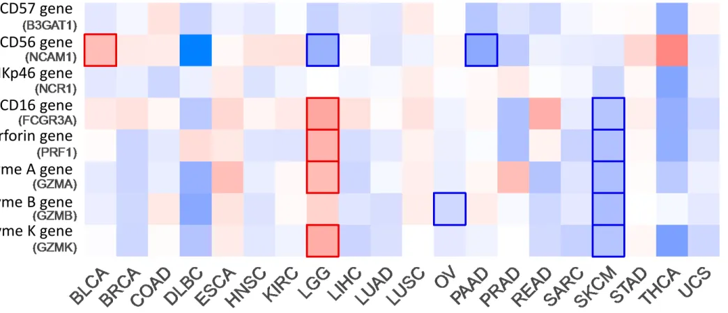
To complete the data from literature, we compared the expression of genes encoding for NK cells markers – CD56 (NCAM1), CD57 (B3GAT1) and NKp46 (NCR1) – versus genes encoding for cytotoxic molecules – Granzyme A (GZMA), Granzyme B (GZMB), Granzyme K (GZMK), Perforin (PRF1), CD16 (FCGR3A) and NKp44 (NCR2) –, and evaluated their impact on overall survival of patients (Figure 3). Using public data of TCGA database and analyzing it using GEPIA2 [86], we were able to compare 20 types of tumors. NCAM1 is the only gene correlating with the prognostic value in the first signature whereas all the genes are linked in prognostic value of Brain Lower Grade Glioma (LGG), Skin Cutaneous Melanoma (SKCM) and uveal melanoma (UVM) patients, showing the potential interest of NK cell activity over NK cell number.
4. Theranostic value of NK cells
As immunotherapies represent the most recent and striking improvement of cancer management, the theranostic value of NK cells has recently been studied with respect to immune checkpoints blockade (ICB). In melanoma, lymphoma and colon carcinoma murine models, Hsu et al. demonstrated that the PD-1/PD-L1 axis modulates NK cell phenotype, as its blockade restores NK cell antitumor functions and allows a better survival of the animals [87]. In a prospective analysis of lung cancer patients, the use of Nivolumab (an anti-PD-1 therapy) increased the NK cell number in patients with better prognosis, whereas the NK declined in the bad prognosis group [88]. Similarly, NCR1 was the only parameter affecting the prognosis in non-small cell lung carcinoma (NSCLC) patients with high PD-L1 expression on tumor cells [84]. In metastatic melanoma, the increased density of activated NK cells into the tumor was correlated with response to PD-1 blockade, concomitant with activation of NK cell cytotoxicity [89]. NK cell activation was also found predictive to non-progressive disease and high progression free survival (PFS) in patients with melanoma, lung cancer and head and neck cancers, in response to anti-PD-1 therapy [90].
The modulation of NK cell activity by ICB could be, in part, explained by regulatory T cells (Tregs) which were shown to be regulators of NK cells, both in pre-clinical models and in humans [88,91]. By reducing the inhibitory environment, immune checkpoint blockade could restore the cytotoxic functions of NK cells. Moreover, NK cells interact with an intra-tumoral subset of DC that are able to re-stimulate T cells, within the melanoma tumor microenvironment [92]. This interaction is predictive of increased overall survival and of the responsiveness to anti-PD-1 immunotherapy. The crosstalk between NK cells, DC and Tregs also play a central role in GIST patients treated with Imatinib. This inhibitor of tyrosine-kinase receptors induces the accumulation of NK cells into the tumor (multiplying the ratio NKp46/FoxP3 by four), triggers DC-mediated NK cell activation and the production of IFNγ, which is considered as a predictive factor of long-term survival in advanced GIST [49,93,94]. Interestingly, in a murine lung adenocarcinoma model resistant to checkpoint inhibition, the combination of IL2/anti-IL2 complexes with anti-PD-1 acts on CD8 exhaustion, whereas the addition of anti-CTLA-4 to IL2/anti-IL2 complexes allows the rescue of NK cell functions [91]. In human, the use of Tremelimumab, an anti-CTLA-4 immunotherapy restored the CD56dim/CD56bright NK ratio, promoted the killing of tumor
cells by NK cells and consequently improved the overall survival of malignant mesothelioma patients [95].
Altogether, these studies reveal the importance of tumor escape to NK cells and emphasizes the interest to target NK cells to reinvigorate their anti-tumor functions.
4. NK cells as therapeutic targets
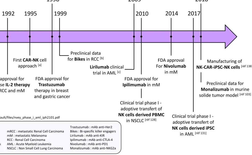
Several therapies targeting NK cells have been proposed to re-activate NK cells. In the 1980s, the first therapy targeting NK has been developed for patients who did not respond to classical treatments: autologous Lymphokine Activated Killer (LAK) cell therapy. Lymphocytes from patients collected by leukapheresis were activated with IL-2 and then re-injected into patients with or without IL-2. This induced the remission of 31% of patients, compared to 15% for those who received IL2 only [96]. This research extended until 1989 for hepatocellular carcinoma (HCC) cancer and lung carcinoma, giving disappointing results [97,98], with undesired expansion of Tregs [99] and side effects [96,100,101].
Immunotherapy is a major breakthrough in cancer treatment. To target NK cells, several approaches are under investigation, based on complementary strategies that consist in blocking inhibitor signals (using anti-KIR, anti-NKG2a or other anti-checkpoint inhibitors), enhancing ADCC (using mAbs against tumor associated antigens (TAAs) and bi- or tri-specific killer engagers), enhancing NK cell proliferation and cytotoxicity (using cocktails of cytokines). Another attractive strategy is to transfer NK cells in tumor-bearing patients. An historical timeline is depicted in Figure 4, which summarizes the major significant NK cell therapies.
4.1. Anti-immune checkpoint inhibitors: pre-clinical models and clinical trials
Functional NK cells can efficiently kill cancer cells that have reduced or lost MHC class I molecules, but not cells maintaining their expression. In addition, as many cells in the TME, NK cells do express immune checkpoints molecules [102]. It is therefore possible to use therapeutic anti-inhibitory monoclonal antibodies (mAbs), such as anti-KIR or anti-NKG2A mAbs to restore their anti-tumor activity. Preclinical studies demonstrated the efficacy of immune checkpoint blockade on NK cells: anti-TIGIT mAbs inhibited tumor growth and prevented exhaustion of infiltrating NK cells in tumor-bearing mice [55], and the use of anti-NKG2A combined with anti-PD-1/PL-L1 promoted antitumor immunity in several mice models [103].
These treatments are part of the « IC blockade » based immunotherapeutic strategies and are currently used in clinical trials in the treatment of solid tumors as summarized in the Table 2. Several clinical trials combining Lirilumab (anti-KIR), Nivolumab (anti-PD-1) and/or Ipilimumab (anti-CTLA-4) are currently being studied for treatment of bladder cancer or several advanced refractory solid tumors (clinical trials numbers: NCT03532451, NCT03203876 and NCT01750580). Phase I trials demonstrated the patients' tolerance to the combination of these antibodies. However, trials in an extended population of patients with squamous cell carcinoma of the head and neck (SCCHN) have not shown any clear
clinical benefit
(https://www.innate-pharma.com/sites/default/files/leidner_sitc_2016_liri_001_efficacy_oral_0.pdf). Other clinical trials have investigated Monalizumab (anti-NKG2a) in combination with Trastizumab (anti-HER2), Durvalumab (PD-L1) and/or Cetuximab (anti-EGFR) in breast cancer, colorectal cancer and NSCLC
(NCT04307329, NCT02671435, NCT04145193, NCT03822351, NCT 03794544 and NCT02643550). Clinical trials of the combination of Monalizumab and Cetuximab showed no additional toxicity compared to monotherapies and the phase II trial gave promising results for head and neck cancer, with an overall response rate (ORR) of 20%. A phase III trial has been planned (https://www.innate-pharma.com/sites/default/files/asco2020_monacetux.pdf). For combination with Durvalumab, the phase II trial in colorectal cancer also gave encouraging preliminary results (https://www.innate-pharma.com/sites/default/files/180205asco_15poster_09.pdf).
4.2. Monoclonal antibodies that promote NK cell ADCC
Several treatments have been developed to increase ADCC, based on the use of CD20 [104], anti-HER2 [105] or anti-CD133 [106]. These treatments, which target various solid tumors or leukemias, are limited by the polymorphism of the CD16 gene which determines its affinity for IgG1 and IgG3 [105]. Approximately 10% of the population has a polymorphism that gives CD16 a greater affinity for the Fc fractions of antibodies and therefore a better ADCC [107]. These differences can be seen in the heterogeneity of clinical trial results [108,109]. Nowadays, Fc-optimized antibodies are created to increase the affinity of the Fc fragment to CD16, as for Fc-optimized anti-CD133 which promotes increased NK cell degranulation in an AML model [106].
Other strategies aimed at reinvigorate NK cell activation by inhibition of MICA/B shedding, using a synergistic combination of HDAC inhibitor and anti-MICA/B mAbs, to enhance the transcription of MICA/B genes and to inhibit MICA/B shedding and enhance its expression at the cell surface [110,111]. 4.3. Bi- and Tri-specific killer engagers
NK cells need direct contact with target tumor cell in order to lyse them. For this, bi-, tri- or tetra-specific “killer engagers” have been created. These are engineered antibodies composed of two or three Fab fragments linked together: a CD16 part to bind the NK and one or two fragments against tumor-associated antigens [99]. This technology was developed as a treatment for leukemia, targeting the CD20, CD19, CD30, CD33 or CD123 antigens in B cell Non-Hodgkin's lymphomas [112,113], mixed lineage leukemia [114] or acute myeloid leukemia [115,116]. It was then used for several solid cancers including RCC, with fragments targeting the epidermal growth factor receptor (EGF-R) [117] or
HER2-prostate, breast, colon, head or neck cell lines carcinomas [120–122]. Stimulation of NK cells by these antibodies had increased ADCC and degranulation of NK cells.
4.4. Cytokines to boost the anti-tumor activity of NK cells
The preliminary stage of NK cells adoptive transfer relies on in vitro stimulation of NK cells. Several cytokines are known to promote the expansion and activation of NK cells [99]. Since the 1980s, clinical trials has been established for treatments of solid cancers such as breast cancer, colorectal cancer, glioblastoma or melanoma, using type I interferon, IL-2, IL-12, IL-15, IL-18 and IL-21 [122–124]. However, most of these cytokines, except IL-21, induced severe side effects, justifying the need to design new recombinant cytokines.
The combination of cytokines with immune checkpoint blockade has also been investigated to tempt to improve response to treatment, as the combination of IL-21 with Nivolumab in several solid tumors (NCT01629758), with Ipilimumab in melanoma (NCT01489059), or very recently using IL-15 with anti-TIGIT mAb in melanoma [50].
Treatments have evolved to design superagonists IL-2 and IL-15 cytokines with improved affinity for their receptor and higher biological affinity. The IL-2 and IL-15 (ALT-803) suprakines improved NK cells proliferation and prevented their suppression by Tregs [99,125]. ALT-803, with a half-life of 25 hours, is now used in clinical trials in combination with Nivolumab in NSCLC (NCT02523469) [126] or with Rituximab in B cell non-Hodgkin lymphoma (NCT02384954).
The last types of treatment involving NK cells consist in adoptive transfer of unmodified or engineered NK cells that harbor a Chimeric Antigen Receptor (CAR), called NK-CAR cells. The aim of this method is to multiply NK cells and make them cytotoxic so that they can destroy tumor cells [127].
4.5. Adoptive transfer of unmodified NK cells
For the case of adoptive transfers of unmodified NK cells, several methods were currently used. The first one consists in stimulating NK cell proliferation, from patient or donor peripheral blood mononuclear cells (PBMC). For this, the cells are cultured for several weeks with artificial antigen-presenting cells, feeder cells and/or cytokines to stimulate proliferation and cytotoxicity [99,127]. Before 2010, adoptive NK cell transfer was limited to leukemia. Nowadays several phase I or II clinical trials have been started in solid tumors, including NSCLC [128], ovarian and breast tumors [129], but
the results are not conclusive. Many patients do not respond to treatment because of reduced NK cells cytotoxicity due to their interaction with the immunosuppressive microenvironment and their inability to reach the tumor nest.
The second method uses stem cell-derived NK cells. Several sources of NK cells were used, like Umbilical cord blood (UCB) [130], embryonic stem cells (ESC) and induced pluripotent stem cells (iPSC), the aim being to obtain as many activated and cytotoxic NK cells as possible. UCB-derived NK cell therapy is currently in phase I clinical trial for AML and myelodysplasic syndromes [131]. In this clinical trial, NK cells derived UCB seems to acquire maturation stage after the infusion in patients. The use of ESC or iPSC looks promising. The advantage of using stem cells is that a continuous and homogeneous source of NK cells is obtained, unlike NK cells derived from PBMC or UCB [132–134]. Pre-clinical studies have demonstrated that these cells are cytotoxic in vitro against myeloma, pancreatic and ovarian cancer cells [135,136] and also effective in vivo against leukemia, breast, prostate, testicular, and glioma cancer in mice models [137]. In 2018 a study succeeded in genetically modifying NK cells from iPSC into NK-CAR-iPSC cells. This success is revolutionary since it may allow standardization and increased efficacy of adoptive transfer therapies [138].
4.6. Adoptive transfer of CAR-NK
CAR-NK cells are cells genetically modified to express transmembrane and extracellular domains molecules that can bind tumor-associated-antigens expressed by tumor cells. These domains, mainly derived from CD3ζ, are associated with one or two co-stimulation protein receptors [139].
The CAR technology was first developed on T cells and has shown efficacy in lymphomas and leukemias [140]. However, studies on solid tumors have not been conclusive [141]. The technology was then tested on NK cells. CAR-NK cells can be produced from autologous cells, NK cell lines or stem cell-derived NK cells. Currently, most studies focus on leukemia [142] and several clinical trials are ongoing in the context of solid tumors: ovarian cancer with anti-mesothelin-CAR-NK cells (NCT03692637), prostate cancer with anti-PSMA-CAR-NK (Prostate-Specific Membrane Antigen) cells (NCT03692663), pancreatic cancer with ROBO1 specific BiCAR-NK cells (NCT03941457) and various solid tumors with CAR-NK cells targeting NKG2D-Ligand or with ROBO1 CAR-NK cells
The advantage of CAR-NK is that these cells have higher cytotoxicity than CAR-T cells. Furthermore, when using NK cell lines (NK-92), the advantage is that these cells express very low level of inhibitory receptors [143]. These NKs have better viability and can be multiplied at a lower cost [142].
5. Conclusions
NK cells play a critical role in immune surveillance of spontaneous tumors and in preventing tumor metastases [144]. This justifies to exploit NK cell functions for a better management of cancer patients. Many studies have demonstrated NK cell impairment in cytotoxic activity, associated with a higher cancer risk [145]. Since tumors have evolved multiple mechanisms to escape NK cell control, it is fundamental to identify optimal therapies - or combination of therapies – to make NK cells able to kill tumor cells. The conclusions that can be drawn from the published data set on NK cells are that in a solid tumor context, the entire tumor microenvironment must therefore be taken into account in order to hope for synergy and better efficacy of treatment. With respect to this, promising therapies targeting NK cells are now being tested either in pre-clinical model or in clinical trials.
References
[1] E. Vivier, E. Tomasello, M. Baratin, T. Walzer, S. Ugolini, Functions of natural killer cells, Nat. Immunol. 9 (2008) 503–510. https://doi.org/10.1038/ni1582.
[2] N.D. Huntington, C.A.J. Vosshenrich, J.P. Di Santo, Developmental pathways that generate natural-killer-cell diversity in mice and humans, Nat. Rev. Immunol. 7 (2007) 703– 714. https://doi.org/10.1038/nri2154.
[3] H. Peng, Z. Tian, Diversity of tissue-resident NK cells, Semin. Immunol. 31 (2017) 3– 10. https://doi.org/10.1016/j.smim.2017.07.006.
[4] B. Hervier, J. Russick, I. Cremer, V. Vieillard, NK Cells in the Human Lungs, Front. Immunol. 10 (2019). https://doi.org/10.3389/fimmu.2019.01263.
[5] D.K. Sojka, L. Yang, W.M. Yokoyama, Uterine Natural Killer Cells, Front. Immunol. 10 (2019). https://doi.org/10.3389/fimmu.2019.00960.
[6] E. Hashemi, S. Malarkannan, Tissue-Resident NK Cells: Development, Maturation, and Clinical Relevance, Cancers. 12 (2020) 1553. https://doi.org/10.3390/cancers12061553. [7] L. Chiossone, P.-Y. Dumas, M. Vienne, E. Vivier, Natural killer cells and other innate lymphoid cells in cancer, Nat. Rev. Immunol. 18 (2018) 671–688.
https://doi.org/10.1038/s41577-018-0061-z.
[8] A. Crinier, E. Narni-Mancinelli, S. Ugolini, E. Vivier, SnapShot: Natural Killer Cells, Cell. 180 (2020) 1280-1280.e1. https://doi.org/10.1016/j.cell.2020.02.029.
[9] T.L. Geiger, J.C. Sun, Development and maturation of natural killer cells, Curr. Opin. Immunol. 39 (2016) 82–89. https://doi.org/10.1016/j.coi.2016.01.007.
[10] B. Fu, F. Wang, R. Sun, B. Ling, Z. Tian, H. Wei, CD11b and CD27 reflect distinct population and functional specialization in human natural killer cells, Immunology. 133 (2011) 350–359. https://doi.org/10.1111/j.1365-2567.2011.03446.x.
[11] S. Kumar, Natural killer cell cytotoxicity and its regulation by inhibitory receptors, Immunology. 154 (2018) 383–393. https://doi.org/10.1111/imm.12921.
[12] L.A. Guethlein, P.J. Norman, H.G. Hilton, P. Parham, Co-evolution of MHC class I and variable NK cell receptors in placental mammals, Immunol. Rev. 267 (2015) 259–282. https://doi.org/10.1111/imr.12326.
[13] E. Vivier, D. Artis, M. Colonna, A. Diefenbach, J.P. Di Santo, G. Eberl, S. Koyasu, R.M. Locksley, A.N.J. McKenzie, R.E. Mebius, F. Powrie, H. Spits, Innate Lymphoid Cells: 10 Years On, Cell. 174 (2018) 1054–1066. https://doi.org/10.1016/j.cell.2018.07.017.
[14] L.L. Lanier, NKG2D Receptor and Its Ligands in Host Defense, Cancer Immunol. Res. 3 (2015) 575–582. https://doi.org/10.1158/2326-6066.CIR-15-0098.
[15] P. Sharma, P. Kumar, R. Sharma, Natural Killer Cells - Their Role in Tumour Immunosurveillance, J. Clin. Diagn. Res. JCDR. 11 (2017) BE01–BE05.
Anderson, J. Eisenmann, K. Grabstein, M.A. Caligiuri, Interleukin (IL) 15 is a novel cytokine that activates human natural killer cells via components of the IL-2 receptor, J. Exp. Med. 180 (1994) 1395–1403. https://doi.org/10.1084/jem.180.4.1395.
[17] A. Poli, T. Michel, M. Thérésine, E. Andrès, F. Hentges, J. Zimmer, CD56bright natural killer (NK) cells: an important NK cell subset, Immunology. 126 (2009) 458–465. https://doi.org/10.1111/j.1365-2567.2008.03027.x.
[18] F. Cichocki, M.R. Verneris, S. Cooley, V. Bachanova, C.G. Brunstein, B.R. Blazar, J. Wagner, H. Schlums, Y.T. Bryceson, D.J. Weisdorf, J.S. Miller, The Past, Present, and Future of NK Cells in Hematopoietic Cell Transplantation and Adoptive Transfer, Curr. Top.
Microbiol. Immunol. 395 (2016) 225–243. https://doi.org/10.1007/82_2015_445.
[19] M.A. Cooper, T.A. Fehniger, S.C. Turner, K.S. Chen, B.A. Ghaheri, T. Ghayur, W.E. Carson, M.A. Caligiuri, Human natural killer cells: a unique innate immunoregulatory role for the CD56bright subset, Blood. 97 (2001) 3146–3151.
https://doi.org/10.1182/blood.V97.10.3146.
[20] N. Dalbeth, R. Gundle, R.J.O. Davies, Y.C.G. Lee, A.J. McMichael, M.F.C. Callan, CD56 bright NK Cells Are Enriched at Inflammatory Sites and Can Engage with Monocytes in
a Reciprocal Program of Activation, J. Immunol. 173 (2004) 6418–6426. https://doi.org/10.4049/jimmunol.173.10.6418.
[21] M.D. Chiesa, M. Vitale, S. Carlomagno, G. Ferlazzo, L. Moretta, A. Moretta, The natural killer cell-mediated killing of autologous dendritic cells is confined to a cell subset expressing CD94/NKG2A, but lacking inhibitory killer Ig-like receptors, Eur. J. Immunol. 33 (2003) 1657–1666. https://doi.org/10.1002/eji.200323986.
[22] B. Morandi, L. Mortara, L. Chiossone, R.S. Accolla, M.C. Mingari, L. Moretta, A. Moretta, G. Ferlazzo, Dendritic Cell Editing by Activated Natural Killer Cells Results in a More Protective Cancer-Specific Immune Response, PLoS ONE. 7 (2012) e39170.
https://doi.org/10.1371/journal.pone.0039170.
[23] F. Finotello, D. Rieder, H. Hackl, Z. Trajanoski, Next-generation computational tools for interrogating cancer immunity, Nat. Rev. Genet. 20 (2019) 724–746.
https://doi.org/10.1038/s41576-019-0166-7.
[24] D. Lambrechts, E. Wauters, B. Boeckx, S. Aibar, D. Nittner, O. Burton, A. Bassez, H. Decaluwé, A. Pircher, K. Van den Eynde, B. Weynand, E. Verbeken, P. De Leyn, A. Liston, J. Vansteenkiste, P. Carmeliet, S. Aerts, B. Thienpont, Phenotype molding of stromal cells in the lung tumor microenvironment, Nat. Med. 24 (2018) 1277–1289.
https://doi.org/10.1038/s41591-018-0096-5.
[25] A. Crinier, P. Milpied, B. Escalière, C. Piperoglou, J. Galluso, A. Balsamo, L. Spinelli, I. Cervera-Marzal, M. Ebbo, M. Girard-Madoux, S. Jaeger, E. Bollon, S. Hamed, J. Hardwigsen, S. Ugolini, F. Vély, E. Narni-Mancinelli, E. Vivier, High-Dimensional Single-Cell Analysis Identifies Organ-Specific Signatures and Conserved NK Single-Cell Subsets in Humans and Mice, Immunity. 49 (2018) 971-986.e5.
https://doi.org/10.1016/j.immuni.2018.09.009.
[26] Y. Lavin, S. Kobayashi, A. Leader, E.D. Amir, N. Elefant, C. Bigenwald, R. Remark, R. Sweeney, C.D. Becker, J.H. Levine, K. Meinhof, A. Chow, S. Kim-Shulze, A. Wolf, C. Medaglia, H. Li, J.A. Rytlewski, R.O. Emerson, A. Solovyov, B.D. Greenbaum, C. Sanders,
M. Vignali, M.B. Beasley, R. Flores, S. Gnjatic, D. Pe’er, A. Rahman, I. Amit, M. Merad, Innate Immune Landscape in Early Lung Adenocarcinoma by Paired Single-Cell Analyses, Cell. 169 (2017) 750-765.e17. https://doi.org/10.1016/j.cell.2017.04.014.
[27] S. Platonova, J. Cherfils-Vicini, D. Damotte, L. Crozet, V. Vieillard, P. Validire, P. Andre, M.-C. Dieu-Nosjean, M. Alifano, J.-F. Regnard, W.-H. Fridman, C. Sautes-Fridman, I. Cremer, Profound Coordinated Alterations of Intratumoral NK Cell Phenotype and Function in Lung Carcinoma, Cancer Res. 71 (2011) 5412–5422.
https://doi.org/10.1158/0008-5472.CAN-10-4179.
[28] R. Remark, M. Alifano, I. Cremer, A. Lupo, M.-C. Dieu-Nosjean, M. Riquet, L. Crozet, H. Ouakrim, J. Goc, A. Cazes, J.-F. Fléjou, L. Gibault, V. Verkarre, J.-F. Régnard, O.-N. Pagès, S. Oudard, B. Mlecnik, C. Sautès-Fridman, W.-H. Fridman, D. Damotte, Characteristics and Clinical Impacts of the Immune Environments in Colorectal and Renal Cell Carcinoma Lung Metastases: Influence of Tumor Origin, Clin. Cancer Res. 19 (2013) 4079–4091. https://doi.org/10.1158/1078-0432.CCR-12-3847.
[29] N. Halama, M. Braun, C. Kahlert, A. Spille, C. Quack, N. Rahbari, M. Koch, J. Weitz, M. Kloor, I. Zoernig, P. Schirmacher, K. Brand, N. Grabe, C.S. Falk, Natural Killer Cells are Scarce in Colorectal Carcinoma Tissue Despite High Levels of Chemokines and Cytokines, Clin. Cancer Res. 17 (2011) 678–689. https://doi.org/10.1158/1078-0432.CCR-10-2173. [30] N.F. Delahaye, S. Rusakiewicz, I. Martins, C. Ménard, S. Roux, L. Lyonnet, P. Paul, M. Sarabi, N. Chaput, M. Semeraro, V. Minard-Colin, V. Poirier-Colame, K. Chaba, C. Flament, V. Baud, H. Authier, S. Kerdine-Römer, M. Pallardy, I. Cremer, L. Peaudecerf, B. Rocha, D. Valteau-Couanet, J.C. Gutierrez, J.A. Nunès, F. Commo, S. Bonvalot, N. Ibrahim, P. Terrier, P. Opolon, C. Bottino, A. Moretta, J. Tavernier, P. Rihet, J.-M. Coindre, J.-Y. Blay, N. Isambert, J.-F. Emile, E. Vivier, A. Lecesne, G. Kroemer, L. Zitvogel, Alternatively spliced NKp30 isoforms affect the prognosis of gastrointestinal stromal tumors, Nat. Med. 17 (2011) 700–707. https://doi.org/10.1038/nm.2366.
[31] P. Carrega, I. Bonaccorsi, E. Di Carlo, B. Morandi, P. Paul, V. Rizzello, G. Cipollone, G. Navarra, M.C. Mingari, L. Moretta, G. Ferlazzo, CD56 bright Perforin low Noncytotoxic
Human NK Cells Are Abundant in Both Healthy and Neoplastic Solid Tissues and Recirculate to Secondary Lymphoid Organs via Afferent Lymph, J. Immunol. 192 (2014) 3805–3815. https://doi.org/10.4049/jimmunol.1301889.
[32] R. Castriconi, P. Carrega, A. Dondero, F. Bellora, B. Casu, S. Regis, G. Ferlazzo, C. Bottino, Molecular Mechanisms Directing Migration and Retention of Natural Killer Cells in Human Tissues, Front. Immunol. 9 (2018). https://doi.org/10.3389/fimmu.2018.02324. [33] R. Castriconi, A. Dondero, F. Bellora, L. Moretta, A. Castellano, F. Locatelli, M.V. Corrias, A. Moretta, C. Bottino, Neuroblastoma-Derived TGF-β1 Modulates the Chemokine Receptor Repertoire of Human Resting NK Cells, J. Immunol. 190 (2013) 5321–5328. https://doi.org/10.4049/jimmunol.1202693.
[34] S. Regis, F. Caliendo, A. Dondero, B. Casu, F. Romano, F. Loiacono, A. Moretta, C. Bottino, R. Castriconi, TGF-β1 Downregulates the Expression of CX3CR1 by Inducing miR-27a-5p in Primary Human NK Cells, Front. Immunol. 8 (2017) 868.
https://doi.org/10.3389/fimmu.2017.00868.
https://doi.org/10.1002/eji.201344272.
[36] J. Cong, X. Wang, X. Zheng, D. Wang, B. Fu, R. Sun, Z. Tian, H. Wei, Dysfunction of Natural Killer Cells by FBP1-Induced Inhibition of Glycolysis during Lung Cancer Progression, Cell Metab. 28 (2018) 243-255.e5. https://doi.org/10.1016/j.cmet.2018.06.021. [37] J. Piñeiro Fernández, K.A. Luddy, C. Harmon, C. O’Farrelly, Hepatic Tumor
Microenvironments and Effects on NK Cell Phenotype and Function, Int. J. Mol. Sci. 20 (2019) 4131. https://doi.org/10.3390/ijms20174131.
[38] H. Song, H. Park, Y.-S. Kim, K.D. Kim, H.-K. Lee, D.-H. Cho, J.-W. Yang, D.Y. Hur, l-Kynurenine-induced apoptosis in human NK cells is mediated by reactive oxygen species, Int. Immunopharmacol. 11 (2011) 932–938. https://doi.org/10.1016/j.intimp.2011.02.005. [39] C. Harmon, M.W. Robinson, F. Hand, D. Almuaili, K. Mentor, D.D. Houlihan, E. Hoti, L. Lynch, J. Geoghegan, C. O’Farrelly, Lactate-Mediated Acidification of Tumor Microenvironment Induces Apoptosis of Liver-Resident NK Cells in Colorectal Liver
Metastasis, Cancer Immunol. Res. 7 (2019) 335–346. https://doi.org/10.1158/2326-6066.CIR-18-0481.
[40] N.D. Huntington, J. Cursons, J. Rautela, The cancer–natural killer cell immunity cycle, Nat. Rev. Cancer. (2020). https://doi.org/10.1038/s41568-020-0272-z.
[41] V. Groh, J. Wu, C. Yee, T. Spies, Tumour-derived soluble MIC ligands impair expression of NKG2D and T-cell activation, Nature. 419 (2002) 734–738.
https://doi.org/10.1038/nature01112.
[42] M.G. Morvan, L.L. Lanier, NK cells and cancer: you can teach innate cells new tricks, Nat. Rev. Cancer. 16 (2016) 7–19. https://doi.org/10.1038/nrc.2015.5.
[43] M. Gillard-Bocquet, C. Caer, N. Cagnard, L. Crozet, M. Perez, W.H. Fridman, C. Sautès-Fridman, I. Cremer, Lung Tumor Microenvironment Induces Specific Gene Expression Signature in Intratumoral NK Cells, Front. Immunol. 4 (2013).
https://doi.org/10.3389/fimmu.2013.00019.
[44] P. Carrega, B. Morandi, R. Costa, G. Frumento, G. Forte, G. Altavilla, G.B. Ratto, M.C. Mingari, L. Moretta, G. Ferlazzo, Natural killer cells infiltrating human nonsmall-cell lung cancer are enriched in CD56 bright CD16(-) cells and display an impaired capability to kill tumor cells, Cancer. 112 (2008) 863–875. https://doi.org/10.1002/cncr.23239.
[45] E. Mamessier, A. Sylvain, M.-L. Thibult, G. Houvenaeghel, J. Jacquemier, R. Castellano, A. Gonçalves, P. André, F. Romagné, G. Thibault, P. Viens, D. Birnbaum, F. Bertucci, A. Moretta, D. Olive, Human breast cancer cells enhance self tolerance by
promoting evasion from NK cell antitumor immunity, J. Clin. Invest. 121 (2011) 3609–3622. https://doi.org/10.1172/JCI45816.
[46] S. Pesce, G. Tabellini, C. Cantoni, O. Patrizi, D. Coltrini, F. Rampinelli, J. Matta, E. Vivier, A. Moretta, S. Parolini, E. Marcenaro, B7-H6-mediated downregulation of NKp30 in NK cells contributes to ovarian carcinoma immune escape, Oncoimmunology. 4 (2015) e1001224. https://doi.org/10.1080/2162402X.2014.1001224.
[47] D. Xu, Q. Han, Z. Hou, C. Zhang, J. Zhang, miR-146a negatively regulates NK cell functions via STAT1 signaling, Cell. Mol. Immunol. 14 (2017) 712–720.
[48] Q.-F. Zhang, W.-W. Yin, Y. Xia, Y.-Y. Yi, Q.-F. He, X. Wang, H. Ren, D.-Z. Zhang, Liver-infiltrating CD11b-CD27- NK subsets account for NK-cell dysfunction in patients with hepatocellular carcinoma and are associated with tumor progression, Cell. Mol. Immunol. 14 (2017) 819–829. https://doi.org/10.1038/cmi.2016.28.
[49] S. Rusakiewicz, M. Semeraro, M. Sarabi, M. Desbois, C. Locher, R. Mendez, N. Vimond, A. Concha, F. Garrido, N. Isambert, L. Chaigneau, V. Le Brun-Ly, P. Dubreuil, I. Cremer, A. Caignard, V. Poirier-Colame, K. Chaba, C. Flament, N. Halama, D. Jäger, A. Eggermont, S. Bonvalot, F. Commo, P. Terrier, P. Opolon, J.-F. Emile, J.-M. Coindre, G. Kroemer, N. Chaput, A. Le Cesne, J.-Y. Blay, L. Zitvogel, Immune infiltrates are prognostic factors in localized gastrointestinal stromal tumors, Cancer Res. 73 (2013) 3499–3510. https://doi.org/10.1158/0008-5472.CAN-13-0371.
[50] J.-M. Chauvin, M. Ka, O. Pagliano, C. Menna, Q. Ding, R. DeBlasio, C. Sander, J. Hou, X.-Y. Li, S. Ferrone, D. Davar, J.M. Kirkwood, R.J. Johnston, A.J. Korman, M.J. Smyth, H.M. Zarour, IL-15 stimulation with TIGIT blockade reverses CD155-mediated NK cell dysfunction in melanoma, Clin. Cancer Res. (2020). https://doi.org/10.1158/1078-0432.CCR-20-0575.
[51] Y. Gao, F. Souza-Fonseca-Guimaraes, T. Bald, S.S. Ng, A. Young, S.F. Ngiow, J. Rautela, J. Straube, N. Waddell, S.J. Blake, J. Yan, L. Bartholin, J.S. Lee, E. Vivier, K. Takeda, M. Messaoudene, L. Zitvogel, M.W.L. Teng, G.T. Belz, C.R. Engwerda, N.D. Huntington, K. Nakamura, M. Hölzel, M.J. Smyth, Tumor immunoevasion by the conversion of effector NK cells into type 1 innate lymphoid cells, Nat. Immunol. 18 (2017) 1004–1015. https://doi.org/10.1038/ni.3800.
[52] V.S. Cortez, T.K. Ulland, L. Cervantes-Barragan, J.K. Bando, M.L. Robinette, Q. Wang, A.J. White, S. Gilfillan, M. Cella, M. Colonna, SMAD4 impedes the conversion of NK cells into ILC1-like cells by curtailing non-canonical TGF-β signaling, Nat. Immunol. 18 (2017) 995–1003. https://doi.org/10.1038/ni.3809.
[53] M. Khan, S. Arooj, H. Wang, NK Cell-Based Immune Checkpoint Inhibition, Front. Immunol. 11 (2020) 167. https://doi.org/10.3389/fimmu.2020.00167.
[54] S.Q. Crome, L.T. Nguyen, S. Lopez-Verges, S.Y.C. Yang, B. Martin, J.Y. Yam, D.J. Johnson, J. Nie, M. Pniak, P.H. Yen, A. Milea, R. Sowamber, S.R. Katz, M.Q. Bernardini, B.A. Clarke, P.A. Shaw, P.A. Lang, H.K. Berman, T.J. Pugh, L.L. Lanier, P.S. Ohashi, A distinct innate lymphoid cell population regulates tumor-associated T cells, Nat. Med. 23 (2017) 368–375. https://doi.org/10.1038/nm.4278.
[55] Q. Zhang, J. Bi, X. Zheng, Y. Chen, H. Wang, W. Wu, Z. Wang, Q. Wu, H. Peng, H. Wei, R. Sun, Z. Tian, Blockade of the checkpoint receptor TIGIT prevents NK cell
exhaustion and elicits potent anti-tumor immunity, Nat. Immunol. 19 (2018) 723–732. https://doi.org/10.1038/s41590-018-0132-0.
[56] M. Terme, E. Ullrich, L. Aymeric, K. Meinhardt, M. Desbois, N. Delahaye, S. Viaud, B. Ryffel, H. Yagita, G. Kaplanski, A. Prevost-Blondel, M. Kato, J.L. Schultze, E. Tartour, G. Kroemer, N. Chaput, L. Zitvogel, IL-18 Induces PD-1-Dependent Immunosuppression in Cancer, Cancer Res. 71 (2011) 5393–5399. https://doi.org/10.1158/0008-5472.CAN-11-0993. [57] M. Terme, E. Ullrich, L. Aymeric, K. Meinhardt, J.D. Coudert, M. Desbois, F.
Prevost-Zitvogel, Cancer-Induced Immunosuppression: IL-18-Elicited Immunoablative NK Cells, Cancer Res. 72 (2012) 2757–2767. https://doi.org/10.1158/0008-5472.CAN-11-3379. [58] H. Komita, S. Koido, K. Hayashi, S. Kan, M. Ito, Y. Kamata, M. Suzuki, S. Homma, Expression of immune checkpoint molecules of T cell immunoglobulin and mucin protein 3/galectin-9 for NK cell suppression in human gastrointestinal stromal tumors, Oncol. Rep. 34 (2015) 2099–2105. https://doi.org/10.3892/or.2015.4149.
[59] A.M. Farkas, F. Audenet, H. Anastos, M. Galsky, J. Sfakianos, N. Bhardwaj, Tim-3 and TIGIT mark Natural Killer cells susceptible to effector dysfunction in human bladder cancer, J. Immunol. 200 (2018) 124.14-124.14.
[60] V. Lougaris, G. Tabellini, M. Baronio, O. Patrizi, L. Gazzurelli, N. Mitsuiki, M.R. Pozzi, B. Grimbacher, S. Parolini, A. Plebani, CTLA-4 regulates human Natural Killer cell effector functions, Clin. Immunol. 194 (2018) 43–45.
https://doi.org/10.1016/j.clim.2018.06.010.
[61] A. Stojanovic, N. Fiegler, M. Brunner-Weinzierl, A. Cerwenka, CTLA-4 Is Expressed by Activated Mouse NK Cells and Inhibits NK Cell IFN- Production in Response to Mature Dendritic Cells, J. Immunol. 192 (2014) 4184–4191.
https://doi.org/10.4049/jimmunol.1302091.
[62] S.Y. Neo, Y. Yang, J. Record, R. Ma, X. Chen, Z. Chen, N.P. Tobin, E. Blake, C. Seitz, R. Thomas, A.K. Wagner, J. Andersson, J. de Boniface, J. Bergh, S. Murray, E. Alici, R. Childs, M. Johansson, L.S. Westerberg, F. Haglund, J. Hartman, A. Lundqvist, CD73 immune checkpoint defines regulatory NK cells within the tumor microenvironment, J. Clin. Invest. (2020) 10.1172/JCI128895. https://doi.org/10.1172/JCI128895.
[63] S.K. Biswas, Metabolic Reprogramming of Immune Cells in Cancer Progression, Immunity. 43 (2015) 435–449. https://doi.org/10.1016/j.immuni.2015.09.001.
[64] C.M. Gardiner, D.K. Finlay, What Fuels Natural Killers? Metabolism and NK Cell Responses, Front. Immunol. 8 (2017). https://doi.org/10.3389/fimmu.2017.00367.
[65] L.A.J. O’Neill, R.J. Kishton, J. Rathmell, A guide to immunometabolism for
immunologists, Nat. Rev. Immunol. 16 (2016) 553–565. https://doi.org/10.1038/nri.2016.70. [66] S.E. Keating, V. Zaiatz-Bittencourt, R.M. Loftus, C. Keane, K. Brennan, D.K. Finlay, C.M. Gardiner, Metabolic Reprogramming Supports IFN-γ Production by CD56 bright NK
Cells, J. Immunol. 196 (2016) 2552–2560. https://doi.org/10.4049/jimmunol.1501783. [67] A. Young, S.F. Ngiow, J. Madore, J. Reinhardt, J. Landsberg, A. Chitsazan, J. Rautela, T. Bald, D.S. Barkauskas, E. Ahern, N.D. Huntington, D. Schadendorf, G.V. Long, G.M. Boyle, M. Hölzel, R.A. Scolyer, M.J. Smyth, Targeting Adenosine in BRAF-Mutant Melanoma Reduces Tumor Growth and Metastasis, Cancer Res. 77 (2017) 4684–4696. https://doi.org/10.1158/0008-5472.CAN-17-0393.
[68] A.C. Markey, D.M. Macdonald, Identification of CD16/NKH-1+ natural killer cells and their relevance to cutaneous tumour immunity, Br. J. Dermatol. 121 (1989) 563–570. https://doi.org/10.1111/j.1365-2133.1989.tb08187.x.
[69] C.M. Nielsen, M.J. White, M.R. Goodier, E.M. Riley, Functional Significance of CD57 Expression on Human NK Cells and Relevance to Disease, Front. Immunol. 4 (2013). https://doi.org/10.3389/fimmu.2013.00422.
[70] I. Takanami, K. Takeuchi, M. Giga, The prognostic value of natural killer cell infiltration in resected pulmonary adenocarcinoma, J. Thorac. Cardiovasc. Surg. 121 (2001) 1058–1063. https://doi.org/10.1067/mtc.2001.113026.
[71] F.R. Villegas, S. Coca, V.G. Villarrubia, R. Jiménez, M.J. Chillón, J. Jareño, M. Zuil, L. Callol, Prognostic significance of tumor infiltrating natural killer cells subset CD57 in patients with squamous cell lung cancer, Lung Cancer. 35 (2002) 23–28.
https://doi.org/10.1016/S0169-5002(01)00292-6.
[72] S. Coca, J. Perez-Piqueras, D. Martinez, A. Colmenarejo, M.A. Saez, C. Vallejo, J.A. Martos, M. Moreno, The prognostic significance of intratumoral natural killer cells in patients with colorectal carcinoma, Cancer. 79 (1997) 2320–2328. https://doi.org/10.1002/(sici)1097-0142(19970615)79:12<2320::aid-cncr5>3.0.co;2-p.
[73] S. Ishigami, S. Natsugoe, K. Tokuda, A. Nakajo, X. Che, H. Iwashige, K. Aridome, S. Hokita, T. Aikou, Prognostic value of intratumoral natural killer cells in gastric carcinoma, Cancer. 88 (2000) 577–583.
[74] J.-Y. Hsia, J.-T. Chen, C.-Y. Chen, C.-P. Hsu, J. Miaw, Y.-S. Huang, C.-Y. Yang, Prognostic Significance of Intratumoral Natural Killer Cells in Primary Resected Esophageal Squamous Cell Carcinoma, 28 (2005) 6.
[75] J.R. Henriksen, F. Donskov, M. Waldstrøm, A. Jakobsen, M. Hjortkjaer, C.B.
Petersen, K.D. Steffensen, Favorable prognostic impact of Natural Killer cells and T cells in high-grade serous ovarian carcinoma, Acta Oncol. 59 (2020) 652–659.
https://doi.org/10.1080/0284186X.2019.1711173.
[76] D. Riemann, M. Cwikowski, S. Turzer, T. Giese, M. Grallert, W. Schütte, B. Seliger, Blood immune cell biomarkers in lung cancer, Clin. Exp. Immunol. 0 (2018).
https://doi.org/10.1111/cei.13219.
[77] G. Erdag, J.T. Schaefer, M.E. Smolkin, D.H. Deacon, S.M. Shea, L.T. Dengel, J.W. Patterson, C.L. Slingluff, Immunotype and Immunohistologic Characteristics of Tumor-Infiltrating Immune Cells Are Associated with Clinical Outcome in Metastatic Melanoma, Cancer Res. 72 (2012) 1070–1080. https://doi.org/10.1158/0008-5472.CAN-11-3218.
[78] G. Sconocchia, S. Eppenberger, G.C. Spagnoli, L. Tornillo, R. Droeser, S. Caratelli, F. Ferrelli, A. Coppola, R. Arriga, D. Lauro, G. Iezzi, L. Terracciano, S. Ferrone, NK cells and T cells cooperate during the clinical course of colorectal cancer, OncoImmunology. 3 (2014) e952197. https://doi.org/10.4161/21624011.2014.952197.
[79] K. Geissler, P. Fornara, C. Lautenschläger, H.-J. Holzhausen, B. Seliger, D. Riemann, Immune signature of tumor infiltrating immune cells in renal cancer, OncoImmunology. 4 (2015) e985082. https://doi.org/10.4161/2162402X.2014.985082.
[80] C.M.L. van Herpen, Intratumoral Recombinant Human Interleukin-12 Administration in Head and Neck Squamous Cell Carcinoma Patients Modifies Locoregional Lymph Node Architecture and Induces Natural Killer Cell Infiltration in the Primary Tumor, Clin. Cancer Res. 11 (2005) 1899–1909. https://doi.org/10.1158/1078-0432.CCR-04-1524.
[81] A. Gras Navarro, A.T. Björklund, M. Chekenya, Therapeutic Potential and Challenges of Natural Killer Cells in Treatment of Solid Tumors, Front. Immunol. 6 (2015).
[82] W. Tian, L. Wang, L. Yuan, W. Duan, W. Zhao, S. Wang, Q. Zhang, A prognostic risk model for patients with triple negative breast cancer based on stromal natural killer cells, tumor-associated macrophages and growth-arrest specific protein 6, Cancer Sci. 107 (2016) 882–889. https://doi.org/10.1111/cas.12964.
[83] A.J. Gentles, A.M. Newman, C.L. Liu, S.V. Bratman, W. Feng, D. Kim, V.S. Nair, Y. Xu, A. Khuong, C.D. Hoang, M. Diehn, R.B. West, S.K. Plevritis, A.A. Alizadeh, The
prognostic landscape of genes and infiltrating immune cells across human cancers, Nat. Med. 21 (2015) 938–945. https://doi.org/10.1038/nm.3909.
[84] L. Fend, S. Rusakiewicz, J. Adam, B. Bastien, A. Caignard, M. Messaoudene, C. Iribarren, I. Cremer, A. Marabelle, C. Borg, M. Semeraro, L. Barraud, J.-M. Limacher, A. Eggermont, G. Kroemer, L. Zitvogel, Prognostic impact of the expression of NCR1 and NCR3 NK cell receptors and PD-L1 on advanced non-small cell lung cancer,
OncoImmunology. 6 (2017) e1163456. https://doi.org/10.1080/2162402X.2016.1163456. [85] K. Imai, S. Matsuyama, S. Miyake, K. Suga, K. Nakachi, Natural cytotoxic activity of peripheral-blood lymphocytes and cancer incidence: an 11-year follow-up study of a general population, The Lancet. 356 (2000) 1795–1799.
https://doi.org/10.1016/S0140-6736(00)03231-1.
[86] Z. Tang, B. Kang, C. Li, T. Chen, Z. Zhang, GEPIA2: an enhanced web server for large-scale expression profiling and interactive analysis, Nucleic Acids Res. 47 (2019) W556–W560. https://doi.org/10.1093/nar/gkz430.
[87] J. Hsu, J.J. Hodgins, M. Marathe, C.J. Nicolai, M.-C. Bourgeois-Daigneault, T.N. Trevino, C.S. Azimi, A.K. Scheer, H.E. Randolph, T.W. Thompson, L. Zhang, A. Iannello, N. Mathur, K.E. Jardine, G.A. Kirn, J.C. Bell, M.W. McBurney, D.H. Raulet, M. Ardolino, Contribution of NK cells to immunotherapy mediated by PD-1/PD-L1 blockade, J. Clin. Invest. 128 (2018) 4654–4668. https://doi.org/10.1172/JCI99317.
[88] G. Mazzaschi, F. Facchinetti, G. Missale, D. Canetti, D. Madeddu, A. Zecca, M. Veneziani, F. Gelsomino, M. Goldoni, S. Buti, P. Bordi, F. Aversa, A. Ardizzoni, F. Quaini, M. Tiseo, The circulating pool of functionally competent NK and CD8+ cells predicts the outcome of anti-PD1 treatment in advanced NSCLC, Lung Cancer. 127 (2019) 153–163. https://doi.org/10.1016/j.lungcan.2018.11.038.
[89] H. Lee, C. Quek, I. Silva, A. Tasker, M. Batten, H. Rizos, S.Y. Lim, T. Nur Gide, P. Shang, G.H. Attrill, J. Madore, J. Edwards, M.S. Carlino, A. Guminski, R.P.M. Saw, J.F. Thompson, P.M. Ferguson, U. Palendira, A.M. Menzies, G.V. Long, R.A. Scolyer, J.S. Wilmott, Integrated molecular and immunophenotypic analysis of NK cells in anti-PD-1 treated metastatic melanoma patients, OncoImmunology. 8 (2019) e1537581.
https://doi.org/10.1080/2162402X.2018.1537581.
[90] A. Prat, A. Navarro, L. Paré, N. Reguart, P. Galván, T. Pascual, A. Martínez, P. Nuciforo, L. Comerma, L. Alos, N. Pardo, S. Cedrés, C. Fan, J.S. Parker, L. Gaba, I. Victoria, N. Viñolas, A. Vivancos, A. Arance, E. Felip, Immune-Related Gene Expression Profiling After PD-1 Blockade in Non–Small Cell Lung Carcinoma, Head and Neck Squamous Cell Carcinoma, and Melanoma, Cancer Res. 77 (2017) 3540–3550. https://doi.org/10.1158/0008-5472.CAN-16-3556.
[91] P. Caudana, N.G. Núñez, P. De La Rochere, A. Pinto, J. Denizeau, R. Alonso, L.L. Niborski, O. Lantz, C. Sedlik, E. Piaggio, IL2/Anti-IL2 Complex Combined with CTLA-4,
But Not PD-1, Blockade Rescues Antitumor NK Cell Function by Regulatory T-cell Modulation, Cancer Immunol. Res. 7 (2019) 443–457. https://doi.org/10.1158/2326-6066.CIR-18-0697.
[92] K.C. Barry, J. Hsu, M.L. Broz, F.J. Cueto, M. Binnewies, A.J. Combes, A.E. Nelson, K. Loo, R. Kumar, M.D. Rosenblum, M.D. Alvarado, D.M. Wolf, D. Bogunovic, N.
Bhardwaj, A.I. Daud, P.K. Ha, W.R. Ryan, J.L. Pollack, B. Samad, S. Asthana, V. Chan, M.F. Krummel, A natural killer-dendritic cell axis defines checkpoint therapy-responsive tumor microenvironments, Nat. Med. 24 (2018) 1178–1191. https://doi.org/10.1038/s41591-018-0085-8.
[93] C. Borg, M. Terme, J. Taïeb, C. Ménard, C. Flament, C. Robert, K. Maruyama, H. Wakasugi, E. Angevin, K. Thielemans, A.L. Cesne, V. Chung-Scott, V. Lazar, I. Tchou, F. Crépineau, F. Lemoine, J. Bernard, J.A. Fletcher, A. Turhan, J.-Y. Blay, A. Spatz, J.-F. Emile, M.C. Heinrich, S. Mécheri, T. Tursz, L. Zitvogel, Novel mode of action of c-kit tyrosine kinase inhibitors leading to NK cell–dependent antitumor effects, J. Clin. Invest. 114 (2004) 379–388. https://doi.org/10.1172/JCI21102.
[94] C. Menard, J.-Y. Blay, C. Borg, S. Michiels, F. Ghiringhelli, C. Robert, C. Nonn, N. Chaput, J. Taieb, N.F. Delahaye, C. Flament, J.-F. Emile, A. Le Cesne, L. Zitvogel, Natural Killer Cell IFN- Levels Predict Long-term Survival with Imatinib Mesylate Therapy in Gastrointestinal Stromal Tumor-Bearing Patients, Cancer Res. 69 (2009) 3563–3569. https://doi.org/10.1158/0008-5472.CAN-08-3807.
[95] R. Sottile, M. Tannazi, M.H. Johansson, C.M. Cristiani, L. Calabró, V. Ventura, O. Cutaia, C. Chiarucci, A. Covre, C. Garofalo, V. Pontén, R. Tallerico, P. Frumento, P. Micke, M. Maio, K. Kärre, E. Carbone, NK- and T-cell subsets in malignant mesothelioma patients: Baseline pattern and changes in the context of anti-CTLA-4 therapy, Int. J. Cancer. 145 (2019) 2238–2248. https://doi.org/10.1002/ijc.32363.
[96] S.A. Rosenberg, Autologous Bone Marrow Transplantation in Non-Hodgkin’s Lymphoma, N. Engl. J. Med. 316 (1987) 1541–1542.
https://doi.org/10.1056/NEJM198706113162409.
[97] A. Kawata, Y. Une, M. Hosokawa, Y. Wakizaka, T. Namieno, J. Uchino, H. Kobayashi, Adjuvant chemoimmunotherapy for hepatocellular carcinoma patients.
Adriamycin, interleukin-2, and lymphokine-activated killer cells versus adriamycin alone, Am. J. Clin. Oncol. 18 (1995) 257–262. https://doi.org/10.1097/00000421-199506000-00014. [98] L.J. Burns, D.J. Weisdorf, T.E. DeFor, D.H. Vesole, T.L. Repka, B.R. Blazar, S.R. Burger, A. Panoskaltsis-Mortari, C.A. Keever-Taylor, M.-J. Zhang, J.S. Miller, IL-2-based immunotherapy after autologous transplantation for lymphoma and breast cancer induces immune activation and cytokine release: a phase I/II trial, Bone Marrow Transplant. 32 (2003) 177–186. https://doi.org/10.1038/sj.bmt.1704086.
[99] F. Fang, W. Xiao, Z. Tian, NK cell-based immunotherapy for cancer, Semin. Immunol. 31 (2017) 37–54. https://doi.org/10.1016/j.smim.2017.07.009.
[100] H. Kimura, Y. Yamaguchi, A phase III randomized study of interleukin-2 lymphokine-activated killer cell immunotherapy combined with chemotherapy or
radiotherapy after curative or noncurative resection of primary lung carcinoma, Cancer. 80 (1997) 42–49.
[101] S. Oh, J.-H. Lee, K. Kwack, S.-W. Choi, Natural Killer Cell Therapy: A New Treatment Paradigm for Solid Tumors, Cancers. 11 (2019).
https://doi.org/10.3390/cancers11101534.
[102] C. Donini, L. D’Ambrosio, G. Grignani, M. Aglietta, D. Sangiolo, Next generation immune-checkpoints for cancer therapy, J. Thorac. Dis. 10 (2018) S1581–S1601.
https://doi.org/10.21037/jtd.2018.02.79.
[103] P. André, C. Denis, C. Soulas, C. Bourbon-Caillet, J. Lopez, T. Arnoux, M. Bléry, C. Bonnafous, L. Gauthier, A. Morel, B. Rossi, R. Remark, V. Breso, E. Bonnet, G. Habif, S. Guia, A.I. Lalanne, C. Hoffmann, O. Lantz, J. Fayette, A. Boyer-Chammard, R. Zerbib, P. Dodion, H. Ghadially, M. Jure-Kunkel, Y. Morel, R. Herbst, E. Narni-Mancinelli, R.B. Cohen, E. Vivier, Anti-NKG2A mAb Is a Checkpoint Inhibitor that Promotes Anti-tumor Immunity by Unleashing Both T and NK Cells, Cell. 175 (2018) 1731-1743.e13.
https://doi.org/10.1016/j.cell.2018.10.014.
[104] M.C. Ochoa, L. Minute, I. Rodriguez, S. Garasa, E. Perez-Ruiz, S. Inogés, I. Melero, P. Berraondo, Antibody-dependent cell cytotoxicity: immunotherapy strategies enhancing effector NK cells, Immunol. Cell Biol. 95 (2017) 347–355. https://doi.org/10.1038/icb.2017.6. [105] S. Varchetta, N. Gibelli, B. Oliviero, E. Nardini, R. Gennari, G. Gatti, L.S. Silva, L. Villani, E. Tagliabue, S. Ménard, A. Costa, F.F. Fagnoni, Elements related to heterogeneity of antibody-dependent cell cytotoxicity in patients under trastuzumab therapy for primary
operable breast cancer overexpressing Her2, Cancer Res. 67 (2007) 11991–11999. https://doi.org/10.1158/0008-5472.CAN-07-2068.
[106] S.P. Koerner, M.C. André, J.S. Leibold, P.C. Kousis, A. Kübler, M. Pal, S.P. Haen, H.-J. Bühring, L. Grosse-Hovest, G. Jung, H.R. Salih, An Fc-optimized CD133 antibody for induction of NK cell reactivity against myeloid leukemia, Leukemia. 31 (2017) 459–469. https://doi.org/10.1038/leu.2016.194.
[107] R.W. Childs, M. Carlsten, Therapeutic approaches to enhance natural killer cell cytotoxicity against cancer: the force awakens, Nat. Rev. Drug Discov. 14 (2015) 487–498. https://doi.org/10.1038/nrd4506.
[108] G. Cartron, L. Dacheux, G. Salles, P. Solal-Celigny, P. Bardos, P. Colombat, H. Watier, Therapeutic activity of humanized anti-CD20 monoclonal antibody and
polymorphism in IgG Fc receptor FcgammaRIIIa gene, Blood. 99 (2002) 754–758. https://doi.org/10.1182/blood.v99.3.754.
[109] W.-K. Weng, R. Levy, Two immunoglobulin G fragment C receptor polymorphisms independently predict response to rituximab in patients with follicular lymphoma, J. Clin. Oncol. Off. J. Am. Soc. Clin. Oncol. 21 (2003) 3940–3947.
https://doi.org/10.1200/JCO.2003.05.013.
[110] L. Ferrari de Andrade, R.E. Tay, D. Pan, A.M. Luoma, Y. Ito, S. Badrinath, D. Tsoucas, B. Franz, K.F. May, C.J. Harvey, S. Kobold, J.W. Pyrdol, C. Yoon, G.-C. Yuan, F.S. Hodi, G. Dranoff, K.W. Wucherpfennig, Antibody-mediated inhibition of MICA and MICB shedding promotes NK cell-driven tumor immunity, Science. 359 (2018) 1537–1542. https://doi.org/10.1126/science.aao0505.
[111] L. Ferrari de Andrade, S. Kumar, A.M. Luoma, Y. Ito, P.H. Alves da Silva, D. Pan, J.W. Pyrdol, C.H. Yoon, K.W. Wucherpfennig, Inhibition of MICA and MICB Shedding
Elicits NK-Cell-Mediated Immunity against Tumors Resistant to Cytotoxic T Cells, Cancer Immunol. Res. 8 (2020) 769–780. https://doi.org/10.1158/2326-6066.CIR-19-0483.
[112] P. Glorius, A. Baerenwaldt, C. Kellner, M. Staudinger, M. Dechant, M. Stauch, F.J. Beurskens, P.W.H.I. Parren, J.G.J. van de Winkel, T. Valerius, A. Humpe, R. Repp, M. Gramatzki, F. Nimmerjahn, M. Peipp, The novel tribody [(CD20)(2)xCD16] efficiently triggers effector cell-mediated lysis of malignant B cells, Leukemia. 27 (2013) 190–201. https://doi.org/10.1038/leu.2012.150.
[113] I. Schubert, C. Kellner, C. Stein, M. Kügler, M. Schwenkert, D. Saul, B. Stockmeyer, C. Berens, F.S. Oduncu, A. Mackensen, G.H. Fey, A recombinant triplebody with specificity for CD19 and HLA-DR mediates preferential binding to antigen double-positive cells by dual-targeting, MAbs. 4 (2012) 45–56. https://doi.org/10.4161/mabs.4.1.18498.
[114] I. Schubert, C. Kellner, C. Stein, M. Kügler, M. Schwenkert, D. Saul, K. Mentz, H. Singer, B. Stockmeyer, W. Hillen, A. Mackensen, G.H. Fey, A single-chain triplebody with specificity for CD19 and CD33 mediates effective lysis of mixed lineage leukemia cells by dual targeting, MAbs. 3 (2011) 21–30. https://doi.org/10.4161/mabs.3.1.14057.
[115] L.M. Silla, J. Chen, R.K. Zhong, T.L. Whiteside, E.D. Ball, Potentiation of lysis of leukaemia cells by a bispecific antibody to CD33 and CD16 (Fc gamma RIII) expressed by human natural killer (NK) cells, Br. J. Haematol. 89 (1995) 712–718.
https://doi.org/10.1111/j.1365-2141.1995.tb08406.x.
[116] M. Kügler, C. Stein, C. Kellner, K. Mentz, D. Saul, M. Schwenkert, I. Schubert, H. Singer, F. Oduncu, B. Stockmeyer, A. Mackensen, G.H. Fey, A recombinant trispecific single-chain Fv derivative directed against CD123 and CD33 mediates effective elimination of acute myeloid leukaemia cells by dual targeting, Br. J. Haematol. 150 (2010) 574–586. https://doi.org/10.1111/j.1365-2141.2010.08300.x.
[117] D. Elsässer, H. Stadick, S. Stark, J.G. Van de Winkel, M. Gramatzki, K.M. Schrott, T. Valerius, W. Schafhauser, Preclinical studies combining bispecific antibodies with cytokine-stimulated effector cells for immunotherapy of renal cell carcinoma, Anticancer Res. 19 (1999) 1525–1528.
[118] G.T. Armstrong, Y. Chen, Y. Yasui, W. Leisenring, T.M. Gibson, A.C. Mertens, M. Stovall, K.C. Oeffinger, S. Bhatia, K.R. Krull, P.C. Nathan, J.P. Neglia, D.M. Green, M.M. Hudson, L.L. Robison, Reduction in Late Mortality among 5-Year Survivors of Childhood Cancer, N. Engl. J. Med. 374 (2016) 833–842. https://doi.org/10.1056/NEJMoa1510795. [119] M. Turini, P. Chames, P. Bruhns, D. Baty, B. Kerfelec, A FcγRIII-engaging bispecific antibody expands the range of HER2-expressing breast tumors eligible to antibody therapy, Oncotarget. 5 (2014) 5304–5319. https://doi.org/10.18632/oncotarget.2093.
[120] D.A. Vallera, B. Zhang, M.K. Gleason, S. Oh, L.M. Weiner, D.S. Kaufman, V.
McCullar, J.S. Miller, M.R. Verneris, Heterodimeric bispecific single-chain variable-fragment antibodies against EpCAM and CD16 induce effective antibody-dependent cellular
cytotoxicity against human carcinoma cells, Cancer Biother. Radiopharm. 28 (2013) 274–282. https://doi.org/10.1089/cbr.2012.1329.
[121] M. Felices, T.R. Lenvik, Z.B. Davis, J.S. Miller, D.A. Vallera, Generation of BiKEs and TriKEs to Improve NK Cell-Mediated Targeting of Tumor Cells, Methods Mol. Biol.