HAL Id: hal-02652303
https://hal.inrae.fr/hal-02652303
Submitted on 29 May 2020
HAL is a multi-disciplinary open access
archive for the deposit and dissemination of
sci-entific research documents, whether they are
pub-lished or not. The documents may come from
teaching and research institutions in France or
L’archive ouverte pluridisciplinaire HAL, est
destinée au dépôt et à la diffusion de documents
scientifiques de niveau recherche, publiés ou non,
émanant des établissements d’enseignement et de
recherche français ou étrangers, des laboratoires
Both plant and bacterial nitrate reductase contribute to
nitric oxide production in Medicago truncatula
nitrogen-fixing nodules
Faouzi Horchani, Marianne Prévôt, Alexandre Boscari, Edouard Evangelisti,
Eliane Meilhoc, Claude Bruand, Philippe Raymond, Samira Aschi-Smiti,
Alain Puppo, Renaud Brouquisse
To cite this version:
Faouzi Horchani, Marianne Prévôt, Alexandre Boscari, Edouard Evangelisti, Eliane Meilhoc, et al..
Both plant and bacterial nitrate reductase contribute to nitric oxide production in Medicago
truncat-ula nitrogen-fixing nodules. Plant Physiology, American Society of Plant Biologists, 2011, 155 (2),
pp.1023-1036. �10.1104/pp.110.166140�. �hal-02652303�
Ctrl
+NO
3-+NO
2-N
O
p
ro
d
u
c
ti
o
n
(
F
lu
o
.u
n
it
h
-1m
g
F
W
-1)
0
2
4
Leaf
Leaf
0
2
4
Root
Root
+Tg
Ctrl
+NO
3-+NO
2-+Tg
21% O
21% O
2Figure S1: Effects of nitrate reductase effectors on M. truncatula leaf and root NO production. NO production, expressed as relative fluorescent units, was measured under either 21% or 1% O2. Effector concentrations were 10 mM NaNO3 (NO3-), 1 mM NaNO2 (NO2-), and 1 mM sodium tungstate (Tg). Data are the means ± SD of 2 independent experiments assayed in duplicates.
Photo coupe
nodosité
NCR001 -GUS
I
II
III
IV
A
B
C
5 mm
5 mm
Figure S2: Histochemical analysis of MtNCR001 expression in nodules and phenotype of nr1/nr2 mutant nodules. A, ProMtNCR001:GUS constructs were used to generate transgenic roots as described in material and methods section. GUS activity was only detected in the N2-fixing zone (zone III) of the nodule. I, meristematic zone; II, infection zone; III, N2-fixing zone; IV, senescence zone. M. truncatula nodules of ProMtNCR001:GUS (B), and ProMtNCR001:nr1/nr2 (C) four weeks after inoculation with 2011 S. meliloti strain.
N
O
p
ro
d
u
c
ti
o
n
(
F
lu
o
.u
n
it
h
-1m
g
F
W
-1)
0
2
4
6
8
10
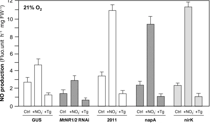
GUS Ctrl +NO2-+Tg MtNR1/2 RNAi Ctrl +NO2-+Tg 2011 Ctrl +NO2-+Tg napA Ctrl +NO2-+Tg nirK Ctrl +NO2-+Tg21% O
2Figure S3: NO production by M. truncatula GUS and nr1/nr2 , and by S. meliloti 2011, napA and nirK nodules under 21% O2. M. truncatula control (GUS) and MtNR1/2 RNAi plants were inoculated with S.
meliloti 2011 strain. NO production is expressed as relative fluorescent units. Effector concentrations
were 1 mM NaNO2 (NO2-), and 1 mM sodium tungstate (Tg). Data are the means ± SD of 2 independent experiments assayed in duplicates.
N
O
p
ro
d
u
c
ti
o
n
(
F
lu
o
.
u
n
it
h
-1m
g
F
W
-1)
+ NO 2-+AA +Rot +Myx +FCCP Ctrl 10 4 2 0 2 8 6 Ctrl 12 14 16 18 20 22Figure S4: Effects of electron transfer chain effectors on NO production of M. truncatula/S.
meliloti nodules in the presence of nitrite. M. truncatula wild type plants were inoculated with S. meliloti 2011 strain. NO production, expressed as relative fluorescent units, was measured under
1% O2. Effector concentrations were 1 mM NO2-, 10 µM rotenone (Rot), 25 µM antimycin A (AA), 25 µM myxothiazol (Myx), and 10 µM FCCP. Data are the means ± SD of 3 independent experiments assayed in duplicates.
Figure S5: Effects of NaTg on nodule nitrate reductase activity. Nodule protein extracts were preincubated for 15 min at ambiant temperature with NaTg before NR activity measurement. Data are the means ± SD of 3 independent experiments.
0
N
R
a
c
ti
v
it
y
(%
o
f
c
o
n
tr
o
l)
40
100
20
60
80
0 0.1 0.3 1 2 3
Tg (mM)
2
8
4
6
0
A
T
P
/A
D
P
r
a
ti
o
10
0 2 5 10 20
CuFl (µM)
B
T 2h
T 0h
A
1
4
2
3
5
6
Figure S6: Histochemical analysis and toxicity test of CuFl treated nodules. A, CuFL detection of NO after Incubation (0 and 2 h) of entire nodules with 5 µM CuFL. Nodules were then cut into 100 µM thick slices and analyzed with a Zeiss LSM 500 confocal laser microscope; 1 and 4, fluorescence-field images; 2 and 5, bright-field images; 3 and 6, merged images. B, nodules were incubated for 2h in the presence of various