ii
Any or all parts of this manual may be reproduced, provided the parts reproduced are free- not for sale. For commercial purposes, no part of this manual may be reproduced
or utilized in any form or by any means, electronic or mechanical, including photocopying and recording, or by any information storage and retrieval system, without permission in writing from the publisher.
The intent of this manual is to be freely used, copied, and distributed
in Developing Countries for the teaching and promotion of basic
anesthesia knowledge/skills.
The purpose of this manual is to provide developing countries with a copyright
free basic anesthesia manual. This manual can be freely copied and translated
into a native language for the promotion of basic anesthesia knowledge/skills.
Contributors with credited pictures and illustrations have graciously given
permission for their material to be used for this specific purpose. The author
and publishers of this manual cannot accept liability from the use of this
material or errors in translation. It is up to each translator to ensure that the
translation is correct. Knowledge about the art and science of anesthesia
continues to change. It is up to each anesthesia provider to continue to learn
and upgrade their knowledge. This manual only contains basic knowledge and
is not a replacement for more comprehensive anesthesia information.
iii
“Every prudent man acts out of knowledge.”
Proverbs 13:15
iv
Acknowledgements
This project would not have been possible without the help of many.
Several anesthesia providers and medical personal gave their time and effort to this worthy task. This group of academic and clinical experts provided valuable insights and gave thoughtful consideration to the content of this manual. Guidance was provided by Chuck Biddle CRNA, PhD.; Charles Reese CRNA, PhD; Carolyn Nicholson CRNA, B.S. Ed; Linda H. Croop RN, CNNP, MSN; Carolyn J. Watts MD, FRCSC; Wayne E. Smith MD; W.C. Petty MD; Thomas Fell MD; Richard Henker PhD, CRNA; Sandra Ouellette CRNA, Med; and Carson F. Frazzini CRNA, MS. Stephen “Human” Pfauter kindly applied his expertise with some digital editing as well as the cover design.
The International Federation of Nurse Anesthetists (IFNA) provided a grant for the professional illustrations contained in this manual. The illustrations were created by Welti & Rose Advertising, Inc. Photographs provided by the author and William H. Hartland Jr. CRNA, PhD.
v
Introduction
This is the second volume of Basic Guide to Anesthesia for Developing Countries. This volume contains three distinct sections: regional anesthesia; obstetric anesthesia; and trauma. Each section contains information for basic and safe care. For advanced and in-depth discussions I would refer the reader to other sources. This is simply a primer.
This journey began in 2004 with a short trip to Afghanistan. I realized that there was an absence of basic anesthesia material in a native language. As time passed I learned that another country, Cambodia, lacked a basic anesthesia text in Khmer. Since 2004 I have worked hard to produce a basic text that would be freely available without copyright restrictions for translation and use in a native language.
It is my prayer that this manual will increase knowledge and improve care of all surgical patients who undergo care under our watchful eyes. Administering anesthesia is a lifelong learning process. This manual is designed to introduce a basic foundation of knowledge to the trainee as well as to serve as a basic review for those who are practicing the art and science of anesthesia.
Every effort was made to ensure that the material and information contained in this manual is correct and up-to-date. The publishers and authors cannot accept liability from errors that may occur from the use of this material.
Please feel free to contact me at [email protected] with comments, questions, recommendations for future editions, and any concerns.
vi
Resources
A number of resources are available for the anesthesia provider in developing countries.
Books
Anaesthesia at the District Hospital, 2nd Edition. Michael B. Dobson. Published by the
World Health Organization in collaboration with the World Federation of Societies of Anesthesiologists. This manual was published to help guide medical officers in small hospitals. It contains a wealth of practical and useful information. This book is available in English.
Internet Resources
Manuals
Safe Anaesthesia. Lucille Bartholomeusz, 3rd edition updated and revised by Jean Lees. This manual is available at http://www.worldanaesthesia.org . This 700+ page manual contains comprehensive information concerning anesthesia. Individual chapters may be downloaded. This manual is available in English.
Basic Guide to Anesthesia for Developing Countries Volume 1, Daniel D. Moos.
This manual is available at http://www.worldanaesthesia.org and http://ifna-int.org . This manual can be freely downloaded, copied, and translated for the promotion of basic anesthesia knowledge and skills. The manual contains 230 pages of information which includes: medical math, documentation, fluid management/replacement, medications, preparation, positioning/monitoring, airway management, basic CPR, cardiac arrest, recovery basics, and pediatric anesthesia.
Basic Guide to Resuscitation for Developing Countries. Daniel D. Moos.
This manual is available at http://www.worldanaesthesia.org and http://ifna-int.org . This manual can be freely downloaded, copied, and translated for the promotion of basic resuscitation techniques. Additional information concerning basic resuscitation may be obtained at http://erc.edu (European Resuscitation Council) and http://americanheart.org
(American Heart Association).
Primary Trauma Care is an excellent resource for basic trauma care. It is available at http://www.primarytraumacare.org . This thirty-nine page manual is available in
vii
Education
World Anaesthesia Online can be accessed at http://www.nda.ox.ac.uk/wfsa/. This web site is dedicated to the promotion of anesthesia knowledge and skills in the developing world. Update in Anaesthesia is “An educational journal aimed at providing practical advice for those working in isolated or difficult environments.” The majority of the updates that are available online are in English. A small number of updates are available online in Russian and French. The print version is available in English, Russian, French, Mandarin, and Spanish.
World Anaesthesia can also be accessed at http://www.neda.ox.ac.uk/wfsa/ . World Anaesthesia is a newsletter of the World Federation of Societies of Anaesthesia. The newsletter allows “for the exchange of views & ideas on advancing the specialty of anaesthesia in the developing world.” It is available in English.
International Organizations
International Federation of Nurse Anesthetists (IFNA) was founded in 1989 and
currently has 34 country members. The IFNA is an international organization whose mission is in part dedicated to the advancement of educational standards and practices of anesthesia. The IFNA website is located at http://www.ifna-int.org .
Additional resources concerning the IFNA include:
Caulk R, Ouellette S M. The International Federation of Nurse Anesthetists A Professional Study and Resource Guide For The CRNA. AANA Publishing 2001; Chapter 19: 381-406. McAuliffe M. Countries where anesthesia is administered by nurses. AANAJ 64 (5), 469-479. Henry B, McAuliffe M. Practice and education of nurse anesthetists. Bulletin of the World Health Organization.The International Journal of Public Health. 77 (3), 267-270.
World Federation of Societies of Anesthesiologists (WFSA) was founded in 1955 and
currently has 122 country members. The objectives of the WFSA is the improve/disseminate knowledge concerning the standards of anesthesia, pain treatment, trauma management and resuscitation to all countries of the world. The WFSA website can be located at http://anaesthesiologists.org .
viii
Table of Contents
Section I
Chapter 1: Local Anesthetics
Chemistry 2
Structure Activity Relationships 3 Clinical Implications- Ionized/Nonionized Forms 6 Peripheral Nerve Anatomy 6 Nerve Conduction Physiology 7 Fiber Types 9 Pharmacokinetics 10 Clinical Pharmacology 15 Clinical Factors/ Local Anesthetic Activity 16 Infiltration Anesthesia 18 Topical Anesthesia 19 Methemoglobinemia and Benzocaine 21
Chapter 2: Introduction to Neuraxial Blockade
Considerations 26
Generic Indications 27 Contraindications 27 Neuraxial Blockade and Anticoagulation 29 Spinal/Epidural Hematoma Formation 30 General Recommendations 31 Antiplatelet Medications 32 Oral Anticoagulants 33 Thrombolytic/Fibrinolytics 35 Herbal Preparations 35
ix
Chapter 3: Neuraxial Blockade Anatomy, Landmarks,
Physiologic Effects & Complications
Anatomy of Vertebral Column 39 Supportive Structures 42 Blood Supply 43 Subarachnoid Space 43 Epidural Space 45 Surface Anatomy 46 Dermatome Levels 48 Assessment 49 Physiologic Effects of Neuraxial Blockade 50 Action, Spread, Uptake, Metabolism, & Elimination 50 Somatic & Autonomic Blockade 51 Cardiovascular Effects 52 Respiratory Effects 53 Renal, Metabolism, & Endocrine Effects 54 Complications 55 High Neuraxial Blockade 56 Cardiac Arrest 57 Urinary Retention 58 Inadequate Anesthesia 58 Intravenous Injection 59 Subdural Injection 60 Backache 60
Postdural Puncture Headache 61
Epidural Blood Patch 63
Neurological Injury 64 Spinal/Epidural Hematoma 65 Meningitis/Arachnoiditis 65 Epidural Abscess 66 Catheter Sheering 67
x
Neuraxial Blockade Anatomy, Landmarks, Physiologic Effects &
Complications continued…
Transient Neurological Syndrome 67 Cauda Equina 68
Chapter 4: Spinal Anesthesia
Advantages & Disadvantages 70 Action, Uptake, & Elimination 71 Factors Affecting Spinal Anesthesia 72 Local Anesthetic Characteristics 72 Patient Characteristics 74 Technique of Injection 75 Characteristics of CSF 76 Short Acting Spinal Anesthetics 77 Long Acting Spinal Anesthetics 78 Baracity 79 Additives 80 Technique 80 Monitoring 88 Obstetrics 88 Postoperative Care 88
Chapter 5: Epidural Anesthesia
Advantages & Disadvantages 91 Action, Spread, Uptake, & Elimination 92 Factors Affecting Height 94 Short Acting Local Anesthetics 95 Intermediate Acting Local Anesthetics 96 Long Acting Local Anesthetics 96 Additives 98 Technique 99
xi
Epidural Anesthesia continued…
Monitoring 106 Obstetrics 107 Postoperative Care 107
Chapter 6: Peripheral Nerve Blocks
Indications 109 Contraindications 110 Sedation 111 Maximum Local Anesthetic Doses 111 Local Anesthetic Toxicity 112 Treatment 114
Chapter 7: Femoral/ 3-in-1 Nerve Block
Indications 117 Anatomy 117 Contraindications 119 Techniques 120 Local Anesthetics 122 Complications 123
Chapter 8: Brachial Plexus Anesthesia
Anatomy 125 Interscalene Approach 129 Axillary Approach 131 Local Anesthetics 136 Complications 136
xii
Chapter 9: Peripheral Nerve Blocks at the Elbow,
Wrist, and Digital Block
Indications 139
Advantages & Disadvantages 139 Peripheral Nerve Block at the Elbow & Wrist 140 Radial Nerve 140 Median Nerve 141 Ulnar Nerve 143 Digital Nerve Block 144 Metacarpal Block 145
Chapter 10: Bier Block (Intravenous Regional
Anesthesia)
Indications 148 Advantages & Disadvantages 148 Contraindications 148 Equipment 149 Local Anesthetic Choice 149 Technique 150 Complications 153
Chapter 11: Ankle Block
Indications 156 Advantages & Disadvantages 156 Contraindications 156 Anatomy 157 Equipment 159 Local Anesthetic Choices 160 Technique 160 Complications 164 Metatarsal Block 165
xiii
Section II
Chapter 12: Obstetric Anesthesia: Anatomy, Physiology
& Anesthetic Implications
Pregnancy Related Physiological & Anatomical Changes 169 Uteroplacental Circulation 175
Labor 177
Anesthesia Considerations for Non-Obstetric Surgical
Intervention During Pregnancy 177
Chapter 13: Anesthesia Implications & Approaches for
Cesarean Section
Indications for Cesarean Section 181 Common Concerns 181 Preparation 182 Local Anesthetic Infiltration 184 Neuraxial Blockade 188 General Anesthesia 191 Failed Intubation 195 Aspiration of Gastric Contents 196
Chapter 14: Obstetric Specific Considerations
Valvular Heart Disease 198
Conditions Associated with Pregnancy 200 Obstetrical Hemorrhage 203
Chapter 15: Neonatal Resuscitation
APGAR Score 208
Neonates at Risk 209
xiv
Section III
Chapter 16: Trauma
Type of Trauma Injury 215 Basic Rules of Trauma Evaluation 217 Initial Evaluation 218 Hemorrhage 225 Secondary Examination 229 Basic Equipment in Trauma Receiving Area 231 Anesthesia for Trauma 231 Emergence and Postoperative Care 235
Appendices
236Glossary
2591 Local Anesthetics Local Anesthetics
2
Local Anesthetics
Local Anesthetics
Chapter One
Local Anesthetics
Local anesthetics produce a reversible loss of sensation in a portion of the body. Local anesthetics may be used as the sole form of anesthesia, in combination with general anesthesia, and/or to provide postoperative analgesia.
Indigenous natives of Peru chewed on leaves of Eryroxylon coca, the source of cocaine, to decrease fatigue and promote a feeling of well being. In 1884, Koller introduced cocaine as a topical anesthetic for the cornea. There were two problems with cocaine, physical dependence and toxicity. In 1905, Einhorn introduced the prototypical ester local anesthetic, procaine. In 1943, Lofgren introduces lidocaine, the prototypical amide local anesthetic.
Chemistry
The basic chemical structure of a local anesthetic molecule consists of 3 parts:
1. Lipophilic group- an aromatic group, usually an unsaturated benzene ring.
2. Intermediate bond- a hydrocarbon connecting chain, either an ester (-CO-) or amide (-HNC-) linkage. The intermediate bond determines the classification of local anesthetic.
3. Hydrophilic group- a tertiary amine and proton acceptor.
N
Lipophilic Group Intermediate Bond Hydrophilic Group
Benzene Ring Ester or Amide Linkage Tertiary Amine & (-CO- ester or –HNC- amide) Proton Acceptor3 Local Anesthetics Local Anesthetics
Amide and ester local anesthetics follow different paths of metabolism. Ester local anesthetics are more likely to cause an allergic reaction. (See Biotransformation & Excretion).
Structure Activity Relationships (Potency, Duration, &
Onset)
The intrinsic potency, duration, and onset of action for a local anesthetic are dependent upon: 1. Lipophilic-hydrophobic balance
2. Hydrogen ion concentration
Lipophilic-Hydrophobic Balance
The term “lipophilic” means “fat” loving, expressing the tendency of the local anesthetic molecule to bind to membrane lipids. The term “hydrophobic” means fear of water. The lipid membrane is a hydrophobic environment. The term “hydrophobicity” is often used to describe the physiochemical property of local anesthetics. Potency as related to local anesthetics correlates with lipid solubility. In clinical practice, the potency of a local anesthetic is affected by several factors including:
• Hydrogen ion balance
• Fiber size, type, and myelination
• Vasodilator/vasoconstrictor properties (affects rate of vascular uptake) • Frequency of nerve stimulation
• pH (an acidic environment will antagonize the block)
• Electrolyte concentrations (hypokalemia and hypercalcemia antagonizes blockade) Duration of action is associated with lipid solubility. Highly lipid soluble local anesthetics generally have a longer duration of action due to decreased clearance by localized blood flow and increased protein binding. Amides Esters Bupivacaine Benzocaine Etidocaine Chloroprocaine Levobupivacaine Cocaine Lidocaine Procaine Mepivacaine Tetracaine Prilocaine Ropivacaine
4
Local Anesthetics
Local Anesthetics
1= least; 4= greatest
Hydrogen Ion Concentration
Local anesthetics are weak bases, containing a positive charge on the tertiary amine at a physiologic pH. Local anesthetics exist in equilibrium between the basic uncharged (non-ionized) form, which is lipid soluble, and the charged (ionized) cationic form, which is water soluble. The measurement pKa expresses the relationship between the non-ionized and ionized concentrations. Specifically, pKa is the pH at which the ionized and non-ionized forms of the local anesthetic are equal.
Non-Ionized Form Ionized Form
=
pKa = pH at which ionized and non-ionized forms of local anesthetic are equal.
Local anesthetics are weak bases and contain a higher ratio of ionized medication compared to non-ionized. Increasing the concentration of non-ionized local anesthetic will speed onset. In general, local anesthetics with a pKa that approximates physiologic pH have a higher concentration of non-ionized base resulting in a faster onset. On the other hand, a local anesthetic with a pKa that is different from physiologic pH will have more ionized medication which slows onset. For example, the pKa for lidocaine is 7.8 and 8.1 for bupivacaine. Lidocaine is closer to physiologic pH than bupivacaine. Lidocaine has a greater concentration on non-ionized local anesthetic than bupivacaine which results in a faster onset. Non-ionized and ionized portions of local anesthetic solution exert distinct actions. Lipid soluble, non-ionized form of the local anesthetic penetrates the neural sheath and membrane. In the cell, the non-ionized and ionized forms equilibrate. The ionized form of the local anesthetic binds with the sodium channel. Once “bound” to the sodium channel, impulses are not propagated along the nerve.
Local Anesthetic Potency and Lipid Solubility/Duration of Action AMIDES Bupivacaine/Levo- Bupivacaine 4/4 Etidocaine 4/4 Ropivacaine 4/4 Mepivacaine 2/2 Lidocaine 2/2 Prilocaine 2/2 ESTERS Tetracaine 4/3 Cocaine 2/2 Procaine 1/1 Chloroprocaine 1/1
5 Local Anesthetics Local Anesthetics
Ionized Non-ionized penetrates the
neural sheath/membrane
Lipid
Ionized- binds with the sodium channel
Ionized form of local
anesthetic molecule
Na+ Channel
Clinically, onset of action is not the same for all local anesthetics with the same pKa. This is due to the intrinsic ability of the local anesthetic to diffuse through connective tissue. Local anesthetics with a pKa closest to the physiological pH generally have a higher concentration of non-ionized molecules and a more rapid onset. Two notable exceptions are chloroprocaine and benzocaine. Chloroprocaine has a high pKa and rapid onset. Benzocaine does not exist in an ionized form and exerts its effects by alternate mechanisms.
Local Anesthetic pKa AMIDES
Bupivacaine and levo- Bupivacaine 8.1 Ropivacaine 8.1 Lidocaine 7.8 Prilocaine 7.8 Etidocaine 7.7 Mepivacaine 7.6 ESTERS Chloroprocaine 9.0 Procaine 8.9 Cocaine 8.7 Tetracaine 8.2
6
Local Anesthetics
Local Anesthetics
Clinical Implications of Hydrogen Ion Concentration
Local anesthetics are prepared as a water soluble hydrochloride salt and generally have a pH of 6-7. If the commercial preparation contains epinephrine, the solution must be acidic to create a stable environment. The corresponding pH is in the range of 4-5. Commercial preparations with epinephrine have less free base, slowing the onset of action. To enhance clinical onset, carbonated solutions of epinephrine containing local anesthetics have been used instead of HCL solutions. Alternatively, adding sodium bicarbonate to commercial preparations of epinephrine containing local anesthetic solutions can hasten the onset. One (1) ml of 8.4% sodium bicarbonate should be added to each 10 ml of lidocaine or mepivacaine, and 0.1 ml of 8.4% of sodium bicarbonate should be added to each 10 ml of bupivacaine. Increasing the volume of sodium bicarbonate added the local anesthetic preparation may lead to precipitation. Altering the pH to a more basic solution will increase the amount of non-ionized compared to ionized which will speed onset. Sodium bicarbonate increases the amount of free base, increases onset, improves the quality of the block, and decreases pain associated with subcutaneous infiltration.
Peripheral Nerve Anatomy
Axolemma- the peripheral nerve axon cell membrane.
Non-myelinated nerves- contain axons within a single Schwann cell (i.e. autonomic postganglionic
efferent and nociceptive afferent C fibers).
Large motor and sensory fibers are enclosed in many layers of myelin.
Myelin insulates the axolemma and speeds conduction to the nodes of Ranvier. The nodes of Ranvier are interruptions in the myelin, allowing current regeneration. High concentrations of Na+ channels are located at the nodes of Ranvier.
7
Local Anesthetics
Non-myelinated fibers have Na+ channels distributed along the axon.
A peripheral nerve contains several axon bundles called fascicles. Endoneurium is the connective tissue that covers an individual nerve. Perineurium is the connective tissue that covers each fascicle.
Epineurium is the connective tissue covering the entire nerve.
Schematic Illustration of Nerve
Epineurium Perineurium
Endoneurium
Nerve Conduction Physiology
The neural membrane contains a voltage difference of +60 mV (inner) to -90 mV (outer). At rest the neural membrane is impermeable to Na+ ions, and selectively permeable to K+ ions. The Na+/K+ pump maintains the ion gradient. The K+ to Na+ gradient is constant at 30:1. Within the cell, the concentration of K+ is kept at 30, and outside the cell it is maintained at 1. Sodium, on the other hand, is at higher concentrations outside the cell.
At Rest
Outside Cell
-90 mV K+ concentration low; Na+ concentration high
Neural Membrane dd
+ 60 mV K+ concentration high; Na+ concentration low
Inside Cell
Local Anesthetics
8
Documentation
Local Anesthetics
During an action potential, the nerve membrane switches its permeability from K+ to Na+, changing the membrane potential from -90 to +60 mV (negative to positive) and back again.
Action Potential
Outside Cell
+60 mV
Na+ Neural Membrane dd -90 mV K+
Inside Cell
Local anesthetics produce a conduction block of neural impulses, preventing the passage of Na+ through Na+ channels.
Local Anesthetic
Molecule
Na+ Channel
Local anesthetics DO NOT alter the resting membrane potential. The Na+ channel acts as a receptor for local anesthetic molecules. Local anesthetics are stereospecific. Their action is dependant on the conformational state of the Na+ channel. Local anesthetics bind more readily to the Na+ channel during depolarization. This may occur during the “open” and “inactivated” state. Local anesthetics may bind during the resting state but not as readily as during the “open” or “inactivated” state.
9
Documentation
Conformational State of Na+ Channel
Open Inactivated Closed
(activated) (resting)
In the “inactivated” state, local anesthetics stabilize the Na+ channel. Local anesthetic molecules may bind within the Na+ channel as well as block the external opening. This action prevents permeability to Na+, slowing the rate of depolarization. The rate of depolarization is slowed; threshold potential is not met or propagated along the nerve membrane. Repeated depolarization increases the number of local anesthetic molecules bound to Na+ channels by increasing the number of available binding sites. Local anesthetic disassociation from inactivated channels occurs at a slower rate compared to resting channels.
Fiber Types
There are several classifications of nerve fibers. The classification of a nerve fiber impacts its sensitivity to local anesthetics. The order of susceptibility to blockade by fiber type is as follows: (least susceptible to most susceptible) small myelinated fibers (Aơ motor) < Aơ type Ia; Aơ type Ib; AƢ type II; Aƣ < AƤ sensory fibers < small, non-myelinated C fibers, and partially myelinated B fibers. Fiber Type Function Diameter (mm) Speed of Conduction Local Anesthetic Sensitivity* Myelination
AĮ Motor 12-20 Fast 1 Yes
AĮ Proprioception 12-20 Fast 2 Yes
AĮ Proprioception 12-30 Fast 2 Yes
Aȕ Touch Pressure/Proprioception 5-12 Medium 2 Yes
AȖ Motor 3-6 Medium-Slow 2 Yes
Aį Pain
Cold Temperature Touch
2-5 Medium-Slow 3 Yes
B Preganglionic autonomic fibers <3 Medium-Slow 4 Some
C (dorsal root)
Pain Warm and Cold
Touch
0.4-1.2 Slow 4 No
C
(sympathetic) Postganglionic sympathetic fibers 0.3-1.3 Slow 4 No
(* local anesthetic sensitivity= 1 is the least sensitive and 4 is most sensitive)
Local Anesthetics
10
Documentation
Local Anesthetics
Summary of Impulse Blockade by Local Anesthetics
1. Local anesthetic is deposited near a nerve. A portion of the local anesthetic is removed due to tissue binding and circulation. If the local anesthetic is an ester, a portion of the deposited local anesthetic will be removed by local hydrolysis in addition to tissue binding and circulation. The remaining local anesthetic penetrates the nerve sheath.
2. Local anesthetic penetrates the axon membranes and axoplasm. This step is dependant on pKa and lipophilicity.
3. Local anesthetic binds to Na+ channels preventing their opening by inhibiting conformational changes resulting in activation. Local anesthetics may also bind to the channel pore and block the passage of Na+.
4. During onset, impulse blockade is incomplete. Partially blocked fibers are inhibited by repetitive stimulation. The reverse is true during recovery.
5. The primary route for local anesthetics is the hydrophobic route, within the axon membrane. 6. Onset is due to the slow diffusion of local anesthetic molecules into the nerve, NOT by binding to ions, which occurs at a faster rate. Recovery occurs in reverse.
Pharmacokinetics
Pharmacokinetics involves the medication/body interaction. It is how the body handles medication. Principles of pharmacokinetics include: absorption, distribution, metabolism, and elimination. The local anesthetic blood concentration is determined by:
• Amount of local anesthetic injected • Absorption rate
• Site of injection
• Rate of tissue distribution • Rate of biotransformation • Excretion rate
Patient related factors include: • Age
• Cardiovascular status • Hepatic function
Systemic Absorption of Local Anesthetics
Systemic absorption of local anesthetics is determined by: • Site of injection
• Dose and volume
• Addition of vasoconstrictor
• Pharmacologic profile of the local anesthetic
11
Documentation
1. Site of Injection
Site of injection impacts blood levels of local anesthetic. Areas of high vascularity result in greater uptake and higher blood concentrations. The uptake of local anesthetic from greatest to least is as follows:
IV> tracheal> intercostal> caudal> paracervical> epidural> brachial> sciatic> subcutaneous
Uptake of Local Anesthetics Based on
Regional Anesthetic Technique
Result in Highest Blood Concentrations
Intravenous Tracheal Intercostal Caudal Paracervical Epidural Brachial Sciatic Subcutaneous
Lower Blood Concentrations of Local Anesthetic
Clinically, the site of injection plays an important role in toxicity. For example, 400 mg of plain lidocaine in the intercostal space may lead to peak blood concentrations of 7 mcg/ml. This may result in CNS toxicity. In contrast, 400 mg of plain lidocaine in the brachial plexus will yield blood levels of 3 mcg/ml, which is not toxic.
Toxicity associated signs and symptoms may vary among local anesthetics. For example, with lidocaine there is a larger disparity in blood concentrations required to cause CNS signs and symptoms compared to concentrations that result in cardiovascular collapse. With bupivacaine there is a small difference in the blood concentrations that may result in CNS signs and symptoms and concentrations that result in cardiovascular collapse. Often seizures occur at the same time as cardiovascular collapse. Ropivacaine is similar to bupivacaine with respect to onset and duration. However, blood concentrations of ropivacaine required to cause cardiovascular collapse are much higher than bupivacaine. Ropivacaine has a larger margin of safety. In addition, metabolism plays a role in toxicity. Amides have a high rate of first pass metabolism as the local anesthetic passes through the liver. Slow absorption from tissue is less likely to result in toxicity. Toxicity is often the result of intravenous/intra-arterial injection or overdose.
Local Anesthetics
12
Local Anesthetic Toxicity Lidocaine
Cardiac depression/arrest
Signs & symptoms Respiratory arrest
based on increasing
blood concentrations Seizures
of lidocaine.
Dizziness, ringing of ears,
‘funny’ taste in the mouth.
2. Dose and volume
Blood concentrations of local anesthetics correspond proportionally to the total dose. Higher blood concentrations are associated with large volumes of dilute local anesthetic compared to the same dose in a smaller volume.
3. Vasoconstrictor
Epinephrine, in concentrations of 5-10 mcg/ml, is commonly used to decrease the absorption of local anesthetics. A 5 mcg/ml (1:200,000) dose of epinephrine will significantly reduce the peak blood levels of lidocaine and mepivacaine. Epinephrine does not affect the vascular absorption of etidocaine and bupivacaine in the epidural space. However, the addition of epinephrine does significantly reduce the vascular absorption of etidocaine and bupivacaine when utilized for peripheral nerve blocks. Benefits of decreased absorption include increased neuronal uptake, enhanced quality of analgesia/anesthesia, prolonged duration of action, and decreased risk of toxicity.
A concentration of 1:200,000 (5 mcg/ml) is commonly used for peripheral nerve blocks to reduce vascular absorption. To add epinephrine to local anesthetic solutions use a 1mg/ml (1:1000) ampoule of epinephrine. Take the total volume of local anesthetic, divide it in half, and move the decimal point two places to the left. For example, 40 ml of 1% lidocaine, divide 40 by 2 and 20 is the result. Next, move the decimal point two places to the left. The result is 0.20. This is the amount of epinephrine added to the local anesthetic solution to yield a 1:200,000 concentration. To check the calculation, multiply 5 mcg/ml by 40 ml, which equals 200 mcg. It is important to always check the concentration of epinephrine and the total dose added to the local anesthetic.
Local Anesthetics
13
A second technique for adding epinephrine to local anesthetic preparations is detailed below: • 1:200,000 epinephrine concentration would equal 5 mcg/ml.
• Dilute epinephrine using a 10 ml syringe. Draw up 1 ml of 1:1000 epinephrine (1 mg per ml) and 9 ml of normal saline.
• Mix it by tilting the syringe back and forth.
• The concentration of epinephrine is now 100 mcg per ml.
• Add epinephrine to the local anesthetic solution (see table below).
1:200,000 Epinephrine Concentration
Volume of Local Anesthetic Amount of Epinephrine Added to Local Anesthetic Solution
20 ml 100 mcg of epinephrine
30 ml 150 mcg of epinephrine
40 ml 200 mcg of epinephrine
50 ml 250 mcg of epinephrine
• Always label the syringe of epinephrine. Once the epinephrine is added to the local anesthetic, discard what remains. Epinephrine can be lethal and should be discarded to avoid inadvertent administration.
• Epinephrine containing local anesthetics should never be injected into end organs such as ears, nose, penis, fingers, or toes. Epinephrine may cause vasoconstriction and subsequent necrosis of tissue.
4. Pharmacologic Profile
Individual local anesthetics exhibit different rates of absorption. For example, it has been found that during brachial plexus blockade, lidocaine is absorbed faster than prilocaine, and bupivacaine is absorbed more rapidly than etidocaine. In general, local anesthetics that are highly tissue bound are absorbed at a slower rate. In addition, absorption is dependant on the individual local anesthetics intrinsic ability to cause vasodilatation.
Distribution of Local Anesthetics
A two compartment model describes the systemic distribution of local anesthetics. The rapid disappearance phase (ơ phase) is related to uptake by rapidly equilibrating tissue (tissue with high vascular perfusion which include the brain, lung, liver, kidney, and heart). The slow phase of disappearance (Ƣ phase) is the function of the individual local anesthetics distribution to muscle tissue and the gut.
Local anesthetics are distributed to all tissues. Higher concentrations of local anesthetics are found in highly perfused organs compared to tissues that receive lower rates of perfusion. The pulmonary system is responsible for extraction of local anesthetics. As local anesthetics are transported through the pulmonary vasculature, levels are greatly reduced. The largest reservoir for local anesthetics is the skeletal muscle.
Local Anesthetics
14
Biotransformation and Excretion of Local Anesthetics
The metabolism of local anesthetics is dependent upon their classification: ester vs. amide. Ester local anesthetics undergo extensive hydrolysis in the plasma by pseudocholinesterase enzymes (plasma cholinesterase or butyrylcholinesterase). Ester hydrolysis is rapid, resulting in water soluble metabolites which are excreted in the urine. The ester that is an exception is cocaine. In addition to ester hydrolysis cocaine is partially metabolized in the liver (N-methylation). Patients with pseudocholinesterase deficiency are at risk for toxicity (genetic or liver disease). This is due to slowed metabolism and accumulation of the ester local anesthetic. Procaine and benzocaine are metabolized to p-aminobenzoic acid (PABA), which has been associated with allergic reactions. Benzocaine may result in methemoglobinemia. When ester local anesthetics are placed in the CSF, metabolism does not occur until there has been vascular absorption of the local anesthetic. CSF does not contain esterase enzymes.
Amide local anesthetics are metabolized primarily by microsomal P-450 enzymes in the liver (N-dealkylation and hydroxylation) and, to a lesser extent, in other tissues. The rate of metabolism among amides varies.
prilocaine> lidocaine> mepivacaine> ropivacaine> bupivacaine
.Prilocaine metabolites include o-toluidine derivatives, which can accumulate after large doses (>10 mg/kg), resulting in the conversion of hemoglobin to methemoglobinemia. Treatment for methemoglobinemia includes the administration of methylene blue. Methylene blue is generally available in a 1% solution. 1-2 mg/kg should be administered over 5 minutes. Methylene blue reduces methemoglobin to hemoglobin.
The excretion of amide local anesthetics occurs in the kidneys. Less than 5% of the unchanged medication is excreted by the kidneys.
Patient Alterations in the Pharmacokinetics of Local Anesthetics
Age is one factor that alters the pharmacokinetics of local anesthetics. Changes in half life have been demonstrated for the elderly and newborns. In both populations, lidocaine has been found to have an increased half life. Newborns have an immature hepatic enzyme system, whereas the elderly have decreased hepatic blood flow. The second factor that affects the pharmacokinetics of local anesthetics includes any disease process (i.e. hepatitis) that diminishes hepatic blood flow or impairs the livers ability to produce enzymes. This may result in elevated levels of amide local anesthetics in these patients compared to patients with normal liver function.
Local Anesthetics
15
Clinical Pharmacology
General Considerations
General considerations related to local anesthetics include the following: • Anesthetic potency
• Onset of action • Duration of action
• Differential sensory/motor blockade
Potency, onset, and duration were covered earlier under structure activity relationships. For completeness they will be briefly covered under clinical pharmacology.
Anesthetic Potency
The primary factor related to potency is the hydrophobicity (lipid solubility) of the local anesthetic. Local anesthetics penetrate the nerve membrane and bind to Na+ channels, which are hydrophobic. Additional factors include:
• Fiber size, type, and myelination • Hydrogen ion balance
• Vasodilator/vasoconstrictor properties (affects the rate of vascular uptake) • Frequency of nerve stimulation
• pH (acidic environment will antagonize the block)
• Electrolyte concentrations (hypokalemia and hypercalcemia antagonizes blockade).
Onset of Action
In the individual nerve, onset is related to the unique physiochemical property of the local anesthetic. Clinically, the onset of action is related to pKa, dose, and concentration.
• pKa – when pKa approximates the physiologic pH a higher concentration of non-ionized base is available, increasing onset of action.
• Dose- the higher the dose of local anesthetic administered, the faster the onset.
• Concentration- higher concentrations of local anesthetic will result in a more rapid onset.
Duration of Action
Duration of action is dependant on individual local anesthetic characteristics. Local anesthetics are classified as follows:
• Short acting: procaine and chloroprocaine
• Moderate acting: lidocaine, mepivacaine, prilocaine
• Long acting: tetracaine, bupivacaine, etidocaine, ropivacaine, levobupivacaine
Local Anesthetics
16
Duration of action is influenced by peripheral vascular effects that local anesthetics exhibit. Local anesthetics exhibit a biphasic effect on vasculature smooth muscle. At low, sub-clinical doses, vasoconstriction is noted. With larger, clinically relevant doses, vasodilatation is seen. The degree of vasodilatation varies among individual local anesthetics. For example, lidocaine> mepivacaine> prilocaine. The effect of local anesthetics on vascular tone and regional blood flow is complex and dependant on the following:
• Concentration • Time
• Type of vascular bed
Ropivacaine is unique among local anesthetics since it exhibits a vasoconstrictive effect at clinically relevant doses.
Differential Sensory/Motor Blockade
Local anesthetics have the ability to produce varying degrees of inhibition for sensory and motor activity. For example, bupivacaine and etidocaine are both potent, long acting local anesthetics. Bupivacaine exhibits a more potent sensory than motor block. Etidocaine exhibits an equally effective sensory and motor block. Ropivacaine, on the other hand, exhibits a potent sensory block similar to bupivacaine but motor blockade appears less intense.
Factors Affecting Local Anesthetic Activity Clinically
Dose
An increase in the dose of a local anesthetic will increase the likelihood of a successful block while decreasing the time to onset. An increase in the volume of local anesthetic will be beneficial in the anatomical spread of anesthesia.
Addition of Vasoconstrictors
Epinephrine is the most commonly used vasoconstrictor. The usual dose and concentration is 5 mcg/ml or 1:200:000. Norepinephrine and phenylephrine have been used as vasoconstrictors but do not exhibit properties that make them superior to epinephrine. Epinephrine acts to decrease vascular absorption, reduces blood concentration of local anesthetics, and decreases the risk of toxicity thus allowing more local anesthetic molecules to reach the nerve membrane. As more molecules reach the nerve membrane, there is an increase in the depth and duration of local anesthetic blockade. Epinephrine prolongs the duration of blockade for most short to moderate acting local anesthetics. The addition of epinephrine for neuraxial blockade has the added benefit of activating endogenous analgesic mechanisms through ơ-Adrenergic receptors. This may increase the intensity of analgesic action. The addition of vasoconstrictors is controversial. Some advocate that vasoconstrictors may result in nerve injury due to decreased blood flow. Epinephrine containing
Local Anesthetics
17
local anesthetics should never be injected into end organs such as ears, nose, penis, fingers, or toes. Epinephrine may cause vasoconstriction and subsequent necrosis of tissue.
Site of Injection
The anatomical location of blockade influences onset and duration. Location affects the rate of diffusion, vascular absorption, and the amount of local anesthetic administered. Subarachnoid blockade exhibits the most rapid onset and shortest duration of action. Rapid onset, within the subarachnoid space, occurs because nerve roots are not covered with a sheath. The short duration of action is related to the small dose and volume of local anesthetic used to produce anesthesia. Brachial plexus blockade, in contrast, has slower onset and longer duration of action. Local anesthetics are deposited in the sheath around the brachial plexus. Diffusion must take place before reaching the site of action. The long duration of action is related to slow vascular absorption, large doses, and increased exposure of neural tissue to local anesthetics.
Carbonation and pH adjustment
In the isolated nerve, adding sodium bicarbonate, or carbon dioxide, may accelerate the onset of action. The addition of bicarbonate increases the pH, which in turn, increases the amount of local anesthetic in the uncharged base form. This theoretically accelerates the rate of diffusion across the sheath and membrane, resulting in a faster onset. Controversy exists concerning the clinical utility of pH adjustment. Studies remain ambiguous concerning the use of sodium bicarbonate to improve the speed of local anesthetic induced anesthesia.
Mixtures of Local Anesthetics
Clinicians occasionally will combine local anesthetics to achieve quick onset and long duration. Often the clinician will combine a local anesthetic with a fast onset with a local anesthetic that has a long duration of action to achieve this goal. Clinical trials have yielded mixed results. Chloroprocaine and bupivacaine, in the brachial plexus region, have achieved a quick onset/prolonged duration. However, when used for epidural anesthesia it was found that the duration of action was shorter than if bupivacaine was administered alone. Clinically there are few advantages to this technique. It should be noted that when mixing local anesthetics the risk of toxicity remains. Care should be exercised not to exceed the maximum dose. Toxicities of local anesthetics are not independent, but additive! A solution containing 50% of the toxic dose of local anesthetic A, and 50% of the toxic dose of local anesthetic B, will have the same implications as 100% of the toxic dose of either local anesthetic alone.
Pregnancy
Hormonal changes during pregnancy are primarily responsible for the enhanced potency of local anesthetics. Mechanical factors such as dilated epidural veins, which decrease the volume of the epidural or subarachnoid space, may play a minor role in later stages of pregnancy. The spread and depth of epidural/spinal anesthesia is greater in the pregnant patient when compared to the patient
Local Anesthetics
18
who is not pregnant. It has been found that the spread of local anesthetics are more extensive with epidural anesthesia as early as the first trimester. There is a correlation between progesterone levels and the mg per segment requirement of lidocaine required for the parturient. Based on current research, the dose of local anesthetics should be reduced for the pregnant patient by 30%, regardless of the trimester of pregnancy.
Medication Interactions with Local Anesthetics
Ester Local Anesthetics Succinylcholine- may potentiate the effects since both are dependent on pseudocholinesterase for metabolism.
Ester Local Anesthetics Cholinesterase inhibitors such as neostigmine and pyridostigmine can lead to a decrease in the metabolism of ester local anesthetics.
Ester Local Anesthetics Decreased pseudocholinesterase activity during pregnancy and postpartum period. Local Anesthetics in
General Opioids and alpha adrenergic agonists potentiate the analgesic effects of local anesthetics. Local Anesthetics in
General
Potentiate the effects of non-depolarizing muscle relaxant blockade. Chloroprocaine
(epidural) May interfere with the analgesic effects of subarachnoid opioids.
Lidocaine Cimetidine and propranolol decrease hepatic blood flow and lidocaine clearance. This acts to increase the risk of systemic toxicity.
Infiltration and Topical Local Anesthetics
Infiltration Anesthesia/Postoperative Analgesia
On occasion, anesthesia providers will place field blocks either as supplementation for a marginal regional anesthetic block or as the sole form of anesthesia. In addition, anesthesia providers should be knowledgeable about this form of anesthesia/analgesia for our surgeon and patient’s sake. It is not uncommon to be asked by the surgeon, “How much can I inject?” As the ‘expert’ in local anesthetics, the anesthesia provider may be called upon to share their knowledge with surgical colleagues.
It is essential to know maximum local anesthetic dosages for plain and epinephrine containing local anesthetic solutions. Knowledge of the local anesthetic concentration/dose and the patients’ weight will allow for rapid calculation of the maximum dose and volume of local anesthetic that can be safely administered. In addition to the maximum dose based on mg/kg, there is a total maximum dose regardless of weight. Avoidance of toxic dosages is essential.
Basic facts about infiltration:
• Almost any local anesthetic can be used for infiltration anesthesia.
• Onset is almost immediate for intradermal and subcutaneous administration.
• Epinephrine will prolong the duration of action of all local anesthetics, but is most pronounced with lidocaine.
Local Anesthetics
19
• The conscious patient will experience some discomfort during infiltration due to the acidic nature of these solutions.
• Epinephrine containing local anesthetics should never be injected into end organs such as ears, nose, penis, fingers, or toes. Epinephrine may cause vasoconstriction and subsequent necrosis of tissue.
Commonly administered local anesthetics for infiltration
Plain local anesthetics (maximum doses based on 70 kg)
Local Anesthetic Type Concentration % Max dose Max dosemg/kg
Duration
Lidocaine amide 0.5-1.0 300 4.5 30-60 minutes
moderate duration
Mepivacaine amide 0.5-1.0 300 4.5 45-90 minutes
moderate duration
Bupivacaine amide 0.25-0.5 175 2.5 120-240 minutes
long duration
Ropivacaine amide 0.1-1 200 3 120-360 minutes
long duration
Local anesthetics with epinephrine (1:200,000) for infiltration
Local Anesthetic Type Concentration % Max dose Max dosemg/kg
Duration
Lidocaine amide 0.5-1.0 500 7 120-360 minutes
moderate duration
Mepivacaine amide 0.5-1.0 500 7 120-360 minutes
moderate duration
Bupivacaine amide 0.25-0.5 225 3 180-420 minutes
long duration
Topical Anesthesia
Several local anesthetics can be used for topical anesthesia. The most common local anesthetics include:
• Lidocaine • Dibucaine • Tetracaine • Benzocaine
• EMLA (eutectic mixture of local anesthetic)
Topical local anesthetics provide effective, short term analgesia when applied to mucous membranes and abraded skin. Lidocaine and tetracaine sprays can be used for endotracheal anesthesia prior to
Local Anesthetics
20
intubation. EMLA is a preparation used to provide cutaneous anesthesia through intact skin. EMLA is a mixture of 2.5% lidocaine and 2.5% prilocaine. The risk of methemoglobinemia is very rare. EMLA is effective in anesthetizing the skin in preparation for the placement of intravenous needles and skin grafting procedures. To be effective, EMLA must be placed under an occlusive dressing for 45-60 minutes.
Topical anesthesia is used in the emergency room for repairing lacerations. TAC is a mixture of 0.5% tetracaine, 1:200,000 epinephrine, and 10-11.8% cocaine. It is safe to use on skin, but should not be used on mucous membranes since rapid absorption may lead to toxicity. The maximum dose for adults is 3-4 ml. For the pediatric population, a dose of 0.05 ml/kg is considered safe. Concerns about cocaine toxicity, abuse, or diversion has led to the creation of an equally effective preparation, LET. LET is a preparation of lidocaine, epinephrine, and tetracaine. In the past ENT surgeons used cocaine for vasoconstriction and anesthesia; however this practice is rapidly being replaced by the use of oxymetazoline or phenylephrine in combination with a local anesthetic, such as 2-4% lidocaine. Dilute solutions should be used in children. Of concern to the anesthesia provider is the systemic absorption of phenylephrine, which can result in hypertension and reflex bradycardia. Oxymetazoline has a larger margin of safety and is absorbed less systemically.
Common Topical Preparations
Anesthetic Concentration % Form Area of useBenzocaine 1.5 20 20 Cream Ointment Aerosol
Skin and mucous membrane Skin and mucous membrane Skin and mucous membrane
Cocaine 4.0 solution Ear, nose, throat
Dibucaine 0.25-1.0 0.25-1.0 0.25-1.0 0.25 2.5 Cream Ointment Aerosol Solution suppositories Skin Skin Skin Ear Rectum Lidocaine 2-4 2 2.5-5 2 10 10 Solution Jelly Ointment Viscous Suppositories aerosol
Oropharynx, trachea, nose Urethra
Skin, mucous membranes Oropharynx Rectum Gingival mucosa Tetracaine 0.5-1.0 0.5-1.0 0.25-1.0 Ointment Cream solution
Skin, rectum, mucous membranes Skin, rectum, mucous membranes Nose, tracheobronchial tree
EMLA Lidocaine 2.5
Prilocaine 2.5
cream Intact skin
TAC Tetracaine 0.5
Epinephrine 1:200,000 Cocaine 11.8
solution Cut skin
LET Lidocaine 4
Epinephrine 1:200,000 Tetracaine 0.5
solution Cut skin
Local Anesthetics
21
Methemoglobinemia and Benzocaine
Benzocaine administration to the mucous membranes can result in the relatively uncommon but potentially fatal complication of methemoglobinemia. The anesthesia provider may encounter methemoglobinemia by assisting in airway management in another department, or in the OR when using this local anesthetic to anesthetize the upper airway.
Benzocaine made its debut into clinical use in 1900. It is used solely as a topical anesthetic. Benzocaine is the most commonly implicated local anesthetic associated with methemoglobinemia. The incidence of methemoglobinemia has been reported as high as 1 in 7,000 exposures. Up to 35% of topical benzocaine, when applied to mucous membranes, can be absorbed systemically. Inflamed areas of the mucous membranes absorb benzocaine at a higher rate. One of the problems with the administration of topical benzocaine sprays is estimating how much local anesthetic has been delivered. Application of topical benzocaine to mucous membranes should be limited to 1 second. Clinicians often fail to realize the significant absorption rate of benzocaine. In addition, clinicians may use multiple sprays or spray for longer than 1 second. In a review of benzocaine induced methemoglobinemia it was found that 46.4% of the cases reported had more than 1 spray of benzocaine.
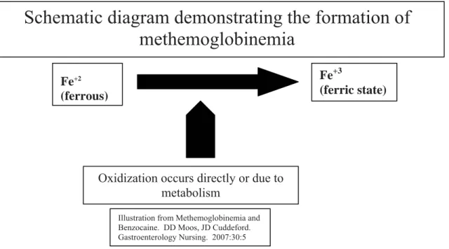
Methemoglobinemia
Hemoglobin contains four heme groups (Fe+2) located on the surface of the molecule. Heme has the ability to reversibly bind with oxygen. Methemoglobin (MHb) is a form of hemoglobin that is unable to bind with oxygen. The ferrous irons (Fe+2) of the heme are oxidized to a ferric iron (Fe+3). The ferric heme is unable to bind with oxygen, resulting in a diminished ability to deliver oxygen to tissue.
Fe+2
(ferrous)
Oxidization occurs directly or due to metabolism
Fe+3
(ferric state)
Schematic diagram demonstrating the formation of
methemoglobinemia
Illustration from Methemoglobinemia and Benzocaine. DD Moos, JD Cuddeford. Gastroenterology Nursing. 2007:30:5
Local Anesthetics
22
Signs and Symptoms of Methemoglobinemia
Signs and symptoms are dependent on the levels of MHb. Patients with anemia and cardiopulmonary disorders may exhibit signs and symptoms earlier. When levels of MHb reach 10% or greater, the patient may appear cyanotic. MHb levels of 15% or greater may demonstrate: cyanosis, headache, weakness, dizziness, lethargy, and tachycardia. Levels between 10-20% are usually well tolerated. At levels of 45% or greater, signs and symptoms may include dyspnea, cyanosis, seizures, coma, dysrhythmias, and heart failure. At levels 70% or greater, mortality can occur.
Diagnosis
Methemoglobinemia should be considered in any patient who develops cyanosis after the use of topical pharyngeal anesthesia. Pulse oximetry readings will be inaccurate and not reflect the degree of hypoxia the patient is experiencing. Readings may range from 80-85% regardless of the severity of methemoglobinemia. A MHb level greater than 10% will result in an oximetry reading that is unreliable. Co-oximetry is able to differentiate between oxyhemoglobin, deoxyhemoglobin, carboxyhemoglobin, and MHb. The gold standard for confirming a diagnosis of methemoglobinemia is co-oximetry. This is available with most, but not all, ABG determinations. It is important to request co-oximetry when sending blood samples to the laboratory, if available.
Treatment
Patients who become cyanotic or hypoxic after the application of benzocaine should have supplemental oxygen placed. If their condition improves, then further evaluation for cardiopulmonary problems should be considered. If their condition does not improve and methemoglobinemia is suspected, then an arterial blood gas (ABG) with co-oximetry should be sent for evaluation. Methylene blue administration is not recommended until the presence of MHb is confirmed by co-oximetry, if available. Methylene blue, 1-2 mg/kg, is the treatment of choice for methemoglobinemia and should be administered over 5 minutes. Methylene blue accelerates the capacity of NADPH MHb reductase to reduce MHb. Reported side effects of methylene blue include: dizziness, confusion, restlessness, headache, abdominal pain, nausea and vomiting, dyspnea, hyper/hypotension, and diaphoresis. If the patient’s condition improves after the administration of methylene blue, the patient should be monitored for the reoccurrence of symptoms. Methylene blue will not improve methemoglobinemia related to G-6-deficiency, NADPH methemoglobinemia, and cytochrome b5 reductase deficiency. Patients with a G-6-deficiency require transfusion or dialysis for treatment and methylene blue administration should be avoided. Patients with NADPH deficiency may acquire hemolytic anemia with the administration of methylene blue. For patients with no contraindications, repeated doses of methylene blue may be required. A second dose may be repeated in an hour. The total dose should not exceed 7 mg/kg since excessive methylene blue administration can result in methemoglobinemia. After initial treatment, the patient should be transferred to the intensive care unit for monitoring. Additional MHb levels should be measured at
Local Anesthetics
23
hours 2 and 8 after the initial dose of methylene blue to monitor the patient for rebound methemoglobinemia.
Summary of Common Local Anesthetics
Local Anesthetic Type Onset of Action Duration Clinical UseProcaine Ester Slow Short Spinal
Bupivacaine Amide Moderate Long Peripheral Nerve Blocks
Infiltration Spinal Epidural
Ropivacaine Amide Moderate Long Peripheral Nerve Blocks
Epidural
Chloroprocaine Ester Fast Short Peripheral Nerve Blocks
Epidural
Etidocaine Amide Fast Long Peripheral Nerve Blocks
Infiltration Epidural
Lidocaine Amide Fast Moderate Peripheral Nerve Blocks
Infiltration Spinal Epidural Bier Block
Mepivacaine Amide Fast Moderate Peripheral Nerve Blocks
Infiltration
Prilocaine Amide Fast Moderate Peripheral Nerve Blocks
Infiltration Bier Block
Practical Application
A.) The surgeon wishes to use 1% plain lidocaine to infiltrate along the incision in a 4 kg pediatric patient. How much can he use?
• What about 0.5% plain lidocaine?
• What about 0.5% lidocaine with epinephrine? • What about 0.5% plain bupivacaine?
• What about 0.5% bupivacaine with epinephrine?
B.) The surgeon wishes to use 1% plain lidocaine to infiltrate the wound in a 55 kg adult patient. How much can he inject?
• What about 0.5% plain lidocaine?
• What about 0.5% lidocaine with epinephrine? • What about 0.5% plain bupivacaine?
Local Anesthetics
24
• What about 0.5% bupivacaine with epinephrine?
References
Tuckley JM. The Pharmacology of Local Anesthetic Agents. Anaesthesia Update. Issue 4, Article 7. 1994. Ezekiel MR. Handbook of Anesthesiology. Current Clinical Strategies Publishing. Laguna Hills, California. 2002. Morgan GE, Mikhail MS, & Murray MJ. Local Anesthetics. Pages 265-270;274. Lange Medical
Books/McGraw-Hill Medical Publishing Division. 2006.
Strichartz GR & Berde CB. Local Anesthetics. In Miller’s Anesthesia 6th edtion. Miller, RD ed. Pages 573-586;589-592.
Elsevier, Philadelphia, Penn. 2005.
Dobson MB. Conduction Anaesthsia. In Anaesthesia at the District Hospital. Pages 86-102. World Health Organization. 2000.
Arias MG. Levobupivacaine. Update in Anaesthesia. Issue 14; Article 7. 2002.
Williams JR. Local Anesthetics. In Nurse Anesthesia 3rd edition. Nagelhout, JJ & Zaglaniczny KL ed. Pages 126-148.
Local Anesthetics
25
Introduction to Neuraxial
Blockade
Intro Neuraxial Blockade
26
Chapter Two
Introduction to Neuraxial Blockade
Neuraxial blockade encompasses both spinal and epidural anesthesia. Neuraxial blockade offers several advantages to the patient when compared to general anesthesia. These include:
• Decreased incidence of nausea and vomiting • Decreased blood loss
• Decreased incidence of graft occlusion
• Improved mobility following major knee surgery
• Superior pain control in the immediate postoperative period
• Decreased alteration in the patient’s cardiopulmonary physiological status • Improved patient satisfaction (especially in elderly)
• Less immunosuppression
• An alternative to general anesthesia for patients with a history of malignant hyperthermia • An alternative for patient’s that may not tolerate a general anesthetic
• Less cognitive impairment (especially in the elderly) • Enhances flexibility/options for anesthetic care
Considerations
There are several factors that the anesthesia provider should consider when deciding on which anesthetic techniques to present to the patient. Examine the patients back for surgical scars, scoliosis, skin lesions, and surface anatomy that may make neuraxial blockade difficult. There are no routine preoperative tests for healthy patients undergoing neuraxial blockade. However, patients with a history of medications/medical conditions that may increase the risk of bleeding should have coagulation studies and platelet counts drawn. The patient should be assessed for thrombocytopenia prior to the initiation of neuraxial techniques. If your setting does not have the ability to perform coagulation studies/platelet counts the following signs and symptom may indicate bleeding tendencies:
• Blood in the urine
• Bleeding around the gums
• Petechiae (small purple colored spots on the skin) In addition the patient should be carefully questioned:
• Do you bruise easily? • Do you bleed easily?
• Do you have problems with forming a blood clot?
Intro Neuraxial Blockade
27
Generic Indications for Neuraxial Blockade
A careful review of the patient’s history will yield valuable information, enabling the anesthesia provider to make an informed decision on the anesthetic technique. Neuraxial blockade may be a suitable option. Neuraxial blockade may be performed as the sole anesthetic (with or without sedation), combined with general anesthesia to decrease anesthetic requirements, or used for postoperative analgesia. Specific indications for epidural and spinal anesthetics will be covered under each technique. General considerations include the following:
• Suitability for the type of surgery being performed • Surgeon’s preference
• Experience in performing neuraxial blockade • Physiological condition of the patient
• Is the patient mentally prepared to accept neuraxial blockade and temporary loss of motor/sensory function?
• No known contraindications to neuraxial blockade
When obtaining informed consent, include all the options and risks/benefits for each anesthetic technique (i.e. general v.s. neuraxial blockade). It is acceptable to present what may be the best choice to the patient. It is important to explain why, based on co-morbidities. The final decision is the patients. Most patients are quite accepting of the anesthesia providers’ opinion, if presented in a manner that can be clearly understood. Never try to scare a patient into a neuraxial block. Be gentle and objective when presenting options. An explanation is often sufficient to help the patient make an informed decision.
Share with the patient specific complications/risks associated with neuraxial blockade. General risks include the following:
• Toxicity of local anesthetics (with epidural techniques) • Transient or chronic paresthesia
• Nerve damage
• Intra-arterial injection, seizures, or cardiac arrest
• Block failure and the need to supplement or convert to general anesthesia
The acceptance of neuraxial blockade will provide the anesthesia provider with a cooperative patient which is essential to success. Carefully explain the procedure and what the patient should expect.
Contraindications for Neuraxial Blockade
Absolute Contraindications: • Patient refusal
• Inability to guarantee sterility of medications/equipment
Intro Neuraxial Blockade
28
• Infection at the site of injection
• Coagulopathy (acquired, induced, genetic)
• Severe hypovolemia. Hypovolemia should be corrected prior to spinal anesthesia. A spinal anesthetic in a severely hypovolemic patient may lead to cardiac arrest.
• Increased intra-cranial pressure (i.e. brain tumor or recent head injury) • Severe aortic stenosis
• Severe mitral stenosis
• Ischemic hypertrophic sub aortic stenosis • Severe uncorrected anemia
• An allergy to local anesthetics. Ensure that it is a “true” allergy. Some patients may report symptoms such as dizziness, nausea, etc. during dental anesthesia. Ask the patient if they had trouble breathing, a rash, and other symptoms that would indicate a “true” allergy. If the patient had a true allergic reaction to a local anesthetic, identify which local anesthetic. Ester local anesthetics have a higher incidence of allergic reactions, related to their metabolism to PABA. Amide local anesthetics have a very low incidence of allergic reactions. There are no cross reactions between amides and esters. A true allergy is an absolute contraindication to a neuraxial blockade with the offending local anesthetic or others in the same class.
Relative Contraindications:
• Sepsis (may spread infection to subarachnoid/epidural space) • Uncooperative patient (dementia, psychosis, emotional instability)
• Preexisting neurological deficits (hard to differentiate natural progression versus neurological trauma related to neuraxial blockade)
• Demyelinating lesions (i.e. multiple sclerosis may be exacerbated by the stress of surgery, temperature changes, or natural progression. However, it may be difficult to differentiate these potential causes from the use of spinal anesthesia.)
• Stenotic valvular heart lesions • Severe spinal deformity Controversial:
• Prior back surgery
• Inability to communicate with the patient
• Complicated surgeries that may involve a prolonged amount of time to perform, major blood loss, and maneuvers that may compromise respiration.
Intro Neuraxial Blockade