HAL Id: tel-01688301
https://tel.archives-ouvertes.fr/tel-01688301
Submitted on 19 Jan 2018HAL is a multi-disciplinary open access archive for the deposit and dissemination of sci-entific research documents, whether they are pub-lished or not. The documents may come from teaching and research institutions in France or abroad, or from public or private research centers.
L’archive ouverte pluridisciplinaire HAL, est destinée au dépôt et à la diffusion de documents scientifiques de niveau recherche, publiés ou non, émanant des établissements d’enseignement et de recherche français ou étrangers, des laboratoires publics ou privés.
The Role of nitric oxide in the remodeling of the
photosynthetic apparatus under abiotic stress in
Chlamydomonas reinhardtii
Marcello de Mia
To cite this version:
Marcello de Mia. The Role of nitric oxide in the remodeling of the photosynthetic apparatus under abiotic stress in Chlamydomonas reinhardtii. Vegetal Biology. Université Paris Saclay (COmUE), 2017. English. �NNT : 2017SACLS516�. �tel-01688301�
THESE DE DOCTORAT
DE L’UNIVERSITE PARIS-SACLAY,
préparée à l’Université Paris-Sud
ÉCOLE DOCTORALE N° 567
Sciences du Végétal : du Gène à l’Ecosystème
Spécialité de doctorat : Biologie
Par
Marcello De Mia
The role of nitric oxide in the remodeling of the photosynthetic apparatus under abiotic stress
in Chlamydomonas reinhardtii
Thèse présentée et soutenue à Paris, le 15 décembre 2017
Composition du Jury :
M.me KRIEGER-LISZKAY Anja, Dr. CNRS CEA-Saclay Université Paris Saclay Présidente du jury
M. ROUHIER Nicolas, Pr. Université de Lorraine, Rapporteur
M. SCHRODA Michael, Pr. Universitat Kaiserslautern Rapporteur M.me HEMSCHEMEIER Anja, Dr. Ruhr-Universitat Bochum Examinatrice M. MAJERAN Wojciech, Dr. Université. Paris Diderot, France Examinateur M. LEMAIRE Stéphane, Dr. CNRS-Université Pierre et Marie Curie Directeur de thèse M. CHOQUET Yves, Dr. CNRS-Université Pierre et Marie Curie Co-Directeur de thèse
Abbreviations A
AG amino guanidine ASC ascorbate
APS ammonium persulfate ATP adenosine TriPhosphate
B
BCA bicinchoninic acid
BPGA 1,3-bisphosphoglycerate BST biotin switch technique
C
cPTIO 2-(4-Carboxyphenyl)-4,4,5,5-tetramethylimidazoline-1-oxyl-3-oxide cyt b6f cytochrome b6f
D
DEANO diethylamine NONOate diethylammonium salt DFMO DL-α-Difluoromethylornithine
DHA dehydroascorbate
DHAR dehydroascorbate reductase DMSO dimethyl sulfoxide
DTT dithiothreitol
E
ECL Enhanced ChemiLuminescence EDTA ethylenediaminotetraacetic acid
F
FBA fructose-1,6-bisphosphate aldolase FBPase fructose-1,6-bisphosphate phosphatase Fd ferredoxin
FNR ferredoxin-NADP reductase FTR ferredoxin-thioredoxin reductase
G
G3P glyceraldehyde-3-phosphate
GAPDH glyceraldehyde-3-phosphate dehydrogenase GRX glutaredoxin GSH reduced glutathione GSNO nitroso-glutathione GSSG oxidized glutathione GST glutathione S-transferase H HPDP-Biotin N-[6-(biotinamido)hexyl]-3’-(2’-Pyridyldithio)-proprionamide HPLC High Performance Liquid Chromatography
I
IAA iodoacetate IAM iodoacetamide
L
L-NAME L-NG-Nitroarginine methyl ester LHC light harvesting complex
M
MALDI-TOF Matrix Assisted Laser Desorption/Ionisation – Time Of Flight
N
NADP Nicotinamide Adenine Dinucleotide Phosphate NADP-MDH NADP malate dehydrogenase
NEM N-Ethylmaleimide NTR NADPH-Thioredoxin Reductase O O2 dioxygen O2 superoxide radical . OH hydroxyl radical 1 O2 singlet oxygen ONOO- peroxynitrite P PC plastocyanin PGA 3-phosphoglycerate PGK 3-phosphoglycerate kinase PQ+ oxidized plastoquinone PQH2 reduced plastoquinone PRK phosphoribulokinase PRX peroxiredoxin PSI photosystem I PSII photosystem II R
RCA rubisco activase
RNS reactive Nitrogen Species ROS reactive Oxygen Species
RPE ribulose 5-phosphate epimerase RPI ribulose 5-phosphate isomerase
Rubisco ribulose-1,5-bisphosphate carboxylase/oxygenase
S
SBPase sedoheptulose-1,7-bisphosphate phosphatase SDS sodium Dodecyl Sulfate
SDS-PAGE SDS - polyacrylamide gel electrophoresis SNAP S-Nitroso-N-acetyl-DL-penicillamine SNP sodium nitroprusside dihydrate
T
TAP Tris Acetate Phosphate TE Tris EDTA
TK Transketolase
TEMED N,N,N',N'-tetramethylethylenediamine TPI triosephosphate isomerase
Tris tris (hydroxymethyl)-aminomethane TRX thioredoxin
Table of Contents
I Introduction en Français ... 7
II Introduction ... 12
II.1 Photosynthesis ... 12
II.1.1 The Chloroplast and its structure ...12
II.1.2 Photosynthetic reactions and their compartmentation ...13
II.2 Regulation of photosynthesis ... 17
II.2.1 Study of photosynthetic function and regulation: a biophysical approach ...18
II.2.2 Photochemical phase regulation ...19
II.2.3 The regulation of the Calvin-Benson cycle ...29
II.3 Stress and photosynthesis: acclimation to avoid photodamage ... 40
II.3.1 Nutrient Starvations ...41
II.3.2 Nitrogen and Sulfur Starvations: some similarities ...42
II.3.3 Light fluctuations and long term acclimation ...45
II.4 The nature of the signal governing environmental responses ... 46
II.5 Objectives of the work ... 48
II.5.1 Chlamydomonas reinhardtii as a model organism ...48
II.5.2 Background ...49
II.5.3 Major Goals ...51
III Materials and Methods ... 52
III.1 Cell cultures and stress treatments ... 52
III.1.1 Chlamydomonas strains and cultures conditions ...52
III.1.2 Culture Media ...52
III.1.3 Culture preparation for stresses ...52
III.1.4 Sulfur starvation ...53
III.1.5 NO-donor /scavenger or NO-synthesis inhibitors ...53
III.1.6 Nitrosative treatments on recombinant proteins ...53
III.2 Methods related to proteins ... 54
III.2.1 Protein preparation, separation and immunoblot analysis ...54
III.2.2 Bicinchoninic acid (BCA) method ...54
III.2.3 Identification and analysis of nitrosylated proteins in whole extracts ...55
III.3 Methods for fluorescence measurements and microscopy ... 58
III.3.1 Fluorescence Measurements ...58
III.3.2 Fluorescence Microscopy ...59
IV Results ... 60
IV.1 The role of Nitric Oxide in the remodeling of the photosynthetic apparatus under Sulfur starvation in Chlamydomonas reinhardtii. ... 60
IV.2 A light switch based on protein nitrosylation fine tunes photosynthetic light harvesting in Chlamydomonas reinhardtii ... 89
V Conclusions and perspectives ... 91 V.1 The role of Nitric Oxide in the remodeling of the photosynthetic apparatus under sulfur starvation in Chlamydomonas reinhardtii ... 91 V.2 A light switch based on protein s-nitrosylation fine-tunes photosynthetic light harvesting in Chlamydomonas reinhardtii ... 95 VI General outlook ... 98 VII Bibliography ... 100
I Introduction en Français
Photosynthèse
La photosynthèse oxygénique est le seul processus dans la nature capable de convertir l'énergie solaire en énergie électrochimique, réalisant une réaction nette où 6 molécules de CO2 sont converties en une molécule de glucose, avec une libération
concomitante de 6 O2. Les photoautotrophes oxygèniques appartiennent aussi bien à
la lignée procaryote qu’à la lignée eucaryote. Parmi les premiers, nous pouvons citer les cyanobactéries, considérées comme le premier groupe de photoautotrophes oxygéniques; et de nombreux eucaryotes appartenant aux groupes des algues et des plantes terrestres. Tous ces organismes partagent des structures et des architectures communes au niveau des systèmes photosynthétiques, même si chaque groupe possède des régulations spécifiques et présente des différences au niveau métabolique, avec une grande plasticité en particulier chez les algues vertes. C'est l'une des raisons pour lesquelles les algues sont utilisées comme organismes modèles.
Le chloroplaste est l'organite eucaryote qui effectue la photosynthèse. Cet organite dérive d'une ancienne cyanobactérie, incorporée par un hôte eucaryote il y a plus d'un milliard d'années dans un événement endosymbiotique qui a permis l’apparition des glaucophytes, des algues rouges, des algues vertes et des plantes. Les réactions photosynthétiques, présentes dans le chloroplaste, sont spatialement cloisonnées et sont divisées en une phase photochimique, se produisant dans les membranes des thylakoïdes, et une phase biosynthétique (ou phase de fixation du CO2), se produisant dans le compartiment stromal.
La phase photochimique consiste en une série de réactions redox entre les complexes photosynthétiques et assure le transfert des électrons et la translocation des protons à travers les membranes des thylakoïdes. Les électrons sont transférés de l'eau au NADP+, suivant un flux d'électrons linéaire. Dans le même temps, les protons sont déplacés du stroma au lumen, créant un ΔpH qui conduit à la synthèse de l'ATP.
L'ATP et le NADPH générés au cours de la phase photochimique sont injectés dans le métabolisme cellulaire et alimentent notamment la phase biosynthétique de la photosynthèse. La fixation du CO2 se produit dans le stroma et permet l'insertion de
l'amidon et du saccharose. La voie de réduction du CO2 en sucres phosphates est
hautement conservée et connue sous le nom de cycle réductif des pentoses phosphate réductrice (RPP) ou cycle de Calvin-Benson (CBC). Le cycle peut être divisé en 3 phases séquentielles appelées Carboxylation, Réduction, Régénération et est réalisé par 11 enzymes différentes catalysant 13 réactions.
Régulation de la photosynthèse
Plusieurs mécanismes existent pour contrôler le taux d'excitation des photosystèmes et l'allocation des électrons, rendant le système hautement dynamique et adaptable à des conditions environnementales variables. Les mécanismes les plus étudiés comprennent la désactivation non photochimique (Non Photochemical Quenching, NPQ), agissant au niveau du PSII; le transfert d'électrons cyclique autour du PSI (CEF); l'activation de voies alternatives d'électrons drainant les électrons vers d'autres puits; et la modulation de la taille des antennes collectrices. Au total, ces mécanismes permettent de libérer l'excès d'énergie du système, évitant ainsi des dommages irréversibles.
La régulation du CBC est certainement le mécanisme de régulation photosynthétique le plus étudié. C'est en effet la première cible dans de nombreuses conditions environnementales et surtout en conditions de stress. Cependant, sa régulation est strictement interconnectée à la phase photochimique et se produit à plusieurs niveaux (expression génique, modifications post-traductionnelles des protéines et dégradation des protéines). Néanmoins, le mécanisme régulateur principal est dépendant de la lumière, avec 4 enzymes (PRK, GAPDH, FBPase, SBPase) présentant une activité faible à l'obscurité et une activité élevée à la lumière. Cette régulation est opérée par le système ferrédoxine / thiorédoxine (Fd / TRX). A côté de ce système régulateur principal, les enzymes du CBC peuvent également être régulées par un mécanisme redox alternatif incluant des modifications redox post traductionnelles, se produisant au niveau des résidus cystéine et induites par les espèces réactives de l’oxygène (ROS) et de l’azote (RNS), en condition basale ainsi qu'en condition de stress.
Stress et photosynthèse
cellulaire et la consommation d'énergie ont un effet direct ou indirect sur cette voie. Les effets néfastes découlent du déséquilibre entre la fixation du CO2 et le transport
des électrons, ce qui entraîne une réduction excessive de la chaine de transfert d’électrons photosynthétique et un blocage du transfert d'électrons, qui aboutit à une production subséquente de ROS et à un stress oxydatif. L'équilibre redox chloroplastique est maintenu efficacement par les mécanismes de régulation déjà décrits, mais lorsque les conditions de stress dépassent la capacité d'acclimatation des cellules, les photo-dommages prennent le dessus, induisant la dégradation des composants photosynthétiques, le blocage de la fixation du CO2 et l'activation des
voies apoptotiques. Les principaux stress abiotiques pour les photoautotrophes oxygénés sont: la carence en eau; le stress thermique / froid; l’irradiation excessive ou insuffisante de la lumière; les carences nutritives; les polluants du sol (par exemple les métaux lourds) et le stress salin.
Carences en nutriments
Le carbone, l'azote, le phosphore et le soufre sont considérés comme des macronutriments, les briques de construction pour toutes les biomolécules comme les protéines, les acides nucléiques, les carbohydrates et les lipides, requis dans à une concentration de l’ordre du millimolaire dans le milieu. Lorsque Chlamydomonas
reinhardtii subit des limitations en macronutriments, des réponses générales
(communes à toutes les limitations) et des réponses spécifiques au nutriment limitant sont activées afin d'atteindre l'acclimatation. La réponse commence généralement par un arrêt du cycle cellulaire, la mise en place de mécanismes permettant l'acquisition de sources alternatives externes et une mobilisation accrue des ressources internes. Ce sont des caractéristiques communes de toutes les limitations, même si chaque type de carence en nutriment active ses propres voies de régulation et possède ses régulateurs spécifiques. Au cours de cette phase initiale, les protéines responsables de l'importation de sources externes et de la dégradation des réserves internes sont exprimées. De manière concomitante, la teneur totale en ARNm codant pour les protéines impliquées dans les voies anaboliques chute considérablement. Si la limitation persiste, les cellules mettent en œuvre des mécanismes de sauvegarde et de recyclage où les protéines essentielles sont remplacées par des isoformes alternatives avec une composition en acides aminés différente, afin de réduire les besoins pour l'élément manquant. Les protéines
non essentielles, riches en acide aminé contenant le nutriment limitant, sont sélectivement dégradées et leurs composants sont recyclés.
Certaines limitations, comme les carences en azote et en soufre, montrent des caractéristiques ayant un intérêt particulier pour des applications biotechnologiques, en particulier si elles sont appliquées à des espèces de cyanobactéries et de microalgues. En effet, ces organismes, sous l'effet de la carece, induisent une réorganisation (ou un remodelage) de la machinerie photosynthétique d'une manière fortement régulée. Le blocage de la photosynthèse provoque alors un basculement vers un métabolisme fermentaire ou respiratoire qui facilite l'accumulation de carbone réduit. Cette stratégie est censée minimiser les photo-dommages, induisant la redirection des électrons vers d'autres puits.
Fluctuations de lumière
Dans la nature, la qualité et la quantité de lumière changent continuellement sur une échelle de temps variant entre des fractions de secondes et des jours. Ainsi, les organismes photosynthétiques ont développé plusieurs réponses pour faire face à ces changements et s'acclimater. Ces réponses peuvent être classées en réponses à court et à long terme qui coopèrent pour affiner la capacité de récolte de la lumière et l'utilisation de l'énergie. Si les changements de lumière se produisent sur des temps courts, non compatibles avec une régulation de la synthèse ou de la dégradation des protéines, les cellules répondent par un mécanisme rapide (NPQ et CEF). Cependant, la lumière peut également changer sur une échelle à long terme (heures ou jours) et, dans une telle situation, l'acclimatation se produit par le remodelage efficace de l'appareil photosynthétique, notamment par la modulation de la taille de l'antenne. Tous les organismes photosynthétiques montrent la même stratégie en réponse aux fluctuations de la lumière à long terme. Sous une lumière faible, les antennes sont plus grandes pour conduire autant d'énergie que possible vers les centres réactionnels des photosystèmes. Inversement, sous une lumière intense, la taille de l'antenne est réduite pour minimiser les effets néfastes de la surexcitation. Les stratégies pour atteindre cette acclimatation sont multiples et varient selon les espèces. Un acteur clé dans ces processus adaptatifs est le système d'antenne photosynthétique LHCII.
La nature du signal régissant les réponses environnementales
L'acclimatation aux stress environnementaux et / ou aux conditions changeantes, ainsi que l'ajustement métabolique aux conditions physiologiques et de développement, nécessite une coordination précise entre l'expression des gènes nucléaires et plastidiaux. Par conséquent, une ou plusieurs voies de signalisation reliant ces organites doivent exister pour accomplir cette tâche.
Nous nous référons communément au terme de "signalisation rétrograde" pour décrire la stratégie assurant l'échange d'informations entre les plastes et / ou les mitochondries avec le noyau. Le signal rétrograde du plaste est responsable du contrôle opérationnel qui synchronise le métabolisme cellulaire à l'état développemental et physiologique du plaste. Le contrôle opérationnel est crucial pour les réponses au stress et, grâce aux progrès de la génétique, au profilage des métabolites et à la bioinformatique; nous avons maintenant une connaissance plus approfondie de cette signalisation dans les organismes photosynthétiques. Même si plusieurs messagers et voies ont été proposés ces dernières années, aucun d'entre eux n'a été démontré de manière convaincante in vivo. Les meilleurs candidats, proposés au cours de la dernière décennie, agissent très probablement en parallèle, formant un réseau complexe de signaux. Ils comprennent les tétrapyrroles; l’état redox du pool de plastoquinone; les hèmes; les phosphonucléotides (PAP); les précurseurs / produits de dégradation du β-carotène ou les ROS / RNS produits dans le plaste. A côté de ces messagers putatifs, l'oxyde nitrique (NO) est un autre acteur clé dans les voies de signalisation, chez les animaux et les plantes, dans des conditions physiologiques aussi bien que lors d’un stress. Le NO est produite dans toutes les cellules et son implication dans la signalisation mitochondriale rétrograde a été établie dans les systèmes nerveux animaux. Compte tenu de sa nature paracrine, c'est un bon candidat pour la signalisation plastid rétrograde ainsi.
Le NO agit principalement en induisant des RPTM dans tous les organismes et est capable de moduler l'activité enzymatique, la localisation et l'interaction des protéines ainsi que l'activation de nouvelles fonctions dites « moonlighting ».
II Introduction
II.1 Photosynthesis
Oxygenic photosynthesis is the only process in nature capable to convert solar energy into electrochemical energy, performing a net reaction where 6 molecules of CO2, taken from atmosphere, are converted into a molecule of glucose, with
concomitant release of 6 O2 (Fig.1A) (Minagawa et al. 2015). Photosynthesis
contributed strongly to make the planet Earth as we know it nowadays, since the entire biomass present on the planet has been fixed by photosynthetic organisms since the first appearance of this peculiar metabolism.
Oxygenic photoautotrophs belong to the prokaryote as well as to the eukaryote lineage. Among the former we can cite cyanobacteria, believed to be the first group of oxygenic photoautotrophs; and many eukaryotes belonging to the algae and land plant groups. All these organisms share common structures and architectures at the level of photosynthetic systems, even though each group possesses peculiar regulatory features and differences at metabolic level, with a great plasticity especially in green algae. This is one of the reasons why algae are used as model organisms.
II.1.1 The Chloroplast and its structure
The chloroplast (Fig.1B) is the eukaryotic organelle that performs photosynthesis. This organelle derives from an ancient cyanobacterium, incorporated by a eukaryotic host more than 1 billion years ago in an endosymbiotic event that gave rise to glaucophytes, red algae, green algae and plants. Therefore, the chloroplast genome exhibits many prokaryotic features even though, during evolution, it has undergone a dramatic reduction in size, mainly as a result of a huge gene loss and a large-scale gene transfer towards the nuclear genome. Thus, the genome of modern chloroplasts (plastomes) contains around 100 genes, most of which encode components of the organelle‘s gene expression machinery and the photosynthetic apparatus (Checchetto et al. 2013; Jensen et al. 2014). The plastome of land plants varies considerably in size among species, ranging from 107 kb (Cathaya argyrophylla) to 2018 kb (Pelargonium) (Daniell et al. 2016). The chloroplast genome of C. reinhardtii
related genes comprise genes for subunits of the major multiprotein complexes in the thylakoid membrane that participate in photosynthetic electron transfer and ATP generation (photosystem II, cytochrome b6f complex, photosystem I, ATP synthase).
The set of genes participating in the chloroplast gene expression machineries encode components of the transcriptional apparatus (subunits of the plastid-encoded RNA polymerase, PEP) and the translational machinery (rRNAs, tRNAs, ribosomal proteins) (Daniell et al. 2016).
The chloroplast is delimited by an envelope formed by two distinct membranes, the outer- and inner-membrane, separated by an inter-membrane space. On the inside, we observe a complex system of membrane discs with a diameter of about 300-600 nm, the thylakoids. Thylakoids are surrounded by an amorphous matrix, called stroma. Each disc delimitates an internal compartment called “the lumen”. The thylakoid architecture is highly dynamic and interconnected. Two macro domains can be distinguished: the “grana”, constituted by stacked thylakoids and the “stroma lamellae”, formed by unstacked thylakoid discs (Jensen et al. 2014).
Chloroplasts are crucial for cell metabolism. Indeed, besides photosynthesis, they are also responsible for nitrate and sulfate assimilation, synthesis of amino acids and fatty acids, as well as chlorophyll, carotenoids and Hemes (Jensen et al. 2014). However, the main function remains the photosynthetic metabolism, controlled in close synergy with the nucleus (Minagawa et al. 2015).
In the following sections the reader will be driven into the photosynthetic machinery, its function and its regulation in response to external cues.
II.1.2 Photosynthetic reactions and their compartmentation
The photosynthetic reactions, occurring in the chloroplast, are spatially partitioned and are divided into the Photochemical phase, occurring in thylakoid membranes, and the biosynthetic phase (or CO2 fixation phase), occurring in the stromal
compartment (Fig.1 C-D) (Eberhard et al. 2008).
II.1.2.a Photochemical phase
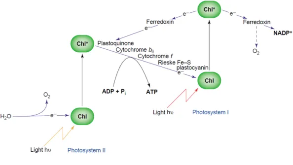
The photochemical phase consists of a series of redox reactions between photosynthetic complexes and ensures the transfer of electrons and the translocation of protons across the thylakoid membranes. The electrons are transferred from water to NADP+, following a linear electron flow (LEF) well described in the “Z-scheme”
(Fig. 2) of photosynthesis (Hill et al. 1960). At the same time, protons are relocated from the stroma to the lumen, creating a ΔpH that drives ATP synthesis.
Photosynthetically produced NADPH and ATP molecules are used for CO2
assimilation in the biosynthetic phase (Mitchell 1966; Ku et al. 1983; Foyer et al. 2012; Minagawa et al. 2015).
The machinery embedded in the thylakoid membrane is commonly called electron transport chain (ETC) and is formed by 4 multimeric protein complexes, encoded by the plastome and the nuclear genome (Fig.1C) (Minagawa et al. 2015). The main complexes are photosystem II and photosystem I (PSII and PSI), chlorophyll-associated multimeric complexes responsible for the charge separation that drives electron flow using light energy. PSII and PSI contain few hundreds of pigments and are structured in a core domain, containing the reaction center (RC) and cofactors, surrounded by light harvesting complexes called antennae, capable of increasing the effective absorption cross-section of the system (Croce et al. 2014).
Figure 1: The photosynthetic metabolism in eukaryotes. A) The net reaction releasing O2 while producing
sugars; B) The chloroplast organelle, where the photosynthetic metabolism takes place; C) The Electron Transport Chain (ETC) responsible for the “Photochemical Phase” in thylakoid membranes; D) The Calvin-Benson cycle (CBC) responsible for CO2 fixation in the stroma of chloroplasts
Each photosystem possesses its particular pigments and cofactors but they are extremely well conserved in all oxygenic organisms.
PSII and PSI are working in series since they are interconnected via the membrane-bound cytochrome-b6f complex and the mobile electron carriers plastoquinone (PQ)
and plastocyanin (PC).
The PSII is associated with the so called light-harvesting complex II proteins (LHCIIs), to constitute a large super-complex with more than 30 subunits. Light energy captured by LHCIIs is transferred to the central core complex of PSII, P680, where it is used to perform charge separation and drive electrons from water to plastoquinone (PQ) with simultaneous injection of H+ into the lumen (Cardona et al. 2012; Tikhonov 2014).
The cytochrome b6f complex is composed by eight polypeptide subunits, including
four major subunits involved in electron transport: the Rieske iron-sulfur protein, the cyt-b6 and cyt-f proteins, and subunit IV. It is responsible for the electron transfer
between PQH2 and PC, activating the so called “Q-cycle” with concomitant pumping
of H+ into the lumen (Stroebel et al. 2003; Cramer et al. 2006; Tikhonov 2014). The PSI possesses its own light-harvesting complex (LHCI) and forms a super-complex with nearly 20 subunits. PSI collects light energy towards its reaction center, P700, where charge separation takes place and drives electrons from plastocyanin to
Figure 2: The Z-scheme for photosynthetic electron transfer as envisaged by Hill and Bendall (1960). Adapted from (Allen 2004).
Ferredoxin (Fd), the final acceptor that will distribute them to several possible acceptor among which NADP+ (Chitnis 2001; Berthold et al. 2012).
The last multimeric complex involved in the process is the ATP-synthase, belonging to the family of F-type ATPases. It is composed of two distinct domains: CF1, the
hydrophilic head group, and CF0, the transmembrane ring (or rotor). ATP-synthase
uses the ΔpH generated by the ETC to produce ATP and to provide energy for cell metabolism (Mitchell 1961; Bottcher et al. 2000; Richter et al. 2005).
II.1.2.b Biosynthetic phase (CO2 fixation)
ATP and NADPH generated in the photochemical phase are injected in the cell metabolism and, notably, feed the biosynthetic phase of photosynthesis. The CO2
fixation occurs in the stroma and allows the insertion of CO2 in carbon skeletons
subsequently used for starch and sucrose biosynthesis (Buchanan 1991). From the simplest prokaryotic cyanobacteria to the most complicated land plants, the pathway for the reduction of CO2 into sugar phosphates is identical and known as reductive
pentose phosphate pathway (RPP) or Calvin-Benson cycle (CBC) (Fig.1D) (Bassham
et al. 1950). The cycle can be divided in 3 sequential phases called Carboxylation,
Reduction, and Regeneration. It is accomplished by 11 different enzymes catalyzing 13 reactions (Fig. 1D). The first phase is initiated by the enzyme ribulose-1,5-bisphosphate carboxylase oxygenase (Rubisco) which catalyzes the carboxylation of the CO2 acceptor molecule, ribulose- 1,5-bisphosphate (RuBP). The two molecules
of 3-phosphoglycerate (3-PGA) formed by this reaction are then utilized to form the triose phosphates, glyceraldehyde phosphate (G-3-P) and dihydroxyacetone phosphate (DHAP) in the reductive phase, via two enzymes that consume ATP and NADPH: phosphoglycerate kinase (PGK) and glyceraldehyde-3-phosphate dehydrogenase (GAPDH), respectively. The regenerative phase of the cycle involves a series of reactions that convert a portion of the triose phosphates into the CO2
acceptor molecule RuBP, and require the sequential action of 9 enzymes, namely triose phosphate isomerase (TPI); fructose-1,6-bisphosphate aldolase (FBA); fructose-1,6-bisphosphatase (FBPase); transketolase (TK); sedoheptulose-1,7-bisphosphatase (SBPase); ribulose-5-phosphate 3-epimerase (RPE); ribose-5-phosphate isomerase (RPI) and phosphoribulokinase (PRK). Thus, a considerable amount of the triose phosphates produced in the cycle remains within the cycle to
trioses), not used to sustain the cycle, constitutes the net result of CO2 fixation.
However, net carbon fixation is sufficient to synthesize sucrose and starch and to ensure growth and development of the organism (Buchanan 1991; Raines 2003; Michelet et al. 2013). It is important to note that the CBC supplies intermediates to an array of other pathways in the chloroplast, including the shikimate pathway for the biosynthesis of amino acids and lignin, isoprenoid biosynthesis and precursors for nucleotide metabolism and cell wall synthesis (Lichtenthaler 1999).
II.2 Regulation of photosynthesis
All metabolic processes occurring in the chloroplasts are sustained by ATP and NADPH generated by the photosynthetic machinery. Among these pathways we can cite nitrate and sulfate metabolism, lipid, amino acids and pigment synthesis (Eberhard et al. 2008; Michelet et al. 2013). Thus, being pivotal for cell metabolism and survival, photosynthesis must be always kept under tight control in all environmental conditions, in order to maintain the balance between metabolic requirements and NADPH/ATP availability. Indeed, any imbalance can lead to damages of the photosynthetic machinery, especially via production of reactive oxygen species (ROS). In order to respond to fluctuating light intensities and temperatures, CO2, water and other nutrients availability, light reactions and CO2
fixation must be always adjusted, to get the maximum efficiency in photon usage and carbon assimilation (Minagawa et al. 2015).
This adjustment between electron/proton fluxes and CO2 fixation acquires a capital
importance under stress conditions, where CBC activity is impaired. To ensure photosynthesis regulation, cells can activate a plethora of different responses at the level of thylakoid complexes or CO2 fixation enzymes. The mechanisms are multiple
and interconnected, going from expression and post-translational gene-regulation, to post-translational modification and degradation of proteins (Eberhard et
al. 2008; Michelet et al. 2013; Minagawa et al. 2015).
In the following sections, an overview of the main approaches used to investigate the photosynthetic function will be provided, together with a description of the most studied regulatory mechanisms. For the purpose of this thesis, the focus will be put on protein post-translational modifications and degradation.
II.2.1 Study of photosynthetic function and regulation: a biophysical approach
Today, after 60 years of functional and genetic studies, the overall picture describing photosynthesis correlates with the early proposal of a Z scheme (Hill et al. 1960). However, currently the view is broader and considers photosynthetic systems as flexible molecular machines capable of rapid adaptation to fluctuating environmental conditions. Dynamic adaptation can be observed and described using multi-disciplinary approaches including genetics and molecular biology, protein crystallography, time-resolved spectroscopy and fluorescence measurements on living organisms, together with bioinformatics for integration of changing functionalities with changes in transcriptome, proteome, and metabolome. All these techniques allow the analysis of photosynthetic function in vivo under precisely regulated growth conditions or in situ, in natural environments.
Among the different approaches currently in use, biophysical analysis of photosynthetic function is crucial for the understanding of the light energy conversion. Indeed the measurements are not destructive, highly sensitive, and allow observation of the function in an entire natural system (Eberhard et al. 2008). Among the great number of biophysical methods that can be applied to photosynthesis, fluorescence spectroscopy is of great importance and will be described below.
The fluorescence is a physical property of chlorophyll (chl) moieties embedded in the thylakoid complexes (Fig. 3A). Once chlorophyll absorbs light energy, it gets excited and there are 3 possible ways to release the excess of energy: photochemistry (charge separation and electron transport to drive photosynthesis); heat dissipation or fluorescence emission. These three mechanisms occur in competition, such that any increase in the efficiency of one will result in a decrease in the yield of the other two (Maxwell et al. 2000).
From a mechanistic point of view, changes in fluorescence yield are linked to the redox state of electron carriers. The reduction of electron acceptors downstream of PSII (namely PQ and QA) is responsible for fluorescence augmentation blocking
temporary reaction centers in a “closed” state (QA−), with a decrease in
photochemistry efficiency and augmented fluorescence emission (Maxwell et al. 2000). Thus, fluorescence yield measures can give information on the redox state of PSII (QA/QA− ratio), a precise picture of light utilization and dissipation rates.
This approach can be efficiently used to screen for photosynthetic mutants, since the fluorescence signature is characteristic for each dysfunction in the photosynthetic machinery (Fig. 3C). The power of this approach relies on the possibility to calculate several parameters from fluorescence curves. These parameters can be easily compared between strains or under different growth conditions, allowing a real-time quantification of photosynthetic efficiency (Fig. 3B).
A great number of mutants defective for photosynthesis-related processes have been identified over the years with fluorescence-based screens. Among them there are strains defective for electron transport or light phase regulation (e.g. NPQ; light-acclimation or Cyclic electron flow) or CBC regulation (e.g. Photorespiration or Carboxylation)(miles 1980; Genty et al. 1990; Matsuda et al. 1996; Walters et al. 2003).
These screenings, coupled with genetic and biochemical tools allowed the discovery of complex gene circuits and post translational mechanisms, often interconnected, responsible for photosynthesis regulation (Eberhard et al. 2008).
II.2.2 Photochemical phase regulation
Several mechanisms exist to control the photosystems excitation rate and electron allocation, making the system highly dynamic and adaptable to varying environmental conditions. The most studied mechanisms include Non Photochemical Quenching (NPQ), acting at the level of PSII; cyclic electron flow around PSI; the activation of alternative electron pathways draining electrons towards other sinks; and antenna size modulation (Eberhard et al. 2008; Foyer et al. 2012; Croce et al. 2014; Minagawa et al. 2015). Altogether, these mechanisms operate to release the excess of energy from the system, avoiding irreversible damage.
II.2.2.a Non Photochemical Quenching
NPQ refers to the mechanisms that allow decreasing the fluorescence yield of PSII, without triggering any photochemical process. It has 3 major components, acting through distinct mechanisms: (i) energy dependent quenching (qE); (ii) state-transition quenching (qT); (iii) photo-inhibition quenching (qI) (Horton et al. 1996). The qE (Fig. 4A) is the major component in plants and is activated within few minutes (1-5) after exposure to light. It is ∆pH-dependent and takes place as a result of over acidification of the lumen (a situation occurring under excess light conditions).
Figure 3: The use of chlorophyll fluorescence in the study of photosynthesis. A) Fluorescence is a physical property of Chl and it is in competition with photochemistry and heat dissipation. B) A typical induction curve for a wild type in optimal growth conditions. F0= fluorescence of
dark-adapted cells; Fs= Stationary fluorescence, when the system is working; FM= Maximal
fluorescence measured after a saturating light pulse. C) Characteristic fluorescence signatures of mutants K.O. for the major photosynthetic complexes.
The decrease in lumenal pH causes conformational changes in the PSII associated antenna, most probably through protonation of surface residues within the lumen. These changes are then transmitted to neighboring pigments, affecting their photo-physical properties and leading to deactivation of over-excited chlorophyll-b (1Chl-b, singlet). The deactivation occurs through the generation of a chl-zeaxanthin dimer that transfers energy away from the reaction center. Zeaxanthin is a carotenoid located in the proximal LHCII antenna and to be effective it requires activation via a thylakoid-bound deepoxidase, responsible for the so called “xanthophyll cycle”, where the precursor violaxanthin is converted into the active form zeaxanthin (Wraight et al. 1970; Briantais et al. 1979; Pfundel et al. 1993; Walters et al. 1994; Horton et al. 1996; Ruban et al. 2007; Eberhard et al. 2008). Beside the xanthophyll
and Lhcsr3 in Chlamydomonas (Petroutsos et al. 2016). These regulators, constitutively expressed in land plants and UV-induced in algae, are able to enhance the qE response even though the molecular mechanism is still unresolved so far (Peers et al. 2009; Tokutsu et al. 2013).
qE is present in most, if not all, photosynthetic organisms and its physiological relevance is supported by the phenotype of mutants defectives for the xanthophyll cycle, PsbS or Lhcsr3, that show reduced fitness under naturally fluctuating light conditions (Kulheim et al. 2002; Petroutsos et al. 2016).
The state transition or qT (Fig. 4B), is the major component of NPQ in green algae such as Chlamydomonas reinhardtii, especially under non-saturating light conditions (Bonaventura et al. 1969; Murata 1969; Wollman 2001). The process balances the light absorption capacity between the two photosystems in response to light quality changes. The qT is slower than qE, being dependent on the redox state of the plastoquinone pool (PQ-pool), rather than on the electron transport rate (Eberhard et
al. 2008; Minagawa et al. 2015). It allows energy redistribution via the reversible
association of LHCII antenna, either with PSII (in state 1) or with PSI (in state 2) (Allen 1992; Wollman 2001; Turkina et al. 2006). This differential association is triggered by phosphorylation of the antenna through the membrane-bound Serine-Threonine kinase STT7 (Depege et al. 2003; Turkina et al. 2006). Thus, excessive stimulation of PSII leads to over-reduction of the plastoquinone pool, binding of a PQH2 molecule to the Q0 site of cytochrome b6f, activation of STT7, phosphorylation
of LHCII antennae and their displacement from PSII towards PSI (state 2). Conversely, the oxidation of the plastoquinone pool by excess stimulation of PSI reverses this process (state 1) stimulating the de-phosphorylation of the antennas (Bellafiore et al. 2005; Turkina et al. 2006). In green algae ~80% of LHCII are involved in this displacement and the qT acquires a predominant role in energy dissipation together with the cyclic electron flow (CEF). However, the interconnection between CEF and qT is still a matter of debate and the most recent studies rather support their independency (Finazzi et al. 2004; Takahashi et al. 2013).
The qI (Fig. 4C) occurs under high light, when photo-damage and degradation of photosynthetic complexes occur. It is a well conserved mechanism, observed in cyanobacteria, algae and land plants (Nixon et al. 2010; Komenda et al. 2012; Jarvi
et al. 2015). It involves, the proteolytic degradation and the replacement of
of the complexes. PSII photo inactivation is strictly linked to reactive oxygen species (ROS), normally produced in oxygenic atmosphere, especially under high light conditions, when the ETC is over reduced (Aro et al. 1993; Eberhard et al. 2008). In normal conditions, the neo-synthesis of the RC efficiently counterbalances the degradation process avoiding irreversible damage. Nevertheless, there are stress conditions (e.g. high light and nutrient limitations) where this balance is not achieved and the net result is PSII-RC degradation. The PSII repair cycle requires a lateral migration of RC from grana towards stroma lamellae and is phosphorylation dependent. The target is the D1 core protein, which dissociates from the RC, migrates in the lamellae, and is degraded by DEG-P2 and FTS-H plastidial proteases (Adam et al. 2001; Antao et al. 2005; Aro et al. 2005; Adam et al. 2006; Eberhard et
al. 2008; Kato et al. 2012).
II.2.2.b Cyclic electron flow around PSI
The cyclic electron flow (CEF) around PSI (Fig. 5) was demonstrated initially by Arnon and coworkers in the late 50’s (Arnon 1959). In this cyclic mode, electrons are reinserted from Ferredoxin (Fd), the PSI acceptor, into the PQ-pool and then, via cytochrome b6f, they flow back into PSI. This cyclic flow generates ΔpH via the
Q-cycle of cytochrome b6f but does not allow reduction of NADP+, thus adjusting the
Figure 4: NON PHOTOCHEMICAL QUENCHING. A) qE component, heat dissipation triggered by 1Chl-b and performed by carotenoids and conformational changes in antenna. B) qT component, State transitions triggered by ΔpH-dependent phosphorylation of LHCII. C) qI component, degradation of damaged D1-core protein and parallel co-translational substitution.
ratio ATP/NADPH (Joliot et al. 2006). Unlike the NPQ, CEF seems to be constitutively active in all conditions (DalCorso et al. 2008). Nevertheless, stresses that decrease carbon fixation capability enhance the CEF rate. This is because the system tries to avoid photo-damage when NADPH, the major sink for electrons, cannot be consumed by the primary metabolism and starts to accumulate. Thus, CEF can be used by cells when primary metabolism is working slowly and some researchers support the hypothesis that it can modulate also the light usage via ΔpH-dependent NPQ (Johnson 2005; Minagawa et al. 2015). The electron pathway of CEF is still under debate. Two major routes have been proposed. One involves the plastidial NAD(P)H-plastoquinone reductase (NDH); the other is dependent on an as-yet unidentified ferredoxin-quinone reductase (FQR) activity. To date, there are no biochemical evidence demonstrating the existence of an “FQR-enzyme” but there are two major hypothesis: the first claims the existence of a super complex involving PSI; Fd; FNR; cyt.b6f; PC; in association with PGR5 and PGRL1 proteins (Nandha et al.
2007; DalCorso et al. 2008). The second hypothesis links FQR activity directly to the cytochrome b6f complex, and in particular to the c’-heme (Stroebel et al. 2003; Joliot et al. 2004).
Beside these two major hypotheses there is a third one, not implying a permanent association between protein complexes, but rather taking advantage of the structural model of thylakoid organization proposed by Albertsson and colleagues (Albertsson 1995; Albertsson 2001). In these models CEF and LEF (linear flow) are spatially separated in “micro-domains”. These domains have different protein composition. Indeed, a large fraction of PSI centers are at the margin and the ends of the grana stacks, close to the appressed region where PSII and cytochrome b6f are localized.
In these regions protein complexes would be preferentially involved in the linear pathway (LEF). The other fraction of the PSI centers is localized in the stroma lamellae, together with a fraction of cytochrome b6f, and would be preferentially
involved in the cyclic pathway. Thus, the segregation of PSI and PSII centers in different membrane regions provides a rationale for both a linear and a cyclic electron flow taking place in the photosynthetic process (Albertsson 2001; Joliot et al. 2004; Johnson 2005).
II.2.2.c Alternative electron sinks
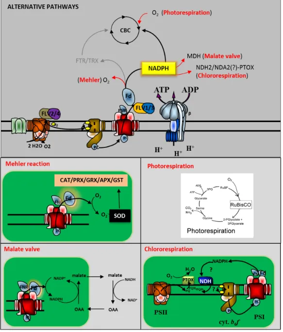
As already mentioned, ATP/NADPH and metabolic requirements must be balanced in all conditions. Therefore, a certain metabolic plasticity is required in order to modulate energy utilization. In general, all external cues that decrease CBC activity lead to full reduction of the photosynthetic electron carriers, since the excess of electrons cannot be injected into the metabolism. Thus, not just energy absorption, but also energy utilization can be tuned to maximize carbon fixation to avoid damages. An effective strategy to release the pressure from photosystems is the redirection of electrons towards alternative sinks. Through this metabolic flexibility a sensitive portion of the LEF is used to reduce other acceptors, sustaining alternative metabolic pathways (Laisk et al. 2007). Alternative flows account for up to 60% of the electrons generated by the water splitting complex and include photorespiration, Mehler reaction, malate shuttle, chlororespiration, as well as other acceptors like flavodiiron proteins (Fig. 6) (Asada 1999; Ort et al. 2002; Kramer et al. 2004; Laisk et
al. 2007; Bailey et al. 2008; Cardol et al. 2008; Eberhard et al. 2008; Gerotto et al.
2016; Alric et al. 2017).
Photorespiration operates alongside carbon assimilation, having a major impact on cellular metabolism, particularly under high light, high temperatures, CO2 and water
deficits (Foyer et al. 2009). It derives from the oxygenation activity of Rubisco (Wingler et al. 2000) that uses ATP and reducing equivalents to convert Ribulose bisphosphate (RuBP) into the CBC intermediate glycerate-3-phosphate (G3-P) and the toxic by-product 2-phoshoglycolate (2PGA) that is then detoxified in peroxisomes and mitochondria in an energy consuming pathway, with net release of CO2 and NH3
(Foyer et al. 2009). In the last years, photorespiration has been considered not only as a wasteful process but also as a protective mechanism that provides an energy
Figure 5: Cyclic electron flow (CEF) around PSI. It is constitutively active to balance the ATP/NADPH ratio in all light conditions. CEF ensures ATP production without NADPH synthesis. The actual pathway has not been discovered yet.
concentration. Furthermore, photorespiration provides metabolites for other metabolic processes, such as glycine for the synthesis of glutathione (Ort et al. 2002; Eberhard
et al. 2008). The importance of photorespiration in the control of photosynthetic
metabolism is demonstrated by the fact that several attempts to engineer Rubisco, in order to reduce its oxygenase activity, failed to enhance the overall photosynthetic fitness, or have given controversial results (John Andrews et al. 2003; Khan 2007; Xin et al. 2015; Basler et al. 2016), but the mutation of other enzymes involved in this pathway leads to severe phenotypes as well (often the plants are viable only under high-CO2 conditions) (Dellero et al. 2016).
The Mehler reaction is important in the dark–light induction phase or during CO2
-limited photosynthesis (Asada 2006; Krieger-Liszkay et al. 2011). The reaction corresponds to direct reduction of dioxygen at the acceptor side of PSI with consequent production of superoxide anions (O2-·), a highly reactive and harmful
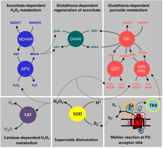
ROS. Superoxide is then promptly dismutated into hydrogen peroxide (H2O2) by
chloroplastic superoxide-dismutase (SOD), located in the vicinity of PSI. The dismutation is the first step of a process commonly called “water-water cycle” which, besides SOD, requires the action of enzymes allowing detoxification of H2O2:
ascorbate peroxidase (APX) coupled to dehydroascorbate reductase (DHAR) and glutathione reductase (GR), glutaredoxins/peroxiredoxins (GRX/PRX), glutathione-S-transferases (GST) and catalase (CAT) (Fig. 6) (Foyer et al. 2011). The final result of the water-water cycle is the reduction of O2-· into H2O, with net electron transport
from water to water. It is commonly accepted that the water-water cycle fulfills two functions especially important under redox-stress conditions: photo-protection and ATP/NADPH ratio balancing (Badger et al. 2000; Forti et al. 2005; Asada 2006; Eberhard et al. 2008; Foyer et al. 2011).
The “Malate Valve” is a shuttle capable of exporting reducing power (NADPH) from the chloroplast toward cytosol and mitochondria, when the NADPH to NADP+ ratio is high, thus poising the NADPH to ATP ratio required for optimal carbon reduction in the light (Scheibe 1987; Laisk et al. 2007). In chloroplasts the NADPH in excess, generated by the ETC, can be used to reduce oxaloacetate (OAA) to malate by malate dehydrogenase (MDH); the malate/OAA transporter then shuttles the substrates between the chloroplast and the cytosol where the malate is oxidized to OAA by the cytosolic NAD+, or is transferred into the mitochondrion to be further metabolized.
Figure 6: The « water-water cycle » and the antioxidant system involved in ROS removal at the level of PSI. APX= ascorbate peroxidase; MDHAR= monodehydroascorbate reductase; DHAR= dehydroascorbate reductase; GR= glutathione reductase; GST= Glutathione S-transferase; GRX= Glutaredoxins; PRX= peroxiredoxins; SOD= superoxide dismutase; CAT= catalase; TRX= Thioredoxin (Modified from Foyer & Noctor 2011).
Although the actual metabolic consequences are still a matter of debate, the physiological role of the malate valve as a sink for photosynthetic electrons is universally accepted and, based on the study of Laisk et al. (2007), its contribution can represent up to 10% of the overall electron flux generated by PSII-driven water oxidation.
Chlororespiration was discovered recently in prokaryotic and eukaryotic marine phytoplankton. The reaction reroutes PSII-generated electrons into a water-water cycle upstream to the cytochrome b6f (Bailey et al. 2008; Cardol et al. 2008). This
process is catalyzed by two enzymes associated with thylakoid membranes: NAD(P)H dehydrogenase (called NDH in land plants and NDA2 in most unicellular green algae) and Plastid-terminal oxidase (PTOX). These enzymes transfer electrons from NADPH (or Fd) to the PQ-pool first and finally to dioxygen, which is reduced to water (Feilke et al. 2014; Nawrocki et al. 2015). The physiological role(s) of
chlororespiration and the interplay with the photosynthetic function remain under debate. Even though many studies have suggested a role in photo-protection, this option does not seem realistic because the electron flow is too low to have an effect on the ETC (Nawrocki et al. 2015).
Another photo-protective strategy involves a class of recently discovered proteins: the flavodiiron proteins (FLVs). These peptides are located in the proximity of PSII and/or PSI and are composed of three domains: an N-terminal β-lactamase-like domain, a flavodoxin-like domain and a C-terminal NAD(P)H-flavin reductase-like domain. FLVs have been found in Archea and anaerobic bacteria, where they possess the first two domains and are able to reduce O2 and NO (Allahverdiyeva et al. 2015). More recently it has been demonstrated that cyanobacteria,
Chlamydomonas, bryophyte and gymnosperms contain four flavodiiron isoforms with the additional C-terminal domain. FLVs assemble, 2 by 2, to form heterodimers called FLV-1/3 and FLV-2/4. FLV-1/3 is able to reduce O2 to water using NADPH as
electron donor. It is believed to protect PSI from light stress, especially at the onset of illumination or under fluctuating light conditions (Allahverdiyeva et al. 2013; Gerotto et
al. 2016). FLV-2/4 instead, has been shown to be active in photo-protection of PSII
Figure 7: ALTERNATIVE ELECTRON SINKS activated when the CO2 fixation is limited by external factors. The brunching point is the acceptor side of PSI. Oxygen, NDH2 or NDA2, as well as MDH can receive electrons directly from Fd or through NADPH activating, respectively, Mehler reaction; Chlororespiration or the Malate valve. Photorespiration is enhanced under CO2 limiting conditions and starts with oxygenation of RUBP. Beside these sinks, Flavodiiron proteins can contribute to the photoprotection draining out the electrons directly from PSI/PSII.
II.2.3 The regulation of the Calvin-Benson cycle
Regulation of the CBC (Fig.8) is certainly one of the most studied mechanisms of photosynthetic regulation. It is indeed the first target under many environmental conditions and especially under stress. However, its regulation is strictly interconnected with Photochemical phase regulations and, as them, its modulation occurs at many levels (gene expression, protein post-translational modifications and protein degradation), but for the scope of this work I will describe only the post-translational regulations.
After the discovery of the pathway by Bashaam et al. (1950), all the enzymes of the cycle have been purified and characterized from diverse sources including plants, algae and cyanobacteria, and this allowed a deep understanding of regulatory mechanisms. Important steps forward were done in the late 60s and 70s, when the
Figure 8: A scheme showing the Calvin-Benson cycle with all the enzymes involved. In blue are highlighted enzymes target for light-dependent redox regulation. In red and green two other proteins, involved in light-dependent regulation of CBC. The other enzymes are target of additional Redox Post Translational Modifications (RPTMs). From Michelet et al., (2013).
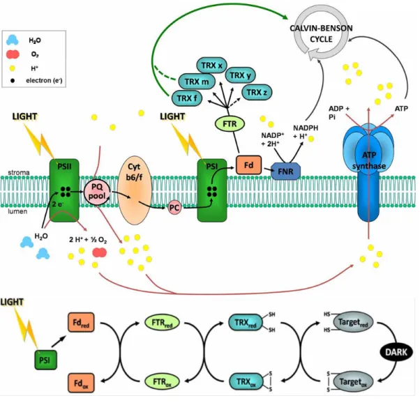
activity of 4 enzymes (PRK; GAPDH; FBPase; SBPase) was shown to be regulated by light, with a low-activity state in the dark and a high-activity state in the light (Schürmann et al. 2008; Buchanan et al. 2012; Michelet et al. 2013). This regulation is operated by the ferredoxin/thioredoxin (Fd/TRX) system that links the activity of CBC enzymes to the redox state of the photosynthetic ETC. Once reduced in the light, TRXs are able to reduce regulatory disulfides present in their target enzymes. This reduction induces a conformational change of the target, converting the protein from a low activity to a high activity conformation. This type of redox regulation is not limited to the photosynthetic metabolism, but is involved in numerous fundamental cellular processes including central metabolic pathways. Besides TRX dependent reduction of disulfide bonds, other types of reversible redox post-translational modifications (RPTMs) of cysteine residues, such as glutathionylation or nitrosylation, have emerged as important mechanisms of enzymatic regulation and metabolic plasticity. RPTMs can modulate protein activity, stability, localization, function or oligomerization (Foyer et al. 2009; Buchanan et al. 2012; Zaffagnini et al. 2012; Zaffagnini et al. 2012). The redox regulation of the CBC is achieved through multiple RPTMs including light dependent activation by the Fd/TRX system.
II.2.3.a The thioredoxin-dependent regulation
The Ferredoxin/Thioredoxin system was discovered by Buchanan and colleagues in the 60’s and 70’s as responsible for the light-dependent regulation of CO2 fixation
(Buchanan et al. 1967; Buchanan et al. 1971; Buchanan 1991). The system is composed of three types of soluble proteins, located in the stroma of chloroplasts, in close connection with thylakoid membranes: Ferredoxin (Fd), Ferredoxin/Thioredoxin reductase (FTR) and thioredoxin (TRX) (Fig.9) (Buchanan 1991; Buchanan et al. 2012). Fd is the final acceptor of the ETC located downstream of PSI. FTR is a thin and flat [4Fe-4S]-enzyme interacting with Fd on one side and TRX on the other side (Dai et al. 2007). TRXs belong to a multigenic family, composed of small (~12 kDa) disulfide-containing redox proteins, ubiquitous in all kingdoms and localized in cytoplasm/nucleus, chloroplasts, mitochondria, flagella or in the secretory pathway (Buchanan et al. 2012). Their active site is composed of a Cys-Gly-Pro-Cys sequence highly conserved throughout evolution (Koharyova et al. 2008).
The TRX enzyme exists in its oxidized form (Trx-S2), where two cysteine residues are
linked by an intramolecular disulfide-bond (-SS-), or in the reduced form (Trx-(SH)2),
containing two thiol groups able to reduce exposed disulfides (Holmgren 1985; Michelet et al. 2013). Once reduced, TRXs activate their targets through a mechanism known as “thiol-disulfide exchange” (Fig.10), involving the formation of a transient mixed disulfide intermediate between the target and TRX. The reaction is reversible and TRX may either break or form disulfides depending on the redox potential of its substrate (Koharyova et al. 2008; Michelet et al. 2013).
Thus, CBC enzymes are oxidized (-SS-) and have a low activity conformation in darkness. Upon illumination, the ETC reduces Fd, thereby allowing NADP+ reduction by Fd-NADP-Reductase (FNR).
Figure 9: The Fd/TRX system and its action mechanism in the dark to light transition. (Adapted from Michelet et al., 2013)
Photoreduced Fd also allows reduction of chloroplastic TRXs by FTR. Consequently reduced thioredoxins are capable of reducing the 4 CBC targets, initiating the CO2
fixation process (Michelet et al. 2013). This regulatory pathway is strongly integrated with regulations operating on the photochemical phase and acts in concert with other modulation processes, to adjust NADPH/ATP consumption with the electron transport rate. Thus, the acceptor side of PSI constitutes the “central hub” of the photosynthetic regulation. According to the redox state of the thylakoids, cells modify either the energy absorption or the energy utilization by the metabolism.
There are 5 highly conserved types of chloroplastic thioredoxins: f; m; y; x; z. Land plants possess several isoforms of each type: 2 TRXf, 4 TRXm, 1 TRXx, 2 TRXy, 1 TRXz in Arabidopsis; 1 TRXf, 8 TRXm, 1 TRXx, 2 TRXy, 1 TRXz in poplar. This variability cannot be observed in unicellular eukaryotic algae, Chlamydomonas
reinhardtii indeed possesses 2 TRXf, 1 TRXm, 1 TRXx, 1TRXy and 1 TRXz, while Ostreococcus lucimarinus 2 TRXf, 1 TRXm, 1 TRXx no TRXy and 1 TRXz. (Lemaire
et al. 2007; Michelet et al. 2013). TRXx and TRXy appear to be reduced by FTR,they were found to be the most efficient TRXs for the reduction of PRXs, GPXs and MSRs and thus they are believed to play a major role as antioxidants rather than in photosynthesis regulation (Michelet et al. 2013). TRXz has been recently
characterized as a subunit of the plastid encoded RNA polymerase and plays an important role in chloroplast transcription and chloroplast development (Arsova et al. 2010; Schroter et al. 2010). In addition to this role, TRXz was suggested to be redox active although its mode of reduction remains controversial (Chibani et al. 2011; Bohrer et al. 2012). Conversely, TRXf and TRXm are reduced by FTR and are clearly dedicated to the regulation of the CBC, although TRXf plays a more prominent role. Indeed, all TRX-dependent enzymes of the CBC are exclusively or preferentially reduced by TRXf. TRXm was historically considered has a regulator of other chloroplastic processes, such as the transfer of reducing equivalents between stroma and lumen, or the regulation of proteins involved in electron transfer (Motohashi et al. 2006; Motohashi et al. 2010; Courteille et al. 2013). However, more recently its role has been revisited, since Okegawa and colleagues (Okegawa et al. 2015) demonstrated how the deficiency of TRXm has a severe impact on photosynthetic CO2 assimilation and on the reduction state of fructose-1,6-bisphosphatase and
sedoheptulose-1,7-bisphosphatase, suggesting a prominent role in photosynthesis regulation.
II.2.3.b Insight into the trx-dependent regulation
The 4 light-regulated enzymes GAPDH, PRK, FBPase and SBPase, are the classical examples of TRX-mediated thiol-disulfide exchange and have been extensively characterized over the last decades.
GAPDH was the first CBC enzyme reported to be activated in the light by reduced TRX (Wolosiuk et al. 1978). Two genes for GAPDH exist: gapA and gapB. They encode two different isoforms that assemble in A4-GAPDH or A2B2-GAPDH
tetramers. Land plants contain both isoforms while other photoautotrophs possess only A4-GAPDH. These two tetramers are differently regulated. Indeed, GAPA and
GAPB subunits differ between each other for a C-terminal extension (CTE), carrying the cysteines (Cys349 and Cys358) target of TRX-f and present only in GAPB (Baalmann et al. 1996; Sparla et al. 2002). The oxidation of A2B2-GAPDH modifies
the CTE conformation, leading to a hairpin in proximity of the coenzyme-binding site, with consequent repression of NADPH-dependent activity (Fermani et al. 2007). Conversely, A4-GAPDH, the only isoform present in cyanobacteria, green and red
algae, is not TRX-regulated per se but acquires this regulation through the interaction with the TRX-regulated proteins CP12 and PRK, forming a multimeric complex where
the small CP12 behaves as the CTE domain of A2B2-GAPDH (Petersen et al. 2006;
Trost et al. 2006). The supramolecular complex formation is promoted by the oxidation of CP12. When oxidized, it forms a binary complex with A4-GAPDH and
ultimately, a ternary complex with the oxidized PRK dimer.
PRK dimers interact with the binary complex CP12-GAPDH after the oxidation of Cys243 and Cys249, two additional residues well conserved across the eukaryotic PRKs (Thieulin-Pardo et al. 2015). Even though PRK has been described as a component of this ternary complex, its light activation was initially reported as a classical thiol-disulfide exchange at the level of Cys16 and Cys55, the residues largely conserved in the eukaryotic and cyanobacterial PRKs. The residues forming the disulfide are located in the catalytic site of each PRK monomer and are target of TRX-f and m (even though TRX-m is less efficient) (Brandes et al. 1996; Michelet et
al. 2013). In land plants both regulatory strategies co-exist, suggesting a fine
regulation in response to redox potential and therefore to light intensity. However, the latter regulation is not present in all photosynthetic organisms: Rhodophytes, Cryptophytes and Heterokonts display very weak or non-existent redox regulation via Cys16 and Cys55, despite the presence of both residues (Thieulin-Pardo et al. 2015). The two phosphatases involved in CBC, FBPase and SBPase, were analyzed in different organisms in the attempt to dissect the molecular mechanism of their redox-regulation. In Physcomitrella patens, they display ∼25% sequence identity at the amino acid level but the homology at the level of their 3D architecture is much higher, although they have different evolutionary histories (Gutle et al. 2016). Both enzymes possess a single disulfide, highly conserved among species. FBPase forms a disulfide between Cys153 and Cys178 in pea, or Cys224–Cys241 in P. patens. SBPase is regulated at the level of Cys52 and Cys57 in wheat, or Cys120 and Cys125 in P. patens (Michelet et al. 2013; Gutle et al. 2016).
The light-dependent activation of FBPase was initially reported in Chlorella (Pedersen et al. 1966). Oxidized FBPase has a basal activity (20–30%) and becomes fully activated upon disulfide reduction which is strictly dependent on TRX-f, with other TRX types being inefficient (Jacquot et al. 1995; Jacquot et al. 1997). In P.
patens, TRX-f and TRX-m are both able to reduce the disulfide but the former is
much more efficient (Gutle et al. 2016). The enzyme is a homotetramer present in two isoforms: a cytosolic FBPase which participates in gluconeogenesis and is not
Gutle et al. 2016). The disulfide formation forces a loop, connecting two antiparallel beta strands, to slide in toward the active site, thereby disrupting the binding sites for the catalytic Mg2+ cations (Chiadmi et al. 1999).
SBPase is a homodimer, unique to the CBC and without cytosolic counterpart (Gutle
et al. 2016). It can be found in all photosynthetic eukaryotes but not in cyanobacteria,
which encode a bifunctional FBPase possessing also SBPase activity (Tamoi et al. 1996). As in the case of GAPDH and FBPase, the light dependent activation of SBPase was initially reported in Chlorella (Pedersen et al. 1966; Bassham 1971; Michelet et al. 2013). However, the biochemical characterization was performed on the wheat enzyme leading to disentangle the molecular mechanism of regulation (Dunford et al. 1998).The oxidized SBPase is completely inactive in darkness and its reactivation absolutely requires the TRX-f-mediated light activation. SBPase in P.
patens requires high levels of TRX-f if compared with FBPase and the activation from
TRX-m is marginal. These results suggest SBP as a bottleneck in the cycle, thus making it a limiting factor for plant productivity together with Rubisco (Tamoi et al. 2006; Gutle et al. 2016). The differences in FBPase versus SBPase activation kinetics may be linked to the fact that FBPase is also involved in starch synthesis while SBP is not. Thus, the cells require a strategy to separate fine control of starch synthesis from the Calvin–Benson cycle (Gutle et al. 2016).
Even though Rubisco is the crucial enzyme involved in biomass production via carbon assimilation, its redox regulation has only been demonstrated around 20 years ago (Zhang et al. 1999; Zhang et al. 2001). In land pants and green algae Rubisco is composed of 8 large subunits, encoded in the plastome by the rbcL gene, and 8 small subunits, encoded by multiple rbcS genes in the nucleus. Two L monomers assemble head-to-tail to form an L2 dimer, with two catalytic sites at the
L-L interface. Four L-L2 dimers interact together to form an octamer, which finally
interacts with 8 small subunits in a (L2)4S8 structure (Whitney et al. 2011). The
abundance of Rubisco can be explained by the fact that it is a slow and highly aspecific enzyme, considered as the main bottleneck in the entire pathway. Its regulation is known to be light dependent but is more complex than the other 4 enzymes described above. Rubisco is not directly targeted by Trx-f, but requires a protein, called Rubisco Activase (RCA), which is the actual Trx-f target (Zhang et al. 1999). The catalytic site of Rubisco indeed, can be occupied by different sugars, naturally produced, which act as inhibitors: 2-carboxy-D-arabinitol-1-phosphate
(CA1P); xylulose-1,5-bisphosphate (which can be produced after a misfired carboxylation reaction); and RuBP itself (Pearce 2006; Parry et al. 2008). Thus, RCA uses the energy of ATP to remove these sugars from the active site. Its ATPase activity can be in turn modulated by Trx-f at the level of two cysteines, Cys392 and Cys411, through the classical thiol-disulfide exchange (Zhang et al. 1999; Zhang et
al. 2001). ATP hydrolysis promotes the interaction of the C-terminal domain of RCA
with the region comprised between residues 89-94 of the L-subunit of Rubisco, with consequent release of sugar from the active site (Portis et al. 2008). At this point, Rubisco should be preactivated via the reaction of CO2 with a conserved Lys201
which leads to the formation of a carbamate, stabilized with a Mg2+ cation, capable to carboxylate RuBP (Andersson et al. 2008).
An additional enzyme of the CBC with a low activity in the dark and a TRX-dependent activation in the light, in green algae but not in land plants, is the PGK (Morisse et al. 2014). PGK exist in two isoforms, one chloroplastic participating in the CBC and one cytosolic, present in most organisms and participating in the glycolytic pathway. The two isoforms show high sequence similarity, suggesting that the molecular structure of the enzyme might be strongly conserved among several organisms. PGK is a monomeric enzyme composed of two domains linked by an α-helix with the N-terminal domain containing the substrate binding site, whereas the ATP cofactor binding site is placed in the C-terminal domain. After substrate binding, a conformational change brings the two domains in close proximity, generating the so-called “closed conformation” that ensures catalytic reactions. PGK from C. reinhardtii can be regulated by TRX-f but not TRX-m. The regulation involves the not strictly conserved Cys227 and Cys361 and the formation of the disulfide was proposed to impose structural constraints in the C-terminal domain of the enzyme that may lower its catalytic efficiency (Morisse et al. 2014).
Although the CBC can be considered as the paradigm of redox regulation, the improvement of proteomic approaches allowed a deeper understanding of the TRX-interactome (or thioredoxome), revealing how other cellular processes, beside CO2
fixation, might be regulated by TRXs (Koharyova et al. 2008; Buchanan et al. 2012; Perez-Perez et al. 2017). Some examples of alternative targets are malate dehydrogenase; Acetyl-CoA carboxylase; ADP-glucose pyrophosphorylase; glucan:water dikinase; beta-amylase; glucose-6-phosphate dehydrogenase;