HAL Id: tel-01628199
https://tel.archives-ouvertes.fr/tel-01628199
Submitted on 3 Nov 2017HAL is a multi-disciplinary open access archive for the deposit and dissemination of sci-entific research documents, whether they are pub-lished or not. The documents may come from teaching and research institutions in France or abroad, or from public or private research centers.
L’archive ouverte pluridisciplinaire HAL, est destinée au dépôt et à la diffusion de documents scientifiques de niveau recherche, publiés ou non, émanant des établissements d’enseignement et de recherche français ou étrangers, des laboratoires publics ou privés.
Synaptic modifications in hippocampal CA3 pyramidal
cells in an Alzheimer’s mouse model
Pei Zhang
To cite this version:
Pei Zhang. Synaptic modifications in hippocampal CA3 pyramidal cells in an Alzheimer’s mouse model. Neurons and Cognition [q-bio.NC]. Université de Bordeaux, 2017. English. �NNT : 2017BORD0628�. �tel-01628199�
THÈSE PRÉSENTÉE
POUR OBTENIR LE GRADE DE
DOCTEUR DE L’UNIVERSITÉ DE BORDEAUX
École DoctoraleSciences de la Vie et de la Santé par Pei ZHANG
Synaptic modifications in hippocampal CA3
pyramidal cells in an Alzheimer's mouse model
Sous la direction de : Christophe MULLE (co-directeur : André FISCHER)
Soutenue le 27. Juin 2017
Membres du jury:
Mme. Yoon CHO Maître de Conférences, CNRS INCIA Président/Examinateur Mme. Hélène MARIE Principal Investigator, CNRS IPMC Rapporteur M. Lionel DAHAN Maître de Conférences, CNRS CRCA Rapporteur M. André FISCHER Principal Investigator, DZNE, Allemagne Co-directeur M. Christophe MULLE Principal Investigator, CNRS IINS Directeur
Synaptic modifications in hippocampal CA3
pyramidal cells in an Alzheimer's mouse model
Dissertation for the award of the degree
Doctor of Philosophy
by Département Sciences du vivant et de la santé, Université de Bordeaux and Division of Mathematics and Natural Sciences, Georg-August-Universität Göttingen
submitted by Pei Zhang
June 2017, Bordeaux
Members of the Jury Committee:
Yoon Cho Maître de Conférences at CNRS INCIA, France President/Examiner Hélène Marie Principal Investigator at CNRS IPMC, France Reporter Lionel Dahan Maître de Conférences at CNRS CRCA, France Reporter André Fischer Principal Investigator at DZNE, Germany Co-supervisor Christophe Mulle Principal Investigator at CNRS IINS, France Supervisor
1
First of all, I would like to express my heartfelt thanks to Dr. Christophe Mulle, for granting me this opportunity to explore in science, also for his patient instruction and help along my way in the last four years.
I would also like to say thanks to all the members of Mulle team, especially to Silvia Viana da Silva, Bernat Gonzalez, Adam Gorlewicz, Vincent Maingret, Jimmy George, Sabine Fièvre, Stefano Zucca for being there to help when I started patch-clamp, to Mario Carta, Marilena Griguoli, Gael Barthet, Thierry Amédée, Christophe Blanchet, Sandrine Pouvreau for generous help and instruction as experienced researchers, to Noelle Grosjean for all the work we’ve down together in behavioral experiments, to Severine Deforges, Audrey Lacquemant, Julie Rumi-masante, Fanny bernadou for technical support on many issues, to Meryl Malezieux for mental support during ups and downs, Ashley Kees, Mariela Escande, Nan Jiang, Dario Cupolillo, Ania Ramalho-Goncalves, Tomas Jorda-Siquier, Ruth Betterton, Eva Rodrigues for daily discussions about science and about life.
Equally important, I want to thank my co-supervisor Dr. Andre Fischer for welcoming me into his group to learn. I really appreciated the everyday supervision of Dr. Cemil Kerimoglu, who helped me to start in molecular biology and answered all of my naïve questions, and to all the team members: Susanne Burkhardt, Daniel Riester, Farahnaz Sananbenesi, Sadman Sakib, Rezaul Islam, Eva Benito, Rashi Halder, Tea Berulava, Hendrik Urbanke, Magdalena Navarro, Christian Schiffmann, Lalit Kaurani, Gaurav Jain, Ashish Rajput, who have helped me a great deal on a daily basis.
I would like to thank Dr. Andreas Frick, Elisabetta Aloisi, Dr. Yoon Cho, Dr. Wenhui Peng, whom I have done collaboration work with.
I’d also like to thank the animal facility staff, without them taking care of the mice, this work would have been impossible, and the BIC team for their very professional support and suggestions.
I am very grateful to my funding, Erasmus Mundus joint doctorate program and the ENC network. Especially Ms. Maaike Leusden, Ms. Laurie, Francois, Ms. Florina Camarasu, and Dr. Michael Hoerner made this happen by dealing with a great amount of paper work.
Finally, I wish to express my gratitude to my family and to all my friends, who supported me patiently and encouraged me endlessly throughout the whole journey. Life is not all about science, but I am very grateful to have this wonderful
2
experience of my doctoral study. It made me more mature, open-minded, and more resilient in the face of frustrations and challenges. To continue in academic or not, I will always cherish this experience at the bottom of my heart. With the advancement of new research tools and numerous devoted scientists, we might be standing in the timepoint where the scope of knowledge of mankind is being broadened at speed never seen, and I feel very lucky to be able to participate in or at least spectate this great cause.
CA3 de l'hippocampe dans un modèle de souris de la maladie
d'Alzheimer
Résumé :
L'encodage de la mémoire dépend de changements durables dans l'activité des circuits synaptiques dans un ensemble de neurones interconnectés. La région CA3 de l'hippocampe reçoit des informations directement ou indirectement (à travers le gyrus denté - GD) en provenance des structures corticales. Des données théoriques et comportementales ont montré que la région CA3 est importante pour l'encodage de la mémoire épisodique, en particulier au stade initial de l'acquisition, en développant vraisemblablement une représentation instantanée d'un contexte. Les neurones pyramidaux CA3 reçoivent une variété de connections afférentes, parmi lesquelles les fibres moussues (FM), les axones des cellules du gyrus denté. Ces connections synaptiques ont attiré une attention par leurs propriétés morphologiques et fonctionnelles uniques. Malgré les nombreuses études, les liens entre plasticité des circuits CA3 et encodage de la mémoire ne sont pas bien compris.
Le cadre général de ce projet de thèse se situe dans l'étude des mécanismes synaptiques de l'encodage de la mémoire épisodique dans des conditions physiologiques ainsi que dans un modèle de souris de la maladie d'Alzheimer (MA). En effet, la MA se caractérise à un stade précoce par une mémoire épisodique altérée, qui peut être associée à une dysrégulation de la plasticité des circuits CA3.
À l'aide de techniques d'enregistrement électrophysiologique, nous avons d'abord exploré les modifications dans les circuits CA3 peu de temps (quelques heures) après conditionnement de la peur contextuelle chez les souris adultes C57Bl6j. Nous avons observé une augmentation de la fréquence des IPSC spontanés accompagnée de changements mineurs dans le nombre de filopodia issus des boutons synaptiques des FM, tandis que les EPSCs et les plasticités à court terme de ces synapses ne sont pas
modifiés.
Nous avons ensuite comparé les propriétés synaptiques des circuits CA3 au stade précoce de la pathologie de la MA, en profitant d'un modèle de souris de MA familliale : les souris APP / PS1 males à 6 mois. Nous avons observé une réduction de la fréquence des IPSC spontanés dans les neurones CA3 avec une diminution de la charge des courants synaptiques évoqués des synapses FM-CA3, alors que la plasticité à court terme de ces synapses et les propriétés intrinsèques des neurones CA3 restent inchangées. En outre, il existe une réduction marquée des courants médiés par les récepteurs kainate (KAR) aux synapses FM-CA3 dans ce modèle de MA. Cette réduction est aussi observée en invalidant la préséniline sélectivement dans les cellules pyramidales de CA3. Ceci suggère que perturber des voies de signalisation de la γ-secretase, en particulier son action protéolytique sur la N-Cadhérine pourrait sous-tendre la perte de KAR aux synapses FM-CA3.
Enfin, j'ai comparé le transcriptome des souris contrôle et APP/PS1, et j'ai pu établir une liste de gènes dont l'expression est modifiée à ce stade précoce de la MA. De plus, nous avons effectué une analyse en ChIP-seq et rapportons ici qu'il existe une diminution des niveaux de H3K4me3, qui s'est révélé être directement lié à la mémoire contextuelle à un coup.
Dans l'ensemble ce travail a révélé que la transmission inhibitrice des circuits locaux CA3 de l'hippocampe pourrait être importante dans l'encodage de la mémoire épisodique. Dans le modèle murin de la MA avec déficit de mémoire, il y a une réduction de la transmission GABAergique et des courants médiés par les KAR réduits cellules pyramidales de CA3. Finalement, avons observé une modification transcriptionnelle d'un certain nombre de gènes dans CA3, à des stades précoces de développement de la pathologie dans notre modèle de MA. Notre étude pourrait contribuer à la compréhension des mécanismes pathologiques précoces de la MA, au niveau synaptique ainsi qu'au niveau transcriptionnel, et fournir des idées nouvelles sur les mécanismes sous-jacents au codage rapide de la mémoire contextuelle.
synaptiques
Title: Synaptic modifications in hippocampal CA3
pyramidal cells in an Alzheimer's mouse model
Abstract:
Memory encoding is thought to proceed from durable changes in the activity of synaptic circuits, leading to the storage of patterns of electrical events in a sparsely distributed ensemble of neurons. Located at the entry level of hippocampal circuitry, the CA3 region is thought to be important for episodic memory encoding, especially at the initial stage of acquisition, by presumably developing an instant representation of a context. CA3 pyramidal cells receive a variety of inputs, among which the mossy fiber (Mf) inputs draw special attention for their peculiar structure and unique synaptic properties. However, the links between the plasticity of CA3 circuits and memory encoding are not well understood.
This thesis project aimed to address the synaptic mechanisms of episodic memory encoding in physiological conditions as well as in a mouse model of Alzheimer's disease (AD).
AD is characterized at an early stage by impaired episodic memory, which may involve dysregulation of the plasticity of CA3 circuits.
First of all, we searched for synaptic deficits in CA3 local circuit in the early stage of AD pathology in acute slices, taking advantage of a familial AD mouse model: 6-month male APP/PS1 mice. We report that there is a reduction in spontaneous IPSC frequency in CA3 pyramidal cells (PCs) together with decreased inhibitory charges of evoked events at Mf-CA3 synapses, whereas the short-term plasticity of these synapses and intrinsic properties of CA3 PCs remain unaffected. Furthermore, there is a robust
reduction in Kainate receptor (KAR) mediated currents at Mf-CA3 synapses. The same results were obtained from PSKO mice, suggesting that disturbed function of γ-secretase and N-Cadherin processing pathways may underlie the dysfunction of KARs at Mf-CA3 synapses.
In the next step, we explored the changes in CA3 circuits shortly after one-trial contextual fear conditioning in adult C57Bl6j mice. We show that despite hardly any changes in filopodia number of Mf terminals, an increase in spontaneous IPSC frequency can be registered, while the EPSCs and short-term plasticities of these synapses are unaltered. However, this increase cannot be seen anymore 24 hours after the contextual learning. We also tried to do simplified computational modeling of the DG-CA3 neuronal networks, to investigate if and to what extent the local interneurons in CA3 region contribute to memory encoding precision.
Finally, to screen for changes at a transcriptome level, we performed RNA-seq with dissected CA3 tissue from APP/PS1 mice and identified up- and down-regulated genes at this early stage of AD. Moreover, we carried out ChIP-seq for a histone modification marker: H3K4me3, which has been shown to be directly related to one-trial contextual memory, and we report that there is a significant decrease in H3K4me3 levels at the promoter areas of various genes in CA3 PCs. However, these genes are hardly overlapping with the down-regulated genes from RNA-seq result, suggesting that other epigenetic mechanisms may play more important roles in expressing early deficits in this AD mouse model.
Taken together, we show that inhibitory connections of hippocampal CA3 circuits may be important for episodic memory encoding, and in early AD mouse model with memory deficits, there is reduced GABAergic transmission and reduced KAR-mediated currents in CA3 PCs, together with many active transcriptional regulations across the genome. Our study may contribute to the understanding of early AD pathologies at a synaptic level as well as a transcriptional level, and provide novel insights into the mechanisms underlying rapid encoding of contextual memory.
Unité de recherche
[Interdisciplinary Institute for Neuroscience CNRS UMR 5297
Centre Broca Nouvelle-Aquitaine Université Bordeaux 38 Rue Albert Marquet 33000 Bordeaux cédex mobile: +33 6 26 01 61 74 office: +33 5 33 51 47 16]
9
Abbreviations
A/C Associative/commissural
AC Adenylate cyclase
aCSF Artificial cerebrospinal fluid
ADAM A disintegrin and metalloproteinase
AMPA α-amino-3-hydroxy-5-methyl-4-isoxazolepropionic acid
AMPAR α-amino-3-hydroxy-5-methyl-4-isoxazolepropionic acid receptor
AP Action potential
APP Amyloid precursor protein ATP Adenosine 5’-triphosphate
Aβ Amyloid β
bp Base pair
CA Cornu Ammonis
CaMK II Ca2+/calmodulin-dependent protein kinase II
CB1R Cannabinoid 1 receptors
CCK Cholecystokinin
CFC Contextual fear conditioning CNS Central nervous system
D-APV D-(-)-2-Amino-5-phosphonopentanoic acid
DG Dentate gyrus
DIC Differential interference contrast
EC Entorhinal cortex
eCB Endocannabinoids
GFP Green fluorescent protein
EGTA Ethylene glycol-bis(β-aminoethyl ether)-N,N,N',N'-tetraacetic acid EPSC Excitatory postsynaptic current
FAD Familial Alzheimer’s disease FF Frequency facilitation
GABA γ-Aminobutyric acid GTP Guanosine 5’-triphosphate
H3K4Me3 Trimethylated lysine 4 on histone 3
HEPES 4-(2-hydroxyethyl)-1-piperazineethanesulfonic acid iGluRs Ionotropic glutamate receptors
10
L-CCG-I (2S,1'S,2'S)-2-(Carboxycyclopropyl)glycine LMT Large mossy fiber terminals
LTD Long-term depression
LTP Long-term potentiation
mEPSC Miniature excitatory postsynaptic current
Mf Mossy fiber
MfBs Mossy fiber boutons
mGluR Metabotropic glutamate receptor
min Minute
mIPSC Miniature inhibitory postsynaptic current
MWM Morris water maze
NBQX 2,3-dihydroxy-6-nitro-7-sulfamoyl-benzo[f]quinoxaline-2,3-dione
NCad N-Cadherin
NFT Neurofibrillary tangles NMDA N-methyl-D-aspartate
NMDAR N-methyl-D-aspartate receptor PBS Phosphate buffered saline
PCs Pyramidal cells
PEN-2 Presenilin enhancer 2
PS1 Presenilin 1
PS2 Presenilin 2
PSD Postsynaptic density
PFA Paraformaldehyde
PKA protein kinase A
PP Perforant path
PPF Paired-pulse facilitation Pr Probability of release
PS1 Presenilin 1
PS2 Presenilin 2
PSD Post synaptic density PTP Post-tetanic potentiation
PV Parvalbumin
RAWM Radial arm water maze S/C Schaffer collateral SEM Standard error of mean
11
sIPSC Spontaneous postsynaptic currents
SOM Somatostatin
ThEs Thorny excrescences TSS transcription start sites
TTX Tetrodotoxin
VIP Vasoactive intestinal polypeptide
Acknowledgements ... 1 Abstract ... 3 Résumé ... 5 Abbreviations ... 9 Table of contents ... 12 1 Introduction ... 17 1.1 Synapses ... 17 1.1.1 Morphology of synapses ... 17 1.1.2 Neuronal transmitters ... 19 1.1.3 Glutamate receptors... 19 1.1.4 Synaptic plasticity ... 22
1.2 Hippocampus and its circuitry ... 26
1.2.1 Learning and memory ... 26
1.2.2 Hippocampus in learning and memory ... 29
1.2.3 Hippocampal anatomy and circuitry ... 32
1.2.4 Hippocampal CA3 circuits ... 33
1.2.5 Computational modeling of CA3 circuits ... 38
1.3 Brain plasticity on a molecular level ... 39
1.3.1 Epigenetics and chromatin plasticity ... 39
1.3.2 Histone Methylation as a transcription regulator ... 40
1.3.3 H3K4me3 in learning and memory ... 42
1.4 Alzheimer’s disease (AD) ... 43
1.4.1 Historical background and clinical aspects of AD ... 43
1.4.2 Familial AD (FAD) on genetic and molecular level ... 46
1.4.3 Mouse models of FAD ... 48
1.4.4 APPswe/PSEN1dE9 (APP/PS1) mice as an FAD model ... 50
13
2 Materials and Methods ... 55
2.1 Animal Use... 55
2.1.1 Ethical Considerations ... 55
2.1.2 Mice ... 55
2.1.3 APP/PS1 Mice Genotyping ... 55
2.2 Electrophysiology ... 57
2.3 Genomic profiling and ChIP-seq ... 59
2.3.1 Tissue Preparation and Neuron Sorting ... 59
2.3.2 RNA Isolation and qPCR ... 61
2.3.3 Chromatin immunoprecipitation (ChIP) ... 63
2.4 Immunohistochemistry and Imaging ... 65
2.4.1 Immunofluorescent Staining for c-Fos ... 65
2.4.2 Confocal Imaging of Mf Terminals ... 65
2.5 Behavioral tests ... 65
2.6 Computational modeling ... 66
2.7 Statistical Analysis ... 66
3 Results... 69
3.1 Input-specific synaptic alterations in CA3 pyramidal cells at an early stage in a mouse model of Alzheimer's disease. ... 69
3.2 Electrophysiological studies of KAR-mediated currents in CA3 PCs of 6-month male APP/PS1 mice ... 97
3.2.1 KAR-EPSCs of Mf-CA3 synapses are reduced in APP/PS1 mice ... 97
3.2.2 KAR-EPSCs of Mf-CA3 synapses is also reduced in PS KO mice ... 99
3.3 CA3 circuits modifications after contextual fear conditioning (CFC) ... 102
3.3.1 One-trial CFC can induce changes in CA3 circuits ... 102
3.3.2 Electrophysiological studies of Mf-CA3 circuit 3h after CFC ... 104
3.3.3 Electrophysiological studies of Mf-CA3 circuit 24h after CFC ... 108
3.4 Genomic and epigenetic studies of APP/PS1 mice ... 111
3.4.1 RNA-sequencing of hippocampal CA3 region in APP/PS1 mice ... 111
3.4.2 H3K4Me3 level is decreased at gene promoters in APP/PS1 mice... 113
4 Discussion ... 119
14
4.1.1 Unaltered presynaptic features and loss of LTP of NMDARs at Mf-CA3 synapses
... 119
4.1.2 Decreased GABAergic transmissions onto CA3 PCs ... 121
4.1.3 Reduced KAR-mediated EPSCs in CA3 PCs ... 122
4.2 CA3 circuits modifications after contextual learning ... 124
4.3 Transcriptome changes in CA3 region in early AD ... 126
4.4 Perspectives ... 127
5 Bibliography ... 131
5.1 Books ... 131
5.2 References ... 132
6 Annexes... 165
6.1 Computational modeling of hippocampal DG-CA3 networks ... 165
6.1.1 Single pyramidal neuron model ... 165
6.1.2 Network design of DG-CA3 circuits ... 166
6.1.3 Test of the model with input/output correlation... 168
6.1.4 NEST code of DG-CA3 model ... 171
6.2 Publication 1 (as co-second author) ... 176
6.3 Publication 2 (as co-second author) ... 187
6.4 Publication 3 ... 213
6.5 List of genes differentially expressed in 6-month APP/PS1 mice by RNA-seq ... 228
6.6 List of genes with low H3K4me3 levels in 6-month APP/PS1 mice by ChIP-seq ... 237
Chapter 1
Introduction
17
1 Introduction
1.1 Synapses
1.1.1 Morphology of synapses
The connection and communication between neurons (or neurons with other faculties) are realized through synapses. A typical synapse consists of two components: a presynaptic compartment, and a postsynaptic compartment, with a synaptic cleft in between. Depending on the nature of pre- and postsynaptic compartments, the synapses can be called axodendritic, axosomatic, axoaxonic, axosecretory, dendrodendritic synapses, etc., with the vast majority in the mammalian nervous system being axodendritic. In addition, astrocytes also exchange information with neurons, responding to synaptic activity, and regulating neurotransmission in a “tripartite synapse” configuration (Perea, Navarrete, & Araque, 2009). Further down this path, a concept of “quadripartite synapse” has also been proposed, arguing that the role extracellular matrix (ECM) in regulating synaptic functions and plasticity (Sykova & Nicholson, 2008)
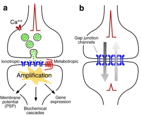
Synapses can be categorized as chemical synapses and electrical synapses, and in some cases, both at the same synapse (Pereda, 2014) (Figure 1.1).
Electrical synapses are also known as gap junctions, propagating action potentials (APs) very rapidly. Gap junctions can conduct electrical signal in both directions, and are usually found at systems requiring fast synchronization of neurons. Due to the nature of transmission here is the passive flow of ionic currents, theses synapses lack gain, and the possibility to modify the synaptic strength is very limited.
The much more common form of synapses is chemical synapse. Although more complex, the chemical synapses are more primitive in an evolutionary point of view (Bennett, 2000), because the sequences of gap-junction-forming proteins are markedly different between vertebrates and invertebrates, while the genes underlying chemical transmission are almost homologs among species (Bennett, 2000; Bruzzone, Hormuzdi, Barbe, Herb, & Monyer, 2003).
18
Figure 1.1. The two main modalities of synaptic transmission.
A. Schematic representation of a chemical synapse. Chemical transmission requires
sophisticated presynaptic machinery that releases neurotransmitters upon depolarization of the presynaptic terminal (e.g. the arrival of an AP). An equally complex postsynaptic machinery includes inotropic and metabotropic receptors is capable of detecting and translating the presynaptic message (neurotransmitter) into various postsynaptic events (e.g. resting potential changes or signaling cascades). B. Schematic representation of an electrical synapse. Electrical transmission is mediated by clusters of gap junctions that linking two adjacent cells, directly allowing the bi-directional traffic of electrical currents as well other small molecules. Adapted from (Pereda, 2014).
The synaptic cleft of a chemical synapse (20~40nm) is much wider than that of an electrical synapse (2~4nm) (Goyal & Chaudhury, 2013). When an AP arrives at presynaptic terminals, vesicles containing neurotransmitters are released into the synaptic cleft. The neurotransmitters can activate receptors on the postsynaptic side, resulting in the opening of the channels and ion flows across the postsynaptic membrane. These alterations in membrane potential on the postsynaptic side can summate in the soma and generate the next AP once over the threshold. Therefore, the signal is conveyed in an electrical-chemical-electrical manner (Pereda, 2014). Chemical synapses are considered to be unidirectional. However, they are subject to certain retrograde modulations, via conventional neurotransmitters, gasses, peptides, growth factors, or membrane-derived lipids (Carta, Lanore, et al., 2014; Regehr, Carey, & Best,
Introduction
19 2009).
1.1.2 Neuronal transmitters
A chemical synapse is usually described according to the type of neurotransmitters it releases (Table 1.1). These neurotransmitters cause different responses on the postsynaptic side depending on the context, which can be developmental, homeostatic, pathological states, etc. Glutamatergic synapses are excitatory in the vertebrate central nervous system (CNS), while GABAergic synapses are mainly inhibitory, albeit a major excitatory role during development (Ben-Ari, 2002). Most of the other neurotransmitters in the CNS are considered to be modulatory, the effect of which can vary depending mainly on the type of receptors they interact with. Furthermore, the co-release of different neurotransmitters at the same synapse is not rare (Hnasko & Edwards, 2012; Trudeau & Gutierrez, 2007).
Table 1.1 An incomplete list of neurotransmitters
Amino acids Glutamate
Aspartate
Glycine (in spinal cord) Amino acid derivatives
(mono amines) GABA Histamine Norepinephrine Epinephrine Dopamine Serotonin (5-HT) Purine derivatives ATP
Adenosine
Gas Nitric oxide
Carbon monoxide Miscellaneous Acetylcholine Neuropeptides Opioid peptides
Somatostatin Substance P Neuropeptide Y
lipid Endocannabinoids
1.1.3 Glutamate receptors
Glutamatergic synapses are the most abundant type of synapses in vertebrate CNS (>50%), comparing to GABAergic synapses (30~40%) and other modulatory synapses
20
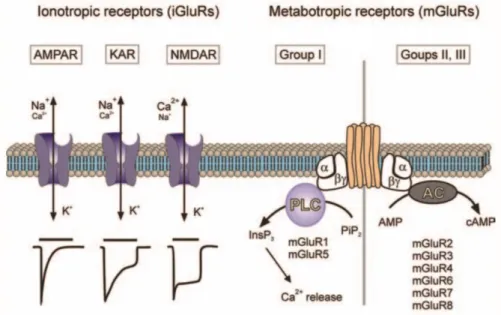
(~10%) (Hendry, Schwark, Jones, & Yan, 1987; Nadim & Bucher, 2014; Roberts, 1986). The actions of glutamate are mediated via a broad range of ionotropic glutamate receptors (iGluRs, including AMPA, NMDA, Kainate and Delta receptors), and metabotropic glutamate receptors (mGluRs, including group I, II, III). These receptors can locate both pre- and post-synaptically. iGluRs are ligand-gated ion channels. Ligand binding leads to rapid changes in membrane potential and cell excitability (Bleakman & Lodge, 1998; Yamakura & Shimoji, 1999). mGluRs are G-protein coupled modulatory receptors, and their activation will trigger a series of reactions in certain signaling pathways and may indirectly open or close other channels (Conn & Pin, 1997; Schoepp, Jane, & Monn, 1999) (Figure 1.2, 1.3).
Figure 1.2. Classification of glutamate receptors.
Ionotropic glutamate receptors are tetramers, including AMPARs (α-amino-3-hydroxy-5methyl-4-isoxazolepropionic acid receptors), NMDARs (N-methyl-D-aspartic acid receptors), KARs (Kainic acid receptors) and Delta receptors. Their properties are defined by the subunit composition and their modifications (Dingledine, Borges, Bowie, & Traynelis, 1999; Dingledine & Conn, 2000).
AMPARs are hetero-oligomers of four subunits: GluA1 to GluA4 (Hollmann & Heinemann, 1994), and are mediating the fast component of excitatory postsynaptic currents (EPSCs), permeable to cations: Na+ and K+. The majority of AMPARs in adult
CNS are GluA1/2 or GluA2/3 heterodimers (Lu et al., 2009; Mansour, Nagarajan, Nehring, Clements, & Rosenmund, 2001; Wenthold, Petralia, Blahos, & Niedzielski, 1996), whereas GluA4-containing AMPARs can be found in hippocampus during development (Zhu, Esteban, Hayashi, & Malinow, 2000). However, there’s a small
Introduction
21
portion of AMPARs lacking the GluA2 subunit (e.g. GluA1/3 heterodimers or homomeric GluA1), and thus permeable to Ca2+ (Seeburg, Higuchi, & Sprengel, 1998;
Wisden & Seeburg, 1993).
NMDARs are heterotetramers composed of subunits GluN1, GluN2A, GluN2B, GluN2C, and GluN2D (in some rare cases also GluN3A and GluN3B), and mediate the slow component of EPSCs (Lieberman & Mody, 1999; Stern, Behe, Schoepfer, & Colquhoun, 1992). As cation channels, NMDARs are highly Ca2+ permeable (Mayer &
Westbrook, 1987). Ca2+ entry triggers several downstream events which are important
for synaptic plasticity (Berridge, 1998). Apart from glutamate binding, the activation of NMDARs also requires the co-agonist glycine and the removal of the blockage by extracellular Mg2+. The blockage is voltage-sensitive and can be unblocked upon
depolarization (Kleckner & Dingledine, 1988; Schoepfer et al., 1994).
Figure 1.3. Ionotropic and metabotropic glutamate receptors.
Schematic representations of the two classes of glutamate receptors. The ionotropic receptors were further subdivided into three distinct subtypes AMPA, NMDA, and Kainate receptors. The iGluRs are assembled from four subunits, which will determine their functional properties. The AMPARs and KARs are predominantly permeable for Na+ and K+, although there are certain Ca2+-permeable subtypes, whereas NMDA receptors are highly Ca2+-permeable. When activated by glutamate, AMPA receptors undergo rapid desensitization (time constant ~100 ms); KA receptors desensitize slower, and NMDA receptors show almost no desensitization. Metabotropic glutamate receptors (mGluRs) control intracellular second messenger signaling cascades as represented. Adapted from (Verkhratsky & Kirchhoff, 2007).
22
KARs are homo- and heteromeric tetramers composed of 5 subunits: GluK1 – 5. The KAR-mediated synaptic current is much smaller and slower compared to AMPAR current (Lerma, 2003; Traynelis et al., 2010). Unlike AMPARs and NMDARs which are normally enriched at excitatory postsynaptic densities (PSDs), KARs can be located at both pre- and postsynaptic sites. In hippocampus, the postsynaptic KARs are predominantly present at Mf-CA3 synapses, where it has been extensively studied (Castillo, Malenka, & Nicoll, 1997; Darstein, Petralia, Swanson, Wenthold, & Heinemann, 2003).
The delta receptors (subunit: GluD1 and GluD2) are regarded as ionotropic glutamate receptors solely because of sequence homology. To date, the wild type delta receptors stay closed in the presence of molecules that have been proved to bind its ligand-binding domain, although the ion channel function has been observed in some mutant or chimera delta receptors (Orth, Tapken, & Hollmann, 2013). Knock-out studies suggested they play important roles in cerebellar function and high-frequency hearing (Schmid & Hollmann, 2008).
1.1.4 Synaptic plasticity
Synaptic plasticity is thought to be one of the mechanisms underlying learning and memory. It exists in many forms, ranging from changes of property in a single synapse to large-scale modifications of synaptic strengths, also including formation or pruning of synaptic connections (Figure 1.4). Synaptic plasticities are normally categorized as short-term and long-term. While short-term synaptic plasticity leads to transient changes in synaptic functions (lasting from milliseconds to minutes) that regulate the moment-to-moment information flow through the circuits, long-term synaptic plasticity leads to persistent changes in synaptic strengths (lasting from hours to the lifetime of the synapse) that adaptively alter synaptic function in response to activities and computational demands (Collingridge, Peineau, Howland, & Wang, 2010; Granger & Nicoll, 2014; Zucker & Regehr, 2002).
Introduction
23
Figure 1.4. Different forms of synaptic plasticity.
Short-term plasticity can be further divided into presynaptic and postsynaptic plasticities. In the presynaptic forms of short-term plasticity, there can be depression (hundreds of milliseconds to seconds), facilitation (hundreds of milliseconds to seconds), as well as augmentation and post-tetanic potentiation (PTP) (tens of seconds to minutes) (Fioravante & Regehr, 2011) (Figure 1.5).
Presynaptic depression can be observed at many synapses when stimulated repetitively at short time intervals. Several factors can account for the reduced synaptic strength, including depletion of the readily releasable pool, inactivation of release sites, and decreased presynaptic calcium influx.
Presynaptic facilitation is often found at synapses with a low initial release probability (e.g. mossy fiber synapses), and repeated stimulation at short time intervals can lead to a transient increase in release probability of these synapses. Increased presynaptic residual calcium level, saturation of endogenous calcium buffers, and facilitation of calcium currents are considered to be contributing to this phenomenon (Fioravante & Regehr, 2011). At Mf-CA3 synapses, this type of plasticity are often registered with two protocols: paired-pulse (PP) or even longer train protocol with repetitive stimulation at short intervals, and frequency facilitation (FF) protocol with increasing stimulation frequency from basal (0.1Hz) to moderate (1-3 Hz) (Gundlfinger, Breustedt, Sullivan, & Schmitz, 2010; Nicoll & Schmitz, 2005; Salin, Scanziani, Malenka, & Nicoll, 1996). Post-tetanic potentiation (PTP) and augmentation is another form of short-term plasticity noticed at Mf synapses, which means enhancement in
24
Figure 1.5. Presynaptic mechanisms of use-dependent short-term plasticity.
Schematic illustrations of different mechanisms for use-dependent short-term plasticity:
A. depression, B. facilitation, C. post-tetanic potentiation (PTP) and augmentation.
RRP: readily releasable pool of vesicles; Cares: residual calcium. Adapted from (Fioravante & Regehr, 2011)
synaptic strength following sustained, high-frequency synaptic activation (Vyleta, Borges-Merjane, & Jonas, 2016).
Other forms of short-term plasticity involving both pre- and postsynaptic elements and retrograde messengers have been reported, such as endocannabinoids mediated depolarization-induced suppression of inhibition (DSI) (Regehr et al., 2009; Wilson & Nicoll, 2002), and depolarization-induced potentiation of excitation (DPE) at the Mf synapses (Carta, Lanore, et al., 2014).
Introduction
25
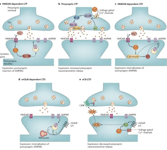
not limited to: NMDA-dependent LTP, presynaptic LTP, NMDA-dependent LTD, mGluR-dependent LTD, eCB-LTD, which were nicely reviewed by Kauer & Malenka (Kauer & Malenka, 2007) (Figure 1.6). Interestingly, albeit the presence of functional NMDA receptors (Weisskopf & Nicoll, 1995), a form of presynaptic NMDA-independent LTP is characterized at Mf-CA3 synapses, which is quite different from the canonical NMDA-dependent LTP found in other regions of the brain (Harris & Cotman, 1986).
In addition, other forms of plasticity such as homeostatic plasticity (up- or down-scaling of excitability), metaplasticity (plasticity of synaptic plasticity), structural plasticity like formation or deletion of synapses are also actively regulating neuronal network activities (Fernandes & Carvalho, 2016; Holtmaat, Randall, & Cane, 2013; Hulme, Jones, & Abraham, 2013).
Figure 1.6. Well-described forms of LTP and LTD.
Highly simplified diagrams of the induction and expression of synaptic plasticity observed in the rodent brain. A. NMDAR-dependent LTP is dependent on postsynaptic NMDAR activation and calcium/CaMKII signaling pathway for its initiation. The voltage-dependent relief of the magnesium block of the NMDAR allows the synapse
26
to detect coincident presynaptic release of glutamate and postsynaptic depolarization, leading to AMPAR insertion into the postsynaptic membrane. B. Presynaptic LTP has been best characterized at hippocampal Mf–CA3 synapses. Repetitive synaptic activity leads to the entry of presynaptic Ca2+, which activates a Ca2+-sensitive adenylate cyclase (AC) leading to a rise in cAMP and the activation of cyclic AMP-dependent protein kinase A (PKA). This in turn modifies the functions of Rab3a and RIM1α leading to a long-lasting increase in glutamate release. C. NMDAR-dependent LTD is triggered by Ca2+ entry through postsynaptic NMDAR channels, leading to increases in the activity of the protein phosphatases calcineurin and protein phosphatase 1 (PP1). The primary expression mechanism involves internalization of postsynaptic AMPARs and a downregulation of NMDARs by an unknown mechanism. D. mGluR-dependent LTD has been best characterized at cerebellar parallel fibre–purkinje cell synapses and hippocampal synapses. Activation of postsynaptic mGluR1/5 triggers the internalization of postsynaptic AMPARs, a process that under some conditions appears to require protein synthesis. E. Endocannabinoid-LTD is the most recently discovered form of LTD. Either mGluR1/5 activation, leading to activation of phospholipase C (PLC) or an increase of intracellular Ca2+ (or both), in the postsynaptic neuron initiates the synthesis of an endocannabinoid (eCB). The eCB is subsequently released from the postsynaptic neuron, travels retrogradely to bind to presynaptic cannabinoid 1 receptors (CB1R), and this prolonged activation of CB1Rs depresses neurotransmitter release via unknown mechanisms. Adapted from (Kauer & Malenka, 2007).
1.2 Hippocampus and its circuitry
1.2.1 Learning and memory
Learning and memory are closely related concepts. Learning is the acquisition of skill and knowledge, while memory is the faculty by which the mind stores and remembers information, and is the retention of information over time for the purpose of influencing future action (Kazdin, 2000). Learning and memory are so immediately relevant to everyone’s daily life, yet remain poorly understood in terms of mechanisms. Questions such as what is the nature of memory, where the memories are stored, how they are engraved and retrieved have intrigued generations of people till today. In 400 B.C., Hippocrates was the first one to declare brain as the major controlling center for the body despite Egyptian, biblical, and earlier Greek views, which were heart-centered theory (Finger, 2005). And the first sign that certain parts of the brain might be involved in memory storage came from the very famous case of patient Henry
Introduction
27
Molaison. He was an American epileptic patient who had a bilateral medial temporal lobectomy to surgically resect the anterior two thirds of his hippocampi, parahippocampal cortices, entorhinal cortices, piriform cortices, and amygdalae in an attempt to cure his epilepsy. After the surgery, he exhibited severe anterograde amnesia characterized by the inability to form new memories, while his remote memories together with his ability of reasoning and acquiring new motor skills seemed intact (Scoville & Milner, 1957). Numerous clinical case-studies have demonstrated in the last century that the anterior hippocampus and hippocampal gyrus, either separately or together, are critically concerned in the retention of current experience (Scoville & Milner, 2000). Altogether, it is now widely-accepted that the ability of learning and memory exist in many different forms involving various regions in the brain.
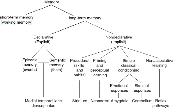
In an effort to categorize forms of memory, Richard Atkinson and Richard Shiffrin proposed in 1968 the Atkinson–Shiffrin model (also known as the multi-store model or modal model) dividing memory as sequential events of: sensory register, short-term memory (also known as working memory, <1min), and long-term memory (Spence & Spence, 1968).
Figure 1.7. Different forms of memory and the brain regions involved.
Scheme of categorization of mammalian memory. Adapted from Squire et al. (Squire & Dede, 2015)
28
Long-term memory involves many processes such as encoding, consolidation, storage and retrieval of memories, and is normally divided into declarative and non-declarative memories, also known as explicit and implicit memories. Declarative memory is the conscious storage and recollection of data, and can be further divided into semantic and episodic memory (Clayton & Russell, 2009; Eichenbaum, 1997). Semantic memory refers to memory that encodes notion, concepts, or specific meaning (Grilli & Verfaellie, 2014), while episodic memory refers to the ability to re-experience a time-and-place-specific event in its original context (Schacter, Addis, & Buckner, 2007; Szpunar, 2010). Declarative memory is usually the primary process thought of when referencing memory (Eysenck, 2012). Declarative memory is a very complicated process involving many different brain regions, among which hippocampus mostly lies in the center of attention (Huijgen & Samson, 2015) (Figure 1.8).
Figure 1.8. Proposed roles of different medial temporal lobe regions in declarative memory tasks by fMRI studies.
Scheme of the anatomical connections between the medial temporal lobe regions and the proposed roles of the hippocampus, entorhinal cortex, parahippocampal cortex, and perirhinal cortex in recognition memory according to the ‘binding of item and context’ (BIC) model (Diana, Yonelinas, & Ranganath, 2007).
Introduction
29
Normally when people refer to memory, declarative memory is usually the one they are thinking of. However, non-declarative memory also plays a significant role in daily life. Non-declarative memory is the storage and recollection of information without conscious attention to learning (Fitts, 1992; Ullman, 2001). An example of a non-declarative learning could be for example acquiring the skills of riding a bicycle or playing the violin. When needed, these memories are unconsciously activated (Tulving & Schacter, 1990).
Memory is not a perfect system, and it can be affected by many factors or even manipulated. However, learning and memory is an essential activity in daily life not only for higher organisms but also for invertebrates such as honeybees (Menzel, 2012) and Drosophila (Ostrowski, Kahsai, Kramer, Knutson, & Zars, 2015).
1.2.2 Hippocampus in learning and memory
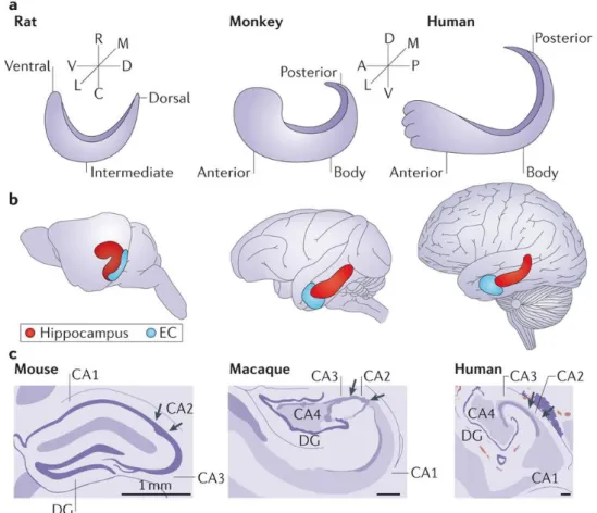
The earliest description of hippocampal formation comes from the Venetian anatomist Julius Caesar Aranzi (1587), who likened it first to a silkworm and then to a seahorse, and that is why it got its name from the Greek word “seahorse”. Humans and other mammals have two hippocampi, one in each hemisphere, located in the limbic system of cerebral cortex. The anatomy and neural circuitry of the hippocampal formation are very similar across different species of mammals (Anderson, Morris, Amaral, Bliss, O’Keefe, 2007) (Strange, Witter, Lein, & Moser, 2014) (Figure 1.9).
Over the years, many theories have been proposed trying to explain the exact role of hippocampus in learning and memory, which are summarized by Bird et al. (Bird & Burgess, 2008) (Table 1.2).
30
Figure 1.9. Scheme showing the comparative structures of hippocampal formation in humans, monkeys, and rodents respectively.
A. Schematic illustrations of hippocampus in the longitudinal axis in rats, macaque
monkeys, and humans. The longitudinal axis is described as ventrodorsal in rodents and anteroposterior in primates. B. The relative locations of the hippocampus (red) in brains of rats, macaque monkeys, and humans, and the entorhinal cortex (EC) is shown in blue.
C. Drawings of Nissl cross-sections of mouse, rhesus, and human hippocampi. A,
anterior; C, caudal; D, dorsal; DG, dentate gyrus; L, lateral; M, medial; P, posterior; R, rostral; V, ventral. Adapted from (Strange et al., 2014).
Introduction
31
Table 1.2 Theories on the role of the hippocampus in learning and memory. Declarative Theory
(Squire, 1986; Squire, Stark, & Clark, 2004)
The hippocampus, acting in concert with other medial temporal lobe regions, is crucial for all forms of consciously accessible memory processes (episodic and semantic, recollection and familiarity) for a time-limited period. Ultimately all memories are consolidated to neocortical sites and are thus unaffected by subsequent medial temporal lobe damage.
Multiple-Trace Theory (Nadel & Moscovitch, 1997)
The hippocampus, together with other medial temporal lobe regions, is crucial for the acquisition of episodic and semantic memories. The recollection of episodic memories remains dependent on the hippocampus for the duration of one's life and becomes more resistant to partial damage with repetition and/or rehearsal, whereas semantic memories become independent of the hippocampus and are stored in other brain regions over time.
Dual-Process Theory (Aggleton & Brown, 1999; Eichenbaum, Yonelinas, & Ranganath, 2007; Norman & O'Reilly, 2003; Rugg & Yonelinas, 2003)
The hippocampus is crucial for episodic recollection of the contextual details of an event. Familiarity-based recognition processes are subserved by other medial temporal lobe regions. Recollection is required for the associative recognition of non-unitized items (for example, voice– face pairs).
Relational Theory (Cohen, Poldrack, & Eichenbaum, 1997)
The hippocampus allows the flexible association of information in neocortical modules that could not otherwise communicate. This enables the relations between elements of a scene or event to be retrieved or used for inference in novel situations, in addition to retrieval of the elements themselves. The Cognitive-Map Theory can be subsumed as a special case of spatial relational processing.
Cognitive-Map Theory (O'Keefe & Nadel, 1978)
A primary role of the mammalian hippocampus is to construct and store allocentric (world-centered) representations of locations in the environment to aid flexible navigation, for example, from a new starting position. In humans, these predominantly spatial processes have evolved to support the spatial–temporal context of episodic memories.
All of these theories acknowledged the importance of hippocampus in learning and memory, especially in consolidation stage of declarative memory and spatial navigation, remaining differences and disputes in whether hippocampus has a time-limited role in episodic memory and hippocampus’s function in the acquisition of non-contextual information.
32
1.2.3 Hippocampal anatomy and circuitry
The hippocampus is made up of four regions or subfields: CA1, CA2, CA3, and CA4. The CA4 region is also known as hilus, or hilar region if considered as a part of the dentate gyrus (Amaral, 1978; Blackstad, 1956). The abbreviation CA comes from “Cornu Ammonis”, which is an earlier name of the hippocampus.
The dentate gyrus is composed of three layers: molecular layer (relatives cell-free, with the exception of some interneurons), granular layer (densely packed with granule cells), and polymorphic layer (contains a number of cell types the most prominent being mossy cells) (Amaral, Scharfman, & Lavenex, 2007). The CA regions are also structured in layers: stratum oriens (SO, inhibitory basket cells and horizontal trilaminar cells, recurrent fibers contacting the basal dendrites of pyramidal neurons here), stratum pyramidale (SP, contains the cell bodies of the pyramidal neurons and some interneurons), stratum lucidum (SL, only found in the CA3 where mossy fibers course through), stratum radiatum (SR, contains some interneurons, and Schaffer collateral fibers, part of recurrent fibers contacting the apical dendrites of pyramidal neurons here), stratum lacunosum-moleculare (SLM, perforant path fibers form synapses onto the apical tufts of the apical dendrites of pyramidal cells) (van Strien, Cappaert, & Witter, 2009) (Anderson, Morris, Amaral, Bliss, & O’Keefe, 2007).
The input of hippocampus comes from neurons in Entorhinal cortex (EC) via the perforant path. Neurons in layers II of entorhinal cortex (EC) project to DG and CA3 (the indirect pathway), whereas layer III neurons project to CA1 and the subiculum (the direct pathway). Subsequently, cells in DG project to CA3 through mossy fibers (Mf), and CA3 PCs project to CA1 through the Schaffer collaterals (Witter, Canto, Couey, Koganezawa, & O'Reilly, 2014). The hippocampal output is made by CA1 and the subiculum, reciprocally projecting to the deeper layers of EC (layer V and VI). There are also substantial projections of CA3 PCs to other CA3 PCs, through associational/commissural or recurrent fibers (Le Duigou, Simonnet, Telenczuk, Fricker, & Miles, 2014; X. G. Li, Somogyi, Ylinen, & Buzsaki, 1994) (Figure 1.10).
Moreover, in rodents the two hippocampi are highly connected at the stems by the commissure of fornix (also called the hippocampal commissure), while in primates this commissural connection is much sparser (Gloor, Salanova, Olivier, & Quesney, 1993).
Introduction
33 Figure 1.10. Basic circuitry of the hippocampus.
Schematic drawing of basic circuitry of the hippocampus. DG: dentate gyrus. Sub: subiculum. EC: entorhinal cortex. Adapted from hand drawings of Santiago Ramón y Cajal (1852 - 1934).
1.2.4 Hippocampal CA3 circuits
As is described in the last chapter, the DG has relatively simple connection, receiving inputs from EC and projecting to CA3. The DG is only sparsely activated for any input (Chawla et al., 2005), probably because DG has significantly more granule cells (~1,000,000) than cells of EC (~200,000) projecting to DG (Amaral, Ishizuka, & Claiborne, 1990; Rebola, Carta, & Mulle, 2017) (Figure 1.11). This type of connection makes it easier for DG to have distinct, non-overlapping activation patterns during each input from EC (O'Reilly & McClelland, 1994). Besides, DG granular cells are interconnected by excitatory mossy cells and inhibitory interneurons located in the hilus (Scharfman & Myers, 2012). However, the DG-CA3 connection is found not to be strictly unidirectional, with CA3 collateral fibers reciprocally influencing DG granule cells through mossy cells and hilar interneurons (Myers & Scharfman, 2011). In contrast to DG, CA3 receives more complicated excitatory inputs: from DG granule cell through mossy fibers (Blackstad, Brink, Hem, & Jeune, 1970; Swanson, Wyss, & Cowan, 1978), directly from layer II of the EC via the perforant path (Witter, 1993), and through recurrent collaterals from CA3 itself (Myers & Scharfman, 2011).
34
Figure 1.11. CA3 circuits and their proposed role in memory.
This schematic illustration shows the different elements of CA3 circuits and their hypothesized involvement in memory encoding and recall. The CA3 PCs receives three types of glutamatergic inputs, including the input from EC through perforant path, from DG through mossy fibers, and from CA3 layer itself through A/C fibers. The CA3 PCs also receive inhibitory signals from local interneurons, this can be either feedforward inhibition coming from DG, or feedback inhibition coming from CA3 cells. Adapted from Rebola, Carta & Mulle, 2017.
The Mf-CA3 connection is especially interesting for its peculiar structure. The mossy fibers (Mfs) were named by Ramon y Cajal for the varicosities all along their axons, giving them a "mossy" appearance. They form three morphologically different synaptic terminals, include the large mossy terminals (LMTs), filopodial extensions of the terminals, and smaller "en passant" synapses (Acsady, Kamondi, Sik, Freund, & Buzsaki, 1998). The mossy fiber boutons are large complex terminals with multiple releasing sites and packed with synaptic vesicles (Blackstad & Kjaerheim, 1961; Laatsch & Cowan, 1966). They form synapses with the proximal dendritic spines of CA3 PCs in stratum lucidum of CA3 region, consisting the feedforward excitation of Mf-CA3 pathways. These spines on CA3 pyramidal cell dendrites are called “thorny excrescences” (ThEs) (Gonzales, DeLeon Galvan, Rangel, & Claiborne, 2001). Meanwhile, the filopodia of LMT form synapses with local interneurons for feedforward inhibition onto CA3 PCs (Ruediger et al., 2011) (Figure 1.12).
Introduction
35 Figure 1.12. Mossy fiber terminals.
A. Schematic representation of the hippocampus with the region of mossy fiber
terminals indicated (dashed box). B. Dil-labeled MfBs. Scale bar, 5μm. C. CA3 neuron with TE spines labeled by intracellular injection of LY dye in fixed tissue. Scale bar, 5μm. D. Reconstructed Mf axons (red) with boutons (regions of vesicle accumulation; blue) in P14 and adult mice. Adapted from (Wilke et al., 2013).
A single mossy fiber axon may make as many as 37 contacts with a single CA3 PC, but innervates only about 14 different CA3 PCs (Claiborne, Amaral, & Cowan, 1986). In contrast, each DG granule cell is innervating as many as 40 to 50 interneurons in CA3 area (Acsady et al., 1998). Intriguingly, these numbers can be dynamically changed by, for example, contextual or spatial learning (Crusio & Schwegler, 2005; Ruediger et al., 2011).
On the other hand, each CA3 pyramidal cell receives input from about 50 different DG granule cells (Crusio, Genthner-Grimm, & Schwegler, 2007), as well as ~3600 excitatory inputs from EC layer II via perforant path (PP), and ~12000 contacts from recurrent collaterals of other CA3 PCs (Rolls, 2013). However, the number of contacts doesn’t necessarily mean stronger innervation, considering the synaptic-distance-dependent scaling in the brain (de Jong, Schmitz, Toonen, & Verhage, 2012; K. J. Lee et al., 2013). Although sparse, Mf-CA3 synapses at the proximal dendrites of CA3 pyramidal cells are a powerful connection on the main path of information flow, and are thought to enhance the signal-to-noise ratio and optimize the selection of populations of CA3 PCs firing in a correlated manner.
In CA3 area, inhibitory neurons play a pivotal role in information transfer as well. The CA3 interneurons receive inputs from various sources, including the associational/commissural fibers (A/C), the mossy fibers (MF), and the perforant path
36
(PP). These interneurons serve both the feed-forward and feed-back circuits and can contact hundreds of CA3 pyramidal cells (Lawrence and McBain, 2003). It has been shown in the last century that MF synapses on interneurons (may be via filopodial extensions or small en passant boutons and occasional large boutons) are significantly more than MF synapses on CA3 pyramidal cells, the ratio being approximately 10 to 1 (Acsady et al., 1998). This MF preferential innervation of interneurons may underlie the overall inhibitory effect of DG innervation of the CA3 network (Bragin et al., 1997, Penttonen et al., 1997).
The interneurons located in the CA3 area are not a homogenous group and can be classified into various subtypes by their morphology, physiology, molecular expression patterns (such as receptors, neuropeptides or calcium-binding proteins), or biophysical features. For the convenience of our MF synapse electrophysiological studies, one functional classification of CA3 interneurons receiving MF inputs by Szabadics and Soltesz is shown below (Figure 1.13) (Szabadics and Soltesz, 2009). To be more specific, there are fast spiking basket cells and spiny lucidum cells receiving more MF inputs, which exhibit low release probability and small EPSC amplitudes; there are also regular spiking basket cells and Ivy cells receiving fewer MF inputs, which have high release probability and large EPSCs amplitudes. Long-term plasticities of MF-interneuron synapses in CA3 area is review by Galván et al. (Galván et al., 2011).
Figure 1.13. Functional classification of some CA3 interneurons.
A. Non-normalized averages from each cell type that received monosynaptic EPSCs
Introduction
37
potentials from a representative MF). Note that no traces are shown for MFAs, since these cells were not found to be receiving monosynaptic inputs from MFs. B. Schematic presentation of the number and the strength of monosynaptic MF inputs (black arrows from the left) (the number of arrows represent the approximate relative abundance of MF inputs to the different cell types and the strength is illustrated by the width of the arrows) to CA3 GABAergic cells (color-coded circles), background synaptic events (gray arrows around the schematic GABAergic cells; number of arrows reflect spontaneous synaptic event frequency), and the GABAergic projections of the cells within and outside of the CA3 area. A representative CA3 pyramidal cell is shown for illustration. MF, mossy fiber; IvyC, Ivy cell; MFA, MF-associated cell; RSBC, regular-spiking basket cell; FSBC, fast-regular-spiking basket cell; SLC, spiny lucidum cell. Adapted from (Szabadics and Soltesz, 2009).
Although there have been many attempts to explain populational activities through cell-type-specific interactions, we still lack the concrete evidence of how different subtypes of CA3 interneurons control information flow in the local circuits.
DG–CA3 connections have been implicated in pattern separation and assistance in memory encoding and the recall of contextual and spatial memory (Rebola et al., 2017). For example, it has been shown that transgenic inactivation of output of adult versus developmentally born DG neurons has differential impacts on pattern separation and completion (Nakashiba et al., 2012). Some other study has demonstrated that pharmacological impairment of DG–CA3 transmission alters novel contextual representation (Daumas, Ceccom, Halley, Frances, & Lassalle, 2009; Lassalle, Bataille, & Halley, 2000). Further evidence includes that mice with impaired Mf LTP are deficient for incremental learning (Otto et al., 2001), exposure to novel context regulates electrically induced plasticity (Hagena & Manahan-Vaughan, 2011), and that there is structural remodeling of Mf boutons upon learning (Holahan, Rekart, Sandoval, & Routtenberg, 2006; Routtenberg, 2010; Ruediger et al., 2011).
However, there are still open questions and missing evidence about the specific role of Mf-CA3 connections in memory encoding. For instance, whether there is a causal link between Mf LTP and memory encoding is still under debate (Kaifosh & Losonczy, 2016). Moreover, other questions remain, such as the impact of NMDAR plasticity and its role in metaplasticity and synaptic integration (Hunt, Puente, Grandes, & Castillo, 2013; Rebola, Carta, Lanore, Blanchet, & Mulle, 2011), and the role of Mf-dependent heterosynaptic plasticity (input-unspecific synaptic plasticity) in memory encoding.
38
1.2.5 Computational modeling of CA3 circuits
The relative simplicity of the hippocampal networks, when compared with e.g. neocortex, strongly appealed to computational neuroscientists to build models and test theories about memory mechanisms as early as since the 1970s. The original idea was built on Hebbian theory, stating “when an axon of cell A is near enough to excite cell B and repeatedly or persistently takes part in firing it, some growth process or metabolic change takes place in one or both cells such that A's efficiency, as one of the cells firing B, is increased” (Hebb, 1949). In the most influential early model of Marr, he constructed an “autoassociator” that learns to associate all components of an input pattern with all other components of the same pattern (Marr, 1971).
With the computational power and accessibility change in the 1990s, the amount of publication surged in this field. Computational models of the hippocampus, especially of DG-CA3 circuits have been numerously proposed, because this network shares many of the basic connectivity requirements for an autoassociator (Hasselmo, Wyble, & Wallenstein, 1996; Korol, Abel, Church, Barnes, & McNaughton, 1993). These models focused mainly on the ability of the hippocampal regions to perform sequential learning, spatial navigation, and the consolidation of episodic memories.
For example, Levy presented a model of hippocampal CA3 region as a sequence predictor, and the model was able to learn and use context by solving sequence and configural learning problems, providing a computational unification of a variety of putative hippocampal-dependent functions (Levy, 1996). Liaw & Berger described a simple hippocampal network consisting of a small number of neurons connected with dynamic synapses could perform speech recognition, suggesting that the dynamic interplay between synapses (facilitative and inhibitory) results in an emergent “temporal chunking” mechanism for sequential pattern recognition (Liaw & Berger, 1996).
In some more recent works, Myers & Scharfman presented a simple computational model of the dentate gyrus to demonstrate that hilar cells may mediate dynamic regulation of pattern separation, by mediating the backprojection from CA3 to DG (Myers & Scharfman, 2009). Hiratani et al. constructed an associative memory network model of spiking neurons that stores multiple memory patterns in a connection matrix with a lognormal weight distribution, demonstrating that heavy-tailed distributions of connection weights can generate noise which is useful for associative memory recall (Hiratani, Teramae, & Fukai, 2012).
Introduction
39
Computational models provide us a chance to connect data at multiple levels including molecular, cellular, anatomical and behavioral levels, no matter they are developed in a top-down (computational models predicting biological details) or bottom-up (computational models simulating biological data) fashion (Gluck & Granger, 1993). The computational and biological fields of neuroscience can inform each other and jointly lead to better understanding of our brain.
1.3 Brain plasticity on a molecular level
1.3.1 Epigenetics and chromatin plasticity
Now diving to an even deeper and more basic level under learning and memory, right to the eukaryotic cell nuclei, scientists have been trying to explain plasticities happening in the brain on an epigenetic level.
The notion “epigenetics” was first proposed by C. H. Waddington in 1942 to describe that the genes might interact with each other and with their environment to produce a phenotype (Waddington, 1953).
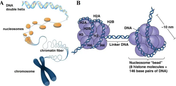
A gene is a locus or region of DNA and the molecular unit of heredity (Alberts, Johnson, Lewis, Raff, Roberts, & Walter, 2002). Genes are compactly stored on chromosomes (the condensed form) or chromatins (the unraveled form) (Figure 1.14). The more condensed form of chromatin is called heterochromatin, and the genes located in heterochromatic region usually are less active or not active, while euchromatin is dispersed and loose, making the genes located at euchromatic regions relatively active (Grewal & Moazed, 2003).
Approximately 146 base pair (bp) of DNA strand is wrapped around a histone octamer core protein, forming a structure called nucleosome. The "linker DNA" between two nucleosomes can be up to 80 bp long. The histone octamer consists of 4 types of histones: H2A, H2B, H3, and H4, with two copies of each (Luger, Mader, Richmond, Sargent, & Richmond, 1997). The histone proteins are overall positively charged, making them attracted to negatively charged DNAs. Importantly, each histone protein has an N-terminal tail sticking out, which is subject to many forms of modifications, such as acetylation, methylation, phosphorylation, ubiquitylation and ADP-ribosylation (Strahl & Allis, 2000; Vaquero, Loyola, & Reinberg, 2003).
40
Figure 1.14. Schematic representation of chromosome, chromatin, nucleosome, and histone octamers.
A. Illustration showing how DNA is packaged into a chromosome. DNA wraps around
the histone cores to form nucleosomes. These units then condense into a chromatin fiber, which condenses further to form a chromosome. Image copyright: Genome Research Ltd. B. Nucleosome as a “beads-on-a-string” structure. The nucleosome core is comprised of a histone octamer with histone 2A, 2B, 3 and 4, two copies each. The DNA double helix strand is wrapped around (~1.7 times) the histone octamer. Image copyright: Pearson Education Inc.
These post-translational modifications on histone tails actively regulate the interaction between DNA and histones. If the positive charge is reduced by the modification, the attractive force between histone and DNA decreases, thus making DNA more accessible for translational machinery; conversely, when the positive charge is increased, DNAs are less likely to be translated.
1.3.2 Histone Methylation as a transcription regulator
Unlike histone acetylation, which almost always facilitates local gene expression, histone methylation can either promotes or represses gene expression, depending on which residue is modified (Justin, De Marco, Aasland, & Gamblin, 2010; Scharf & Imhof, 2011; Shi & Whetstine, 2007; Shilatifard, 2008). It occurs on lysine (K) or arginine (R) residues of histone tails, and it can happen more than once on the same
Introduction
41
residue, resulting in monomethylation, dimethylation or trimethylation (Mosammaparast & Shi, 2010; Santos-Rosa et al., 2002; Schneider et al., 2005). A summary of known histone methylation types is listed in Table 1.3 (Di Lorenzo & Bedford, 2011).
Table 1.3 Histone tail methylations.
Histone Modification Role Reference
H3 H3K4me2 Permissive euchromatin (Santos-Rosa et al., 2002) H3K4me3 Transcriptional elongation; active
euchromatin
(Roguev et al., 2001; Strahl, Ohba, Cook, & Allis, 1999) H3K9me3 Transcriptional repression; imprinting; DNA
methylation
(Rea et al., 2000; K. Zhang et al., 2002)
H3R17me Transcriptional activation (Bauer, Daujat, Nielsen, Nightingale, & Kouzarides, 2002; Chen et al., 1999) H3K27me3 Transcriptional silencing; X-inactivation;
bivalent genes/gene poising
(K. Zhang et al., 2002)
H3K36me3 Transcriptional elongation (K. Zhang et al., 2002) H4 H4R3me Transcriptional activation (Wang et al., 2001)
H4K20me Transcriptional silencing (Nishioka et al., 2002) H4K20me3 Heterochromatin (Schotta et al., 2004)
H3K4 is the one of the most extensively targeted positions among all possible histone methylation sites (Vallianatos & Iwase, 2015), and is always facilitating translation. With the advances of technological nowadays, especially chromatin immunoprecipitation (ChIP) together with quantification of associated DNA by next-generation sequencing (ChIP-seq), it has been shown that H3K4 methylation has wide-ranging roles on a genome-wide scale, and is closely related to transcriptional regulation and epigenetic tagging of promoter and enhancer sequences in both neurodevelopment and neuropsychiatric diseases (Barski et al., 2007).
ChIP-seq studies have shown that the distribution of the trimethylated form of histone 3 (H3K4me3) in the human brain shows approximately 30 000 sharp “peaks” genome-wide (Cheung et al., 2010; Shulha, Crisci, et al., 2012). These “peaks” (1–2 kb in length) can differ among different cell types, and are mostly located around transcription start sites (TSSs) of proximal gene promoters and other regulatory