HAL Id: tel-01635239
https://tel.archives-ouvertes.fr/tel-01635239
Submitted on 14 Nov 2017
HAL is a multi-disciplinary open access archive for the deposit and dissemination of sci-entific research documents, whether they are pub-lished or not. The documents may come from teaching and research institutions in France or abroad, or from public or private research centers.
L’archive ouverte pluridisciplinaire HAL, est destinée au dépôt et à la diffusion de documents scientifiques de niveau recherche, publiés ou non, émanant des établissements d’enseignement et de recherche français ou étrangers, des laboratoires publics ou privés.
Dental composite properties evaluation : from
experimental approaches to the prerequisite of a
chewing bench
Hazem Abouelleil Sayed
To cite this version:
Hazem Abouelleil Sayed. Dental composite properties evaluation : from experimental approaches to the prerequisite of a chewing bench. Human health and pathology. Université de Lyon, 2017. English. �NNT : 2017LYSE1054�. �tel-01635239�
N°d’ordre NNT :
THESE de DOCTORAT DE L’UNIVERSITE DE LYON
délivrée par
l’Université Claude Bernard Lyon 1
Ecole Doctorale N°205
(ECOLE DOCTORALE INTERDISCIPLINAIRE
SCIENCES-SANTE)
Spécialité de doctorat
: BiomatériauxSoutenue publiquement le 03/04/2017, par :
(M. Hazem ABOUELLEIL)
Dental composite properties evaluation:
From experimental approaches to the
prerequisite of a chewing bench
Devant le jury composé de :
Yves AMOURIQ PU-PH Université de Nantes Invité
Marc BOLLA PU-PH Université de Nice Sophia Antipolis Examinateur
Denis BOURGEOIS PU-PH Université Lyon1 Examinateur
Pierre COLON PU-PH Université Paris Diderot Directeur de thèse
Brigitte GROSGOGEAT PU-PH Université Lyon1 Examinatrice
Christophe JEANNIN MCU-PH Université Lyon1 Co-directeur Invité
Bruno TAVERNIER PU-PH Université Paris Diderot Rapporteur
2
UNIVERSITE CLAUDE BERNARD - LYON 1 Président de l’Université
Président du Conseil Académique
Vice-président du Conseil d’Administration
Vice-président du Conseil Formation et Vie Universitaire Vice-président de la Commission Recherche
Directrice Générale des Services
M. le Professeur Frédéric FLEURY
M. le Professeur Hamda BEN HADID M. le Professeur Didier REVEL M. le Professeur Philippe CHEVALIER M. Fabrice VALLÉE
Mme Dominique MARCHAND
COMPOSANTES SANTE
Faculté de Médecine Lyon Est – Claude Bernard
Faculté de Médecine et de Maïeutique Lyon Sud – Charles Mérieux
Faculté d’Odontologie
Institut des Sciences Pharmaceutiques et Biologiques Institut des Sciences et Techniques de la Réadaptation
Département de formation et Centre de Recherche en Biologie Humaine
Directeur : M. le Professeur G.RODE
Directeur : Mme la Professeure C. BURILLON Directeur : M. le Professeur D. BOURGEOIS Directeur : Mme la Professeure C. VINCIGUERRA Directeur : M. X. PERROT
Directeur : Mme la Professeure A-M. SCHOTT
COMPOSANTES ET DEPARTEMENTS DE SCIENCES ET TECHNOLOGIE
Faculté des Sciences et Technologies Département Biologie
Département Chimie Biochimie Département GEP
Département Informatique Département Mathématiques Département Mécanique Département Physique
UFR Sciences et Techniques des Activités Physiques et Sportives
Observatoire des Sciences de l’Univers de Lyon Polytech Lyon
Ecole Supérieure de Chimie Physique Electronique Institut Universitaire de Technologie de Lyon 1 Ecole Supérieure du Professorat et de l’Education Institut de Science Financière et d'Assurances
Directeur : M. F. DE MARCHI
Directeur : M. le Professeur F. THEVENARD Directeur : Mme C. FELIX
Directeur : M. Hassan HAMMOURI
Directeur : M. le Professeur S. AKKOUCHE Directeur : M. le Professeur G. TOMANOV Directeur : M. le Professeur H. BEN HADID Directeur : M. le Professeur J-C PLENET Directeur : M. Y.VANPOULLE
Directeur : M. B. GUIDERDONI Directeur : M. le Professeur E.PERRIN Directeur : M. G. PIGNAULT
Directeur : M. le Professeur C. VITON
Directeur : M. le Professeur A. MOUGNIOTTE Directeur : M. N. LEBOISNE
Acknowledgments
I would like to express my gratitude and present my thanks to all who helped me accomplish this work.
To my team of Biomaterials and its director Professor Brigitte GROSGOGEAT for hosting me as one of its team members and allowing me to conduct all the research studies included in this work, I would also like to thank her for the encouragement and for her support during all the time I was preparing my thesis.
To my Thesis director Professor Pierre COLON, I would like to express my deepest gratitude for his patience, for his advice and for his thorough observations.
To my Thesis Co-director Doctor Christophe JEANNIN, thank you for your help, your support and for your implication in this work.
To my reporters Professor Bruno TAVERNIER, and Doctor Yannick TILLIER who accepted spontaneously to report this work, I would like to express my deep gratitude and my sincere thanks for your observations, and for the disposition and the readiness with which you accepted to evaluate this work.
To Professor Denis BOURGEOIS, the dean of Lyon 1 faculty of dentistry, for hosting me as a staff member during the past years, and for his kindness and his support.
To my jury members Professor Marc BOLLA and Professor Yves AMOURIQ for participating in my thesis defense, for their observations and their commitment.
For my wife Hoda, thank you for your love, your help and for your patience, without which I could never have finished this work.
For the light of my life Aya, Ahmed and Mariam, it is from you that I get my motivation to do my best and try to achieve the most.
For my late father and mother, and to my sister and my brother, who taught me to work hard and to try to excel in whatever I do.
For my team member; Nina ATTIK, for her help, her friendship and for her precious advices.
For my team members and my colleagues:
Professor Dominique SEUX, Doctor Cyril VILLAT, Doctor Alexis GOUJAT, Doctor Kerstin GRITSCH, Doctor Nelly PLASSE-PRADELLE, Professor Jean-Christophe MAURIN, Mademoiselle Fatima CHERCHALI and Monsieur Frank HALLAY.
For their help, their support and their friendship.
To the laboratory of multi-materials and interfaces, its members and its Director professor Arnaud BRIOUDE for accepting me as one of its members.
Even though I started late in the domain of scientific research, and even though the road was long and winding, it was with your help and support that I was able to complete this work.
I
Table of contents
1. Introduction ... 1
2. Dental restorative materials ... 7
Types and composition of restorative dental materials ... 7
2.1. Restorative resin composite ... 8
Original Research Article: Comparison of mechanical properties of a new fiber reinforced composite and bulk filling composites 2.2. Ceramics and CAD/CAM restorations ... 25
2.2.1. Glass-based systems (mainly silica) ... 27
a. In-Ceram® spinell ... 29
c. In-Ceram® zirconia ... 29
2.2.2. CAD/CAM ... 31
Original Research Article: Mechanical properties and internal fit of four CAD CAM block materials 2.3. Metals and alloys ... 49
3. Mechanical and Physical properties of dental materials ... 50
3.1. Thermal properties ... 50
3.1.1. Thermal conductivity ... 51
3.1.2. Thermal diffusivity ... 52
3.1.3. Coefficient of thermal expansion ... 52
3.2. Mechanical properties ... 53
3.2.1. Fracture strength ... 54
II
3.2.3. Flexural modulus (Elastic modulus) ... 56
3.2.4. Hardness ... 57
Original Research Article: Thermal shock effect on enamel – resin composite interface 4. Dental Materials evaluation ... 70
4.1. Types of tests used to evaluate dental materials ... 70
4.2. ISO Tests ... 71
4.3. Tooth-material interface ... 76
4.3.1. Direct analyses of bond strength ... 76
4.3.2. Indirect analyses of bond strength ... 77
Original Research Article: Evaluation of interfacial type of fracture using two adhesive systems Original Research Article: Tensile Bond Strengths of Two Adhesives on Irradiated and Nonirradiated Human Dentin 5. Oral simulators ... 94
5.1. Human oral cavity ... 94
5.1.1. Temperature ... 94
5.1.2. pH……….. ... 96
5.1.3. Bacteria... 97
5.1.4. Mandibular movements ... 98
5.2. Biocompatibility and Material release in the oral cavity ... 100
5.2.1. Bisphenol A... 103
III
Dental composites and Bisphenol A - Potential toxicity and ways to reduce it - Systemic review
5.3. Chewing bench ... 121 5.3.1. Design ... 121 5.3.2. Construction and Working Principle ... 122
Original Research Article: Development of a chewing simulator for testing dental materials: a pilot study
6. Discussion ... 133 7. Conclusion ... 142 8. References ... 144
1
1. Introduction
The oral health is an integral part of the human health, which is necessary to maintain a healthy oral function and dental structures. The ability to process food to be adequately digested and absorbed, the ability to speak and communicate, and more over the ability to acquire a presentable social appearance, are all essential in the modern day life [1].
Historically, teeth were replaced and restored using natural human or animal teeth, stones or sea shells, ancient Egyptians, for example, had a dental specialist for the king. But the modern day dentistry did not start except in the 17th century, and even though the first attempts to restore teeth had mostly humble results, by the 20th century much development took place [2].
The last 30 years have seen a big development in the restorative materials technology and techniques; part of this is due to the advanced development in computer, technology and digital imaging. Another part is the type of material itself, the dental composite resin enables the addition and the diversification of its components whether the polymer or the filler [3]. This can be considered equally true with the glass ionomer cement and CAD/CAM block materials [4,5]. Dental products companies have seen large development with the growth in the dental field market, thus pushing forward for innovations and new patents and discoveries, together with more demand from patients for more efficient and more esthetic restorations. Modern biomaterial research resulted in the development of a wide variety of restorative materials and the innovation of new compositions and techniques [6,7].
Several factors seem to add to the complex process of making the adequate choice of restoration. The increased patient demand for esthetic at the age of selfies,
2
social networks and excessive photographing, esthetics seem to gain more ground as one of the principle factors that drive the choice of restorative material, sometimes on the cost of efficiency and survival rate. On the other hand, the dentist plays a role in guiding the patient to the correct choice of material in order not to sacrifice function for looks [8].
Another rising feature is the minimal intervention dentistry for managing dental caries and the remineralization materials and methods. The conservation of the dental tissue and pulp vitality is the scope of new research as the future of dental restorative dentistry [9].
The rising patient awareness of efficient oral hygiene, dental tissue conservation as well as good esthetics brought about more awareness and concerns about the biological effects of dental materials whether real or imagined. The media seem to play a major role in that issue, dentists are being routinely interrogated by their patients about mercury in dental amalgam and Bisphenol A in dental composite. While most of the concern is ungrounded, other issues like endocrine disrupting compound effects should be examined in depth before the development of new dental restorative materials [10].
Another important decision to be taken by the dental practitioner is the choice between direct and indirect restorations, each of which has its benefits and disadvantages. Direct restorations are inserted at the clinic within the same session, no laboratory procedures are needed, and no extra costs are required. Indirect restorations, on the other hand, are fabricated outside the oral cavity after taking an impression of the cavity (manual or digital) and are then inserted. This allows for better shaping of teeth contours and contact points, and also allows for the use of
3
extremely hard and tough materials in the fabrication process. Consequently, they are mainly indicated in large defects or replacement of large compromised existing restorations.
No ideal restorative material has been developed so far whether direct or indirect. The number of basic properties that should be fulfilled by dental restorative materials makes the task more challenging. The American dental association (ADA) required a list of basic criteria for restorative dental materials. These included that it should not be poisonous or harmful to the body, nor be harmful or irritating to the tissues of the oral cavity. It must resemble the natural dentition and help protect the tooth and tissues of the oral cavity. It must be easily formed and placed in the mouth and withstand the biting and chewing force in the posterior area of the mouth. Finally all of these criteria should not degrade by time.
Because of the above mentioned arguments and because of the aggressive oral environment, and in order to predict the behavior and the clinical performance of these materials, multiple thorough and complex tests are carried out to characterize their physical, mechanical, biological as well as their esthetic properties. Dental restorations have to function without failure or degradation while tolerating the humid oral environment and the microbial oral flora and being subjected to the masticatory and physical stresses caused by the fluctuating temperature and pH, the same holds true for the interfacial cement or adhesive layer that keeps the restoration in place.
Ferracane questions the ability to predict clinical success of resin based restorations only through laboratory testing, he claims that only clinical trials are able to provide the needed information. As a researcher in the field of chemistry and laboratory
4
testing, he exhibits the limits of such in vitro evaluation regarding the high number of parameters involved in clinical behavior of dental restorations [11].
However, in vitro characterization of physical properties and modulation of aging of restoration remain a prerequisite to develop new materials regarding costs and security before in vivo evaluation. Moreover, the present work claims that laboratory testing is essential in many ways, and can actually predict to a large extent the material performance. When properly designed and conducted, laboratory testing can provide valuable information about the actual causes of failure, and how different correlations between various properties can modify the restoration behavior.
The objective, through this work, was to develop transversal competences for a better understanding of in vitro evaluation as a way to predict clinical behavior.
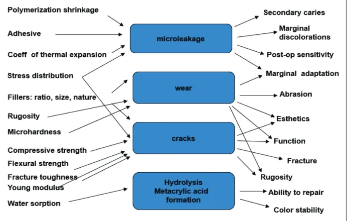
Many parameters involved in the physical and mechanical properties of dental materials (dental composite as an example) have been compiled in figure 1, together with different tests used to predict its performance and longevity in the oral environment.
As can be shown, that sophisticated interaction between the different variants adds to the complexity of defining the role played by each of these properties.
We decided to investigate most of the parameters that impact the behavior of the restoration once in function.
The relevance of this work resides in the transversal approach it adopts, through simultaneously studying the restorative materials characteristics, to improve the
5
knowledge of their behavior and to create specific investigative tools (numerical, analogical) that we seek to develop in our laboratory.
Figure 1: Physical and mechanical properties of composite and their clinical implications (courtesy Pr Colon)
Different factors present on the left hand side of figure 1 were examined through different studies presented in this work. We selected the mechanical properties that correlate effectively with clinical outcomes the flexural strength, flexural modulus, microhardness and fracture toughness. These tests were performed on CAD/CAM block materials (indirect restorations) with different material compositions. They were also conducted on bulk composites and fiber composite (direct restorations) being
6
polymerized in bulk. They present the advantage of ease of insertion, and comprise different types of monomer composition and filler volumes including the fiber reinforced composite. Filler ratio, size and nature were then analyzed for their impact on the outcome of different materials.
The role of adhesive layer on the integrity of the restoration was the subject of several studies discussed in this work. Thus, while the restorative material may have mechanical properties sufficient to resist masticatory stresses, differences in the coefficient in thermal expansion, and in modulus of the adhesive layer would lead to marginal leak, secondary caries and eventual failure of the restoration.
With the continuous development of new dental restorative products, many important properties are overlooked. We can logically presume that using a limited number of tests cannot be sufficient to adequately predict the longevity of the restoration and that studies that characterize only one aspect of the restorative material would ultimately draw inaccurate conclusions. Hence, a more global method for evaluating dental materials should be adopted. The final aim, furthermore, is to launch, through an ANSM project (Agence Nationale de la Sécurité du Médicament), a new device that would integrate all the critical parameters needed for testing new materials.
This thesis is built through different scientific papers, each of them comprise part of our knowledge in the field of dental restorations. The overview of all these different topics is the basis of the manuscript leading to the final target of medialization of the clinical behavior of dental restorations.
7
2. Dental restorative materials
Types and composition of restorative dental materials
The restoration of dental tissue is a complex problem. The shape, function and appearance of the dental tissue to be replaced have to be restored in an efficient and simple way. The loss of dental tissue could be a result of caries process, attrition, abrasion, erosion or trauma, and regardless of the causative agent. It is mostly the amount, the location and the type of tissue to be replaced that dictates the technique of restoration and the material used, and sometimes the simple observation.
The complex anatomy of the dental structures and the amount of physical and mechanical stresses to which they are submitted, calls for a material that could withstand such a challenge.
Another equally important requirement is the handling of the material. Direct restorative materials have to go through a process of trituration, in which the material is inserted in a plastic phase into a tooth cavity (shaped to conform to the material used), then the materials becomes solid and begin to harden and acquire its final properties. Each of these steps is operator sensitive and requires time and effort. The other solution is the indirect restoration in which fabrication of the restoration outside the oral cavity (indirect technique) in the lab or using the CAD/CAM technology. The restoration is then cemented in place using the appropriate cement or adhesive. The insertion of the restoration again requires much time and effort to assure the entry in position and to avoid the affection of the weak interface between the material and the dental tissue.
8
The choice of the material of restoration and the technique of insertion depends on a multitude of factors, beginning with biologic and esthetic as well as economic factors. Moreover, the patient oral hygiene may dictate the kind of restoration to be used.
2.1. Restorative resin composite
Resins in dentistry, comprise a large variety of materials and functionalities, whether restorative, prosthetic or else. Composite, as a definition, is a physical mixture of materials. Dental composites typically involve a dispersed phase of filler particles distributed within a continuous phase (matrix phase). Historically, restorative composite resin began with the first successful innovation by Bowen in 1962 and the introduction of bis-GMA as monomer, able to form a resistant cross linked matrix, as well as the introduction of organic silane as a coupling agent to bind the filler particles to the resin matrix [2].
Restorative resin composite is basically composed of three distinct phases: the polymer resin matrix, the inorganic filler (mostly) and the filler resin interface. The polymer resin matrix is composed initially of a liquid monomer that converts to a highly cross linked polymer upon initiation of polymerization, whether chemically or using light. The filler particles enhance the mechanical and physical properties and reduce the fraction of resin in the final restoration. Finally, the filler resin interface couples these two components together [12].
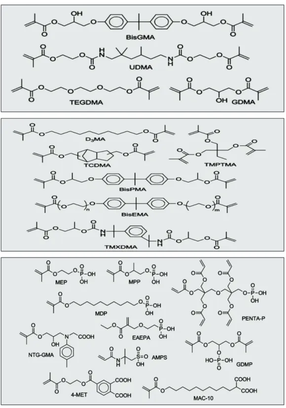
The most used monomers are the di-functional long chained BIS-GMA and UDMA making them extremely viscous. Therefore, they are diluted with another di-functional monomer, TEGDMA, of much lower viscosity, in order to practically be able to add larger portion of filler particles and to manipulate the material. The
9
corresponding monomer composition can be shown in Figure 2. Much development has taken place in monomer types and compositions, for example, several bisphenol A free composites are now available in the market.
Figure 2: The composition of several monomer types used in dental composite resin restorations; (Moszner et al. 2012) [13].
10
The filler particle mainly used in dental composite is silica, but the filler composition is often modified with other ions to produce desirable changes in properties. Lithium and aluminum ions make the glass easier to crush to generate small particles. Barium, zinc, boron, zirconium, and yttrium ions have been used to produce radio opacity in the filler particles. On the other hand, excessive modification (by replacement of the silicon in the structure) can reduce the efficacy of silane coupling agents [2].
Composites, generally, are classified with respect to the components, amounts, and properties of their filler or matrix phases or by their handling properties. The most common classification method is based on filler content (weight or volume percent), filler particle size, and method of filler addition.
Composites also could be defined on the basis of the matrix composition. The corresponding size and distribution of filler particles can be shown in Figure 3.
11
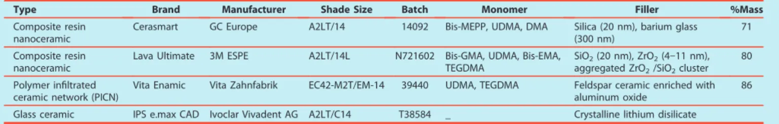
Almost all important properties of composites are improved by using higher filler levels. The only practical problem is that, as the filler level is increased, the fluidity decreases. The degree of filler addition is represented in terms of the weight percent or volume percent of filler. As the overall filler content increases, the physical, chemical, and mechanical properties generally improve. Relevant properties of different types of dental composite can be shown in Table 1.
Polymerization of acrylic resin monomers used in dentistry takes place in stages named activation, initiation, propagation, and termination. Activation involves the production of free radicals. Initiation is the step in which free radicals react with monomer units to create the initial end of a polymer chain. Propagation is the addition of monomer to the growing chain. Termination is the conclusion of the process as a result of steric hindrance, lack of monomer, or other problems.
Composites originally were designed for restoration of Class III, IV, and V tooth preparations, but now are used in modified forms for most other restorative dental uses. Based on their intended application, they can be used in all Classes (I-VI) of restorations, cements, bases, cores, veneers, or repair materials.
Table 1: Relative properties off different composite resin restorations, the values are reported from a variety of sources including manufacturer’s product bulletins (Sturdevant's Art and Science of Operative Dentistry) [14].
12
Developments in resin composite restorations
Currently, the high demand for these restorations and their structural composition allow for significant room for advancements, particularly with respect to their mechanical properties: polymerization shrinkage, thermal expansion coefficient, fracture resistance, marginal leakage, and toxicity [15,16].
These shortcomings reduce the lifetime of composite resin restorations and represent the driving force for improvement in dental composites. Clinical evaluations and laboratory-based studies continue to highlight the need for improved dental restorative composites [17]. Each component of the resin composite represents an opportunity for improvement and is the target of recent research.
In fact, one of the main advantages of restorative composite resin is its limitless ability for development and modification. Some examples of the potential advances, that could develop restorative resin composite, include:
Development of the initiator system
The use of benzoyl germanium initiators in place of the Camphorquinone / Amine. Systems show better shelf stability and depth of cure [18].
Development of the polymerization reaction
x Soft start curing was studied in depth, and though it was originally hypothesized that it allowed for stress relaxation, it was found that there was subsequent reduction in the degree of conversion and mechanical properties [19].
x Polymerization-induced Phase Separation was considered to decrease the volumetric shrinkage taking place during the polymerization reaction [20].
x Hybrid polymerization reactions forming interpenetrating polymer network (IPN) where the material is formed from two distinct polymerizations with
13
significant bonds between the two. These materials exhibit low toxicity and reduced shrinkage stress [21].
x Ring-opening Polymerization reaction, that depend on opening of a cyclic structure to facilitate intermonomer bonding and crosslinking, lead to less volumetric shrinkage. The most pronounced example is the silorane composite [22].
x Bulk filling composites are claimed to be more efficiently polymerized, and are then to be inserted in thicker increments in posterior cavities.
Development in the Polymer component
x Bile acids and Fluorinated derivatives of BisGMA into BisGMA / TEGDMA resins were found to reduce water sorption and volumetric shrinkage. But their high viscosity led to limited commercial incorporation of these monomers [23].
x Ultra rapid Monomethacrylates like Monovinyl (meth) acrylate monomers, showed rapid polymerization rate and resulted in adequate mechanical properties and higher degree of conversion compared to conventional composite systems [24].
x Acidic monomers enabled the adhesive layer to be eliminated. On the other hand, they increased the hydrophilicity of these materials limiting their use.
Development in the filler system and addition of components
x Eliminating the silane-mediated interface between filler and matrix by direct mechanical interlocking by using mesoporous silica fillers or single-walled carbon nanotubes (SWCNT) as a secondary filler [25].
14
x Calcium phosphate nanoparticles with silicon nitride whiskers or other composites to promote remineralization [26].
x Fiber reinforced composites (FRCs): Composite made of a polymer matrix that is reinforced by fine thin fibers
More than 50% of the glass fibers used for reinforcement is E-glass. They are a mixture of amorphous phases and silicon oxide, calcium oxide, barium oxide, aluminum oxide and some oxides of alkali metals. Orientation of fiber plays an important role in increasing the strength. The reinforcing effect of the fiber fillers is based, not only on stress transfer from polymer matrix to fibers, but also on the behavior of individual fibers as crack stoppers. Xu et al. showed that increasing the glass fiber length generally increased the ultimate strength and fracture resistance. These properties have clinical significance and would affect the longevity of restoration [27]. Their conclusion was that the Quantity of glass fibers should be defined by volume percentage and not in weight percentage.
Coating of fiber with polymer matrix avoid voids between the matrix and the fiber that would lead to decreasing the load bearing capacity and water sorption. Other important factors are the adhesion of fiber to the polymer matrix and the distribution of fibers. Mechanical properties such as strength, stiffness, toughness and fatigue resistance as well as the linear coefficient of thermal expansion (LCTE) depend upon the geometry of the reinforcement and the fiber orientation (Krenchel's factor) [28].
Short fiber reinforced composite (everX Posterior™) has been introduced as a dental restorative composite resin. Studies showed improvements in the load bearing capacity short glass fiber composite
15
resin, has also exhibited control of the polymerization shrinkage stress by fiber orientation. Thus, marginal microleakage was reduced compared with conventional particulate filler restorative composite resins. The specific mechanical and physical strength and the specific modulus of these fiber reinforced composite materials may be markedly superior to those of existing resin-based composites and metallic materials.
A study was conducted by our group to test and compare fiber composite to other bulk fill composites. Several mechanical properties, correlated to the composite longevity in the oral cavity, were chosen. The results obtained showed that the mechanical properties of the fiber composite were among the best, making it suitable to be used in posterior stress bearing regions in the oral cavity.
Original Research Article: Comparison of mechanical properties of a new fiber reinforced composite and bulk filling composites
©Copyrights 2015. The Korean Academy of Conservative Dentistry.
262
This is an Open Access article distributed under the terms of the Creative Commons Attribution Non-Commercial License (http://creativecommons.org/licenses/ by-nc/3.0) which permits unrestricted non-commercial use, distribution, and reproduction in any medium, provided the original work is properly cited.
Comparison of mechanical properties of a new fiber
reinforced composite and bulk filling composites
Objectives: The aim of this study was to evaluate the mechanical and physical properties of a newly developed fiber reinforced dental composite. Materials and Methods: Fiber reinforced composite EverX Posterior (EXP, GC EUROPE), and other commercially available bulk fill composites, including Filtek Bulk Fill (FB, 3M ESPE), SonicFill (SF, Kerr Corp.), SureFil (SDR, Dentsply), Venus Bulk Fill (VB, HerausKultzer), Tetric evoceram bulk fill (TECB, Ivoclar Vivadent), and Xtra Base (XB, Voco) were characterized. Composite samples light-cured with a LED device were evaluated in terms of flexural strength, flexural modulus (ISO 4049, n = 6), fracture toughness (n = 6), and Vickers hardness (0, 2, and 4 mm in depth at 24 hr, n = 5). The EXP samples and the fracture surface were observed under a scanning electron microscopy. Data were statistically analyzed using one-way ANOVA and unpaired t-test. Results: EXP, FB, and VB had significantly higher fracture toughness value compared to all the other bulk composite types. SF, EXP, and XB were not statistically different, and had significantly higher flexural strength values compared to other tested composite materials. EXP had the highest flexural modulus, VB had the lowest values. Vickers hardness values revealed SF, EXP, TECB, and XB were not statistically different, and had significantly higher values compared to other tested composite materials. SEM observations show well dispersed fibers working as a reinforcing phase. Conclusions: The addition of fibers to methacrylate-based matrix results in composites with either comparable or superior mechanical properties compared to the other bulk fill materials tested. (Restor
Dent Endod 2015;40(4):262-270)
Key words: Bulk composite; Fiber composite; Mechanical properties
Introduction
Dental composite resin recently became the material of choice for most patients and dental practitioners.1 However, volumetric shrinkage and fracture are still considered as major concerns with dental composites.2,3 In order to overcome these weaknesses, attempts have been made toward increasing both their physical and mechanical properties.4 This necessitates the comprehensive appraisal of each of its components such as the resin matrix, the filler or the filler-resin interface, and their role in affecting the material properties. Different studies have investigated this in order to improve composite properties, either by varying the particle size, percentage, or by development of the polymer matrix chemistry.4,5
Hazem Abouelleil1*, Nelly Pradelle1,2, Cyril Villat1,3, Nina Attik1, Pierre Colon1,2, Brigitte Grosgogeat1,3 1
Laboratoire des Multimatériaux et Interfaces, UMR CNRS 5615, Université Lyon1, Villeurbanne, France
2
UFR D’odontologie, Université Paris Diderot, APHP, Hôpital Rothschild, Service d’Odontologie, Paris, France
3
UFR Odontologie, Université Lyon1, Service de Consultations et de Traitements Dentaires, Hospices Civils de Lyon, Lyon, France
Received April 2, 2015; Accepted June 30, 2015.
1
Abouelleil H; Pradelle N; Villat C; Attik N; Colon P; Grosgogeat B, Laboratoire des Multimatériaux et Interfaces, UMR CNRS 5615, Université Lyon1, Villeurbanne, France
2
Pradelle N; Colon P, UFR D’odontologie, Université Paris Diderot, APHP, Hôpital Rothschild, Service d’Odontologie, Paris, France
3
Villat C; Grosgogeat B, UFR
Odontologie, Université Lyon1, Service de Consultations et de Traitements Dentaires, Hospices Civils de Lyon, Lyon, France
*Correspondence to
Hazem Abouelleil, DDS, MD. Dental practitioner and Lecturer, Laboratoire des Multimatériaux et Interfaces, UMR CNRS 5615, Université Lyon1, Villeurbanne, France
TEL, +33(0)478778689; FAX, +33(0)478 778712; E-mail, [email protected]
Research article
ISSN 2234-7658 (print) / ISSN 2234-7666 (online) http://dx.doi.org/10.5395/rde.2015.40.4.262
263 www.rde.ac
Evolution in both filler and polymer technology in dental composite resins led to a wide selection of materials that provide the adequate properties required for each clinical situation.4 Yet, the use of dental composites in high stress
bearing areas remains to be a challenge for the dental practitioner, since bulk fracture is still considered one of the primary reasons for failure.2,6 Bulk fill composites were
introduced in an effort to improve the performance of composite resin restorations, which was inserted in 4 mm increments mainly in the posterior areas and considered to have higher physical and mechanical properties to endure the higher masticatory stresses. Moreover, the reduced treatment time decrease the risk of air entrapment or moisture contamination.7 They are also claimed to reduce
cuspal deflection and promote light transmittance.7,8
Currently, various studies reveal the difficulty in comparing between the available materials due to variation in composition and viscosity.9-11
Bulk filling composites usually have higher filler volume percentage, and sometimes a modified initiator system to ensure better curing in depth, as compared to conventional composites. While no long term clinical studies are available regarding their intraoral performance, Ilie et al. found bulk filling composites to have lower mechanical properties, except for flexural strength as compared to nanohybrid and microhybrid resin based composites.9
However, other studies found them equally successful compared to conventional composites.7,12 Many bulk fill
composite resins have been investigated regarding different parameters like degree of conversion, polymerization stress or microleakage. Such studies have shown that bulk fill composites resins have similar properties as conventional dental composite resins.12-16
Finan et al. studied the degree of conversion, biaxial flexural strength and Vickers hardness of two flowable bulk composites (SDR and XB), and despite the differences between the two materials, found that the properties justify their use in 4 mm increments.17 The variation in material composition and viscosity, whether flowable or non flowable bulk composites, leads to differences in physical and mechanical properties among the bulk fill composites available in the market.7 Fiber reinforcement of conventional dental composites were also introduced with the aim of enhancing their physical and mechanical properties, and increasing their resistance to fracture. The enhancement of the material properties was due to the stress transfer from the matrix to the fibers depending on the fibers length and diameter. Garoushi et al. studied their effect, and found a significant improvement in the materials physical properties.18
It was deemed important to investigate the role of fibers added to composite compared to other commonly used bulk fill composites, and to examine the extent to which fiber reinforcement would enhance the mechanical properties of
the materials. Multiple laboratory investigations have been used to evaluate dental composite resins; standardized tests present the advantage of being easily reproducible in laboratories, and allowing values obtained by different institutes to be compared. Moreover, they provide preliminary information about the material suitability in the oral environment and the extent to which they conform to the indications prescribed by the manufacturer.19 Heintze
et al. found that flexural strength and flexural modulus
tests can be used as a good indicator for the material durability under stress, and correlate well with the clinical longevity.19 Fracture toughness test was considered by
Ilie et al. as another important method that investigates the material’s ability to endure stress without fracture and monitor the crack propagation inside the material before failure.11 On the other hand, Vickers hardness assay,
one of the most used mechanical experiments examines the material surface hardness, and scanning electron microscope observations reveal important information about the samples used and the mode of failure of the material.9 Standard ISO flexural strength and modulus
tests consider only 2 mm thickness samples. However bulk fill composites are indicated to be used clinically in 4 mm thick increments, and accordingly investigating the material at this thickness seems more appropriate.
The aim of this study was to investigate the mechanical properties of a fiber reinforced composite compared to other commonly used bulk fill composites, and to consider its performance under laboratory settings. The null hypothesis was that there is no significant difference in mechanical properties (flexural strength, flexural modulus, fracture toughness, and Vickers hardness) among the fiber reinforced composite and other bulk fill composites.
Materials and Methods
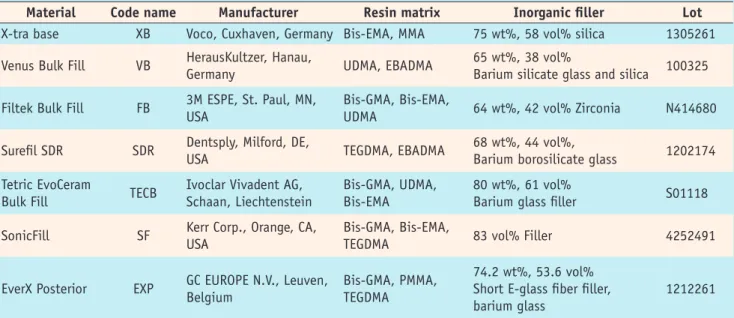
Bulk fill dental composites used in the study were X-tra base (XB, Voco GmbH, Cuxhaven, Germany), Venus bulk fill (VB, HerausKultzer, Hanau, Germany), Filtek bulk fill (FB, 3M ESPE, St. Paul, MN, USA), Surefil SDR (SDR, Dentsply, Milford, DE, USA), Tetric evoceram bulk fill (TECB, Ivoclar Vivadent AG, Schaan, Liechtenstein), SonicFill (SF, Kerr Corp., Orange, CA, USA), and a fiber reinforced bulk fill dental composite resin, EverX Posterior (EXP, GC EUROPE NV, Leuven, Belgium). The compositions of bulk fill materials used, their shade as well as their lot numbers are listed in Table 1.
For the fracture toughness test, flexural strength and modulus tests, the number of samples for each of the materials used was 6. The tested samples were polymerized using GC G-light unit (GC EUROPE NV) from both sides for 40 seconds. A modified flexural strength test was performed using bulk fill samples with 4 mm2 cross sectional areas polymerized only from the top side as done Mechanical properties of a fiber reinforced composite
264 www.rde.ac
Table 1. Materials, manufacturers, chemical composition of the matrix, fillers and filler contents
Material Code name Manufacturer Resin matrix Inorganic filler Lot X-tra base XB Voco, Cuxhaven, Germany Bis-EMA, MMA 75 wt%, 58 vol% silica 1305261 Venus Bulk Fill VB HerausKultzer, Hanau,
Germany UDMA, EBADMA
65 wt%, 38 vol%
Barium silicate glass and silica 100325 Filtek Bulk Fill FB 3M ESPE, St. Paul, MN,
USA
Bis-GMA, Bis-EMA,
UDMA 64 wt%, 42 vol% Zirconia N414680 Surefil SDR SDR Dentsply, Milford, DE,
USA TEGDMA, EBADMA
68 wt%, 44 vol%,
Barium borosilicate glass 1202174 Tetric EvoCeram
Bulk Fill TECB
Ivoclar Vivadent AG, Schaan, Liechtenstein
Bis-GMA, UDMA, Bis-EMA
80 wt%, 61 vol%
Barium glass filler S01118 SonicFill SF Kerr Corp., Orange, CA,
USA
Bis-GMA, Bis-EMA,
TEGDMA 83 vol% Filler 4252491
EverX Posterior EXP GC EUROPE N.V., Leuven, Belgium
Bis-GMA, PMMA, TEGDMA
74.2 wt%, 53.6 vol% Short E-glass fiber filler, barium glass
1212261
Bis-EMA, ethoxylatedbisphenol A dimethacrylate; MMA, methylmethacrylate; UDMA, urethane dimethacrylate; EBADMA, ethoxylatedbisphenol A dimethacrylate; Bis-GMA, bisphenylglycidyldimethacrylate; TEGDMA, triethylene glycol dimethacrylate; PMMA, polymethyl methacrylate.
in the clinical situation. The wavelength of the light was between 380 and 520 nm with maximal intensity at 470 nm and light intensity was 1,150 mW/cm2. The specimens
from each group were stored in water at 37 for 48 hours before testing.
Fracture toughness
To measure the fracture toughness (KIC), rectangular
glass molds that were lined with polyester strips (Striproll, Kerrhawe SA, Bioggio, Switzerland) were used to prepare single-edge-notched specimens. The cured samples (3 mm × 6 mm × 25 mm) were removed without using force. A sharp central notch of specific length (a) was produced by inserting a razor blade into an accurately fabricated slot at mid-height in the mold. The slot extended down half the height to give a/W = 0.5. The crack plane was perpendicular to the specimen length. The length of the crack was checked using a stereomicroscope.
Fracture toughness KIC was calculated from the following
formula:
KIC = [ 3PL ]Y
2BW
Where P is the peak load at fracture, L is the length, B is the width, W is the height, a is the average notch depth, and Y is the calibration functions for given geometry
3 2
Y = 1.93[a/W]1/2 − 3.07[a/W]3/2 + 14.53[a/W]5/2 −
25.11[a/W]7/2 + 25.80[a/W]9/2
Flexural strength and flexural modulus
According to the ISO 4049, samples for a three point bending test were prepared in Teflon molds between two glass slabs, resulting in bar shaped specimens (2 mm × 2 mm × 25 mm). The test was conducted under a cross-head speed of 0.5 mm/min, with a span length of 20 mm and an indenter diameter of 2 mm. All specimens were loaded in a Universal Mechanical testing machine (Servo hydraulic - Adamel Lhomargy DY-34, MTS, Roissy-en-Brie, France). Flexural strength and modulus tests, were repeated on larger samples (n = 6, 4 mm × 4 mm × 25 mm), that were cured only from the top, using the same light and stored in water at 37°C for 48 hours before testing.
Flexural strength (Of) and flexural modulus (Ef) were
calculated from the following formulas:
Of = 3FmI 2bh2 Ef = SI 4bh3
Where Fm is the applied load (N) at the highest point of load–deflection curve, I is the span length (20 mm), b
3
Abouelleil H et al.
http://dx.doi.org/10.5395/rde.2015.40.4.262
265 www.rde.ac
Mechanical properties of a fiber reinforced composite
examine the fracture mode, and to measure the fiber’s diameter and length. Samples were dried, sputter-coated with metal, and observed. The type of fracture was determined for each specimen.
Statistical analysis
The statistical analysis of the current data was performed using the application of one-way analysis of variance (ANOVA). The results were compared between each test and between each material type using unpaired t-test. The results are reported as mean ± SD. Statistical significance was accepted at p < 0.05.
Results
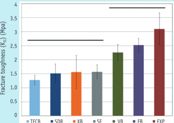
Fracture toughness and Vickers hardness of tested composite materials are presented in Figures 1 and 2. Flexural strength and flexural modulus are presented in Table 2. The fiber reinforced composite EXP had significantly higher fracture toughness value (3.1 MPa·m1/2), compared to other bulk composites except
for FB (2.52 MPa·m1/2) and VB (2.26 MPa·m1/2) where
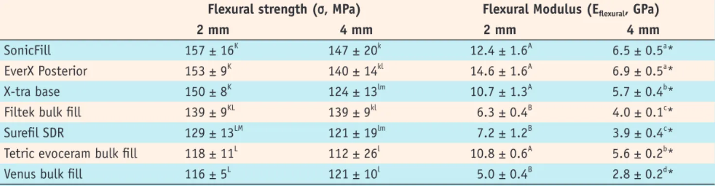
no significant difference was found with EXP. In the normalized flexural strength test (2 mm x 2 mm x 25 mm), SF (157.6 MPa), EXP (153.6 MPa), XB (150.4 MPa) and FB (140.0 MPa) were not statistically different, and these have significantly higher flexural strength values compared to other tested composite materials, except for FB which was similar to SDR. EXP had significantly higher flexural modulus (14.6 GPa), while SF (12.47 GPa), TECB (10.87 GPa), and XB (10.65 GPa) were not significantly different and came in second position. On the other hand, VB (5.02 GPa) had the lowest statistically significant value.
Table 2. Flexural strength (Ȁ, MPa) and Flexural Modulus (Eflexural, GPa) for the 2 mm and 4 mm sample groups
Flexural strength (Ȁ, MPa) Flexural Modulus (Eflexural, GPa)
2 mm 4 mm 2 mm 4 mm SonicFill 157 ± 16K 147 ± 20k 12.4 ± 1.6A 6.5 ± 0.5a* EverX Posterior 153 ± 9K 140 ± 14kl 14.6 ± 1.6A 6.9 ± 0.5a * X-tra base 150 ± 8K 124 ± 13lm 10.7 ± 1.3A 5.7 ± 0.4b * Filtek bulk fill 139 ± 9KL 139 ± 9kl 6.3 ± 0.4B 4.0 ± 0.1c* Surefil SDR 129 ± 13LM 121 ± 19lm 7.2 ± 1.2B 3.9 ± 0.4c* Tetric evoceram bulk fill 118 ± 11L
112 ± 26l
10.8 ± 0.6A
5.6 ± 0.2b
* Venus bulk fill 116 ± 5L
121 ± 10l
5.0 ± 0.4B
2.8 ± 0.2d
* Uppercase letters identify statistically homogenous groups for 2 mm thickness samples. Lower case letters identify statistically homogenous groups for 4 mm thickness samples. Asterisks identify statistical difference between 2 mm and 4 mm thickness samples of the same material (p < 0.05).
is the width of test specimens and h is the thickness of test specimens. S is the stiffness (S = F/d, N/m) and d is the deflection corresponding to load F at a point in the straight-line portion of the trace.
Vickers hardness test
The Vickers hardness test was performed with Leitz microhardness device (Leitz, Wetzlar, Germany), under a force of 200 g for 30 seconds. Ten samples for each material were prepared using a 5 mm diameter Teflon mold, with either 2 mm (n = 5) or 4 mm thickness (n = 5), placed between 2 glass plates. The materials were polymerized only on one side for 40 seconds. The excesses were removed by polishing the 2 surfaces using abrasive paper discs of decreasing coarseness from 2,400 to 4,000 grits (Struers SAS, Champigny sur Marne, France) at 3,000 rpm under water irrigation. The top surface (polymerized) and the bottom surface (non-polymerized) were marked to be identified. Each sample was tested 5 times on each side, at 24 hours after immersion in distilled water at 37. The Vickers hardness was calculated using the formula:
HV = 1854.4 P
d2
Where P (g) is the load applied, and d is the average of the 2 diagonals of the surface of the diamond indentation.
Scanning electron microscopy
Scanning Electron microscope (S800-1, Hitachi Europe Ltd., Whitebrook, Berkshire, UK) observations were conducted under x80, x100, and x250 magnification to
266 www.rde.ac
GPa), which were less affected in comparison with original test.
Vickers hardness values revealed that SF had the highest value followed by EXP. The decrease in hardness between the surface and 2 mm and 4 mm depths were not significant for EXP, TECB, and VB, while other bulk composites revealed a significant difference between the curing depths.
SEM analysis revealed that the fibers stop the crack propagation along the fracture line, as shown in Figure 3. Considering the modified test (4 mm × 4 mm × 25 mm),
there was no significant difference from those obtained in the original test, except for XB and they had the same order of the strength values from SF (147.67 MPa), EXP (140.04 MPa), and FB (139.62 MPa). On the other hand, the flexural modulus values decreased significantly in comparison with the normalized test, attaining almost half the original value, while remaining in the same order, with the highest flexural modulus for EXP (6.89 GPa), together with SF (6.55 GPa), followed by XB (5.7 GPa) and FB (4.01 Figure 1. Bar graph illustrating fracture toughness (KIC).
Straight line indicates that there was no statistically significant difference between the groups.
TECB, Tetric evoceram bulk fill; SDR, SureFil SDR; XB, Xtra Base; SF, SonicFill; VB, Venus bulk fill; FB, Filtek bulk fill; EXP, EverX Posterior.
4 3.5 3 2.5 2 1.5 1.0 0.5 0 Fr actur e toughn ess (K IC ) (Mpa) TECB SDR XB SF VB FB EXP
Figure 2. Bar graph illustrating Vickers hardness (N/mm2) at different curing depths of 4 mm, 2 mm and at the surface. Dotted line ( ) indicates that there was no statistically significant difference between the materials. Straight line ( ) indicates that there was no statistically significant difference within the same material at different curing depths.
TECB, Tetric evoceram bulk fill; SDR, SureFil SDR; XB, Xtra Base; SF, SonicFill; VB, Venus bulk fill; FB, Filtek bulk fill; EXP, EverX Posterior.
90.00 80.00 70.00 60.00 50.00 40.00 30.00 20.00 10.00 0.00 Vi ck ers H ar d n ess (N/mm 2 ) VB FB SDR XB TECB EXP SF
Figure 3. Scanning electron photomicrograph of fracture toughness sample (a) after failure; (b) the fiber orientation across the failure line are shown at higher magnification.
(a) (b)
Abouelleil H et al.
http://dx.doi.org/10.5395/rde.2015.40.4.262
267 www.rde.ac
Mechanical properties of a fiber reinforced composite
Discussion
According to the results obtained in the current study, the null hypothesis was rejected, that is, fiber insertion into composite leads to significant increase in physical and mechanical properties, such as flexural strength, flexural modulus, fracture toughness, and Vickers hardness.
In this study, flexural strength and modulus were investigated. These tests are considered to be good indicators of the material resistance to fracture in normal masticatory conditions, taking in account the great variability in the results obtained between studies.19,20 The
results obtained are in accordance with previous studies conducted on bulk composites. SF, EXP, XB, and FB had significantly higher flexural strength values, compared to VB and TECB which had the lowest.9,18,21 Moreover, as shown
in previous works, the filler volume percentage is closely related to the flexural strength and flexural modulus values.9,18,21 This can be shown for SF with the highest filler
volume percentage (83%) ranking the highest, TECB (61%) and XB (58%) follow next, while VB with the lesser filler volume percentage (38%) ranking the lowest. Interestingly, EXP (53.6%) performed relatively better in these two tests compared to its filler volume percentage, showing the role of the fibers in increasing the material stiffness and resistance to bending force during testing and probably during function.
In this work, the modified flexural strength and modulus tests were done on 4 mm increments cured only from the top side in an effort to mimic the clinical situation. This would eventually mean less matrix polymerization and, accordingly, a larger role of the filler type and percentage in the material behavior. The results obtained show that the flexural strength values remained significantly unchanged. In comparison to the original test, significant decrease in the flexural modulus values of the composites tested indicated a marked decrease in rigidity. This is probably due to an increase in thickness of the increments and decrease in the overall matrix polymerization. A probable explanation would be that, as a result of less matrix polymerization and the consequent lack of rigidity, the modified test samples were able to withstand flexure even at greater load relative to greater sample thickness (hence unchanged flexural strength) but with more deformation before final failure (hence lower modulus of flexure). These results, when confirmed with further studies, would throw more insight on an important aspect regarding the amount of deformation and the distortion of the material due to the decreased stiffness, most notably at the interface region. This would also provide some explanation for the discrepancies found between results obtained in the laboratories and those from clinical studies in which bulk materials are inserted in larger and thicker increments and cured only from one side.19
Results obtained acknowledge the role of fibers in increasing the material’s resistance to fracture, and coincide with those of previous studies.18,22 The single edge
notched beam method used in this study is one of the most commonly used fracture toughness test methods, which are used to predict resistance to fracture. The method is widely used in dental material research and is usually conducted by means of a 3 point bending apparatus, and the sharp crack created could be easily measured. This method is also very sensitive to the notch width and depth, thus making comparison difficult between different studies.11,18
In the present work, no correlation was found between the fracture toughness value and the filler volume percentage or the filler particle size.
The enhancement of the material properties was explained to be due to the stress transfer from the matrix to the fibers and also due to the action of the fibers in stopping crack propagation through the material.23 It was found
that the mere insertion of fibers is not enough to enhance the composite properties, that is, the fibers length and diameter play a critical role in this mechanism. Peterson found that fibers incorporated into a material, greatly enhances its mechanical properties, on the condition that the fibers have a length that exceeds a certain minimum length. This is known as the critical fiber length,which could be calculated using the following formula:24
lc = f
d
2c
Where the critical length (lc) equals the ultimate tensile strength of the fiber (f) multiplied by the fiber diameter (d), and divided by twice the shear strength of the matrix interface (c)
The physical explanation of the strengthening and stiffening mechanism is that since the matrix has a much lower modulus than the fiber, the matrix strains more. The critical fiber length is therefore the minimum length at which the center of the fiber reaches its ultimate tensile strength when the matrix reaches its maximum shear strength. Accordingly, composite with fibers below critical length fail to show enhanced properties.18 In the present study, we were able to measure the fiber length and diameter using stereomicroscopy and SEM, and we found that EXP had a fiber diameter of 16 μm and a wide range of fiber length, with the average length lying between 1 and 2 mm similar to the values found in previous studies, thus exceeding the fiber length required.18 The fiber length and orientation can be shown in Figure 4.
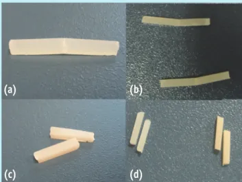
One interesting observation found from the fracture toughness and the flexural strength test samples alike was that all fiber reinforced composite EXP samples remained attached, even after failure of the sample and formation of crack line, unlike the samples from other bulk fill brands
268 www.rde.ac
which separated in two pieces the time the failure load was reached, as can be seen in Figure 5. Scanning electron microscope observations performed on fractured samples show the fibers traversing the crack line and between the fractured parts, as can be seen in Figure 3. Further investigation of this property is important clinically, since not only it shows the material resistance to fracture, but also its resistance to displacement at the more vulnerable interface, thus preventing cavitation and food impaction. Moreover, this property would render the material with better potential for repair.
The Vickers microhardness test samples show that SF, EXP, TECB, and XB have the highest values compared to SDR, FB, and VB. It is worth noting that though this method was criticized as an unreliable indicator of the curing quality, and that it overestimates the depth of cure.Flury et al. have shown that Vickers microhardness could be considered as an accurate tool for estimating the polymerization depth for bulk composite resins.25,26 Moreover, only SF and EXP had bottom surface hardness values that exceeded the 50 VHN considered ideal.27 However, EXP is the only composite with Vickers hardness value at 4 mm depth that exceeded the 80% ratio compared to the top surface hardness as required in literature.26-29 The results thus obtained provide evidence that EXP could be used in 4 mm increments for tooth cavity fillings.
The present results were obtained in optimized laboratory settings, however, clinical conditions are not similar and the aspects like insertion and handling could have a potential effect on the mechanical properties of the materials and their performance in vivo. Another important
factor that should be taken into consideration as one of the limitations of the current study is the fiber alignment inside the composite in relation to the acting force, which is not necessarily consistent with the laboratory simulations performed during in vitro testing. Some of the important aspects considering the materials polymerization contraction and contraction stress were not included in the study. Further investigations should be conducted to test other material properties. According to the results obtained in this work, the fiber reinforced composite tested may be used as a restorative material in stress bearing areas. In order to acknowledge the results obtained with the present study, this should be followed by long term clinical studies to assure the materials performance under normal clinical conditions.
Conclusions
In the current study, fiber reinforced composite EXP had either comparable or superior resistance to fracture, flexural strength and modulus, as well as high microhardness values, compared to other bulk fill composite resins.
Acknowledgement
The dental companies GC, Voco, 3M/ESP, Dentsply, Ivoclar Vivadent, Kerr, Heraus Kultzer are gratefully acknowledged for the generous donation of the dental composites. The SEM study was supported by the Microstructures Technology Center of University Claude Bernard Lyon1.
Figure 5. Samples of (a) fracture toughness and (b) flexural strength tests for EverX Posterior remained connected after failure, compared to other bulk composite samples after (c) fracture toughness and (d) flexural strength, which were completely separated into two fragments.
(a) (b)
(c) (d)
Figure 4. Microscopic image of EverX Posterior showing fiber length extending to the length of one millimeter and up to two milimeters.
Abouelleil H et al.
http://dx.doi.org/10.5395/rde.2015.40.4.262
269 www.rde.ac
Mechanical properties of a fiber reinforced composite
Conflict of Interest: No potential conflict of interest relevant to this article was reported.
References
1. Pallesen U, van Dijken JW, Halken J, Hallonsten AL, Höigaard R. Longevity of posterior resin composite restorations in permanent teeth in Public Dental Health Service: a prospective 8 years follow up. J Dent 2013;41:297-306.
2. Demarco FF, Corrêa MB, Cenci MS, Moraes RR, Opdam NJ. Longevity of posterior composite restorations: not only a matter of materials. Dent Mater 2012;28:87-101. 3. Sarrett DC. Clinical challenges and the relevance of
materials testing for posterior composite restorations.
Dent Mater 2005;21:9-20.
4. Cramer NB, Stansbury JW, Bowman CN. Recent advances and developments in composite dental restorative materials. J Dent Res 2011;90:402-416.
5. Ferracane JL. Resin composite-state of the art. Dent
Mater 2011;27:29-38.
6. Da Rosa Rodolpho PA, Donassollo TA, Cenci MS, Loguércio AD, Moraes RR, Bronkhorst EM, Opdam NJ, Demarco FF. 22-Year clinical evaluation of the performance of two posterior composites with different filler characteristics. Dent Mater 2011;27:955-963. 7. Walter R. Critical appraisal: bulk-fill flowable composite
resins. J Esthet Restor Dent 2013;25:72-76.
8. Moorthy A, Hogg CH, Dowling AH, Grufferty BF, Benetti AR, Fleming GJ. Cuspal deflection and microleakage in premolar teeth restored with bulk-fill flowable resin-based composite base materials. J Dent 2012;40:500-505.
9. Ilie N, Bucuta S, Draenert M. Bulk-fill resin-based composites: an in vitro assessment of their mechanical performance. Oper Dent 2013;38:618-625.
10. Leprince JG, Palin WM, Vanacker J, Sabbagh J, Devaux J, Leloup G. Physico-mechanical characteristics of commercially available bulk-fill composites. J Dent 2014;42:993-1000.
11. Ilie N, Hickel R, Valceanu AS, Huth KC. Fracture toughness of dental restorative materials. Clin Oral
Investig 2012;16:489-498.
12. El-Safty S, Silikas N, Watts DC. Creep deformation of restorative resin-composites intended for bulk-fill placement. Dent Mater 2012;28:928-935.
13. Alshali RZ, Silikas N, Satterthwaite JD. Degree of conversion of bulk-fill compared to conventional resin-composites at two time intervals. Dent Mater 2013; 29:e213-e217.
14. Van Ende A, De Munck J, Van Landuyt KL, Poitevin A, Peumans M, Van Meerbeek B. Bulk-filling of high C-factor posterior cavities: effect on adhesion to cavity-bottom dentin. Dent Mater 2013;29:269-277.
15. Roggendorf MJ, Krämer N, Appelt A, Naumann M,
Frankenberger R. Marginal quality of flowable 4-mm base vs. conventionally layered resin composite. J Dent 2011;39:643-647.
16. Poggio C, Dagna A, Chiesa M, Colombo M, Scribante A. Surface roughness of flowable resin composites eroded by acidic and alcoholic drinks. J Conserv Dent 2012;15: 137-140.
17. Finan L, Palin WM, Moskwa N, McGinley EL, Fleming GJ. The influence of irradiation potential on the degree of conversion and mechanical properties of two bulk-fill flowable RBC base materials. Dent Mater 2013;29:906-912.
18. Garoushi S, Säilynoja E, Vallittu PK, Lassila L. Physical properties and depth of cure of a new short fiber reinforced composite. Dent Mater 2013;29:835-841. 19. Heintze SD, Zimmerli B. Relevance of in vitro tests of
adhesive and composite dental materials, a review in 3 parts. Part 1: approval requirements and standardized testing of composite materials according to ISO specifications. Schweiz Monatsschr Zahnmed 2011;121: 804-816.
20. Alander P, Lassila LV, Vallittu PK. The span length and cross-sectional design affect values of strength. Dent
Mater 2005;21:347-353.
21. Czasch P, Ilie N. In vitro comparison of mechanical properties and degree of cure of bulk fill composites.
Clin Oral Investig 2013;17:227-235.
22. Xu HH, Schumacher GE, Eichmiller FC, Peterson RC, Antonucci JM, Mueller HJ. Continuous-fiber preform reinforcement of dental resin composite restorations.
Dent Mater 2003;19:523-530.
23. Garoushi S, Vallittu PK, Lassila LV. Short glass fiber reinforced restorative composite resin with semi-inter penetrating polymer network matrix. Dent Mater 2007;23:1356-1362.
24. Petersen RC. Discontinuous fiber-reinforced composites above critical length. J Dent Res 2005;84:365-370. 25. Leprince JG, Leveque P, Nysten B, Gallez B, Devaux
J, Leloup G. New insight into the "depth of cure" of dimethacrylate-based dental composites. Dent Mater 2012;28:512-520.
26. Flury S, Hayoz S, Peutzfeldt A, Hüsler J, Lussi A. Depth of cure of resin composites: is the ISO 4049 method suitable for bulk fill materials? Dent Mater 2012;28:521-528. 27. Galvão MR, Caldas SG, Bagnato VS, de Souza Rastelli
AN, de Andrade MF. Evaluation of degree of conversion and hardness of dental composites photo-activated with different light guide tips. Eur J Dent 2013;7:86-93. 28. Polydorou O, Manolakis A, Hellwig E, Hahn P. Evaluation
of the curing depth of two translucent composite materials using a halogen and two LED curing units.
Clin Oral Investig 2008;12:45-51.
29. Yap AU, Seneviratne C. Influence of light energy density on effectiveness of composite cure. Oper Dent 2001;26: 460-466.
http://dx.doi.org/10.5395/rde.2015.40.4.262
24
In the light of the above study, and according to our results and those of various similar studies, it can be concluded that the tests performed on these composites reveal the variation in their mechanical properties. On the other hand, the presence of the mastication bench device would allow the mechanical testing of these composites under more relevant conditions and under forces that closely resembles these experienced in the human oral cavity.
The dental composites studied would be inserted and polymerized inside the mastication bench using the same clinical protocol utilized during a real clinical session. This would disclose more about the actual performance of the material, as could be observed in the example of the 2 and 4 mm thickness flexural test samples.
Moreover an important feature as fiber alignment in reinforced composite would be closely monitored with relevant ease.
25
1.1. Ceramics and CAD/CAM restorations
The word ceramics is used to designate materials having both metallic and nonmetallic ions in their compositional formula.
Dental ceramics are nonmetallic, inorganic structures, primarily containing compounds of oxygen with one or more metallic or semi metallic elements (aluminum, boron, calcium, cerium, lithium, magnesium, phosphorus, potassium, silicon, sodium, titanium and zirconium). Many dental ceramics contain a crystal phase and a silicate glass matrix phase [2].
They consist of silicate glasses, porcelains glass ceramics or highly crystalline solids. The properties of ceramics are customized for dental applications by precisely controlling the types and amounts of the components used in their production.
Ceramics do not react readily with most liquids gases or alkalis and weak acids, and they remain stable over long time periods. Dental ceramics are characterized by excellent strength, high hardness, biocompatibility, chemical inertness and esthetic potential.
Conversely, they are generally brittle and may fracture without warning when flexed extensively or when quickly heated and cooled, and they cause intensive wear of the opposing dentition.
The two reasons that seemed to limit the use of ceramics: their brittleness and the great effort and time required for processing. Recent advances in ceramic processing methods have led to much improvement in their mechanical properties and expanded the scope for their use in dentistry.
26
Classification
While several classifications exist for the dental ceramic materials as shown in figure 4, only the classifications based on microstructural composition and processing method will be discussed in this chapter.
Figure 1:Different classifications of dental ceramics(Philips dental materials) [2].
Microstructural classification
Ceramics can be differentiated according to their composition of glass to crystalline ratio, into four compositional categories:
x Glass-based systems
x Glass-based systems (mainly silica) with fillers, usually crystalline (typically leucite or, more recently, lithium disilicate),
x Crystalline- based systems with glass fillers (mainly alumina) x Polycrystalline solids (alumina and zirconia).
![Figure 3: Differences in filler size and distribution in dental composite resin (Ferracane 2011) [3]](https://thumb-eu.123doks.com/thumbv2/123doknet/14494610.718121/18.892.120.772.646.1092/figure-differences-filler-distribution-dental-composite-resin-ferracane.webp)
![Table 1: Relative properties off different composite resin restorations, the values are reported from a variety of sources including manufacturer’s product bulletins (Sturdevant's Art and Science of Operative Dentistry) [14].](https://thumb-eu.123doks.com/thumbv2/123doknet/14494610.718121/19.892.116.777.788.1092/relative-properties-different-restorations-manufacturer-sturdevant-operative-dentistry.webp)
![Figure 2: The CAD/CAM concept and the block material (Montazerian et al. 2016) [41].](https://thumb-eu.123doks.com/thumbv2/123doknet/14494610.718121/41.892.96.796.165.1060/figure-cad-cam-concept-block-material-montazerian-et.webp)