HAL Id: tel-01920913
https://tel.archives-ouvertes.fr/tel-01920913
Submitted on 13 Nov 2018HAL is a multi-disciplinary open access
archive for the deposit and dissemination of sci-entific research documents, whether they are pub-lished or not. The documents may come from teaching and research institutions in France or abroad, or from public or private research centers.
L’archive ouverte pluridisciplinaire HAL, est destinée au dépôt et à la diffusion de documents scientifiques de niveau recherche, publiés ou non, émanant des établissements d’enseignement et de recherche français ou étrangers, des laboratoires publics ou privés.
Regulation of lipogenesis in human adipose tissue : effect
of metabolic stress, dietary intervention and aging
Veronika Sramkova
To cite this version:
Veronika Sramkova. Regulation of lipogenesis in human adipose tissue : effect of metabolic stress, dietary intervention and aging. Human genetics. Université Paul Sabatier - Toulouse III, 2017. English. �NNT : 2017TOU30234�. �tel-01920913�
Declaration
Hereby I declare that this doctoral thesis is based on experiments performed during my Ph.D. studies at the Department for the Study of Obesity and Diabetes, at Third Faculty of Medicine, Charles University, in Prague and in Obesity Research Laboratory, INSERM, Institut des Maladies Métaboliques et Cardiovasculaires (I2MC - UMR1048), in Toulouse. Also, I declare that this thesis was written by me, that I cited properly all mentioned sources and literature and that this work was not used to obtain any other or same degree.
This thesis was supported by GAP301/11/0748 of the Grant Agency of the Czech Republic, IGA NT 14486 of the Internal Grant Agency of the Ministry of Health of the Czech Republic, AZV 16-29182A of Ministry of Health/Grant Agency for Health Research of the Czech Republic, PRVOUK, Obelip from Agence Nationale de la Recherche, sponsorship from Region of Midi-Pyrénées, scholarship from Czech Ministry of Education, Youth and Sports in Czech Republic and scholarship for cotutelle from French government. I thank sincerely all the parties for their support.
I agree with permanent deposition of electronic version of my work in the database of inter-university project Theses.cz in order to constantly control similarities of qualification thesis.
In Prague, 6.7.2017
Veronika Šrámková
Identification record
ŠRÁMKOVÁ, Veronika. Regulace lipogeneze v lidské tukové tkáni: Efekt metabolického stresu, kalorické restrikce a stárnutí. [Regulation of lipogenesis in
human adipose tissue: Effect of metabolic stress, dietary intervention and aging].
Praha, 2017. 160 stran, 2 přílohy. Dizertační práce. Univerzita Karlova, Praha, 3. lékařská fakulta, Oddělení pro studium obesity a diabetu. 3. LF UK. Vedoucí závěrečné práce Mgr. Lenka Rossmeislová, Ph.D. a Prof. Dominique Langin, D.V.M., Ph.D..
Klíčová slova: tuková tkáň, lipogeneze, adipogeneze, endoplazmatické retikulum,
kalorická restrikce, stárnutí
Key words: adipose tissue, lipogenesis, adipogenesis, endoplasmic reticulum, calorie
Acknowledgement
This thesis was performed as a cotutelle under the supervision of Lenka Rossmeislová and Dominique Langin, in the franco-czech laboratories of Department for the study of Obesity and Diabetes (earlier Department of Sports Medicine), Third Faculty of Medicine, Charles University, Prague, Czech Republic and at the Institute of Cardiovascular and Metabolic Diseases (I2MC), INSERM, U1048, Toulouse, France.
There are no words to express my immense thanks and gratitude to my supervisor Lenka Rossmeislová. Thank you for your professional guidance, advices, quick responses, positive attitude and psychological support. And all of that not only in Prague, but also in Toulouse! During troubled times when I did not see the things in pink and nearly lost confidence in my work, you still believed in me and had no doubts. All that made me somehow to continue. Your kindness, straightness and fair-minded actions during the leadership of the lab were very inspiring and motivating!
My heartfelt thanks belong to my second supervisor Dominique Langin. Thank you for giving me this opportunity (without even knowing me!) to work on very interesting projects in the laboratory in Toulouse which has such a stimulating environment. Thank you for understanding, time, help, rapid and apt responses. Your knowledge in the field of obesity and tiredness enthusiasm is more than only appreciated. Last, but not least, thank you for having shown me the right internet site, where I came across a good bicycle to drive me every day into the lab!
Special thank belongs to Vladimír Štich for giving me the occasion to join his research group and for his though-provoking scientific remarks during the lab meetings.
Next, I would like to thank all the members from the Department for the Study of Obesity and Diabetes for their help, cooperation and friendly attitude during the time of everyday hard-work. Particularly, to Jana Kračmerová for her endless sense of humour, to Eva Krauzová for her interest and concern about good health of all members of the laboratory, to Lenka Beranová for her amusing stories which made the hours in cell culture room much faster, to Barbora Svobodová for her skilful assistance with FACS and of course for our pizza Thursdays (!), to Iveta Humlová for her everyday willingness to help, to Marek Štěpán for positive attitude, to Michal Koc for his training in the field of ERS biology and to Míša Šiklová for her peaceful aura which diffuses around always at the right time.
Big thanks belong also to people on the French side of the lab Tremendous thanks and respect to Aline Mairal, for your generosity, laboratory ease and skills, support, optimistic mind, ability to be at the right place when people need you the most, ability to transfer the „know-how“, ability to listen and empathize and for your specific sense of humour. Thank you Aline for all of that! I would like to thank also to Marie-Adeline Marques for her skilful assistance with cell culture. Big thanks to Corinne Lefort for her lessons of French language during the „four o´clock coffees“, help with French translations and her super-detailed protocols. Thanks to Geneviève Tavernier for your help with professional French translation.
Enormous thank to Sophie Bonnel for your friendship and help, especially after our visit of „Warrior Adventure“. You were like my sister and I will never forget it! I would like to thank to the „girls“: Marianne Houssier, Pauline Morigny, Claire Laurens, Diane Beuzelin, Marine Coué and Claire Ghilain to take me among them. Thanks for a friendship to girls from the „next door“: Gwendoline Astre, Nancy Geoffre and Ophélie Pereira.
My sincere thank and deep gratitude belongs to Nathalie Viguerie for her kindness, care, inspiration, open mind and very interesting discussions from which I learnt a lot! Thank you for taking me into that great ApoM project! Also, I would like to express my gratitude for the possibility to go on BioHC Summer School into Archamps, it was a great experience. Finally, thanks for borrowing me the crushes, because without them, I would not be able to come home last time.
I would like to thank to my dear parents to always supporting me. Thanks also to my sister Baruška, brother Filip and friends (Kristy and Pavel) for your support and funny comments!
I thank to my formidable husband Martin. Thanks for your endless care, understanding, listening, support, courage, patience and love.
Contents
Declaration ... 2 Identification record ... 3 List of abbreviations... 9 Summary ... 12 Résumé ... 14 Shrnutí ... 161 Introduction into biology of adipose tissue ... 18
1.1. Adipose tissue ... 19
1.1.1 White adipose tissue ... 19
1.1.2 Brown and brite adipose tissue... 20
1.1.3 Cellular composition of white adipose tissue ... 21
1.1.3.1 Adipocytes ... 21
1.1.3.2 Stromal vascular fraction ... 21
1.1.3.2.1 Adipose tissue stem cells ... 21
1.1.3.2.2 Immune cells ... 23
1.1.4 Extracellular matrix ... 24
1.2 Physiology of adipocytes and adipose tissue ... 25
1.2.1 Lipogenesis... 25
1.2.1.1 Fatty acid transport ... 25
1.2.1.2 Triglyceride formation ... 26
1.2.1.3 De novo lipogenesis... 26
1.2.1.3.1 Transcriptional regulation of de novo lipogenesis ... 29
1.2.1.3.2 Relevance of de novo lipogenesis ... 29
1.2.2 Lipolysis ... 30
1.2.2.1 Regulation of lipolysis ... 30
1.2.3 Secretory function of adipose tissue... 31
1.2.3.1 Adipokines ... 31
1.3 Pathophysiology of adipocytes and adipose tissue ... 35
1.3.1 Obesity ... 35
1.3.2 Adipose tissue dysfunction... 35
1.3.2.1 Insulin resistance ... 35
1.3.2.3 Endoplasmic reticulum stress ... 36
1.3.2.4 Inflammation ... 39
1.3.2.5 Senescence ... 41
1.3.2.5.1 Senescence in the context of adipose tissue ... 42
1.3.2.5.2 Therapeutic clearance of senescent cells ... 43
1.3.2.6 Aging ... 44
1.4 Obesity management ... 46
1.4.1 Dietary and lifestyle interventions ... 46
1.4.1.1 Hypocalorie diets ... 46
1.4.1.2 Lifestyle modification... 47
1.4.1.2.1 Diet ... 47
1.4.1.2.2 Physical activity ... 47
1.4.1.2.3 Behaviour therapy ... 48
1.4.1.3 Weight loss in the elderly ... 48
2 Aims ... 50
3 Material and methods ... 51
3.1 Material ... 51
3.1.1 Cohorts, clinical investigation and intervention protocols ... 51
3.1.2 List of used chemicals ... 52
3.2 Methods ... 53
3.2.1 Explant cultures of adipose tissue ... 53
3.2.2 Analysis of metabolites and cytokines in plasma... 53
3.2.3 Cell culture ... 53
3.2.3.1 Isolation and culture of stromal-vascular cells ... 53
3.2.3.2 Wst-1 assay ... 54
3.2.3.3 Sensitivity of preadipocytes to proliferative stimuli... 54
3.2.3.4 Analysis of senescence ... 54
3.2.3.5 Sensitivity of adipocytes to insulin ... 55
3.2.3.6 Oil-Red-Oil staining ... 55
3.2.4 Gene expression ... 55
3.2.4.1 RNA isolation ... 55
3.2.4.2 Gene expression analysis ... 56
3.2.5 Analysis of metabolites ... 63
3.2.5.1 De novo lipogenesis... 63
3.2.5.2 Separation of lipid species by thin layer chromatography ... 64
3.2.5.3 Glucose transport ... 64
3.2.6 Statistical analysis ... 64
4 Results ... 65
4.1 Experimental part A... 65
4.1.1 Introduction to experimental part A ... 65
4.1.2 Results: experimental part A ... 66
4.2 Experimental part B ... 72
4.2.1 Introduction to experimental part B ... 72
4.2.2 Results: experimental part B ... 73
4.3 Experimental part C ... 78
4.3.1 Introduction to experimental part C ... 78
4.3.2 Results: experimental part C ... 79
4.4 Experimental part D... 88
4.4.1 Introduction to experimental part D ... 88
4.4.2 Results : Experimental part D ... 89
5 Discussion... 106
5.1 Discussion to experimental part A... 106
5.2 Discussion to experimental part B ... 108
5.3 Discussion to experimental part C ... 111
5.4 Discussion to experimental part D... 115
6 Conclusions and future perspectives ... 120
7 Bibliography ... 122
Annexe 1 ... 138
List of abbreviations
ACC Acetyl-CoA-carboxylase
ACLY ATP citrate lyase
ACSL Acyl-CoA synthetase
AGPAT AcylCoA acylglycerol-3-phosphate acyltransferase
AKT AKT Serine/Threonine Kinase
ASC Adipose tissue-derived stem cell
ASK1 Apoptosis signal-regulating kinase 1
AT Adipose tissue
ATF Activating transcription factor
ATGL Adipose triglyceride lipase
ATM ATM serine/threonine Kinase or Ataxia Telangiectasia Mutated
BAT Brown adipose tissue
BCL-2 B-cell lymphoma 2
BCL-XL B-cell lymphoma-extra large
BMI Body mass index
BrdU Bromodeoxyuridine
C/EBP CCAAT/enhancer-binding protein
cAMP Cyclic adenosine monophosphate
c-Cbl c-Cbl Proto-Oncogene
CD36 Fatty acid translocase
CRP C-reactive protein
CS Citrate synthase
DAG Diacylglycerol
DGAT Diacylglycerol acyl-transferase
DHAP Dihydroxyacetone-3P
DNA Deoxyribonucleic acid
DNL De novo lipogenesis
ECM Extracellular matrix
EDEM ER degradation-enhancing α-mannosidase-like protein eIF2α Eukaryotic translational factor 2α
ELOVL Elongase
ER Endoplasmic reticulum/Endoplazmatické retikulum
ERS Endoplasmic reticulum stress
ERAD ER-associated degradation
ERO1 Endoplasmic reticulum oxidoreductase 1
FABP4 Fatty acid transport protein 4 FABP5 Fatty acid transport protein 5
FABPpm Plasma membrane fatty acid binding protein FAHFA Fatty acid-hydroxy-fatty acid
FAS Fatty-acid synthase
FATP Fatty acid transport protein
GADD34 Growth arrest and DNA damage-inducible protein
GK Glycerokinase
GLUT Glucose transporter
GM-CSF Granulocyte-macrophage colony-stimulating factor GPAT Glycerol-3-phosphate acyltransferase
GPDH Glycerophosphate dehydrogenase
GRP78 Glucose regulated protein 78
GRP94 Glucose regulated protein 94
HDL High-density lipoproteins
HOMA-IR Homeostasis model assessment for insulin resistance
HSL Hormone-sensitive lipase
HSPA5 Heat shock protein family A member 5
CHOP C/EBP homologous protein
ChREBP Carbohydrate response element binding protein
IDL Intermediate-density lipoproteins
IKK Nuclear factor-κB-IκB kinase
IL Interleukin
INF Interferon
IRE1 Inositol-requiring enzyme-1
IRS Insulin receptor substrate
IκB Inhibitor of κB JNK c-Jun-N-terminal kinase KLF Krüppel-like factor LCD Low-calorie diet LDL Low-density lipoproteins LPL Lipoprotein lipase MAG Monoacylglycerol
MAPK Mitogen-Activated Protein Kinase
MCP1/CCL2 Monocyte chemoattractant protein 1
MGL Monoacylglycerol lipase
mTOR Mammalian target of rapamycin
NF-κB Nuclear factor-κB
NK cells Nature killer cells NKT cells Nature killer T-cells
PAHSA Palmitic acid and stearic acid
PAP Phosphohydrolase
PDK Pyruvate Dehydrogenase Kinase
PERK PKR-like eukaryotic initiation factor 2a kinase
PH Pleckstrin homology
PI3K Phosphatidylinositol 3-kinase
PIP3 Phosphatidylinositol-2,4,5-triphosphate
PKA Protein-kinase A
PKB Protein kinase B
PPAR Peroxisome proliferator-activated receptor QUICKI Quantitative insulin sensitivity check index Rab-GAB Rab-GTPase activating protein
ROS Reactive oxygen species
SAAT Subcutaneous abdominal adipose tissue
SASP Senescence-associated secretory phenotype SAβgal Senescence-associated β-galactosidase
SCD Stearoyl-CoA-desaturase
SDHA Succinate Dehydrogenase Complex Flavoprotein Subunit A
SHC Src-homology-2-containing protein
SREBP1c Sterol regulatory element binding protein 1c
SVF Stromal vascular fraction
TAG Triglycerides
TG Thapsigargin
TLR4 Toll-like receptor 4
TM Tunicamycin
TNF Tumor necrosis factor
TRAF2 Tumor necrosis factor receptor-associated factor 2
TREG T regulatory cells
TT Tuková tkáň
UCP1 Uncoupling protein 1
UPR Unfolded protein response
UQCRC2 Ubiquinol-Cytochrome C Reductase Core Protein II
VEGF Vascular Endothelial Growth Factor
VLCD Very-low calorie diet
VLDL Very-low density lipoproteins
WAT White adipose tissue
WHO World Health Organisation
XBP1 X-box protein 1
XBP1s X-box protein 1 spliced
Summary
Adipose tissue (AT) is a complex organ specialised in safe storage and release of energy as lipids. The adipose organ is therefore essential for the maintenance of energy homeostasis. The prototypical cells of AT are adipocytes, emerging from the precursors in a process called adipogenesis. Adipogenesis itself is tightly connected with lipogenesis, i.e. with the synthesis of fatty acids and triglycerides. Various stimuli can disturb adipocyte differentiation and lipogenesis and thus contribute to AT dysfunction and development of associated metabolic diseases.
This thesis was focused on the investigation of lipogenesis in the context of endoplasmic reticulum stress (ERS), calorie restriction and aging.
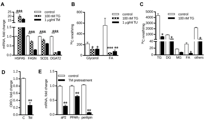
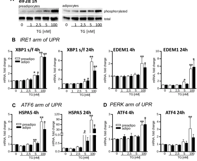
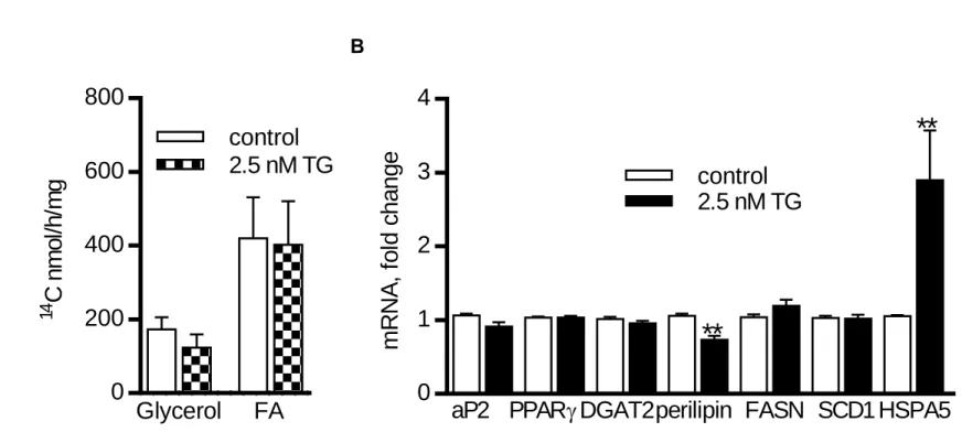
In Project A, we showed that exposition of adipocytes to high acute ERS inhibits expression of lipogenic genes and glucose incorporation into lipids. Moreover, chronic exposure of preadipocytes to ERS impaired both, lipogenesis and adipogenesis. On the other hand, chronic low ERS had no apparent effect on lipogenesis in adipocytes. These effects of ERS could therefore contribute to the worsening of AT function seen in obesity.
The capacity of AT to store lipids decreases in aging, possibly due to the accumulation of senescence cells or higher ERS. In Project B, we investigated lipogenic capacity of human AT in relation to senescence and markers of ERS. AT and adipose cells from young and elderly women were investigated. While mRNA expression of major senescent markers was increased in AT from the elderly compared to young individuals, mRNA expression of lipogenic enzymes and chaperones was decreased in AT from elderly individuals. These results were also partly observed in vitro in differentiated adipocytes from AT of the same individuals suggesting the reduced capability to cope with ERS in aging.
Very-low calorie diet (VLCD) is first line lifestyle intervention to achieve rapid weight loss. The improvement of whole body insulin sensitivity can be seen as soon as after 2 days of VLCD. However, little is known about AT metabolic changes in those early days. Thus, in Project C, we compared metabolic and inflammation-related characteristics of subcutaneous AT in the early (2 days) and later (28 days) phase of a VLCD. In the early phase of VLCD, the expression of lipolytic genes was increased, whereas the expression of lipogenic genes was suppressed. The inflammatory markers remained unchanged in AT. The changes in AT gene expression in the early phase of VLCD could not explain the effect of short calorie restriction on the improvement of insulin sensitivity. At the later phase, expression of genes involved in lipogenesis and β-oxidation was markedly suppressed,
whereas the expression of inflammatory markers was increased. Thus, we found that the early and later phases of VLCD differ with respect to metabolic and inflammatory responses in subcutaneous AT.
In Project D, we compared and defined the effects of moderate calorie restriction on preadipocytes and in vitro differentiated adipocytes in two groups of obese men: juniors and seniors. We did not observe any effect of the intervention on metabolism of preadipocytes in either group. However, we observed an intervention-driven improvement in adipocyte metabolism selectively in the group of seniors. Therefore, our data suggest that moderate calorie restriction could initiate positive changes in metabolism of adipocytes in seniors.
In conclusion, this thesis brought several pieces of evidence that lipogenesis in human AT can be inhibited by ER stress, severe caloric restriction and aging.
Résumé
Le tissue adipeux (TA) est un organe complexe specialisé dans le stockage et la libération d´énergie sous forme de lipides. Cet organe adipeux est essentiel pour le maintien de l´homéostasie énergétique. Les adipocytes sont les cellules prototypiques du TA. Elles se forment durant la différenciation de précurseurs, un processus appelé adipogenèse. L’adipogenèse est intimement associée à la synthèse des acides gras et de triglycérides lors de la lipogenèse. Néanmoins, divers facteurs peuvent perturber l’adipogénèse et la lipogenèse, contribuant au dysfonctionnement du TA et au développement des maladies métaboliques.
Le but de cette thèse a été d´étudier la lipogenèse dans le contexte du stress du réticulum endoplasmiques (SRE), de la restriction calorique et du vieillissement.
Dans le projet A, nous avons montré que l´exposition d’adipocytes à un SRE aigu inhibe l´expression des gènes liés à la lipogenèse et empêche l´incorporation du glucose dans les lipides. En plus, l´exposition des préadipocytes à un SRE chronique, détériore à la fois la lipogenèse et l´adipogenèse. Par contre, pour les adipocytes, un SRE chronique mais modéré n´a pas d´effet évident sur la lipogenèse. Ces effets du SRE pourraient contribuer à la détérioration de la fonction de TA vue dans l´obésité.
La capacité du TA à stocker des lipides diminue avec l´âge, probablement à cause de l´accumulation de cellules sénescentes ou un SRE plus élevé. Dans le projet B, nous avons étudié la capacité lipogénique du TA humain en relation à la sénescence et aux marqueurs du SRE au sein d’une cohorte de femmes obèses jeunes ou âgées. Tandis que l´expression des principaux marqueurs de la sénescence était augmentée dans le TA des femmes âgées, l´expression génique des enzymes de lipogenèse et des chaperonnes était diminuée dans le TA des personnes âgées. Ces résultats étaient partiellement retrouvés dans les adipocytes differenciés in vitro des mêmes individus ce qui suggère une moins bonne capacité à faire face au SRE lors du vieillissement.
Le régime à très basses calories (VLCD) est souvent prescrit en première intention pour une rapide perte de poids. L’amélioration de la sensibilité à l’insuline se voit dès 2 jours de VLCD. Néanmoins, on ne sait quasiment rien des modifications métaboliques du TA survenant durant les premiers jours. Dans le projet C, nous avons donc comparé les réponses métaboliques et inflammatoires du TA sous-cutané précocément (2 jours) et plus tardivement (28 jours) lors d’un VLCD. A 2 jours de régime, l´expression des gènes lipolytiques était augmentée, alors que l´expression des gènes lipogéniques était diminuées. Les marqueurs d´inflammation n´étaient pas changés dans le TA. Néanmoins, les changements d´expression
dans le TA lors de la phase précoce du régime ne pouvait pas expliquer l´effet de ce régime court à l´amélioration de la sensibilité à l´insuline. Dans la phase tardive, l´expression des gènes impliqués dans la lipogenèse et la β-oxydation était largement réduite, tandis que l´expression des marqueurs inflammatoires était augmentée. Nous avons donc montré que les réponses métaboliques et inflammatoires du TA sous-cutané à 2 jours et 28 jours de VLCD sont différentes.
Dans le projet D, nous avons comparé et défini les effets de la restriction calorique modérée sur la physiologie des préadipocytes et des adipocytes différenciés in vitro chez des jeunes obèses ou des personnes âgées obèses. De façon surprenante, on n´a observé aucun effet de l´intervention sur le métabolisme des préadipocytes dans les deux groupes. Par contre, un effet bénéfique de l’intervention sur le métabolisme adipocytaire n’a été observé que chez les personnes âgées. Nos données montrent donc qu’une restriction calorique modérée peut avoir un effet positif sur le métabolisme adipocytaire des séniors.
Pour conclure, cette thèse montre que la lipogenèse dans le TA humain peut être inhibée par le SRE, la restriction calorique sévère et le vieillissement.
Shrnutí
Tuková tkáň (TT) je komplexní orgán specializovaný pro bezpečné skladování a uvolňování energie ve formě lipidů. TT je tedy nezbytná pro udržování energetické homeostázi. Základní funkční jednotky TT se nazývají adipocyty a vznikající z prekurzorových buněk procesem adipogeneze. Adipogeneze jako taková je velmi úzce spojena s lipogenezí, neboli syntézou mastných kyselin a triglyceridů. Nejrůznější faktory mohou ovšem narušit diferenciaci a lipogenezi adipocytů a přispívat tak k dysfunkci TT a rozvoji metabolických onemocnění.
Proto byla tato dizertační práce zaměřena na zkoumání lipogeneze v kontextu stresu v endoplasmatickém retikulu (ER), kalorické restrikce a stárnutí.
V projektu A jsme ukázali, že vystavení adipocytů silnému akutnímu stresu ER snižuje expresi lipogenních genů a inkorporaci glukózy do lipidů. Chronický stres ER negativně ovlivňoval jak lipogenezi, tak vlastní diferenciaci preadipocytů, i když v již maturovaných adipocytech neměl chronický stres ER na lipogenezi zjevný efekt. Tyto efekty stresu ER na lipogenezi a adipogenezi tak mohou přispívat ke zhoršení funkce TT pozorované u obézních jedinců.
Kapacita TT skladovat lipidy se snižuje s věkem, pravděpodobně kvůli akumulaci senescentních buněk nebo zvýšenému stresu v ER. V projektu B jsme zkoumali lipogenní kapacitu lidské TT ve vztahu k senescenci a markerům stresu ER. K analýze byly použity vzorky TT a adipocyty mladších žen a seniorek. Zatímco mRNA exprese hlavních senescentních markerů byla zvýšená v TT seniorek ve srovnání s mladšími ženami, mRNA exprese lipogenních enzymů a šaperonů ER byla v TT u seniorek snížena. Tyto výsledky byly částečně potvrzeny v in vitro diferencovaných adipocytech z TT identických žen. Tyto výsledky naznačují sníženou odpověď na stres v ER ve stáří.
Velmi přísná nízkoenergetická dieta (VLCD, z anglického very low-calorie diet) patří mezi primární intervence používané k rapidnímu poklesu hmotnosti u obézních. Zlepšení celotělové inzulinové sensitivity je možno pozorovat již po 2 dnech VLCD. Nicméně o změnách probíhajících v TT během těchto prvních dnech diety se neví prakticky nic. V projektu C jsme proto srovnávali metabolické a zánětlivé charakteristiky subkutánní TT během rané (2 dny) a pozdější (28 dní) fáze VLCD. Během rané fáze VLCD došlo ke zvýšení exprese lipolytických genů, kdežto exprese lipogenních genů byla potlačena. Zánětlivé markery zůstaly v TT nezměněny. Změny na úrovni genové exprese v TT v rané fázi VLCD nicméně nevysvětlily efekt krátké kalorické restrikce na zlepšení inzulinové sensitivity. V
pozdější fázi byla exprese genů zapojených do lipogeneze a β-oxidace markantně snížena, zatímco exprese zánětlivých markerů byla zvýšena. Tento projekt ukázal, že raná a pozdější fáze VLCD se liší s ohledem na metabolickou a zánětlivou odpověď subkutánní TT.
V projektu D jsme srovnávali a definovali efekty mírné kalorické restrikce na preadipocyty a in vitro diferencované adipocyty u dvou skupin obézních mužů: mladších mužů a seniorů. Zatímco jsme nepozorovali žádný efekt intervence na metabolismus preadipocytů v žádné ze dvou skupin, ve skupině seniorů jsme po intervenci zaznamenali zlepšení metabolismu adipocytů. Naše výsledky tedy naznačují, že mírná kalorická restrikce může vest k zahájení pozitivních změn v metabolismu adipocytů u seniorů.
Závěrem je možné shrnout, že tato dizertační práce přinesla několik důkazů o tom, že lipogeneze v lidské TT může být inhibována stresem ER, přísnou kalorickou restrikcí a stárnutím.
1 Introduction into biology of adipose tissue
Traditional perception of adipose tissue (AT) as a passive organ dedicated to energy storage, insulation and thermoregulation has changed dramatically in the last decades, when the extraordinary complex role of AT in various physiological processes started to be recognised and appreciated. Nowadays, it is known that apart from the regulation of whole body energy homeostasis AT is involved in inflammation, angiogenesis, reproduction, atherogenesis or regeneration (Figure 1). These pleiotropic functions of AT rely not only on the paracrine communication between various cell types within AT itself but also on the cross-talk with distant organs through secreted factors, called adipokines and lipokines.
In the introductory section of this thesis, two elementary types of AT and its cellular composition will be described at first. Next, the most important physiological functions of AT and adipocytes will be explained. Lipogenesis, the process of lipid synthesis, will be described in detail, as it is the main subject of this thesis. Third part of the theore tical introduction will be dedicated to the description of processes which contribute to AT dysfunction in obesity and aging. The last part will briefly outline the possibilities of fight against obesity through lifestyle and dietary interventions.
Figure 1: Adipose tissue is an organ with a plethora of functions. This picture illustrates some of principal
physiological and metabolic processes with which adipose tissue is involved through the secretion of various adipokines from adipocytes. The interactions may be autocrine, paracrine, or endocrine. Adapted from [7].
1.1. Adipose tissue
1.1.1 White adipose tissue
AT organ, in some individuals the largest organ in the body, is distributed in many different depots throughout the body. Different cellular characteristics and anatomical location predetermine specific properties of each depot and its particular function. In mammals, the AT pool is composed of at least two functionally and histologically distinct types of fat: white and brown. Major white AT (WAT) depots are situated in subcutaneous region in both, the upper (superficial and deep abdominal) and lower (gluteal-femoral) body, as well as in the visceral region (omental, mesenteric, mediastinal and epicardia l depot) (Figure 2) [2].
Subcutaneous WAT, a major energy storing depot, is located under the skin to provide a layer of insulation preventing heat loss and protecting against mechanical stress. On the other hand, visceral WAT coats vital organs within the peritoneum and rib cage. In addition, WAT can be found in many other areas, including retro-orbital space, on the face and extremities, and within the bone marrow [8].
Fat distribution is markedly altered by many factors, such as sex, hormonal status, age and disease state [9]. Female body type tends to be a pear shape, because subcutaneous fat is
Figure 2: White adipose tissue depots in humans, shown in orange. Major subcutaneous white adipose tissue
includes superficial and deep abdominal depots and gluteal-femoral depot. Major visceral white adipose tissue includes epicardial, omental and mesenteric. Adapted from [2, 3].
preferentially deposited around the hips and thighs [10, 11]. Pregnancy often emphasizes this sexually dimorphic fat distribution [12]. In contrast, men (and postmenopausal women) accumulate fat around the waist (so called apple shape) and tend to accumulate more visceral fat [13, 14]. Gradually, these gender-based differences in the AT deposition become less prominent later in life due to decreasing influence of steroid hormones.
Also, the aging per se affects the distribution of body fat mass. The peak of fat depot sizes is reached by middle or early old age (40-70 years), followed by a substantial decline in advanced old age (>70 years) [15]. The volume of subcutaneous fat declines first, followed much later by loss of fat in visceral depots. However, the observed decrease in total body fat with old age does not coincide with a decline in percent body fat, which may remain constant or even increase. The age-associated decline in sizes of adipose depots is accompanied by the accumulation of fat outside AT and loss of lean body mass (particularly muscle). Ectopic fat accumulation occurs in bone marrow, muscle, liver and at other sites. This ectopic fat deposition causes lipotoxicity and worsens age-dependent dysfunction of these tissues.
1.1.2 Brown and brite adipose tissue
In comparison with WAT, that is present in humans throughout whole lifetime, brown AT (BAT) is in human present mainly in newborns, predominantly in the interscapular region. BAT uses the chemical energy from lipids and glucose to produce heat through non-shivering thermogenesis via mitochondrial uncoupling [16]. This is possible by the presence of uncoupling protein 1 (UCP1) that uncouples electron transport from ATP production, leading to the generation of heat [17]. Because of the high mitochondria content and dense vascularisation, BAT appears brown compared to WAT.
For many years it was generally accepted that BAT postnatally disappears and that BAT depots are absent in human adults. Nevertheless, a few years ago, the use of 18
F-fluorodeoxyglucose led to discovery of small areas of tissue functionally resembling BAT in the thorax region, chest and abdomen in adult humans (Figure 3) [1]. This metabolically active tissue responds to temperature similarly as BAT, however it is still distinct from the dorsal interscapular BAT in children. Subsequent studies suggested that brown fat cells might be interspersed within the WAT (brown in white, i.e. brite, or beige AT).
Figure 3: Sites of 18F- fluorodeoxyglucose uptake corresponding to BAT in adult humans. The black areas are those that are most frequently described; the gray areas are not always found, even in humans positive in the black areas. Adapted from [1].
This brite AT is considered as a subtype of WAT that has adopted features of BAT upon the stimulation by low temperatures in a process known as browning. Brite AT occurrence and activity in adult decrease with age and higher adiposity [18]. These results suggest that the decreased BAT activity might be associated with the accumulation of classical WAT with age.
1.1.3 Cellular composition of white adipose tissue
Histologically, AT is composed of adipocytes, i.e. the mature fat cell, and stromal-vascular fraction (SVF), comprising stem cells, preadipocytes, immune cells, endothelial cells and extracellular matrix (ECM).
1.1.3.1 Adipocytes
White adipocytes are rounded cells containing a single large lipid droplet that occupies usually over 90% of the cell volume. The cytoplasm and organelles, such as nucleus and mitochondria, are displaced to the periphery of the cell. Lipids stored in the droplet are primarily triglycerides (or triacylglycerols, TAG) and cholesteryl esters. The degree of lipid accumulation determines adipocyte size which is in average 80-90 µm but can reach up to 200 µm [19].
1.1.3.2 Stromal vascular fraction
1.1.3.2.1 Adipose tissue stem cells
Adipose tissue stem cells (ASCs) are precursors of adipocytes, and as such they are prerequisite to hyperplastic growth of AT mainly in the early childhood and puberty. In adulthood, when the total number of adipocytes remains relatively constant [20], they ensure replenishment of aged non-functional adipocytes. In fact, lifespan of adipocytes was estimated to be approximately 10 years. ASCs tend to be associated with blood vessels and may be derived from AT pericytes (cells that wrap around endothelial cells) [21-23]. Since ASCs are multipotent cells, they are capable to differentiate not only into brown and white adipocytes through the precursor stage of preadipocytes but also to other cell types, including macrophages, muscle or bone progenitors [24-28].
1.1.3.2.1.1 From ASC to mature adipocyte: An insight into adipocyte differentiation
The process of adipocyte differentiation from ASC to mature adipocytes includes many cellular intermediates. Although there have been efforts to describe these distinct intermediates, they have been difficult to characterise at the molecular level. Therefore,
adipogenesis is generally presented as a two-phase process including determination and
terminal differentiation phase.
Determination involves the commitment of a pluripotent stem cell to the adipocyte
lineage [29]. This stage results in the conversion of the stem cell to a preadipocyte. Preadipocyte cannot be distinguished morphologically from its precursor cell but has lost the potential to differentiate into other cell types.
During terminal differentiation, the preadipocyte acquires the characteristics of mature adipocyte. These include building machinery necessary for lipid transport and synthesis, insulin sensitivity and the secretion of adipocyte-specific proteins.
The molecular regulation of terminal differentiation is more extensively characterised than determination. It is known that differentiation requires the activation of numerous transcription factors that are responsible for coordinated expression or silencing of more than 2000 genes related to the regulation morphology and physiology of adipocyte [30].
Two members of CCAAT/enhancer-binding proteins (C/EBPs), C/EBPβ and C/EBPδ, are dramatically induced during the first stage of adipogenesis, at least in vitro, in response to the exposition of cells to hormonal differentiation cocktail [31]. Their primary function in adipogenesis is to provoke expression of peroxisome proliferator-activating receptor γ (PPARγ) and C/EBPα, the key transcriptional regulators of adipocyte differentiation [32, 33]. PPARγ and C/EBPα initiate a positive feedback loop in which they induce their own expression and expression of a large number of adipocyte specific genes.
PPARγ, the master regulator of adipogenesis, is a member of the nuclear-receptor superfamily. It was shown that PPARγ is both necessary and sufficient for adipogenesis, as C/EBPα or other transcription factors cannot promote adipogenesis in its absence [34, 35].
In addition to PPARγ and C/EBPs, several other transcription factors are likely to play an important role in the molecular control of adipogenesis. In general, pro-adipogenic factors seem to function at least in part by inducing PPARγ expression or enhancing its activity. These include certain Krüppel-like factors (KLFs), such as KLF4, 5, 6, 9 and 15. On the other hand, KLF2, 3 and 7 are adipogenic [36]. GATA2 and GATA3 also belong to anti-adipogenic factors. They are expressed in preadipocytes and downregulated during terminal maturation [37]. Constitutive expression of GATA2 and GATA3 blunts adipocyte differentiation and trap cells at the stage of preadipocyte. Therefore, the process of adipocyte differentiation is a result of a balance between these intervening factors. Their expression can be influenced by the present cellular state. For example, in the experimental part A of this thesis, we show that adipocyte differentiation is blunted by stress of endoplasmic reticulum
(ER). If the milieu for adipogenesis is favourable, newly formed adipocyte gain a rounded-cell shape with one lipid droplet inside and the whole machinery for handling and synthesis of lipids.
1.1.3.2.2 Immune cells
Immune cells, which reside in AT, include almost the full spectrum of known immune cell types. Their primary physiological role is to maintain AT homeostasis. This includes ECM remodelling, angiogenesis, activation of inflammatory response, removal of molecular debris and apoptotic cells [38, 39].
The most abundant population of AT immune cells are macrophages [40]. Although the spectrum of macrophage phenotypes is continuous, there is no nomenclature that could provide all of the required definitions. Thus investigators generally accept the simplified consensus which distinguishes two principal phenotypes: M1-like (classically activated) and like (alternatively activated). The function of both phenotypes is different: resident M2-like AT macrophages have a role in AT homeostasis, whereas recruited M1-M2-like macrophages contribute to inflammation and insulin resistance, described later. Nevertheless, it should be noted that this M1-/M2-like distinction is artificial and macrophages can possess features of both phenotypes [41].
Neutrophils are myeloid cells with short lifetime that are essential for the initial response to bacterial infections and injury [42, 43]. This is because they facilitate the recruitment of macrophages, dendritic cells and lymphocytes into the site of infection. In metabolically healthy animals, neutrophils represent less than 1 % of total AT immune cells [43]. Dendritic cells are the major antigen presenting cells and can induce proliferation of lymphocyte population. Their gene expression profile is very similar to that one of macrophages [43], yet dendritic cells are still probably the least characterized myeloid cells of AT. Mast cells are known to mediate allergic reactions [44]. They can modulate AT inflammation and possibly fibrotic state found in obesity and diabetes [45]. Eosinophils are classic effectors of anti-helminth responses. In the context of AT, they can assist the induction into M2-like macrophages via interleukin 4 (IL-4) [46]. Other cells originating from lymphoid lineage found in AT are B cells, T cells, nature killer (NK) cells and their numerous subtypes. Activities of T and B lymphocytes rely on the antigen recognition and include diverse populations of cells with proinflammatory or regulatory functions. In human AT, T cells, B cells, NK cells, NKT cells and innate lymphoid cells, group 2 account for up to 10% of non-adipocyte cells [47].
The dynamic nature of immune cells in AT during the progression of obesity is briefly mentioned in 1.3.2.4 Inflammation.
1.1.4 Extracellular matrix
Each adipocyte is surrounded by a thick ECM, also called basal lamina. Basal lamina can decrease the mechanical stress by spreading the forces over a larger area of the tissue and therefore protect adipocyte/lipid droplet from disruption.
The composition of ECM depends on the developmental stage of preadipocyte and/or adipocyte, viability and subtype of the adipocyte, but the two main classes of ECM proteins are proteoglycans and fibrous proteins. Major component of basal lamina is collagen IV [48]. Compared to other ECM components, collagen VI seems to be more specific for adipocytes and there is evidence of its contribution to the pathology of obesity-related disease [49, 50]. Importantly, even in mature adipocyte, ECM is under constant turnover.ECM remodelling is an energy demanding process mediated by a balance between various remodelling enzymes and their enhancers or inhibitor. It is regulated not only by mechanical forces but also by insulin, redox status of the cell and activity of ER [51].
This chapter summarised various function and anatomical location of adipose tissue. Two main types of adipose tissue, white and brown, were introduced. White adipose tissue has plethora of functions, but primarily it is dedicated to safe storage and release of lipids. Brown adipose tissue uses the chemical energy from lipids and glucose to produce heat. Adipose tissue is composed from adipocytes and stromal-vascular fraction which includes stem cells, preadipocytes, immune cells, endothelial cells and extracellular matrix. The physiological function of AT will be the subject of the next chapter.
1.2 Physiology of adipocytes and adipose tissue
Besides the thermogenic and mechanical protection, AT serves as a biological reservoir of calories that expands in response to overnutrition and releases lipids in response to lack of energy. Thus, two main metabolic processes – lipogenesis and lipolysis – help to maintain energetic demands of organism.
1.2.1 Lipogenesis
High fat and/or carbohydrate intake stimulate lipogenesis, a process of fatty acid and TAG synthesis. Fatty acids are synthesised from acetyl-CoA and TAGs are formed by esterification of free fatty acids to glycerol. The majority of fatty acids used for TAG synthesis in AT are derived from the diet.
1.2.1.1 Fatty acid transport
Dietary fat (TAG), when ingested with food, is absorbed by the gut. Because of hydrophobic nature of lipids, they are transported in plasma mostly as parts of specific lipoprotein complexes. Dietary lipids are transported by chylomicrons, whereas endogenous fat and cholesterol are carried by lipoproteins of different densities (very-low density lipoproteins: VLDL, intermediate-density lipoproteins: IDL, low-density lipoproteins: LDL, high-density lipoproteins: HDL). At the site of AT beds, fatty acids are liberated from these complexes through the action of lipoprotein lipase (LPL) that is secreted by adipocytes. Released fatty acids are bound by albumin and in this form they are available for the uptake by adipocytes where the TAG are resynthesized and stored in cytoplasmic lipid droplets (Figure 4).
Figure 4: Systemic transport of lipids and lipoproteins. Dietary lipids are transported by
chylomicrons, whereas lipids from liver are trasported to other tissues via VLDL. At the site of mammary, muscle or AT beds, fatty acids are liberated from lipoprotein complexes by the action of lipoprotein lipase. Then, free fatty acid bind to albumin (not shown) and can be uptaken inside the cell. Remnants of VLDL may transform to IDL or LDL, travelling back to the liver or became (in the case of LDL) another source of lipids for extragepatic tissues. HDL deliver cholesterol to the liver. Adapted from [4].
The transport of fatty acids across the adipocyte plasma membrane appears to be highly complex but still somehow elusive process. There is an evidence for two different (but not mutually exclusive) processes: diffusion and protein-mediated uptake. Proteins implicated in fatty acid transport are plasma membrane fatty acid binding protein (FABPpm) and fatty acid translocase (CD36). FABPpm is a 43 kDa protein which is expressed on the surface of various cell types, including adipocytes [52]. Evidence supporting the role of FABPpm in adipocyte fatty acid uptake came from the finding that anti-FABPpm antibodies selectively inhibit uptake of oleate in 3T3-L1 adipocyte without alteration of 2-deoxyglucose uptake [53]. CD36 is a transmembrane glycoprotein which belongs to a family of class B scavenger receptor. In addition to fatty acids, it is thought to bind a wide variety of hydrophobic molecules such as thrombospondin, sickle cell erythrocytes, collagen, apoptotic cells and oxidized LDLs [54, 55]. Except for FABPpm and CD36, also other proteins like caveolin-1, fatty acid transport protein (FATP) or acyl-CoA synthetase (ACSL) have been found to be functionally linked to fatty acid transport [56-58].
Once inside the adipocyte, fatty acids can be converted into acyl-CoA by acyl-CoA synthetase [56, 57], bound and sequestered by intracellular fatty-acid binding proteins, FABP4 and FABP5, and oriented into diverse metabolic pathways, including restoration of TAG.
1.2.1.2 Triglyceride formation
TAG synthesis involves the activation of three molecules of fatty acids through formation of acyl-CoA and then the synthesis of monoacylglycerol (MAG) and diacylglycerol (DAG) by reacting with glycerol-3-phosphate (G3P) [59]. In AT, the main source of G3P is catabolism of glucose via glycolysis since the activity of glycerokinase (GK), the enzyme that transforms glycerol into G3P, is low.
1.2.1.3 De novo lipogenesis
Biochemical process of fatty acid and TAG synthesis from non-lipid precursors is called de novo lipogenesis (DNL). DNL takes place primarily in the liver and AT [60, 61]. The contribution of AT to DNL was considered for a long time negligible, but newer methods have shown that DNL contributes to the synthesis of approximately 20% TAG [61].
Insulin, a peptide hormone secreted by the β cells of the pancreatic Langerhans islets, is a potent regulator of carbohydrate, lipid and protein metabolism [62]. At the level of adipocytes, insulin promotes all steps necessary for lipid synthesis. Therefore, before going further, insulin signalling will be described in brief. Insulin acts through insulin receptor that
becomes autophosphorylated and thus forms binding sites for docking proteins such as IRS1 [63]. Tyrosine phosphorylation of IRS1 is then necessary for the activation of phosphatidylinositol 3-kinase (PI3K)-AKT/protein kinase B (PKB) pathway, resulting in the translocation of glucose transporter 4 (GLUT4)-containing vesicles to the cell surface and glucose transport inside the cells [64]. Glucose can be then used in both, fatty acid as well as in TAG synthesis. Because carbohydrate metabolism, especially that of glucose, is the most commonly involved in the supplementation of carbon units for DNL, we will use glucose as an example of a primary source to describe DNL pathway.
Following glucose transport into adipocyte, glucose is converted via glycolysis into pyruvate. Pyruvate from cytosol enters mitochondria where it is converted into citrate by citrate synthase (CS). Citrate then passes through the inner mitochondrial membrane by the citrate transporter into the cytosol. There, citrate cleavage by ATP citrate lyase (ACLY) regenerates acetyl-CoA. Thus, ACLY represents a first enzyme of DNL, linking the metabolism of carbohydrates and the production of fatty acids. Acetyl-CoA then serves as a substrate for acetyl-CoA-carboxylase (ACC). This enzyme, containing a biotin prosthetic group, catalyses the conversion of acetyl-CoA into malonyl-CoA in two-step reaction. Malonyl-CoA is a substrate for fatty acid synthase (FAS), a multienzyme complex responsible for the formation of long carbon chains of fatty acids by a repetition of four-step sequence. A saturated acyl group produced by this set of reactions becomes the substrate for subsequent condensation with an activated malonyl group. With each passage through the cycle, the fatty acyl chain is extended by two carbons until the chain reaches 16 carbons length. Then the product (palmitate, 16:0) leaves the cycle.
The further destiny of palmitate can vary. In ER, palmitate can be elongated by fatty acid elongases (ELOVLs) and desaturated at the ∆9 position by stearoyl-CoA desaturase (SCD). In mice, four SCD isoforms (SCD1-SCD4) have been found, whereas only two SCD isoforms have been identified in human (SCD1 and SCD5) [65]. In human AT, only SCD1 isoform is expressed [66]. The presence of at least one functional isoform is critical, as SCD is a rate-limiting enzyme in the production of monounsaturated fatty acids, specifically oleate and palmitoleate [67]. Importantly, the degree of fatty acid unsaturation in cell membrane lipids determines membrane fluidity whose alteration has been implicated in a variety of diseases [68]. SCD1 has been found to be located in proximity with another DNL enzyme – diacylglycerol acyltransferase (DGAT) [69]. DGAT is enzyme that catalyses the final reaction in the synthesis of TAG. Two DGAT genes, DGAT1 and DGAT2, have been identified [70, 71]. Although these enzymes catalyse similar reactions, their nucleotide and
Figure 5: Simplified pathway of de novo lipogenesis. Glucose enteres inside the cell and undergoes
glycolysis. Pyruvate enters mitochondria, where it is metabolised to citrate. In cytoplasm, citrate is converted to acetyl-CoA, metabolised by acetyl-CoA-carboxylase (ACC) into malonylCoA. MalonylCoA is utilised by fatty-acid synthase (FAS) to create palmitate, which enters into endoplasmic reticulum where it can be modified by elongase (ELOVL) or stearoyl-CoA-desaturase (SCD) and finally incorporated into various kind of lipids, including TAG. Glucose also serves as a main source of glycerol-3-phosphate, which subsequently provides backbone of TAG. TAG (triacylglycerides), DGAT (diacylglycerol acyl-transferase), GPDH (glycerophosphate dehydrogenase), GPAT (glycerol-3-phosphate acyltransferase), AGPAT (acylCoA acylglycerol-3-phosphate acyltransferase), PAP (phosphohydrolase).
which is the main source of glycerol-3-phosphate, serving later as a glycerol backbone for TAG. Pyruvate
amino acid sequence differ [72]. Studies performed on mice lacking DGAT1 (DGAT-/-) have suggested that DGAT1 does not have a profound effect on TAG metabolism in general and is not essential for life [73]. In contrast, mice with a disruption of the DGAT2 gene (DGAT2-/-) have severely reduced TAG content in their tissues and survive only early postnatal periods [72]. These results demonstrate that DGAT2 is essential for the fundamental synthesis of TAG in mammals and is crucial for survival.
The overall pathway of DNL is depicted on Figure 5. The analysis of mRNA expression changes of major DNL enzymes in response to calorie deficit is described in the result part C of this thesis.
1.2.1.3.1 Transcriptional regulation of de novo lipogenesis
As master regulators of DNL, two transcriptional factors have been identified – sterol regulatory element binding protein 1c (SREBP1c) and carbohydrate response element binding protein (ChREBP).
SREBP1c belongs to the basic-helix-loop-helix leucine zipper (bHLH/LZ) family of transcription factors and is synthesized as an inactive precursor bound to the membrane of the ER [74]. To become a mature transcription factor, SREBP1c must undergo proteolytic cleavage to liberate its N-terminal domain from the membrane. The cleaved fragment then moves to the nucleus to initiate transcription of target genes.
ChREBP (also known as MondoB or MLXIPL), similarly as SREBP1c, belongs to the family of bHLH/LZ transcription factor. Recently, a novel ChREBPβ isoform have been discovered [75]. ChREBPβ more potently induces DNL genes and its expression in human AT correlates with insulin sensitivity.
Interestingly, although both of these DNL transcriptional factors regulate expression of key lipogenic genes such as FASN or ACC [76], they are activated by different mechanisms. Experiments showed that SREBP1c gene expression is strongly stimulated by insulin [74, 77, 78], whereas ChREBP activity is regulated by glucose and other carbohydrates [79, 80]. Nevertheless, the distinct regulation and functional roles of SREBP1c and ChREBP in AT are still being worked out.
1.2.1.3.2 Relevance of de novo lipogenesis
Considering the huge quantities of lipids stored in adipocytes, DNL is unlikely to contribute essentially to the lipid mass of AT. Thus the intrinsic engagement of adipocytes in fatty acid synthesis raises the possibility that in comparison with their dietary counterparts, adipocyte-derived fatty acids may have unique functions beyond energy storage and may be involved in important biological processes.
Indeed, recent research revealed that lipids have wide-ranging actions as signalling molecule and that particularly the products of DNL can be essential for metabolic homeostasis.
Work of Cao et al. led to the identification of AT-derived lipokine palmitoleate (C16:1n7) which directly links DNL to beneficial systemic effects [81]. Similarly, another independent group has shown insulin-sensitizing effects of palmitoleate on muscle [82]. Considering the clinical research, numerous studies which assessed serum palmitoleate in humans have revealed both positive and negative associations with metabolically adverse
conditions [83-86]. These discrepant results could be related to the measurement of various forms of palmitoleate: while the free fatty acid form of palmitoleate seems to act as a lipokine with systemic beneficial effects, esterified palmitoleate probably loses this distinct function or may simply reflects the hepatic output [87].
Notably, palmitoleate is not the only mediator which provides systemic beneficial effects of elevated AT DNL. Recently, a novel class of AT-derived lipids was described: fatty acid-hydroxy-fatty acids (FAHFAs). FAHFAs were found to be elevated 16-18 fold mice which overexpress GLUT4 transporter in adipocytes and have elevated lipogenesis [88]. The concentration of one specific isomer of FAHFAs, consisting of palmitic acid and stearic acid (PAHSA), was correlated with improved insulin sensitivity. Remarkably, exogenous PAHSA treatment improved glucose tolerance and overall glucose metabolism in mice. In addition, PASHA administration exerted anti-inflammatory effects on AT-resident immune cells. Finally, PAHSA levels were reduced in the serum and subcutaneous AT of insulin resistant human subjects.
In addition to DNL products, many other lipid species as sphingolipids [89], cardiolipins [90] and prostaglandins [91, 92] also act as mediators of metabolism with both beneficial and deleterious effects and undoubtedly many more wait to be discovered.
1.2.2 Lipolysis
Lipolysis is a catabolic pathway that promotes fat mobilization from AT to peripheral tissues. Lipolysis involves hydrolysis of TAG that results in the release of fatty acids and glycerol into the circulation. Complete hydrolysis of TAG requires actions of three different lipases [93]. The first step, involving TAG hydrolysis into diacylglycerol (DAG) and one free fatty acid, is catalysed by adipose triglyceride lipase (ATGL). Next, hormone-sensitive lipase (HSL) cleaves DAG into MAG. Complete hydrolysis accomplishes monoacylglycerol lipase (MGL) by the conversion of MAG into fatty acid and glycerol.
1.2.2.1 Regulation of lipolysis
In response to changing metabolic conditions and nutrient intake, nutritional regulation of lipolysis occurs at multiple levels. Insulin inhibits lipolysis [94], whereas fasting and exercise acutely stimulate lipolysis and the primary activators are catecholamines (norepinephrine, epinephrine) [95, 96]. Norepinephrine binds to β-adrenergic receptors on the plasma membrane of adipocytes. These receptors are coupled with Gs-proteins that transmit a
stimulatory signal to adenylyl cyclase in order to generate cyclic adenosine monophosphate (cAMP). cAMP activates protein-kinase A (PKA) [97]. PKA phosphorylates HSL on multiple sites, which causes activation and subsequent translocation of HSL from cytosol to lipid droplet [98-100]. Except for HSL, PKA also phosphorylates perilipin, a major lipid droplet coating protein in adipocytes. As a result, perilipin moves away from the lipid droplet [101]. This increases the area on the surface available for lipolytic attack of the droplet [102]. Moreover, phosphorylated perilipin assists in the translocation of HSL from cytosol to lipid droplet [103].
Insulin is a suppressor of lipolysis and this regulation involves cAMP-dependent, as well as cAMP-independent mechanisms. cAMP-dependent suppression of lipolysis involves activation of phosphodiesterase 3B which degrades cAMP in adipocytes [104]. The second mechanism involves the stimulation of protein phosphatase-1. This enzyme, when activated, rapidly dephosphorylates and deactivates HSL, causing a fall in the rate of lipolysis [100, 105-107].
1.2.3 Secretory function of adipose tissue
AT functions as an active endocrine organ and releases multiple bioactive molecules known as lipokines [87] and adipokines [5, 108, 109]. Via these molecules, AT is able to communicate with distinctly located metabolically active organs and influence the systemic metabolism. As lipokines were already mentioned in the section below (1.2.1.3.2), the text will continue directly with adipokines.
1.2.3.1 Adipokines
A group of cytokines secreted by AT, i.e. adipokines, comprises today hundreds of molecules, including both anti-inflammatory (f.e. adiponectin) and pro-inflammatory mediators (f.e.tumor necrosis factor α: TNFα, monocyte chemoattractant protein 1: MCP1, interleukin 6: IL6). Key adipokines, their source and function are depicted in Table 1. Because issue of adipokines exceeds the scope of this thesis, only selected adipokines will be mentioned in more detail.
Adipokine Primary source Function
Adiponectin Adipocytes Insulin sensitizer, anti- inflammatory
Angiopoietin-like protein 2 Adipocytes, other cells Local and vascular inflammation
C-X-C motif chemokine ligand 5 SVF cells (macrophages) Antagonism of insulin signalling through the JAK-STAT pathway
Inte rleukin 18 SVF cells Broad-spectrum inflammation
Inte rleukin 6 Adipocytes, SVF cells, liver, muscle Changes with source and target tissue
Leptin Adipocytes Appetite control through the central nervous system
Lipocalin 2 Adipocytes, macrophages Promotes insulin resistance and inflammation through TNF secretion from adipocytes
Monocyte chemotactic protein 1 Adipocytes, SVF cells Monocyte recruitment
Nicotinamide
phosphoribosyltransferase Adipocytes, macrophages, other cells Monocyte chemotactic activity Retinol Binding Protein 4 Liver, adipocytes, macrophages Implicated in systemic insulin resistance
Resistin Monocytes/macrophages (human), adipocytes
(rodent)
Promotes insulin resistance and inflammation through IL6 and TNF secretion from macrophages
Secreted Frizzled Related Protein 5 Adipocytes Suppression of pro- inflammatory WN T signalling
Tumor necrosis factor SVF cells, adipocytes Inflammation, antagonism of insulin signalling
Adiponectin
Adiponectin is almost exclusively produced by adipocytes and is present at high levels in blood (3-30 µg/ml) [108]. It has a collagen-like domain followed by a globular domain [5]. Adiponectin forms trimers, through collagen-like domain interactions, that can further associate to form stable multimeric oligomers (hexamers and a high molecular weight form). All three forms are detectable in the blood. Plasma levels of adiponectin are negatively associated with the accumulation of body fat, particularly visceral fat [110] and with plasma levels of IL6 and C-reactive protein (CRP) [111, 112], i.e. adiponectin levels in plasma are lowered in obese individuals with signs of systemic inflammation. On the other hand, bodyweight reduction in obese women through calorie restriction and lifestyle changes is associated with an increase in adiponectin levels. Thus, adiponectin is a unique adipokine expressed at the highest levels by the functional and insulin-sensitive adipocytes and downregulated in dysfunctional adipocytes frequently found in obese body [113].
Leptin
Leptin is the product of the obese gene (ob; also known as lep), which was identified in ob/ob mice by the method of positional cloning [114]. This adipokine is important in regulating feeding behaviour through central nervous system. Leptin levels, contrary to adiponectin levels, correlate positively with adiposity and thus, leptin functions as a measure of long term energy reserves. The ob/ob mice, which lack leptin, suffer from hyperphagia leading to obesity and insulin resistance [115]. Administration of recombinant leptin in leptin deficient animals or humans reverses these changes. Nevertheless, in diet induced obesity, increased leptin levels do not exert the expected anorexic responses that would be capable to prevent further weight gain which indicates the occurrence of leptin resistance [116].
In addition, leptin has multiple roles in the immune system. It is an inflammatory molecule that is capable of activating both adaptive and innate immunity [117].
Interleukin 6
IL6 belongs to a group of pro-inflammatory cytokines. It is estimated that AT produces approximately one-third of total circulating IL6 [118]. Clinically, plasma IL6 levels positively correlate with the degree of adiposity in human populations [118] and weight loss leads to a reduction in IL6 levels [112, 119]. Plasma levels of IL6 are also increased in type 2 diabetic patients and elevated IL6 plasma concentration predicts the development of type 2 diabetes [120]. Increased secretion of IL6 may disturb proper insulin signalling [121] and
thus diminish not only glucose uptake by insulin-sensitive tissues but also increase AT lipolysis, which can contribute to ectopic lipid accumulation and lipotoxicity [122].
In healthy AT, the expression of anti-inflammatory and pro-inflammatory adipokines is balanced. However, in most obese humans, the expression of pro-inflammatory adipokines predominate and promote insulin resistance which is manifested by decreased insulin-stimulated glucose transport and metabolism in adipocytes and skeletal muscle and by impaired suppression of hepatic glucose output [123]. In addition to pro-inflammatory adipokines, various pathological conditions may disturb well-ordered AT milieu. Next chapter will introduce the global problem of obesity and analyse some of the possible culprits AT dysfunction.
Adipocytes are specialised to safely energy storage and release. This is due to lipogenesis and lipolysis. Lipogenesis is the process of fatty acid and triglyceride synthesis. The term de novo lipogenesis means fatty acid formation from non-lipid precursors. These precursors come from a various cellular pathways, most commonly from carbohydrate metabolism. Lipolysis is the opposite process, responsible for the breakdown of triglycerides stored in lipid droplets. Newly synthetised and released lipids can serve as signalling molecules. In addition to lipokines, adipocytes and others cells of adipose tissue secrete plethora of molecules called adipokines with anti-inflammatory or pro-inflammatory properties. However, pro-pro-inflammatory environment, which is often seen in obesity, can disrupt proper functions of adipocytes and adipose tissue.