RESEARCH OUTPUTS / RÉSULTATS DE RECHERCHE
Author(s) - Auteur(s) :
Publication date - Date de publication :
Permanent link - Permalien :
Rights / License - Licence de droit d’auteur :
Institutional Repository - Research Portal
Dépôt Institutionnel - Portail de la Recherche
researchportal.unamur.be
University of Namur
An interdisciplinary study around the reliquary of the late cardinal Jacques de Vitry
Decorte, Ronny; Polet, Caroline; Boudin, Mathieu; Tilquin, Françoise; Matroule, Jean Yves;
Dieu, Marc; Charles, Catherine; Carlier, Aurore; Lebecque, Fiona; Deparis, Olivier
Published in: PLoS ONE DOI: 10.1371/journal.pone.0201424 Publication date: 2019 Document Version
Publisher's PDF, also known as Version of record Link to publication
Citation for pulished version (HARVARD):
Decorte, R, Polet, C, Boudin, M, Tilquin, F, Matroule, JY, Dieu, M, Charles, C, Carlier, A, Lebecque, F & Deparis, O 2019, 'An interdisciplinary study around the reliquary of the late cardinal Jacques de Vitry', PLoS ONE, vol. 14, no. 2, e0201424. https://doi.org/10.1371/journal.pone.0201424
General rights
Copyright and moral rights for the publications made accessible in the public portal are retained by the authors and/or other copyright owners and it is a condition of accessing publications that users recognise and abide by the legal requirements associated with these rights. • Users may download and print one copy of any publication from the public portal for the purpose of private study or research. • You may not further distribute the material or use it for any profit-making activity or commercial gain
• You may freely distribute the URL identifying the publication in the public portal ? Take down policy
If you believe that this document breaches copyright please contact us providing details, and we will remove access to the work immediately and investigate your claim.
An interdisciplinary study around the
reliquary of the late cardinal Jacques de Vitry
Ronny Decorte1,2, Caroline Polet3, Mathieu Boudin4, Franc¸oise Tilquin5, Jean-Yves Matroule5, Marc Dieu6, Catherine Charles7, Aurore Carlier8, Fiona Lebecque8, Olivier DeparisID9*
1 Forensic Biomedical Sciences, Department of Imaging & Pathology, KU Leuven (KUL), Leuven, Belgium, 2 Laboratory of Forensic Genetics and Molecular Archaeology, Department of Forensic Medicine, UZ Leuven (UZL), Leuven, Belgium, 3 Department of Palaeontology, Royal Belgian Institute of Natural Sciences, Brussels, Belgium, 4 Radiocarbon Dating Laboratory, Royal Institute for Cultural Heritage (KIK-IRPA), Brussels, Belgium, 5 Research Unit in Microorganisms Biology (URBM), Narilis Institute, ILEE Institute, University of Namur, Namur, Belgium, 6 Mass Spectrometry Facility (MaSUN), University of Namur, Namur, Belgium, 7 Moretus Plantin University Library, University of Namur, Namur, Belgium, 8 Archeological Society of Namur (SAN), Namur, Belgium, 9 Heritages, Transmissions, Inheritances (PaTHs) Institute & Department of Physics, University of Namur, Namur, Belgium
Abstract
The reliquary of Jacques de Vitry, a prominent clergyman and theologian in the early 13th century, has experienced several transfers over the last centuries, which seriously question the attribution of the remains to the late Cardinal. Uncertainty about the year of his birth poses an additional question regarding his age at death in 1240. The reliquary, located in the Saint Marie d’Oigines church, Belgium, was reopened in 2015 for an interdisciplinary study around his relics as well as the Treasure of Oignies, a remarkable cultural heritage notably built from Jacques de Vitry’s donation. Anthropological, isotopic and genetic analy-ses were performed independently on the remains found in the reliquary. Results of the analyses provided evidence that the likelihood that these remains are those of Jacques de Vitry is very high: the remains belong to the same human male individual and the historical tradition about his age is confirmed. In addition, a separate relic (left tibia) was analysed and found to match with the remains of the reliquary (right tibia). The unique Jacques de Vitry’s mitre, made of parchment, was sampled non-destructively and the extracted parchment col-lagen was analysed by a proteomic method in order to determine the animal species. The results showed that, surprisingly, not all parts of the mitre were made from the same spe-cies. All together, these findings are expected to fertilize knowledge carried by historical tra-dition around the relics of Jacques de Vitry and his related cultural heritage.
Introduction
Jacques de Vitry was a prominent clergyman and theologian, successively regular canon, bishop and cardinal of the Roman Catholic Church, who was active in Europe and Middle East during the first part of the thirteen-century (S1A Fig). His life and personality are mainly known from his writings (e.g. Historia Orientalis), crusade preaches and sermons.
a1111111111 a1111111111 a1111111111 a1111111111 a1111111111 OPEN ACCESS
Citation: Decorte R, Polet C, Boudin M, Tilquin F,
Matroule J-Y, Dieu M, et al. (2019) An interdisciplinary study around the reliquary of the late cardinal Jacques de Vitry. PLoS ONE 14(2): e0201424.https://doi.org/10.1371/journal. pone.0201424
Editor: David Caramelli, University of Florence,
ITALY
Received: July 4, 2018 Accepted: January 18, 2019 Published: February 22, 2019
Copyright:© 2019 Decorte et al. This is an open access article distributed under the terms of the
Creative Commons Attribution License, which permits unrestricted use, distribution, and reproduction in any medium, provided the original author and source are credited.
Data Availability Statement: All relevant data are
within the paper and its Supporting Information files.
Funding: The work was funded by a grant from the
Fonds Jean-Jacques Comhaire of the Fondation Roi Baudouin (www.kbs-frb.be), Belgium.
Competing interests: The authors have declared
Unfortunately, no autobiography is available and facts about his youth are scarce. His date of birth is uncertain with two hypotheses coexisting: 1165–1170 (anonymous source, ca. 1250 [1]) or 1175–1180 (contemporary scholar Jean Donnadieu [2]), the former source being ques-tionable (Jacques de Vitry neither studied theology in Paris in 1187 nor was the confessor of the king of France, as reported by the anonymous source). The date and place of his death, on the other hand, are known precisely as 1st May 1240 in Rome [3].
Having studied theology in Paris (between 1190 and 1208), he received episcopal consecra-tion around 1210. He left Paris around 1208 to join the priory Saint Nicolas d’Oignies (funded in 1187) of the Diocese of Liège as an Augustinian canon regular. There, he met Marie d’Oig-nies (S1B Fig) and became the confessor and the biographer of the visionary, ecstatic beguine who died in 1213 at the age of 36 and was subject to popular devotions [4,5]. Between 1212 and 1216, he preached the crusade against the Albigenses. Following his election as bishop of Saint John of Acre, he left the priory for the Holy Land in 1216. Following Damietta defeat in 1221, he decided to come back to Europe. A year after his return, in 1225, he resumed his itin-erant life as preacher of the sixth crusade. In 1229, he was elevated to the College of Cardinals and settled in Rome where he died on May 1st 1240. His body was buried in the Dominican headquarter convent in Rome [6].
According to his testimonial will to lay at rest near to Marie d’Oignies, his remains were transferred to Oignies a year later and put in a marble monument, close to Marie’s one in the monastery church [7]. Several transfers of his relics took place in the next centuries. In 1636, the reliquary was open for transfer of his relics to a new place. Two teeth were removed on that occasion and given to clergymen for devotion [8]. On July 25th 1759, the reliquary was open for transfer to another place inside the church [9]. During the demolition of the priory in 1808, the relics were transferred to the Saint Martin church (Aiseau, Belgium) where they were put in a lead container, dated from 1844. After the collapse of the Saint Martin church building in 1970, the reliquary was opened in 1971 for the transfer of the remains to the Saint Marie d’Oignies church, Belgium, which was built in 1908 near the place of the ancient priory. The reliquary (and its content) is still displayed in this church today.
Throughout his life but also by testimony, Jacques de Vitry enriched the priory of Oignies with books, relics, tissues, vessels and other religious artworks [10]. He granted, among others, an ivory cross, a portable altar and two mitres, one of which being a unique object as it features miniatures on parchment. All these objects and others collected later constitute the Treasure of Oignies, a unique cultural heritage ensemble, which was recognized as such by the Belgian Federal State in June 2010 [11].
In 2015, the Archaeological Society of Namur (SAN), which has the scientific responsibility of the Treasure of Oignies, set up a consortium in partnership with several Belgian universities and research institutes in order to undertake an ambitious, interdisciplinary scientific study around the reliquary of Jacques de Vitry (referred as the CROMIOSS project). The study was motivated by the fact that the reliquary of the prominent clergyman and theologian has experi-enced several transfers during the last centuries, which seriously question the attribution of the remains to the late Cardinal. Uncertainty about the year of his birth poses an additional ques-tion regarding his age at death. Given the patrimonial importance of the Treasure of Oignies, the research consortium decided to englobe within its inquiry a material study of one of the bishop’s mitre (Fig 1), the only known example of a mitre composed of parchment. In spite of this exceptional feature, this mitre has so far received little attention from art historians.
Following the opening of the reliquary on September 8th 2015, human remains were found in the lead container. The interdisciplinary study reported hereafter relies on a critical con-frontation between the historical tradition and the scientific results obtained from anthropo-logical, isotopic, genetic and proteomic analyses.
Materials and methods
Putative remains of Jacques de Vitry
With the approval of the competent authorities, the reliquary (S2A Fig) was opened on Sep-tember 8th 2015, in the presence of the Bishop of the Diocese of Tournai, journalists and scien-tists. Caution was taken in order to avoid contamination of the relics by human DNA:
restricted access, use of gloves and masks. Because the reliquary was previously open and relics were transferred several times since the second burial, it was first necessary to determine the human nature of the relics and to check whether or not they belonged to a single individual. A primary inspection of the lead container content was performed during the opening ceremony (S2B Fig). A wooden frame containing a tibia, which was supposed to belong to Jacques de Vitry, was also exhibited on that occasion. In fact, the story of this tibia is rather tumultuous. It was probably displayed in a private place and stolen at an unknown date. Found by the police in the 20thcentury, it was first given back to the village of Oignies erroneously (actually, Oig-nies-en-Thie´rache located in the Province of Namur, about 70 km from Oignies) before finally
Fig 1. Jacques de Vitry’s mitre made of parchment. Donation of Jacques de Vitry to the priory of Oignies (collection of the Treasure of Oignies, Belgium). (A)
Early photography (1879). (B) Contemporary photography with sampling spots indicated for parchment proteomic analyses (1, 2: cap; 3 cap border; 4, 5: left and right lappets). Reprinted under a CC BY license with permission from Vedrin, Guy Focant, original copyright 2012.
reaching the church Saint Marie of Oignies in the 20th century. In the reliquary, the remains were wrapped in a textile, which also contained small fragments of bone, plant remains, textile fibres, golden flakes and insect remains belonging to various groups of beetles (xylophagous (Anobium punctatum), detritivorous (Ptinus and Tenebrio sp.), necrophagous (Trox scaber) and granivorous (Sitophilus granarius)). The first three groups are part of taxa that are occa-sionally found in a funerary context and the presence ofTrox scaber would suggest that organic material remnants, i.e. muscles, skin or hair, were still present. The reliquary, the frame and their contents were then transferred to the University of Namur (Laboratory of Anatomy) in order to take bone samples for further genetic and isotopic analyses.
The specimens consisted of 26 bones and 7 teeth (remains found in the reliquary con-tainer), 1 bone (tibia in a wooden frame considered as a separate relics) and 1 mitre (TreM.a– Muse´e des Arts anciens, Namur, Belgium, inv. n˚ TO28). The list of bone samples used for genetic and isotopic analyses is given inTable 1.
The container and its remains (see inventory,Fig 2A) together with the framed tibia, here-after denoted as the remains, are property of the Church Wardens of the Saint Marie d’Oignies church, Belgium. Permits for the study on the remains were obtained from the Church War-dens and from the Bishop of the Diocese of Tournai, Guy Harpigny. The minutes of the reli-quary opening ceremony and of the remains inventory were recorded by Sophie Bellotto, Notary in Chaˆtelet, Belgium. Permit for the transfer of the remains to the University of Namur was obtained from the Civil Registrar of Namur. The mitre, recorded as a classified artwork by the Wallonia-Brussels Federation (FWB), is property of the Fondation Roi Baudouin (FRB), Belgium. Permits for the study on the mitre were obtained from FRB and from the Ministry of Cultural Affairs of the FWB.
Anthropological study
A detailed inventory of the remains found in the reliquary was first established in order to esti-mate the minimum number of individuals. The sex determination of the studied individual was complicated by the absence of pelvic bones, which show the greatest sexual dimorphism. For this reason, we applied a sex determination method using cranium characteristics [12] and discriminant functions based on tibia measurements [13]. In order to estimate the age at death, we used the dental wear [14], the closure of the cranial sutures [13] and cementum anal-ysis [15]. The stature was estimated using equations of Olivier et al., and computed from the length of long bones originating from a French sample [16].
Table 1. Bone samples used for analyses.
Type of analysis Laboratory Bone samples Remarks
Isotopic analysis (δ13C,δ15N,14C) KIRK-IRPA Left tibia 2 samples
Right tibia 1 sample Skull fragment 1 sample Genetic analysis (nuclear DNA) UNamur Left tibia Powder
Right tibia Powder Petrous bone Powder Genetic analysis (nuclear DNA, mtDNA) KUL Left tibia Powdera
Reused tooth Previously used for cement chrono-analysis Upper tooth 1
-Upper tooth 2
-Petrous bone Powder and fragmentsa
aSampled at UNamur and sent to KUL.
Isotopic study
Collagen extraction was performed following Longin’s (1971) method. A 1% NaOH wash step (15 minutes) was introduced between demineralization and hydrolization steps. First, all the bone samples were demineralized in 10 ml 8% HCl for 20 minutes, and rinsed with MilliQ-water. After that, each sample was immersed for 15 minutes in 1% NaOH, and again rinsed with MilliQ-water. Then, after adding 1% HCl for neutralization, it was washed with MilliQ-water. For all the steps mentioned above, Ezee-filters were used. Gelatinization of the extract was done in water (pH 3), at 90˚ C for 12 hours. The resulting gelatin was filtered with a Millipore 7 micro-metre glass filter, and freeze-dried. Age determinations (14C analyses) were carried out on the AMS instrument at the Royal Institute for Cultural Heritage (KIRK-IRPA), Brussels (Lab code RICH-) [17]. CO2was released by sample combustion in the presence of CuO and Ag. Graphiti-zation was performed with H2over a Fe catalyst. Targets were prepared at KIK-IRPA [18].14C calibrations were performed using OxCal (version 3.1) [19] and the IntCal13 calibration curve date [20]. The C:N ratio,δ13
C andδ15
N analyses were performed on a Thermo Flash EA/HT ele-mental analyser, coupled to a Thermo Delta V Advantage Isotope Ratio Mass Spectrometer via ConFlo IV interface (Thermo Fischer Scientific, Bremen, Germany). Standards used were IAEA-N1, IAEA-C6, and internally calibrated acetanilide. Analytical precision was 0.25‰ for bothδ13
C andδ15
N based on multiple measurements of the standard acetanilide.
Genetic study
DNA extraction and amplification were performed at UNamur and KULeuven in dedicated laboratories for ancient DNA analysis. Standard precautions for contamination were taken including separated areas for pre- and post-PCR procedures, restricted access to pre-PCR
Fig 2. Remains found in the reliquary. (A) Inventory of the remains. (B) Radiography of the left and right tibias. (C) Left tibia (top) and right tibia (bottom).
(D) Evidences of cuts on the bones (articulations).
areas, multiple negative controls in the DNA extraction and amplification reactions, replica-tion of DNA extracreplica-tions and PCR reacreplica-tions, contaminareplica-tion control with DNA profiles of all laboratory staff.
At UNamur laboratory, bone nuclear DNA was extracted according the procedure
described by Mundorff and Davoren [21]. Briefly, the petrous bone and the tibias were cleaned with 10% bleach and bone powder was withdrawn by drilling with a decontaminated 8-mm drill under a previously decontaminated chemical hood. Nuclear DNA was then extracted from �3 g of bone powder using the Set Buffer Trace Bone kit and Nucleospin DNA Trace from forensic sample kit (Macherey-Nagel, Germany). Nine different microsatellite markers were amplified on the ancient bone DNA and contemporary DNA (used as control DNA) using the AmpFlSTR Minifiler PCR Amplification kit (Applied Biosystems) (see PCR protocol below) and were further analysed with GeneMapper on the ABI 3130xl Genetic Analyser (Applied Biosystems) at URBE (UNamur). Standard PCRs of 5 Y chromosome short tandem repeats (DYS19, DYS389II, DYS448, DYS456, DYS635) were performed using sets of primers designed by Kwon and coworkers [22] The PCR for microsatellite markers was realized with the AmpFlSTR miniFiler PCR Amplification Kit. Briefly, 10μl of sample DNA (in low-TE) were mixed with 10μl of master mix and 5 μl of primer set in a final volume of 25 μl. The PCR mix was then subjected to PCR (95˚C, 11 min + 40 cycles: 94˚C, 20 sec– 59˚C, 2 min– 72˚C, 1 min + 60˚C, 45 min). Negative controls corresponded to low-TE alone while positive controls corresponded to kit control DNA mixed with low-TE. Standard PCR for Y chromosome STR was realized by mixing 2μl of sample DNA with 200 μM of dNTP and 400 nM of each primer in a master mix containing Taq polymerase, Taq buffer and DMSO. PCR conditions were: 95˚C, 11 min + 50 cycles: 94˚C, 20 sec– 60˚C, 1 min– 72˚C, 30 sec + 72˚C, 7 min).
At KULeuven laboratory, the procedures described by Ottoni et al. [23] were used for decontamination and grinding of the two upper teeth samples, as well as for DNA extraction through silica-based spin columns (QIAquick PCR Purification Kit, Qiagen). One of the teeth was reused for genetic analysis after cement chrono-analysis. This tooth was embedded in epoxy resin and cut into two so that only the dentin and the root channel could be removed with a decontaminated dental drill. Multiplex DNA amplification of autosomal short tandem repeats (STRs) and STRs from the Y chromosome was done respectively according to Dog-naux et al. and Larmuseau et al. [24–26] except for the number of cycles, which was raised to 34. The autosomal multiplex of 9 STRs (D1S1656, D1S1677, D2S441, D10S1248, D12S391, D18S51, D21S11, D22S1045 and FGA—fragment size between 70 and 275 bp) includes also primers to amplify a sequence in an intron of the Amelogenin gene present on the sex chromo-somes (123 bp on the X and 129 bp on the Y) [27]. Three multiplexes were used for the Y-chro-mosome STRs including in total 40 STRs (DYS19, DYS385 a/b, DYS388, DYS389I, DYS389II, DYS390, DYS391, DYS392, DYS393, DYS426, DYS437, DYS438, DYS439, DYS442, DYS447, DYS448, DYS449, DYS454, DYS455, DYS456, DYS458, DYS459 a/b, DYS460, DYS464 a/b/c/ d, DYS481, DYS533, DYS549, DYS570, DYS576, DYS607, DYS635, DYS643, DYS724 a/b and GATA H4) with a size range between 74 and 420 bp. Fragment analysis was performed on an Applied Biosystems 3130XL Genetic Analyzer (Thermo Fisher Scientific) with data analysis using GeneMapper ID v3.2 software (Thermo Fisher Scientific). Consensus profiles were established from at least two PCR reactions. Analysis of the first and the second hypervariable segments (HV-I and HV-2) of the mtDNA control region was accomplished by amplification of, respectively, five and two overlapping fragments ranging in size from 109 to 166 bp, fol-lowed by direct sequence analysis according to Ottoni et al. [23]. Forward and reverse sequenc-ing was performed ussequenc-ing the BigDye Terminator v3.1 Cycle Sequencsequenc-ing Kit (Thermo Fisher Scientific) according to the protocol of the manufacturer. Sequence analysis was done on an Applied Biosystems 3130XL Genetic Analyzer (Thermo Fisher Scientific) with data analysis
using DNA Sequence Analysis Software v5.2 (Thermo Fischer Scientific) and aligning of the sequences against the revised Cambridge Reference Sequence using BioEdit v7.0.4 [28,29].
Proteomic study
Non-invasive collagen extraction and sample preparation were done following the ZooMS method [30].
Sampling procedure. Sampling was performed in a clean area (fume hood) inside
biologi-cal laboratory, with the mitre laid down on a clean table. Sampling consisted in gentle rubbing of the surface of parchment parts of the mitre (S3 Fig). For each sampling, a new piece of PVC erasers (Mars, Staedler) and new nitrile gloves were used, and the fume hood table was cleaned with isopropanol in order to avoid any contamination by human proteins. Samples (eraser crumbs containing parchment collagen) were taken in duplicate at different locations where the parchment was bare (non-decorated parts): on the cap as well as on the right and left lap-pets (Fig 1B). Eraser crumbs were collected in a 1.5ml Eppendorf tube and were stored at 4˚C until collagen extraction.
Collagen extraction and digestion. 50μl of NH4HCO3 50mM buffer was added to each sample. Eppendorf tubes were spin down at maximum speed in a centrifuge. 200 ng of trypsin (Promega) was added to each sample and incubated during 4 hours under light agitation. The samples were acidified with a solution of trifluoroacetic acid (TFA) to a final concentration of 1% (vol./vol.). Eppendorf tubes were centrifuged during 5 min at maximum speed and super-natant containing the peptides were transferred to another Eppendorf vial.
Peptides desalting and concentration. ZipTip C18 (Millipore) pipettes were used. After
washing and conditioning of the ZipTip according the manufacturer’s instructions, the pep-tides were loaded and desalted with a solution of H2O, 0,1% TFA (vol./vol.). Peptides elution was done with 10μl of 80% actonitrile (ACN) / 0.1% TFA (vol./vol.). All samples were then vacuum dried (Heto) and recovered with a solution of 2% ACN/ 0.1% TFA (vol./vol.).
Mass spectrometry analysis. All samples were analysed using liquid chromatography
(UltiMate 3000, Thermo Systems) coupled to electrospray tandem mass spectrometry (MaXis Impact UHR-TOF, Bruker) (LC-MSMS). The digests were separated by reverse-phase liquid chromatography using a 75μm X 150-mm reverse phase column (Acclaim PepMap 100 C18). Mobile phase A was 95% H2O/5% ACN, 0.1% formic acid. Mobile phase B was 80% ACN/20% H2O, 0.1% formic acid. The digest was injected, and the organic content of the mobile phase was increased linearly from 5% B to 40% B in 15 min and from 40% B to 100% B in 5min. The column effluent was connected to a Captive Spray (Bruker). In survey scans, MS spectra were acquired for 0.5 s in the m/z range between 50 and 2200. The 10 most intense peptides 2+or 3+ ions were sequenced. The collision-induced dissociation (CID) energy was automatically set according to mass-to-charge (m/z) ratio and charge state of the precursor ion. MaXis and Thermo Systems instruments were piloted by Compass HyStar 3.2 (Bruker). Peak lists were created using DataAnalysis 4.0 (Bruker) and saved as mgf file for use with ProteinScape 3.1 (Bruker) with Mascot 2.4 as search engine (Matrix Science). Enzyme specificity was set to tryp-sin, and the maximum number of missed cleavages per peptide was set at one. Hydroxylation (KP) and oxidation (M) were allowed as variable modification. Mass tolerance for monoisoto-pic peptide window was 7 ppm and MS/MS tolerance window was set to 0.05Da. The peak lists were searched against a home-made collagen protein database and a contamination protein database. Scaffold software (Proteome Software) was used to validate protein and peptide iden-tifications, and also to perform the search of species marker peptides. Our species marker data-base contained specific peptides that allowed us to differentiateCapra hircus, Ovis aries and Bos taurus.
Results and discussion
Results
Anthropological analysis of the remains. Inside the container, a cranium, 26 bones and
7 teeth were found, wrapped in a black satin shroud sealed with staples and a red wax seal. Mandible, ribs, pectoral and pelvic girdle bones were absent (Fig 2A), precluding efficient identification of the sex. No lower teeth were found and 9 upper teeth were missing: 7 were lost post mortem and 2 ante mortem.
Visual and metric inspection of the remains led us to hypothesize that they belong to the same individual. Metric comparison between the right and left tibias (Fig 2C) revealed almost no difference, except the discoloured aspect of the right tibia (the one put in the frame as a dis-tinct relic), which might be the result of cleaning treatment applied before framing. Radiogra-phy of both tibias (Fig 2B) showed that the internal structures of both tibias exhibit strong similarity (concordance of Harris lines), which supports their belonging to the same individual.
Five cranial female characteristics (slightly delimited glabella, arched traces of nuchal lines and delimited supraorbital ridge; smooth external occipital protuberance and vertical frontal bone) and two male features (large mastoid process and quadrangular orbits) were identified. Inspection of tibias morphologies produced male or female diagnostics, depending on the morphological character considered.
Dental wear indicated an age ranging from 45 to 55. Cranial vault sutures indicated death between 30 and 60. Analysis of tooth cementum gave an age estimate from 53 to 62. The “con-sensus biological age” was around 55, which was supported by the fact that the specimen pre-sented a few signs of degenerative osteoarthritis. Stature was estimated, based on the left tibia length. If the individual were a male (female), his (her) height would have been around 1.66 (1.62) meter.
Close visual inspection of the bones revealed peculiar, unexpected and interesting features: cut marks (Fig 2D) in the periphery and within articulations of the right shoulder, the right and left elbows, the right and left knees. These cuts were most likely made in order to remove muscles and ligaments.
Isotopic analysis of tibias and skull fragments. Isotopic analyses (δ13C,δ15N and14C) were carried out on two samples taken from the left tibia, one sample from the right tibia and one sample from the skull. The results are shown inTable 2.
The C:N ratio of the collagen of all samples felt within the C:N range proposed by De Niro [31] for well-preserved collagen, namely between 2.9 and 3.6. The stable isotopes (δ13C,δ15N) results suggested that both tibias and the skull might belong to the same individual. The14C results strengthened this hypothesis by passing positively theχ2-test. The average of the four 14C dates was calculated: 919±13BP (χ2-test: df = 3, T = 3.3(5% 7.8)), which led to a skeleton date between 1040 and 1170AD (95.4% probability) after calibration (Fig 3).
Table 2. Results of isotopic analyses: Sample laboratory codes and names,14
C ages (BP), calibrated ages (2σ), stable isotope fractionations (δ13C andδ15N), C:N
ratio of collagen extracted from different bone samples from the remains.
Lab-code Sample name 14C age (BP) Calibrated age (2σ) δ13C (‰) δ15N (‰) Atomic C:N ratio
RICH-22318.1.1 Left Tibia 1 915±28 1030AD (95.4%) 1190AD -19.9 10.8 3.3 RICH-22318.2.2 Left Tibia 2 903±28 1030AD (95.4%) 1210AD -20.0 11.0 3.3 RICH- 22319.2.1 Right Tibia 896±26 1040AD (95.4%) 1220AD -19.8 11.2 3.2 RICH-23856 Skull 957±26 1020AD (95.4%) 1160AD -19.0 11.0 2.9 BP, Before Present.σ, standard deviation.
Genetic analysis of the remains. Analysis of nuclear DNA from the petrous bone and
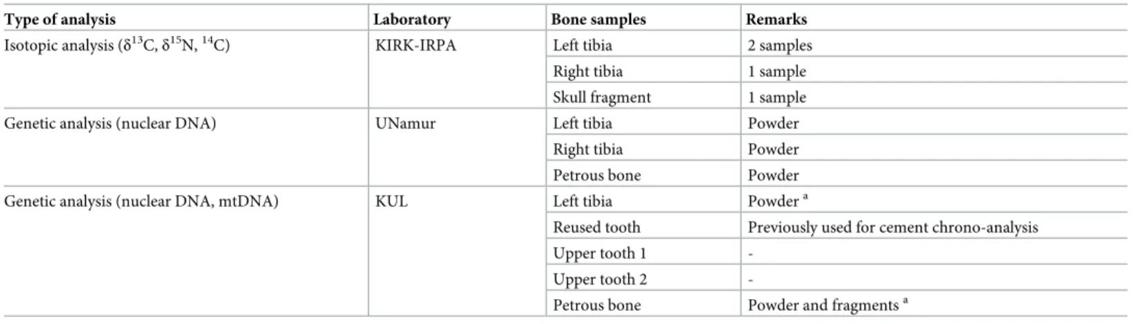
both tibias was first performed at UNamur using 9 human DNA microsatellite markers. How-ever, the analysis of DNA extracts from the remains turned out to be not reproducible among the different samples and the presence of the Y chromosome could not be established (data not shown). A standard PCR was then used to amplify 5 short tandem repeats (STR) of the Y chromosome using the same DNA samples. The Y chromosome was found in the petrous bone DNA, whereas the tibias DNA did not give reproducible results (Fig 4). These results
Fig 3. Radiocarbon dating results from the remains. (A) Calibrated ages (2σ) of the left tibia, right tibia and skull. (B) Average age of the four14C dates
determined for these remains.
suggest that the genetic material is strongly damaged in the tibias and, to a lesser extent, in the petrous bone. It could also be possible that the petrous bone sample contained a higher level of endogenous DNA compared to the tibia sample [32,33].
Genetic analysis of the remains was performed independently at KULeuven following stan-dard ancient DNA (aDNA) protocols. Five samples were analysed: petrous bone fragments and tibia bone powder sampled by UNamur, tooth embedded in epoxy resin (sample reused after cement chrono-analysis), and two upper teeth. Analysis of short tandem repeats from the Y chromosome on the tibia DNA extract gave no results, indicating that the quantity and/or quality of the extracted nuclear DNA were insufficient or that the DNA originated from a female. Identical results were obtained for the resin embedded tooth and the petrous bone DNA extracts. The results obtained by UNamur and KULeuven for nuclear DNA are consis-tent with the treatment of the remains attributed to Jacques de Vitry (discussed below) which would have impacted the preservation of the DNA [34,35]. In contrast, partial profiles for both autosomal (including sex determination with amelogenin gene) and Y-chromosome STRs were obtained with the two upper teeth DNA extracts (Table 3).
The autosomal STR results were reproducible between the DNA extracts as well between different PCR reactions indicating that the two teeth originated from the same individual. The characteristics of these profiles (drop-out of alleles or loci, drop-in of one or two alleles, high stutter frequency, heterozygote imbalance) were consistent with ancient DNA, where DNA damage and fragmentation lead to non-amplification of alleles or loci, and where low amounts of DNA lead to stochastic effects (drop-out, high stutter effect and heterozygote imbalance) that will influence the results [36]. The fact that very partial profiles were obtained for the Y-chromosome STRs can be explained by the higher sensitivity of the autosomal STR multiplex for low amounts of degraded DNA (personal data), except for the Amelogenin amplicons where the X-allele could not be amplified in the DNA extracts of the two upper teeth. As the two upper teeth have been subjected to a strict decontamination procedure [23] that would remove contaminating DNA or cellular material on the outer surface, any remaining contami-nation would have been visible as additional alleles in the genotyping results, and probably
Fig 4. PCR amplification of 5 STRs from the Y chromosome. Left tibia (LT), right tibia (LT), petrous bone (S), negative control (-), positive control (+).
would have revealed also more results for the STRs with an amplicon length greater than 150 base pairs.
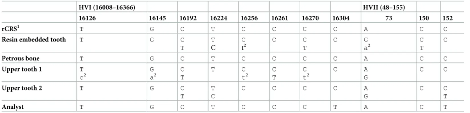
All five DNA extracts were also subjected to the analysis of the non-coding region of human mtDNA. In contrast to the nuclear DNA results, all DNA extracts with the exception of the DNA extract from the tibia powder revealed a mitochondrial (mt) sequence (Table 4). The samples of the reused tooth and the two upper tooth samples showed at several posi-tions the presence of two nucleotides, which is evidence for the presence of exogenous DNA in the DNA extracts. The origin of this DNA remains unknown (laboratory contamination can be excluded) and can probably be related to the handling of the remains during the different historical transfers. The fact that four out of five DNA extracts showed preservation of human mtDNA can be explained by the higher copy number of mtDNA and, to a lesser extent, addi-tional protection against degradation by the double membrane of the mitochondrion which would increase the chance to extract preserved mtDNA from archaeological remains [37,38]. The mtDNA sequence of the DNA extract from the petrous bone fragment revealed no evi-dence for exogenous DNA and was identical to the revised Cambridge Reference Sequence [28]. This mtDNA sequence, the most frequent (about 10%) in West Eurasian populations (https://empop.online/; v3/R11), could also be present in the other DNA extracts, which would be consistent with the hypothesis that the skeletal remains belong to the same
Table 3. Consensus genotyping results of the analysis of nine autosomal STRs, Amelogenin and 40 Y-chromosome STRs in DNA extracts from two upper teeth.
Autosomal STRs AMEL1 D2S441 D1S1656 D12S391 D10S1248 D21S11 D22S1045 D18S51 D1S1677 FGA Amplicon length (bp) 123–129 72–112 115–168 169–227 78–142 150–217 68–119 139–219 74–118 119–278 Upper tooth 1 Y 11 16.3 - 14 - - 14 13,15 18,(22) [20] Upper tooth 2 Y (11),14 - - 14 [13] 32.2 - 14,(20) [19] 13,(15) 18,22
Y-STRs DYS438 DYS392 DYS458 DYS385 DYS460 DYS481 Amplicon length (bp) 87–140 90–130 130–160 237–316 95–123 105–168
Upper tooth 1 9 11 14 12,13 - 22
Upper tooth 2 - - 14 - 10
-1Amelogenin.
[] PCR fragments attributed to stochastic effects of either high stutter or drop-in; () alleles with lower peak height (heterozygote imbalance).
https://doi.org/10.1371/journal.pone.0201424.t003
Table 4. Results of mtDNA Sanger sequencing for different bone and teeth samples from the remains.
HVI (16008–16366) HVII (48–155)
16126 16145 16192 16224 16256 16261 16270 16304 73 150 152
rCRS1 T G C T C C C C A C C
Resin embedded tooth T G C T T C C t2 C C T C G a2 C T C Petrous bone T G C T C C C C A C C Upper tooth 1 T c2 G a2 C T T C t2 C T C t2 C A G C C Upper tooth 2 T G C T T C C C C C A G C C T Analyst T G C T C C C T A C T
1cCRS, revised Cambridge Reference Sequence [28]. 2minor presence of nucleotide in sequence.
Analyst is the person who performed the DNA extraction and sequence analysis.
individual. This hypothesis would also imply that the origins of the exogenous DNA in the other samples are different, which is supported by the different mtDNA sequences observed for the two upper teeth where reproducible nuclear DNA profiles were obtained.
Proteomic analysis of collagen from parchment parts of the mitre. Identification of the
origin species of parchment parts of the mitre was carried out by proteomic analysis of the col-lagen protein extracted using a non-invasive sampling method (S3 Fig). Searches for species biomarkers gave unambiguous results on samples taken from four different locations on the mitre: cap, lower part of cap, left and right lappets (Fig 1B). Origin species (sheep, calf or goat) were identified in parchment samples according to the number of occurrences of species-spe-cific peptide markers obtained during sequencing of collagen I and III proteins (Table 5).
Discussion
History of the remains. In order to ease interpretation of the results of the scientific
stud-ies, we develop here additional historical considerations apart from the biography of Jacques de Vitry given above. Before Jacques de Vitry settled in Oignies, the relatively recent and poor pri-ory did not attract attention. No doubt the arrival of Jacques de Vitry changed the destiny of the priory, favouring its enrichment by the acquisition of relics [39,40]. The meeting with Marie d’Oignies had important consequence for the destiny of the future bishop and cardinal since it was at the origin of his decision to be buried in Oignies. Actually, it was a current practice in the high society of that time to notify by testimony the will to be buried in homeland in case of death occurred abroad. Repatriation of dead human bodies from long distances had practical consequences in terms of treatments applied to the corpse in order to ensure hygienic and safe transfer. This implied dismembering of the corpse and use of recipes to treat the remains [41–
43]. It is noteworthy that this practice was forbidden in 1299 by Pope Boniface VIII as it was regarded as an abominable custom for Christians. About 50 years before the ban, in due respect to his testimonial will, it is reasonable to assume that the dead body of Jacques de Vitry has expe-rienced dismembering and subsequent treatment of the remains. It is also noteworthy that a left tibia put in a wooden frame was kept as a separate relic (Fig 2C). No clear explanation has been given so far about the origin of this relic, except that it is attributed to Jacques de Vitry thanks to a label present in the frame and bearing the following notice in French: “Ceci est un ossement du Ve´ne´rable Jacques de Vitry. Cardinal et Evêque d’Acre en Palestine. Mort à Rome le 30 Avril 1240. Transporte´à son Monastère D’Oignies l’anne´e suivante, d’ou` provident cette pre´cieuse Relique” which means: “This is a bone from the Respectable Jacques de Vitry. Cardinal and Bishop of Acre in Palestine. Died in Rome on May 1st, 1240. Transported to his Monastery of Oignies next year, from where this invaluable Relic comes.” Therefore, a question naturally arises regarding the belonging of this framed tibia to the remains found in the reliquary.
Are the remains those of Jacques de Vitry?. In the absence of historical portrait or
description, traits of Jacques de Vitry remain unknown. No information is available regarding his stature, physiognomy and health. Moreover, we face the case of a secondary burial, not the
Table 5. Results of species identification in parchment parts of the mitre.
Sample Location Species-specific peptides (SSP) detected Number of SSP validated and sequenced Identified species
1 Cap 1 19 Ovis aries (sheep)
2 Cap 1 12 Ovis aries (sheep)
3 Lower part of cap 2 3 Bos Taurus (calf)
4 Left lappet 7 52 Bos Taurus (calf)
5 Right lappet 2 5 Bos Taurus (calf)
original one. His age at death is uncertain: between 60 and 75. Regarding the sex, it turned out that the morphology of the (incomplete) skeleton did not allow us to determine it. Therefore, we decided to resort to genetic analysis with the hope to be able to determine the sex from pre-served DNA. Regarding the age, anthropological results (dental wear, cranial vault and tooth cementum) tend to support the contemporary hypothesis regarding the birth year of Jacques de Vitry, with a slightly lower age estimate. The biological profile (age, sex, stature) obtained from the anthropological study did not allow us to deliver a verdict about the attribution of the remains to Jacques de Vitry. Cut marks on bones, on the other hand, are compatible with con-temporary medieval practices used for hygienic and safe repatriation of dead human bodies. Evidences of cuts on several bones and around joints suggest dismembering of the corpse [44–
46] in order to facilitate post mortem transfer. This hypothesis is supported by historical reports of contemporary dismembering practices used for remote burial and agrees with the translation of Jacques de Vitry remains from Rome to Oignies, on a distance of more than 1400 km. According to historical recipes, dismembering was followed by boiling of bones in water, wine or vinegar [41–43]. Such treatments would have damaged seriously the genetic material in the bones [47,48].
The results of isotopic analyses of the remains (Table 2) show that the left tibia, the right tibia and the skull might be from the same individual. Taking into account the standard devia-tions on the14C ages, there are no significant differences between the dates. The upper bound of average14C dates (1170AD) is anterior (70 years offset) to the year of death of Jacques de Vitry (1240AD). On the other hand, the stable isotopes analyses indicate a mainly terrestrial diet. If this happened to be the case, the skeleton could not be attributed to Jacques de Vitry. However, theδ15
N value is quite enriched, which may indicate a small amount of fish con-sumption since a carnivore has aδ15N value of +8‰ (S1 Table) [49]. Based on the depleted δ13
C value, it can be assumed this comes from freshwater fish and not marine fish, which has a more enrichedδ13C value (compareTable 2andS1 Table). Freshwater fish can have reservoir ages ranging between several hundreds of years and two thousand years, as demonstrated in [50]. A minimal consumption of freshwater fish with a large reservoir age can explain the offset between the year of death of Jacques de Vitry and the obtained radiocarbon ages. If this reser-voir effect is present, then the remains are likely to belong to Jacques de Vitry.
The objective of the genetic analysis was to determine the sex of the individual whose remains were found in the reliquary. Based on the DNA results for the petrous bone and two upper teeth (sex determination with amelogenin gene and analysis of Y-chromosomal STRs), we are able to conclude that the skull originates from a male individual.
Jacques de Vitry’s mitre. Jacques de Vitry bequeathed several personal objects, which
constituted, among others, the Treasure of Oignies. Among these, the mitre having miniatures on parchment is not only unique but also intriguing: questions about his usage, origin and fab-rication have not been addressed so far. The results of proteomic analyses showed that parch-ment of the cap was made from sheep, except the lower part where calf was identified. On the other hand, parchment of both lappets was made of calf. This difference of origin species between different parchment parts of the mitre is puzzling and has not been reported before. Was this choice of different animal species intentional or not? If yes, was it motivated by differ-ences in parchment quality, price or availability according to the species? All these questions certainly deserve further investigations from the point of view of the history of art.
Conclusion
An interdisciplinary study was carried out around the reliquary of the late cardinal Jacques de Vitry, a prominent clergyman and theologian who was active in Europe and Middle East
during the first part of the thirteen century. Results of anthropological, isotopic and genetic analyses provided evidence that the likelihood that the remains found in the reliquary are those of Jacques de Vitry is very high. Parchment parts of a mitre having belonged to Bishop Jacques de Vitry were analysed non-invasively by proteomic techniques and found to be made of different animal species. These findings are expected to fertilize knowledge carried by his-torical tradition around the relics of Jacques de Vitry and his related cultural heritage.
Supporting information
S1 Fig. Jacques de Vitry and Marie d’Oignies. (A) Cardinal Jacques de Vitry on his deathbed
(A. Marminia and E. Borne, engraving). (B) Saint Marie d’Oignies (engraving). Republished from “De B. Maria Oigniacensi in Namurcensi Belgii dioecesi. Appendix” in “Acta Sancto-rum” under a CC BY license, with permission from Socie´te´ des Bollandistes, original copyright 1867.
(TIF)
S2 Fig. Reliquary of Jacques de Vitry. (A) The reliquary. (B) The remains found in the
reli-quary after opening on 8thSeptember 2015. Reprinted under a CC BY license with permission from Vedrin, Guy Focant, original copyright 2015.
(TIF)
S3 Fig. Non-invasive sampling of parchment parts of Jacques de Vitry’s mitre. Gentle
rub-bing of the parchment surface with a PVC eraser for proteomic analyses. Reprinted under a CC BY license with permission from Vedrin, Guy Focant, original copyright 2012.
(TIF)
S1 Table. Isotopic fractionations of carbon (δ13C) and nitrogen (δ15
N) according to diet.
(DOCX)
Acknowledgments
The CROMIOSS project (www.lasan.be/la-recherche/projet-cromioss) was funded by a grant from the Fonds Jean-Jacques Comhaire of the Fondation Roi Baudouin (FRB), Belgium. Anne De Breuck (FRB) and Prof. Jean-Jacques Cassiman (KU Leuven, Belgium) are acknowledged for their intellectual support to the project. Jacques de Vitry’s mitre (donation from the Soeurs de Notre-Dame de Namur to FRB) belongs to FRB collections and is hold at TreM.a–Muse´e des Arts anciens, Namur, Belgium.
We thank Caroline Tilleux (Universite´ Catholique de Louvain and Muse´es Royaux d’Art et d’Histoire, Belgium) for her help during examination of the remains. We also want to thank Jean-Franc¸ois Nisolle (Centre Hospitalier Universitaire Dinant Godine, Belgium) for radiogra-phies of the tibiae, Benoıˆt Bertrand (University of Lille, France) for age estimation using cementum analysis, and Anne-Marie Wittek (Association pour la Diffusion de l’Information Arche´ologique, Belgium) for drawing the sketch of bones with cut marks.
We thank Prof. Karine Van Doninck, Jonathan Marescaux and Catherine Demazy (URBE, University of Namur, Belgium) for giving access to the genetic analyser and for providing tech-nical support.
Prof. Pierre Garin (Laboratory of Anatomy, University of Namur, Belgium) is acknowl-edged for secured hosting of the remains during the period of the project.
Finally, we are indebted to Jean Donnadieu (Universite´ de Provence Aix-Marseille I, France), expert of Jacques de Vitry’s history.
Author Contributions
Conceptualization: Aurore Carlier, Fiona Lebecque. Funding acquisition: Aurore Carlier, Fiona Lebecque.
Investigation: Ronny Decorte, Caroline Polet, Mathieu Boudin, Franc¸oise Tilquin, Jean-Yves
Matroule, Marc Dieu, Catherine Charles, Fiona Lebecque.
Project administration: Aurore Carlier. Writing – original draft: Olivier Deparis.
Writing – review & editing: Ronny Decorte, Caroline Polet, Mathieu Boudin, Franc¸oise
Til-quin, Jean-Yves Matroule, Marc Dieu, Fiona Lebecque, Olivier Deparis.
References
1. Huygens RBC, editor. Historia fundationis Venerabilis Ecclesiae Beati Nicolai oigniacensis ac Ancilla Christi Mariae Oigniacensis. Turnhout: Brepols Publishers; 2012. (Latin)
2. Donnadieu J. Jacques de Vitry (1175/1180-1240). Entre l’Orient et l’Occident:l’Evêque aux trois vis-ages. Turnhout: Brepols Publishers; 2014. (French)
3. Auvray L, editor. Les registres de Gre´goire IX, publie´s et analyse´s d’après les manuscrits originaux du Vatican. Paris: Fontemoing; 1896. n˚ 5179. (Latin)
4. Huygens RBC, editor. De Vitry J. Vita Mariae de Oegnies. Turnhout: Brepols Publishers; 2012. (Latin) 5. Buisseret F. Histoire de la vie, miracles et translations de S. Marie d’Oignies. Louvain: Gerard Rivius;
1609. (French)
6. De Cantimpre´ T. Vita Sanctae Lutgardis. Paris: Acta Sancotorum Juin/III; 1867, p. 257. (Latin) 7. De Trois-Fontaines A. Chronica. Scheffer-Boischost P editor. Leipzig; 1925, p. 950. (Latin)
8. Moschus F. Caenobiarchia Ogniacensis, sive Antistitum Ogniacensium Catalogus; Auctore Francisco Moscho, Accessere Elenchus sacrarum reliquiarum, quae ibidem in cimeliarchiopie adservantur; et sanctorum vitae, qui ibidem qui escunt, donec illucescat dies a eternitatis in futurum. Omnia cura et labore Arnoldi Raissi. Douai: Bartholome´ Bardou; 1636, p.p. 9–44. (Latin)
9. Chartes du prieure´ d’Oignies de l’ordre de Saint-Augustin. Poncelet E, editor. In: Annales de la Socie´te´ arche´ologique de Namur. Namur: Socie´te´ arche´ologique de Namur; 1912, pp. 31–101. (French) 10. Rayssius A. Catalogus Sanctarius Reliquiarum. In: Moschus F. Caenobiarchia Ogniacensis, sive
Anti-stitum Ogniacensium Catalogus; Auctore Francisco Moscho, Accessere Elenchus sacrarum reli-quiarum, quae ibidem in cimeliarchiopie adservantur; et sanctorum vitae, qui ibidem qui escunt, donec illucescat dies a eternitatis in futurum. Omnia cura et labore Arnoldi Raissi. Douai: Bartholome´ Bardou; 1636, pp. 48–53. (Latin)
11. Actes de la journe´e d’e´tude Hugo d’Oignies. Contexte et perspectives. Namur: Socie´te´ arche´ologique de Namur; 2012, p.58. (French)
12. Ferembach D, Schwindezky I, Stoukal M. Recommendation for age and sex diagnoses of skeletons. J Hum Evol. 1980; 9: 517–549.
13. Krogman WM, Işcan M.Y. The Human Skeleton in forensic medicine, 2nd edition. Springfield: Thomas CC Publisher; 1986.
14. Lovejoy CO. Dental wear in the Libben population: its functional pattern and role in the determination of adult skeletal age at death. Am J Phys Anthropol. 1985; 68(1): 47–56.https://doi.org/10.1002/ajpa. 1330680105PMID:4061601
15. Colard T, Bertrand B, Naji S, Delannoy Y, Be´cart A. Toward the adoption of cementochronology in forensic context. Int J Legal Med. 2015; 129: 1–8.https://doi.org/10.1007/s00414-014-0996-y
16. Olivier G, Aaron C, Fully G, Tissier G. New estimations of stature and cranial capacity in modern man. J Hum Evol. 1978; 7(6): 513–518.
17. Boudin M, Van Strydonck M, van den Brande T, Synal H-A, Wacker L. A new AMS facility at the Royal Institute for Cultural Heritage, Brussels, Belgium. Nuclear Instruments and Methods in Physics Research B. 2015; 361: 120–123.
18. Van Strydonck M, Van der Borg K. The construction of a preparation line for AMS-targets at the Royal Institute for Cultural Heritage, Brussels. Bulletin Koninklijk Instituut voor Kunstpatrimonium. 1990–1991; 23:228–234.
19. Bronk Ramsey C. Radiocarbon calibration and analysis of stratigraphy: the OxCal program. Radiocar-bon 1995; 37(2): 425–430.
20. Reimer PJ, Bard E, Bayliss A, Beck JW, Blackwell PG, Bronk Ramsey C, et al. IntCal13 and Marine13 radiocarbon age calibration curves 0–50,000 years cal BP. Radiocarbon 2013; 55(4): 1869–1887. 21. Mundorff A, Davoren JM. Examination of DNA yield rates for different skeletal elements at increasing
post mortem intervals. Forensic Science International: Genetics. 2017; 8: 55–63.
22. Kwon SY, Lee HY, Kim EH, Lee EY, Shin KJ. Investigation into the sequence structure of 23Y chromo-somal STR loci using massively parallel sequencing. Forensic Science International: Genetics. 2016; 25: 132–141.
23. Ottoni C, Ricaut F-X, Vanderheyden N, Brucato N, Waelkens M, Decorte R. Mitochondrial analysis of a Byzantine population reveals the differential impact of multiple historical events in South Anatolia. Eur J Hum Genet. 2011; 19: 571–576.https://doi.org/10.1038/ejhg.2010.230PMID:21224890
24. Larmuseau MHD, Vanderheyden N, Jacobs M, Coomans M, Larno L, Decorte R. Micro-geographic dis-tribution of Y-chromosomal variation in the central-western European region Brabant. Forensic Sci Int Genet. 2011; 5: 95–99.https://doi.org/10.1016/j.fsigen.2010.08.020PMID:21036685
25. Dognaux S, Larmuseau MHD, Jansen L, Heylen T, Vanderheyden N, Bekaert B, et al. Allele frequen-cies for the new European Standard Set (ESS) loci and D1S1677 in the Belgian population. Forensic Sci Int Genet. 2012; 6: e75–e77.https://doi.org/10.1016/j.fsigen.2011.05.003PMID:21664209
26. Larmuseau MHD, Bekaert B, Baumers M, Wenseleers T, Deforce D, Borry P, et al. Biohistorical materi-als and contemporary privacy concerns-the forensic case of King Albert I. Forensic Sci Int Genet. 2016; 24.https://doi.org/10.1016/j.fsigen.2016.07.008PMID:27470949
27. Sullivan KM, Mannucci A, Kimpton CP, Gill P. A rapid and quantitative DNA sex test: fluorescence-based PCR analysis of X-Y homologous gene amelogenin. Biotechniques. 1993; 15: 636–8, 640–1. PMID:8251166
28. Andrews RM, Kubacka I, Chinnery PF, Lightowlers RN, Turnbull DM, Howell N. Reanalysis and revision of the Cambridge reference sequence for human mitochondrial DNA. Nat Genet. Nature Publishing Group; 1999; 23: 147–147.https://doi.org/10.1038/13779PMID:10508508
29. Hall TA. BioEdit: a user-friendly biological sequence alignment editor and analysis program for Windows 95/98/NT. Nucl Acids Symp Ser. 1999; 41: 95–98.
30. Fiddyment S, Holsinger B, Ruzzier C, Devine A, Binois A, Albarella U, et al. Animal origin of 13th-cen-tury uterine vellum revealed using noninvasive peptide fingerprinting. Proc Natl Acad Sci. 2015; 112 (49):15066–71.https://doi.org/10.1073/pnas.1512264112PubMed Central PMCID:
PMCPMC4679014. PMID:26598667
31. DeNiro MJ. Postmortem preservation and alteration of in vivo bone collagen isotope ratios in relation to palaeodietary reconstruction. Nature. 1985; 317(6040): 806–809.
32. Hansen HB, Damgaard PB, Margaryan A, Stenderup J, Lynnerup N, Willerslev E, et al. Comparing Ancient DNA Preservation in Petrous Bone and Tooth Cementum. PLoS One. 2017; 12: e0170940.
https://doi.org/10.1371/journal.pone.0170940PMID:28129388
33. Furtwa¨ngler A, Reiter E, Neumann GU, Siebke I, Steuri N, Hafner A, et al. Ratio of mitochondrial to nuclear DNA affects contamination estimates in ancient DNA analysis. Sci Rep. 2018; 8: 14075.https:// doi.org/10.1038/s41598-018-32083-0PMID:30232341
34. Pruvost M, Schwarz R, Correia VB, Champlot S, Braguier S, Morel N, et al. Freshly excavated fossil bones are best for amplification of ancient DNA. Proc Natl Acad Sci U S A. National Academy of Sci-ences; 2007; 104: 739–44.https://doi.org/10.1073/pnas.0610257104PMID:17210911
35. Ottoni C, Koon HE, Collins MJ, Penkman KE, Rickards O, Craig OE. Preservation of ancient DNA in thermally damaged archaeological bone. Naturwissenschaften. 2009; 96: 267–278.https://doi.org/10. 1007/s00114-008-0478-5PMID:19043689
36. Ottoni C, Bekaert B, Decorte R. DNA degradation: Current knowledge and developments. In: Schots-mans EMJ, Ma´rquez-Grant N, Forbes SL, editors. Taphonomy of human remains: forensic analysis of the dead and the depositional environment. John Wiley & Sons Ltd, Chichester, West Sussex; 2017. pp. 65–80.
37. Schwarz C, Debruyne R, Kuch M, Mcnally E, Schwarcz H, Aubrey AD, et al. New insights from old bones: DNA preservation and degradation in permafrost preserved mammoth remains. Nucleic Acids Res. 2009; 37: 3215–3229.https://doi.org/10.1093/nar/gkp159PMID:19321502
38. Higgins D, Rohrlach AB, Kaidonis J, Townsend G, Austin JJ. Differential Nuclear and Mitochondrial DNA Preservation in Post-Mortem Teeth with Implications for Forensic and Ancient DNA Studies. PLoS One. 2015; 10: e0126935.https://doi.org/10.1371/journal.pone.0126935PMID:25992635
39. Huygens RBC, editor. De Vitry J. Vita Mariae de Oegnies. Turnhout: Brepols Publishers; 2012, p.144. (Latin)
40. Wankenne A. La vie de Marie d’Oignies par Jacques de Vitry. Supple´ ment par Thomas de Cantimpre´. Namur; 1989, p. 169. (French)
41. Paravicini-Bagliani A. De´membrement et inte´grite´ du corps au XIIIe siècle. Terrain; 1992, p. 18, pp. 26–32. (French)
42. Georges P. Mourir c’est pourrir un peu. . .Intentions et techniques contre la corruption des cadavresà
la fin du Moyen Age. Micrologus, Nature, Sciences and Medieval Societies. 1999; VII: 359–382. (French)
43. Weiss-Krejci E. Excarnation, evisceration, and exhumation in medieval and post-medieval Europe. In: Rakita GFM, Buikstra J, Beck L, Williams S, editors. Interacting with the dead. Perspectives on mortu-ary archaeology for the new millennium. Gainesville: University Press of Florida. 2005; pp. 155–172. 44. Porta D, Amadasi A, Cappella A, Mazzarelli D, Magli F, Gibelli D, et al. Dismemberment and
disarticula-tion: A forensic anthropological approach. J Forensic Leg Med. 2016; 38: 50–57.https://doi.org/10. 1016/j.jflm.2015.11.016PMID:26708349
45. Morcillo-Me´ndez MD, Campos IY. Dismemberment: Cause of death in the Colombian armed conflict. Torture. 2012; 22 (Suppl 1): 5–13.
46. Rutty GN, Hainsworth SV. The Dismembered Body. In: Rutty GN, editor. Essentials of Autopsy Prac-tice: Innovations, Updates and Advances in Practice. London: Springer 2014; pp. 59–87.
47. Arismendi JL, Baker LE, Matteson KJ. Effects of Processing Techniques on the Forensic DNA Analysis of Human Skeletal Remains. J Forensic Sci. 2004; 49(5): 1–5.
48. Rennick SL, Fenton TW, Foran DR. The effects of skeletal preparation techniques on DNA from human and non-human bone. J Forensic Sci. 2005; 50(5):1016–1019. PMID:16225205
49. Lanting JN, van der Plicht J. Wat hebben Floris V, skelets Swifterbant S2 en visotters gemeen? Palaeo-historia. 1996; 37/38: 491–520.
50. Ervynck A, Boudin M, Van Neer W. Assessing the radiocarbon freshwater reservoir effect for a north-west-european river system (the schelde basin, Belgium). Radiocarbon 2018; 60(2): 395–417