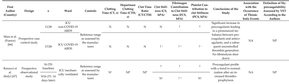
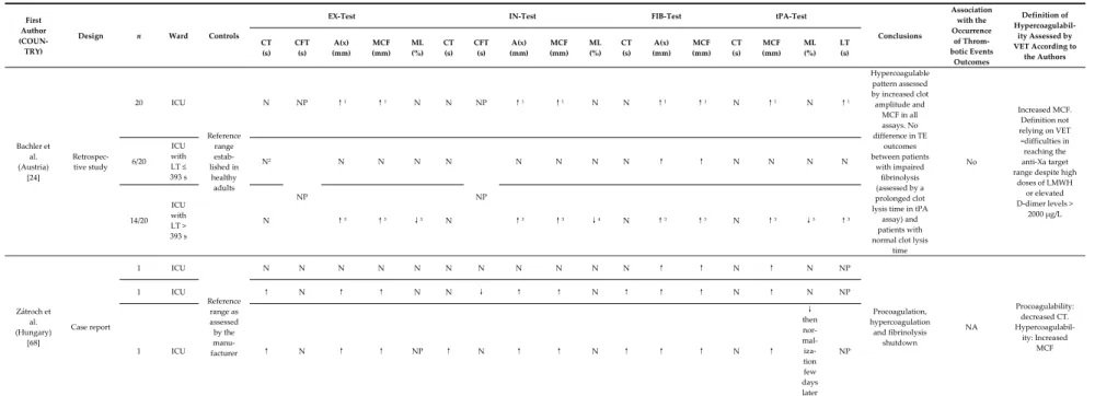
RESEARCH OUTPUTS / RÉSULTATS DE RECHERCHE
Author(s) - Auteur(s) :
Publication date - Date de publication :
Permanent link - Permalien :
Rights / License - Licence de droit d’auteur :
Bibliothèque Universitaire Moretus Plantin
Institutional Repository - Research Portal
Dépôt Institutionnel - Portail de la Recherche
researchportal.unamur.be
University of Namur
Viscoelastometric Testing to Assess Hemostasis of COVID-19
Bareille, Marion; Hardy, Michaël; Douxfils, Jonathan; Roullet, Stéphanie; Lasne, Dominique;
Levy, Jerrold H; Stépanian, Alain; Susen, Sophie; Frère, Corinne; Lecompte, Thomas;
Mullier, François
Published in:
Journal of clinical medicine DOI:
10.3390/jcm10081740
Publication date: 2021
Document Version
Publisher's PDF, also known as Version of record
Link to publication
Citation for pulished version (HARVARD):
Bareille, M, Hardy, M, Douxfils, J, Roullet, S, Lasne, D, Levy, JH, Stépanian, A, Susen, S, Frère, C, Lecompte, T & Mullier, F 2021, 'Viscoelastometric Testing to Assess Hemostasis of COVID-19: A Systematic Review', Journal of clinical medicine, vol. 10, no. 8. https://doi.org/10.3390/jcm10081740
General rights
Copyright and moral rights for the publications made accessible in the public portal are retained by the authors and/or other copyright owners and it is a condition of accessing publications that users recognise and abide by the legal requirements associated with these rights. • Users may download and print one copy of any publication from the public portal for the purpose of private study or research. • You may not further distribute the material or use it for any profit-making activity or commercial gain
• You may freely distribute the URL identifying the publication in the public portal ?
Take down policy
If you believe that this document breaches copyright please contact us providing details, and we will remove access to the work immediately and investigate your claim.
Review
Viscoelastometric Testing to Assess Hemostasis of COVID‐19:
A Systematic Review
Marion Bareille 1,*, Michaël Hardy 2, Jonathan Douxfils 3,4, Stéphanie Roullet 5,6, Dominique Lasne 7, Jerrold H. Levy 8, Alain Stépanian 9, Sophie Susen 10, Corinne Frère 11, Thomas Lecompte 12
and François Mullier 1 1 CHU UCL Namur, Namur Thrombosis and Hemostasis Center (NTHC), Université Catholique de Louvain, 5530 Yvoir, Belgium; [email protected] 2 Service d’anesthésiologie, Namur Thrombosis and Hemostasis Center (NTHC), CHU UCL Namur, Univer‐ sité Catholique de Louvain, 5530 Yvoir, Belgium; [email protected] 3 Namur Thrombosis and Hemostasis Center (NTHC), Département de Pharmacie, Université de Namur, 5000 Namur, Belgium; [email protected] 4 Qualiblood S.A., 5000 Namur, Belgium 5 CHU Bordeaux, Service d’Anesthésie‐Réanimation Tripode, 33000 Bordeaux, France; stephanie.roullet@chu‐bordeaux.fr 6 Biologie des Maladies Cardiovasculaire, University Bordeaux, INSERM U1034, 33600 Pessac, France 7 Laboratoire d’hématologie générale, Hôpital Universitaire Necker‐Enfants Malades, AP‐HP, 75015 Paris, France; [email protected] 8 Departments of Anesthesiology, Critical Care, and Surgery (Cardiothoracic), Duke University School of Medicine, Durham, NC 27710, USA; [email protected] 9 Hôpital Lariboisière, Service d’Hématologie Biologique, Institut de Recherche Saint‐Louis, Université de Paris, AP‐HP Nord‐Université de Paris, EA 3518, 75010 Paris, France; [email protected] 10 Laboratoire d’Hématologie‐Hémostase, Université de Lille, CHU Lille, 59037 Lille, France; sophie.susen@chru‐lille.fr 11 Department of Hematology, Pitié‐Salpêtrière Hospital, Assistance Publique Hôpitaux de Paris, INSERM UMRS_1166, Sorbonne Université, 75013 Paris, France; [email protected] 12 Départements de Médecine, Service d’angiologie et d’hémostase et Faculté de Médecine Geneva Platelet Group (GpG), Université de Genève et Hôpitaux Universitaires de Genève, 1205 Genève, Switzerland; [email protected] * Correspondence: [email protected]
Abstract: Infection by SARS‐CoV‐2 is associated with a high risk of thrombosis. The laboratory
documentation of hypercoagulability and impaired fibrinolysis remains a challenge. Our aim was to assess the potential usefulness of viscoelastometric testing (VET) to predict thrombotic events in COVID‐19 patients according to the literature. We also (i) analyzed the impact of anticoagulation and the methods used to neutralize heparin, (ii) analyzed whether maximal clot mechanical strength brings more information than Clauss fibrinogen, and (iii) critically scrutinized the diag‐ nosis of hypofibrinolysis. We performed a systematic search in PubMed and Scopus databases until December 31st, 2020. VET methods and parameters, and patients’ features and outcomes were ex‐ tracted. VET was performed for 1063 patients (893 intensive care unit (ICU) and 170 non‐ICU, 44 studies). There was extensive heterogeneity concerning study design, VET device used (ROTEM, TEG, Quantra and ClotPro) and reagents (with non‐systematic use of heparin neutralization), timing of assay, and definition of hypercoagulable state. Notably, only 4 out of 25 studies using ROTEM reported data with heparinase (HEPTEM). The common findings were increased clot mechanical strength mainly due to excessive fibrinogen component and impaired to absent fibri‐ nolysis, more conspicuous in the presence of an added plasminogen activator. Only 4 studies out of the 16 that addressed the point found an association of VETs with thrombotic events. So‐called functional fibrinogen assessed by VETs showed a variable correlation with Clauss fibrinogen. Abnormal VET pattern, often evidenced despite standard prophylactic anticoagulation, tended to normalize after increased dosing. VET studies reported heterogeneity, and small sample sizes do not support an association between the poorly defined prothrombotic phenotype of COVID‐19 and thrombotic events. Citation: Bareille, M.; Hardy, M.; Douxfils, J.; Roullet, S.; Lasne, D.; Levy, J.H.; Stépanian, A.; Susen, S.; Frère, C.; Lecompte, T.; Mullier F. Viscoelastometric Testing to Assess Hemostasis of COVID‐19: A Systematic Re‐ view. J. Clin. Med. 2021, 10, x. https://doi.org/10.3390/xxxxx Academic Editor: Angelo Claudio Molinari Received: 26 March 2021 Accepted: 12 April 2021 Published: 16 April 2021
Publisher’s Note: MDPI stays
neutral with regard to jurisdic‐ tional claims in published maps and institutional affiliations.
Copyright: © 2021 by the au‐ thors. Licensee MDPI, Basel, Switzerland. This article is an open access article distributed under the terms and conditions of the Creative Commons Attribution (CC BY) license (http://creativecommons.org/lic enses/by/4.0/).
Keywords: viscoelastic test; thromboelastometry; thromboelastography; sonorheometry; ROTEM;
TEG; Quantra; ClotPro; coronavirus disease 2019; COVID‐19; severe acute respiratory syndrome coronavirus 2; SARS‐CoV‐2 1. Introduction In contrast to conventional clotting tests, viscoelastic tests (VETs) monitor changes of viscoelastic properties of a forming and evolving clot from whole blood, before and be‐ yond the clotting point; they are often referred to as a global hemostasis test, although some aspects of hemostasis are not explored [1–3]. Coagulation occurs in the presence of platelets and red blood cells, and fibrinolysis can translate into a decrease in clot me‐ chanical strength after its maximum has been reached, but clot retraction seems to play a role here as well [4–6]. VETs are based on the mechanical properties of the clot, like me‐ chanical strength, and are influenced by its composition in platelets, fibrin, red blood cells, and factor XIII [7–9]. To our knowledge, the assessment of factor XIII by VETs has not been investigated in COVID‐19 patients. VETs have been considered to provide a comprehensive assessment of the dynamic process of blood clot formation and subsequent lysis. As they can be performed bedside as point‐of‐care testing and can give useable results about clot formation and a potential hyperfibrinolysis within one hour, they are chiefly considered as convenient tools for re‐ al‐time assessment of coagulation and fibrinolysis in whole blood and have been gaining in popularity in various hemorrhagic situations, such as cardiac surgery, obstetrics, and traumatology over decades, for the management of acutely bleeding patients [3]. By contrast, COVID‐19 disturbance of hemostasis is likely a combination of hypercoagula‐ bility and impaired fibrinolysis (a prothrombotic laboratory phenotype), at least in part, contributing to the thrombotic risk and the prothrombotic laboratory phenotype, but VETs have been nevertheless suggested to be potentially useful, in line with previous works on sepsis [10] and trauma [11,12], for example.
Of note, VETs share the same limitations as all currently available clinical lab tests, i.e., negligible effect of endogenous anticoagulants, absence of endothelium, and very low shear in a close system. Furthermore, there are good reasons to challenge the inter‐ pretation of hypercoagulability and to question the ability to sensitively detect and ac‐ curately quantify hypofibrinolysis, especially when a value equal to zero belongs to the manufacturer’s reference range.
Our aim was to assess the potential clinical usefulness of VETs to predict clinical outcomes (mainly thrombotic events) in COVID‐19 patients through this systematic re‐ view. We also (i) analyzed the impact of anticoagulation and the methods used to neu‐ tralize heparin (in other words, was heparin duly neutralized?), (ii) disentangled re‐ ported alterations in clotting dynamics and analyzed whether maximal clot mechanical strength brings more information than Clauss fibrinogen, and (iii) critically scrutinized the documentation of hypofibrinolysis with VET under various reactive conditions. The term ‘hypercoagulable state’ will be uniformly used to refer to the investigators’ inter‐ pretation of VET findings; we will discuss to what extent this is an appropriate interpre‐ tation.
The preanalytical aspects, which are crucial in laboratory hemostasis but scarcely mentioned among the retrieved studies, are beyond the scope of this review and will not be addressed.
2. Materials and Methods
2.1. Search Methodology
We performed a systematic literature search in PubMed and Scopus databases, re‐ gardless of publication status, using the following keywords ‘viscoelastic test OR
thromboelastometry OR thromboelastography OR sonorheometry OR ROTEM OR TEG OR Quantra OR ClotPro’ AND ‘coronavirus disease 2019 OR COVID‐19 OR severe acute respiratory syndrome coronavirus 2 OR SARS‐CoV‐2’. Search strategy is provided as Data S1. We also searched the reference lists of selected articles for additional relevant works, and we did not restrict our search to articles published in English and found some articles in Russian and Hungarian. In addition, reviewers performed manual searches and cross‐references in the retrieved papers. The last search was conducted on 31 De‐ cember 2020. Our review followed the PRISMA (Preferred Reporting Items for System‐ atic Reviews and Meta‐analysis) guidelines [13], and the PRISMA summary table can be found as Data S2. Due to a considerable heterogeneity among the retrieved studies, we did not extend our systematic review to a meta‐analysis.
2.2. Study Selection
All references retrieved from our search were screened based upon their title and abstract to assess eligibility. If they were considered relevant, the full‐text articles were analyzed to check if they met the selection criteria as follows. As COVID‐19 pandemic is a recent phenomenon, and due to the relatively small number of published data on VETs, we did not restrict eligibility according to patients’ characteristics, disease severity, or treatment modalities. Studies of any design and case reports, including original data from VETs in COVID‐19 patients with neither pregnancy nor known history of coagula‐ tion disorder, were deemed eligible. All relevant studies regardless of methodological quality were included when the full‐text article was available (Table 1).
Table 1. Eligibility criteria.
PICOS Inclusion Exclusion
Participants All patients with confirmed COVID‐19 infection regardless of age Pregnancy Pre‐existing coagulation disorder Intervention Viscoelastometric testing performed ‐ Comparison Reference values (manufacturer’s based or healthy controls) ICU COVID‐19 patients and non‐ICU COVID‐19 patients ICU COVID‐19 patients and ICU non‐COVID‐19 patients ‐ Outcomes VET parameters in COVID‐19 patients Difference in VET parameters between ICU COVID‐19 patients and non‐ICU COVID‐19 patients Difference in VET parameters between ICU COVID‐19 patients and ICU non‐COVID‐19 patients Association between VET parameters and clinical outcomes Association between VET parameters and Clauss fibrinogen ‐ Study design Randomized controlled trials Observational clinical studies Case reports Opinion papers Review papers Healthcare guidelines Protocol Non‐human or in vitro studies Abbreviations: VET: viscoelastometric testing; ICU: Intensive care unit. Reviews, position articles, and guidelines were excluded. All kind of VETs were in‐ cluded but were analyzed separately.
2.3. Data Extraction
For each study, data regarding author identification, geographic location, study de‐ sign, number of patients and their characteristics (including comorbidities and throm‐ botic events), prospective design or not, timing of blood collection and anticoagulation status, type of VET device used and results, and the results of other conventional hemo‐ stasis tests (platelet count, fibrinogen and D‐dimers plasma levels), and C‐reactive pro‐ tein were extracted with the aid of a systematic chart. 2.4. A Concise Overview of the Different VET Devices Viscoelastometric testing (VET) should be performed either immediately with native whole blood or within four hours after drawing if performed with whole citrated blood, as most often done [1,3]. ROTEM devices and TEG5000 all rely on the movement of a pin and a cup relative to each other; in the former, the cuvette is fixed, and the pin oscillates, and vice versa in the latter. The oscillations are recorded and graphically displayed with the characteristic normal tuning fork shape [3]. The conventional clotting point roughly corresponds to the reaction time R for TEG, and to the clotting time CT for ROTEM, ClotPro, and Quantra; extended fibrin polymerization is monitored with the kinetics time K and α angle for TEG and with CFT and α angle for ROTEM and ClotPro; the eventual result is maximal mechanical strength (maximal amplitude MA for TEG, maximal clot firmness MCF for ROTEM and ClotPro and clot stiffness CS for Quantra) and its subsequent decrease, as a result of ‘endogenous’ fibrinolysis monitored by lysis of the clot at given time x LY(x) for TEG and by maximal lysis ML or lysis of the clot at a given time x (LI(x)) for ROTEM and ClotPro, at least in part [2,3,14]. Coagulation can be initiated through the contact phase or the tissue factor pathway (often referred to as intrinsic or extrinsic pathways, respectively) and needs recalcifica‐ tion when citrated blood is used [3]. If the nature of the initiating agents is known, their concentrations are not disclosed. Regarding the former pathway, the limitations of aPTT testing apply, although ‘clotting times’ are longer, suggesting a lower amount of contact phase activator (kaolin, celite, or ellagic acid) and higher calcium concentration. The dif‐ ferent well‐known behaviors of those reagents in case of defective contact phase, ab‐ normal factor VIII levels, high CRP (C‐reactive protein) levels, lupus anticoagulant, or heparin must be borne in mind. Two reagents can be used to neutralize heparin, either polybrene (hexadimethrine bromide) or heparinase; two to inhibit the platelet contribu‐ tion to mechanical clot properties, namely cytochalasin D and abciximab, sometimes both together; lastly, two to inhibit fibrinolysis, either aprotinin or tranexamic acid [3]. To what extent those inhibitions are fully achieved is not entirely clear.
2.4.1. ROTEM
Three versions of the ROTEM device exist: from the oldest to the most recent, ROTEM‐gamma, ROTEM‐delta, and the brand‐new version ROTEM‐sigma. The main difference between them is that ROTEM‐gamma and ‐delta need manual pipetting of the blood sample and the reagents into cups, whereas ROTEM‐sigma is a completely auto‐ mated, closed system. For the latter, reagents consist of a consumable ready‐to‐use car‐ tridge with four parallel channels prefilled with specific lyophilized reagents [15]. All ROTEM versions can perform the same assays, namely INTEM, HEPTEM, EXTEM, FIBTEM, and APTEM, to investigate the intrinsic pathway (with and without hepa‐ rinase), the extrinsic pathway, the fibrinogen component, and the fibrinolysis with apro‐ tinin, respectively. Of note, EXTEM, FIBTEM, and APTEM reagents contain polybrene and HEPTEM contain heparinase to neutralize heparin (Table A1) [2,16]. They report the same parameters: clotting time (CT), clot formation time (CFT), α angle, “amplitude of the clot” at a given time x (A(x)), maximum clot firmness (MCF), clot lysis index (LI(x)), and maximum lysis (ML) (Table A2).
2.4.2. TEG Briefly, regarding TEG5000 a blood sample is pipetted into a cup; liquid reagents are added; ultimately, a fixed pin connected to a detector system is then put in the cup. The graphical representation is called TEMogram. TEG6s for its part is a completely closed and automated system. In contrast to its predecessor TEG5000, it relies on sonorheome‐ try. Reagents consist of a consumable, ready‐to‐use cartridge with four parallel channels prefilled with specific lyophilized reagents (Table A3) [17,18].
The two versions of the TEG device can perform the same assays, namely Kaolin TEG with (CKH) or without heparinase (CK), RapidTEG (CRT), and TEG Functional Fi‐ brinogen (CFF), and offer the same parameters: reaction time (R), kinetics time (K), α angle, maximum amplitude (MA), and fibrinolytic activity (Ly) [3,19]. Of note, heparin neutralization differs between TEG500, where neutralization can occur in virtually any channel by using heparinase‐coated cups, and TEG6s, where neutralization occurs only in the CKH channel thanks to heparinase (Table A4) [3]. 2.4.3. Quantra The Quantra device also uses sonorheometry. Briefly, an acoustic radiation force is applied to the blood sample. As the blood clot forms, it starts to resonate: oscillations are then correlated with the shear modulus of the blood sample. The resistance of the sample to shear forces can be quantified by the time delay between the ultrasound pulse emis‐ sion and the returning echoes [20–22]. Reagents consist of a consumable, ready‐to‐use cartridge with four parallel channels prefilled with specific lyophilized reagents [22]. There are currently two kinds of car‐ tridges: the QPlus cartridge and the QStat one dedicated to exploring fibrinolysis [23]. Measurements of clot coagulation time with (CTH) or without (CT) heparinase and co‐ agulation initiation with kaolin, clot stiffness (CS) after initiation with thromboplastin, and fibrinogen contribution to the overall clot stiffness (FCS) after platelet inhibition with abciximab are performed simultaneously in four parallel channels. Of note, channel 2 contains heparinase, and channels 3 and 4 contain polybrene to neutralize heparin. Platelet contribution to clot stiffness (PCS) results from the difference between total CS and FCS (Tables A5 and A6).
2.4.4. ClotPro
The ClotPro device uses rotational technology similar to ROTEM®, but some dif‐ ferences exist between the two devices. First, in contrast with ROTEM, the cuvette rotates and the pin is stationary [24,25]. Second, reagents for each assay are present in dry form in a sponge located in the pipette tip; during pipetting of the patient sample, the reagent is automatically added to the blood [25]. This device can perform the same kind of assays as the ROTEM device (EX‐test, IN‐test, HI‐test, FIB‐test, AP‐test) plus some other specific ones (RVV‐test, ECA‐test), and offer similar parameters. Of note, EX‐test, tPA‐test, and FIB‐test contain polybrene to neutralize heparin (Tables A7 and A8) [24]. 3. Results 3.1. Literature Search Our literature search and selection flow chart according to PRISMA statement [13] is summarized in Figure 1.
Figure 1. Literature search and selection flow chart according to PRISMA statement.
We identified 140 references, resulting in 97 unique citations after duplicates re‐ moval. Two additional articles were identified through other sources. Each title and ab‐ stract were screened, and 36 references were excluded either because they were not re‐ lated to the subject (n = 16), because they were position articles or guidelines (n = 7) or reviews (n = 5), or because there was no full‐text available at this time (n = 5) or no pos‐ sible translation (n = 5). A total of 63 potentially eligible articles were considered for in‐ clusion, and the full‐text articles were retrieved. The most common reasons for exclusion after the full‐text evaluation were that papers were reviews (n = 19), not related to the subject (n = 6), or position articles or guidelines (n = 4). Finally, 44 references [24,26–68] met the eligibility criteria.
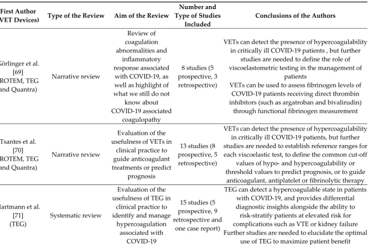
3.2. Originality of Our Systematic Review as Compared to the Existing Ones on the Subject Reviews have already been published recently, two of them only being systematic [69–72], but none has so far investigated the four major commercially available VET de‐ vices (i.e., ROTEM, TEG, ClotPro, and Quantra) or included such a large number of studies (n = 44). Characteristics of each review are summarized in Table 2.
Overall, case reports were excluded (except for one systematic review [71]); few studies were available and presented extensive heterogeneity.
Table 2. Characteristics of the reviews already published.
First Author(Title) Type of the Review Aim of the Review Number and Type of Studies
Included VET Devices Görlinger et al. [69] (COVID‐19 associated coagulopathy and inflammatory response: what do we know already and what are the knowledge gaps?) Narrative review Review of coagulation abnormalities and inflammatory response associated with COVID‐19 8 studies (5 prospective, 3 retrospective) ROTEM, TEG, Quantra Tsantes et al. [70] (COVID‐19 Infection‐Related Coagulopathy and Viscoelastic Methods: A Paradigm for Their Clinical Utility in Critical Illness) Narrative review Evaluation of the usefulness of VETs in clinical practice to guide anticoagulant treatments or predict prognosis 13 studies (8 prospective, 5 retrospective) ROTEM, TEG, Quantra Hartmann et al. [71] (The Role of TEG Analysis in Patients with COVID‐19‐Associated Coagulopathy: A Systematic Review) Systematic review Evaluation of the usefulness of TEG in clinical practice to identify and manage hypercoagulation associated with COVID‐19 15 studies (5 prospective, 9 retrospective and one case report) TEG Słomka et al. [72] (Hemostasis in Coronavirus Disease 2019‐Lesson from Viscoelastic Methods: A Systematic Review) Systematic review Evaluation of the performance of TEG and TEM in the assessment of blood coagulation and fibrinolysis in patients with COVID‐19 10 studies (2 prospective, 8 retrospective) ROTEM, TEG 3.3. Characteristics of the Selected Studies
Quality assessment of the selected study was performed using the Scottish Inter‐ collegiate Guidelines Network (SIGN) grading system [73]. Overall, the retrieved studies were of low (3, “non analytic studies”) to moderate quality (2+, “well‐conducted case control or cohort studies with a low risk of confounding or bias and a moderate proba‐ bility that the relationship is causal”), and details can be found as Data S3. Characteristics of the selected studies are summarized in Table 3.
Table 3. Characteristics of the included studies.
First Author
(Country) Device Study Design Ward n
Number of Patients with Viscoelastic Test Performed Timing of Assay Number of Patients with Invasive Mechanical Ventilation (n) Number of Patients under ECMO (n) Number of Patients with Renal Replace‐ ment Therapy (n) Age 1 Number of COVID‐19 Patients with Thrombotic Events Diagnosis of Throm‐ botic Events Anticoagulation Iwasaki et al.
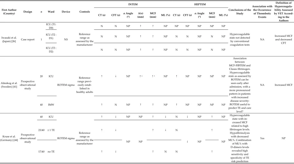
(Japan) [26] ROTEM (NS) Case report ICU 1 1
1 day after
ICU admission 1 NP NP 57 None NP None until TE, then UFH 10,000 IU/d
Pavoni et al. (Italy) [27] ROTEM gamma Retrospective observational study ICU 40 40 ICU admis‐ sion, then 5 and 10 days later 4/40 NP NP 61 ± 13 20/40 patients (6 DVT, 2 TE, 12 catheter related thrombosis) Systematic screening from common femoral vein by ultrasound Enoxaparin 40–60 mg/d according to local protocol Boscolo et al. (Italy) [28] ROTEM delta Prospective observa‐ tional study ICU 32 32
NP 21/32 NP NP 68 (62–75) 11/32 patients No systematic screen‐ing NP IMW 32 32 None None None 61 (53–71) 3/32 patients
Corrêa et al. (Brazil) [29] ROTEM delta Prospective observa‐ tional study ICU 30 30 ICU admis‐ sion, then 1, 3, 7 and 14 days later 27/30 NP 10/30 61 (52–83) 6/30 patients (4 DVT, 2 PE) NP At least prophylactic UFH or LMWH Madathil et al. (USA) [30] ROTEM delta Prospective observa‐ tional study ICU 11 11 ICU admis‐ sion, then 24– 48 h later 11/11 NP NP 53 (45.5–65.5) NP NP NP Spiezia et al. (Italy) [31] ROTEM delta Prospective observa‐ tional case control study
ICU 22 22 ICU admission 19/22 NP NP 67 ± 8 5/22 patients (DVT) NP Prophylactic LMWH
Tsantes et al. (Greece) [32] ROTEM delta Prospective observa‐ tional study ICU COVID‐19 patients 11 11 NP NP NP NP 78 (67–71) NP NP Enoxaparin 1 mg/kg bid ICU non COVID‐19 patients 9 9 NP NP NP NP Enoxaparin 1 mg/kg od IMW COVID‐19 patients 21 21 NP NP NP 73 (50–88) Enoxaparin 1 mg/kg od Al‐Ghafry et al. (USA) [33] ROTEM delta Retrospective observational study PICU (n = 5) and PW (n = 3) 8 8 1 to 4 days after hospital admission
None None None 12.9 (2–20) None NP
Prophylactic enoxaparin 0.5 mg/kg bid according to oxygen require‐ ment and D‐dimers levels, escalated to therapeutic dose (1 mg/kg bid) if clinical deterioration Creel‐Bulos et al. (USA) [34] ROTEM delta Retrospective observational study ICU 25 25 NP NP NP NP 63 (53–77) 9/25 patients (7 DVT, 4 PE, 1 arterial thrombosis) Ultrasound or CT imaging based on clinical suspicion Prophylactic LMWH or UFH Hoechter et al. (Germany) [35] ROTEM delta Retrospective observational case control study ICU COVID‐19 pneumonia 22 11 Within 48 h after ICU admission 22/22 NP NP 64 (52–70) NP NP Prophylactic UFH according to local guidelines ICU non COVID‐19 pneumonia 14 14 NP 14/14 NP NP 49 (36–57) Roh et al. (USA) [36] ROTEM delta Retrospective observational case control study ICU 30 30 ICU admission NP NP NP 63 ± 12 10/30 patients (3 DVT, 1 PE, 1 both DVT and PE, 4 arterial thrombosis, 1 both arterial throm‐ bosis and DVT) Ultrasound or CT imaging based on clinical suspicion At least prophylactic UFH or LMWH
Kong et al. (United Kingdom) [37] ROTEM delta Case report ICU 1 1 2 h after ICU
admission No No No 48 None NP None until ROTEM analysis
ICU 1 1 NP 1 No 1 68 None NP
Raval et al. (USA) [38]
ROTEM
delta Case report ICU 1 1 ICU admission 1 No No 63 None NP
None at admission, then UFH 7500 IU/8 h Nougier et al. (France) [39] Modified ROTEM delta (TEM‐tPA) Prospective observa‐ tional case control study ICU 40 19 NP 33/40 NP 7/40 62.8 ± 13.1 14/40 patients (8 PE, 5 DVT, 1 arterial thrombosis) Ultrasound or CT imaging based on clinical suspicion At least prophylactic UFH or LMWH IMW 38 4 None None None 60.2 ± 14.6 NP
Weiss et al. (France) [40] Modified ROTEM delta (TEM‐tPA) Prospective observa‐ tional case control study
ICU 5 5 NP NP NP NP 57 ± 15 3/5 patients NP Thromboprophylaxis according to current guidelines
Almskog et al. (Sweden) [41] ROTEM sigma Prospective observa‐ tional study ICU 20 20 1 day after hospital admission NP NP NP 62 (55–66) NP NP At least prophylactic tinzaparin IMW 40 40 NP NP NP 61 (51–74) Collett et al. (Australia) [42] ROTEM sigma Prospective observa‐
tional study ICU 6 6 NP 5/6 None 2/6 69 (64.2–73)
3/6 patients (1 PE, 1 catheter related thrombosis, 1 TE not clinically suspected) NP Enoxaparin 40 mg od Ibañez et al. (Spain) [43] ROTEM sigma Prospective observa‐ tional study ICU 19 19 24–48 h after ICU admission NP NP NP 61 (55–73) 5/19 patients (2 DVT, 2 PE, 1 arterial thrombosis) NP Enoxaparin 40–80 mg/d according to local protocol Kruse et al. (Germany) [44] ROTEM sigma Prospective observa‐
tional study ICU 40 40 ICU admission 31/40 10/40 21/40 67 (57.3–76.6)
23/40 patients (14 DVT, 4 PE, 3 ischemic stroke, 1 clotted ECMO cannula, 1 complete thrombosis of the ECMO circuit) Systematic screening by ultrasound once a week At least prophylactic LMWH (or argatroban if ECMO) Pavoni et al. (Italy) [45] ROTEM sigma Prospective case controls observation‐ al study ICU COVID‐19 pneumonia 20 20 ICU admis‐ sion, then 5 and 10 days later 2/20 NP NP 60.3 ± 15.2 NP NP Enoxaparin 40–60 mg/d according to local protocol ICU non COVID‐19 pneumonia 25 25 8/25 NP NP 66.5 ± 18.8 NP Spiezia et al. (Italy) [46] ROTEM sigma Prospective case controls observation‐ al study IMW COVID‐19 pneumonia 56 56 Within 6 h after hospital admission NP NP NP 64 ± 15 NP NP NP IMW non COVID‐19 pneumonia 56 56 76 ± 11 NP Van der Linden et al. (Sweden) [47] ROTEM sigma Cross‐sectional study ICU before enhanced anticoagulation 12 12 13 (7–16) days after ICU admission 12/12 NP 6/12 54 ± 9 7/12 patients (6 PE, 1 DVT) Ultrasound or CT imaging based on clinical suspicion LMWH 129 ± 53 IU/kg/24 h or UFH infusion ICU after enhanced anticoagulation 14 14 18 (13–29) days after ICU admission 14/14 NP 8/14 59 ± 8 5/14 patients (3 PE, 2 DVT) LMWH 200 ± 82 IU/kg/24 h or UFH infusion Blasi et al. (Spain) [48] ROTEM sigma Retrospective observational study ICU 12 12 4 days after hospital admission 12/12 NP NP 69 (57–76) NP NP At least prophylactic LMWH IMW 11 11 None NP NP 58 (42–74) Van Veenendaal et al. (The Netherlands) [49] ROTEM sigma Retrospective
observational study ICU 47 47 NP 47/47 NP NP 63 (29–79) 10/47 patients (10 PE)
Ultrasound or CT imaging based on clinical suspicion
At least prophylactic UFH or LMWH
Lazar et al. (USA) [50] ROTEM sigma Case report IMW 1 1 Hospital admission
No No No NP NP
NP
None at admission, then prophylac‐ tic UFH
IMW 1 1 No No No NP NP None at admission, then enoxaparin 60 mg od Wright et al. (USA) [51] TEG (NS) Retrospective observational study ICU 44 44 NP 43/44 20/44 NP 54 (42–59) 11/39 TE, 6/39 thrombotic stroke, 16/39 acute renal failure requiring dialysis Ultrasound or CT imaging based on clinical suspicion At least enoxaparin 40–60 mg od or UFH 10,000–15,000 IU per day Panigada et al. (Italy) [52] TEG5000 Prospective observa‐ tional study ICU 24 24 NP 24/24 NP NP 56 (23–71) NP NP At least prophylactic dose of LMWH or UFH Cordier et al. (France) [53] TEG5000 Retrospective observational study ICU 24 24 ICU admis‐ sion, then at discharge from the ICU NP NP NP 69 (61–71) 6/24 patients (4 isolated PE, 1 ischemic stroke, 1 both PE and ischemic stroke) Ultrasound or CT imaging based on clinical suspicion Thromboprophylaxis according to current guidelines Hightower et al. (USA) [54] TEG5000 Retrospective
observational study ICU 5 5 NP 4/5 None None 59 (38–69.5) 2/5 patients
Ultrasound or CT imaging based on clinical degradation Enoxaparin 40 mg od or therapeutic UFH Maatman et al. (USA) [55] TEG5000 Retrospective multi‐center observa‐ tional study ICU 109 12 3.5 days after hospital admission 102/109 NP 16/109 61 ± 16 31/109 patients: 2/31 upon admission and 29/31 despite antico‐ agulation (26 isolated DVT, 1 isolated PE, 4 both DVT and PE) Ultrasound or CT imaging based on clinical suspicion UFH 5,000 IU/8 h, 40 mg enoxaparin od or 30 mg enoxaparin bid Mortus et al. (USA) [56] TEG5000 Retrospective cohort
study ICU 21 21 ICU admission NP 2/21 18/21 68 ± 11
13/21 patients for a total of 46 recorded events NP Standard DVT chemoprophylaxis upon admission with subsequent therapeutic anticoagulation (UFH or enoxaparin 2 mg/kg/d) if thrombotic complications Sadd et al. (USA) [57] TEG5000 Retrospective observational cohort study
ICU 10 10 ICU admission 2.5 days after 10/10 NP 3/10 58 (49–70) 4/10 patients (3 AKI, 1 CRRT) NP
Standard UFH or LMWH prophy‐ laxis with subsequent therapeutic anticoagulation according to local guidelines Yuriditsky et al. (USA) [58] TEG5000 Retrospective observational study ICU 64 64 Within 72 h after ICU admission NP NP NP 64 (57–71) 20/64 TE, 31/64 acute renal failure Ultrasound or CT imaging based on clinical suspicion Standard UFH or LMWH prophy‐ laxis with subsequent therapeutic anticoagulation according to D‐dimers levels or if thrombotic events Bocci et al. (Italy) [59] TEG6s Prospective observa‐ tional study ICU 40 40 Within 24 h after ICU admission, then 7 days later 29/40 NP NP 67.5 (55–77) 2/40 patients (2 PE) Ultrasound and CT imaging not routinely used Full‐dose anticoagulation according to local protocols (enoxaparin 0,5 mg/kg/12 h, UFH 7500 IU/8 h or UFH infusion) Stattin et al. (Sweden) [60] TEG6s Prospective observa‐
tional study ICU 31 31 NP 24/31 NP NP 65 (51–70) 5/31 patients NP
Prophylactic dalteparin (75–100 IU/kg) with anti‐Xa levels target 0.2– 0.4 IU/mL Vlot et al. (The Netherlands) [61]
TEG6s Prospective observa‐tional study ICU 16 16 NP 16/16 NP 6/16 67 (56–73) None No systematic screen‐ing
Increase prophylactic dose of LMWH : nadroparin 5700 IU bid (or 7,600 IU according to body weight) instead of 2,850 IU od Patel et al. (United Kingdom) [62] TEG6s Retrospective observational study ICU 39 39 NP 39/39 20/39 NP 52.5 (29–79) 15/39 patients with acute PE, 4/22 with DVT Systematic screening by CT pulmonary angi‐ ography At least prophylactic dose of LMWH or UFH with anti‐Xa levels of 0.2–0.3 IU/mL
Salem et al. (United Arab Emirates) [63] TEG6s Retrospective observational study ICU 52 52 NP 46/52 7/52 16/52 53 (39–62) 14/52 patients (8 DVT, 6 PE, 2 arterial thrombosis) NP Standard UFH or LMWH prophy‐ laxis with subsequent therapeutic anticoagulation according to local guidelines Shah et al. (United Kingdom) [64] TEG6s Multicenter retro‐ spective observation‐ al study ICU 187 20 NP 166/187 6/187 80/187 57 (49–64) 81/187 patients (42 PE, 22 DVT, 25 arterial thrombosis) Extracorporeal circuit disruption n = 23 Ultrasound or CT imaging based on clinical suspicion Standard weight‐based LWMH prophylaxis with subsequent therapeutic anticoagulation if thrombotic events Fan et al.
(Singapore) [65] TEG6s Case report IMW 1 1
13 days after admission, 1 h
after clinical sign of TE
No No No 39 1
Ultrasound or CT imaging based on clinical suspicion None until TE, then therapeutic UFH 1,300 IU/h (anti‐Xa levels 0.4–0.6 IU/mL) Masi et al. (France) [66] Quantra Prospective sin‐ gle‐center cohort study ICU COVID‐19 ARDS 17 17 ICU admission 17/17 NP NP 48 (42–58) 3/17 patients (3 PE) NP Thromboprophylaxis according to current guidelines ICU non COVID‐19 ARDS 11 11 11/11 NP NP 34 (28–55) NP NP Ranucci et al. (Italy) [67] Quantra Prospective observa‐ tional study ICU 16 16 2–5 days after ICU admis‐ sion, then 14 days after 16/16 NP NP 61 (55–65) None NP Nadroparin 4,000 IU bid then 6,000 or 8,000 IU bid according to BMI Bachler et al.
(Austria) [24] ClotPro Retrospective study ICU 20 20
8.5 (4.5–15) days after ICU admission NP NP NP 61.5 (56.25– 68) 2/20 patients NP Enoxaparin 80 (60–100) mg/day (n = 16) or argatroban (n = 4) Zátroch et al.
(Hungary) [68] ClotPro Case report ICU
1 1 NP No No No 62 1
NP
Enoxaparin 80 mg bid
1 1 NP 1 No 1 80 1 Enoxaparin 60 mg od
1 1 NP 1 No No 84 1 Enoxaparin 20 mg od
1 Variables are reported as number, as median with interquartile range (median (IQR)) or as mean with standard deviation (mean ± SD). Abbreviations: ICU: In‐ tensive care unit; IMW: Internal medicine ward; PICU: Pediatric intensive care unit; PW: Pediatric ward; UFH: Unfractionated heparin; LMWH: Low molecular weight heparin; od: once a day; bid: twice a day; IMV: Invasive mechanical ventilation; ECMO: Extracorporeal membrane oxygenation; RRT: Renal replacement therapy; CRRT: Continuous renal replacement therapy; TE: Thrombotic events; DVT: Deep vein thrombosis; PE: Pulmonary embolism; AKI: Acute kidney injury; NP: Not provided; NS: Not specified; TEG: Thromboelastography; ROTEM: Rotational thromboelastometry; TEM: Thromboelastometry; tPA: tissue plasminogen activator.
A total of 1538 inpatients were studied, of which 1393 were COVID‐19‐positive, among whom 1189 were ICU patients. At least one VET was performed during the hos‐ pital stay of 1208 patients, of whom 1063 were COVID‐19 patients hospitalized either in an ICU (893 patients) or in a medical ward (IMW, 170 patients). The remaining 145 pa‐ tients were sex‐ and age‐matched non‐COVID‐19 controls hospitalized either in the ICU (89 patients) or in IMW (56 patients) for ARDS (acute respiratory distress syndrome) or pneumonia non‐related to SARS‐CoV‐2, or for postoperative care. One article [33] re‐ ported data about eight hospitalized children either in a pediatric ward or in a pediatric ICU (PICU).
Among the 44 retrieved studies, 19 were prospective [28–32,39–46,52,59–61,66,67], 18 were retrospective [24,27,33–36,48,49,51,53–58,62–64], one was a cross‐sectional study [47], and six were case reports [26,37,38,50,65,68]. There was no randomized controlled trial (VET versus no VET).
VETs were performed using ROTEM (25 studies), TEG (15 studies), Quantra (two prospective studies [66,67]) and ClotPro (one retrospective study [24] and one case report [68]); no study compared two devices. Among articles reporting data about TEG, four were prospective studies [52,59–61], ten were retrospective studies [51,53–58,62–64], and one was a case report [65]. Among articles dealing with ROTEM, thirteen were prospec‐ tive studies [28–32,39–46], seven were retrospective studies [27,33–36,48,49], one was a cross‐sectional study [47], and four were case reports [26,37,38,50].
Testing was carried out either on admission or within the following days, but the timing of blood collection for VET was specified only for 29 studies [24,26,27,29–31,33,35– 38,41,43–48,50,53,55–59,65–67]. In some studies, the measurements were repeated during the patientʹs stay, either because of a pre‐established protocol [26,27,29,45,53,59,60,67] or because of the occurrence of a thromboembolic event [65,68]. Number of VETs performed during a patient’s stay ranged from 1 to 5 [29]. 3.4. Characteristics of the Included Patients Characteristics of the included patients are shown in Table 4.
Table 4. Characteristics of the included patients. First Author
(Country) Device n Ward Age M:F Ratio SOFA Score APACHE II Score SAPS II Score SAPS III Score DIC Score SIC Score BMI (18.5–24.9 kg/m²) Comorbidities CRP (mg/L) (<5 mg/L) * Fibrinogen (mg/dL) (200–400 mg/dL)* D‐Dimers (μg/L) Platelets (103/μL) (150–450 × 103/μL) * Iwasaki et al.
(Japan) [26] ROTEM (NS) 1 ICU 57 F NP NP NP NP NP NP NP NP 391 334 1500 203 Pavoni et al.
(Italy) [27]
ROTEM
gamma 40 ICU 61 ± 13 24 M: 16 F 4 ± 1 NP NP NP NP NP 28.4 ± 4.7 Yes 5 NP 896 ± 110 1556 ± 1090 318 ± 168 Boscolo et al. (Italy) [28] ROTEM delta 32 ICU 68 (62–75) 26 M: 6 F 3 (3–6) NP NP NP 1 (0–2) 2 (2–2) 29 (27–32) NP 110 (55–167) 500 (450–570) 315 (164–1326) 283 (194–336) 32 IMW 61 (53–71) 24 M: 8 F 2 (1–2) NP NP NP 0 (0–1.8) 2 (1–2) 29 (24–32) 46 (16–96) 450 (330–530) 263 (193–598) 234 (197–290) Corrêa et al.
(Brazil) [29] ROTEM delta 30 ICU 61 (52–83) 15 M: 15 F 10 (7–12) NP NP 49 (41–61) / / 29.3 (24.4–32.2) Yes 10 NP 600 (480–680) 1287 (798–2202) 226 (176–261) Madathil et al.
(USA) [30] ROTEM delta 11 ICU
53 (45.5–
65.5) 7 M: 4 F NP NP NP NP NP NP 28.1 (27.1–34.6) Yes 11 NP NP NP NP Spiezia et al.
(Italy) [31] ROTEM delta 22 ICU 67 ± 8 20 M: 2 F 4 ± 2 NP NP NP NP NP 30 ± 6 Yes 4 NP 517 ± 148 5343 ± 2099 240 ± 119
Tsantes et al. (Greece) [32] ROTEM delta 11 ICU COVID patients 78 (67–71) 10 M: 1 F NP NP NP NP NP NP NP NP 48 (23–128) 439 (313–440) 2420 (1470–7320) 262 (120–350) 9 ICU non COVID patients NP NP NP NP NP NP NP NP NP NP NP NP NP 21 IMW COVID patients 73 (50–88) 11 M: 10 F NP NP NP NP NP NP NP 32 (9–55) 437 (399–503) 860 (540–1210) 253 (207–396) Al‐Ghafry et al. (USA) [33] ROTEM delta 8 PICU (n = 5) and PW (n = 3) 12.9 (2–20) 4 M: 4 F NP NP NP NP NP NP 21.9 (13.3–31.9) NP 86 (4–130) 540 (329–732) 932 (151–2451) 258 (104–446) Creel‐Bulos et al.
(USA) [34] ROTEM delta 25 ICU 63 (53–77) NP NP NP NP NP NP NP NP NP 276 (229–326) NP
7287 (4939– 23,912) NP
Hoechter et al.
(Germany) [35] ROTEM delta
22 (ROTEM n = 11) ICU COVID+ 64 (52–70) 19 M: 3 F 11.5 (10.3–12) NP NP NP 1 (1–1) NP 27 (24–31) Yes 4 156 (103–188) 709 (530–786) 2400 (2000–3900) 227 (175–324)
14 ICU COVID‐ 49 (36–57) 9 M: 5 F 15 (13.3–15) NP NP NP 3 (1–4) NP 26 (22–32) NP 274 (160–328) 598 (502–645) 11,300 (4100–
31,000) 175 (113–347) Roh et al.
(USA) [36] ROTEM delta 30 ICU 63 ± 12 15 M: 15 F NP NP NP NP NP NP 33 ± 8.1 Yes 12 NP NP 11,400 ± 7300 255 ± 103 Kong et al.
(United Kingdom) [37] ROTEM delta
1 ICU 48 F NP NP NP NP NP NP 28.3 Yes 12 196 840 510 307
1 ICU 68 M NP NP NP NP NP NP 27.1 Yes 4 336 680 >20,000 126
Raval et al.
(USA) [38] ROTEM delta 1 ICU 63 M NP NP NP NP NP NP NP NP NP NP 2143 NP
Nougier et al. (France) [39] Modified ROTEM delta (TEM‐tPA) 40 ICU (ROTEM n = 19) 62.8 ± 13.1 NP 5.4 ± 3.1 NP 37.9 ± 13 NP NP NP 29 ± 5.5 NP NP 610 ± 190 3456 ± 2641 NP 38 IMW (ROTEM n = 4) 60.2 ± 14.6 NP / / / / / / 26.2 ± 4.8 NP 560 ± 170 874 ± 539 NP Weiss et al. (France) [40] Modified ROTEM delta (TEM‐tPA) 5 ICU 57 ± 15 5 M: 0 F 9 ± 2 NP NP NP NP NP NP NP NP 740 ± 240 1975 ± 1623 440 ± 270 Almskog et al. (Sweden) [41] ROTEM sigma 20 ICU 62 (55–66) 12 M: 8 F NP NP NP NP NP NP 28 (25–32) Yes 5 NP 680 (480–760) 1500 (700–4000) 252 (206–341) 40 IMW 61 (51–74) 28 M: 12 F / / / / / / 26 (24–32) NP 540 (430–650) 600 (500–1000) 212 (175–259) Collett et al. (Australia) [42] ROTEM sigma 6 ICU 69 (64.2–73) 5 M: 1 F 7.5 (6.25– 11.75) 75.5 (65.75– 105.5) NP NP NP NP NP NP NP 750 (721–808) 6100 (2585–9660) 291 (213–338) Ibañez et al. (Spain) [43] ROTEM
sigma 19 ICU 61 (55–73) 10 M: 9 F 4 (2–6) NP NP NP 1 (0–3) 1.8 (0.9) 28 (27–32) Yes
Kruse et al. (Germany) [44] ROTEM sigma 40 ICU 67 (57.3– 76.6) 35 M: 5 F 9 (6.3–11.8) 28 (22–33) NP NP NP 3 (2–4) 28.1 (24.8–32.8) Yes 10 124 (84–217) 667 (470–770) 3950 (2600–5900) 194 (131–316) Pavoni et al. (Italy) [45] ROTEM sigma 20 ICU COVID‐19 pneumonia 60.3 ± 15.2 11 M: 9 F 4.4 ± 0.8 NP NP NP NP NP 28.4 ± 4.7 Yes 4 NP 698 ± 8 1364 ± 965 289 ± 155 25 ICU non COVID‐19 pneumonia 66.5 ± 18.8 10 M: 15 F 2.8 ± 1.1 NP NP NP NP NP 25.2 ± 2.3 NP 349 ± 81 1476 ± 770 183 ± 70 Spiezia et al. (Italy) [46] ROTEM sigma 56 IMW COVID‐19 pneumonia 64 ± 15 37 M: 19 F 2 ± 1 NP NP NP NP NP 30 ± 4 Yes 4 60 ± 56 451 ± 168 1079 ± 666 277 ± 131 56 IMW non COVID‐19 pneumonia 76 ± 11 35 M: 21 F 3 ± 1 NP NP NP NP NP 27 ± 6 114 ± 77 488 ± 198 1296 ± 8 274 ± 89 Van der Linden et al. (Sweden) [47] ROTEM sigma 12 ICU before enhanced anticoagulation 54 ± 9 12 M: 0 F NP NP NP NP NP NP 30.3 ± 5.6 Yes 12 258 (135–348) 870 ± 200 6900 (5700– 10,000) 393 ± 151 14 ICU after enhanced anticoagulation 59 ± 8 14 M: 0 F NP NP NP NP NP NP 28.2 ± 4.2 57 (37–137) 630 ± 250 3900 (2200–6800) 320 ± 93 Blasi et al. (Spain) [48] ROTEM sigma 12 ICU 69 (57–76) 6 M: 6 F 5.5 (3.3–7.8) 15.5 (12–17.8) NP NP NP NP 32 (27–35) Yes 1 0.77 (0.42– 2.59) 393 (300–488) 2535 (860–7848) 196 (127–293) 11 IMW 58 (42–74) 8 M: 3 F / / / / / / 29 (27–31) 3.28 (2.33– 8.96) 502 (172–552) 565 (425–2188) 167 (154–239) Van Veenendaal et al. (The Netherlands) [49] ROTEM
sigma 47 ICU 63 (29–79) 38 M: 9 F / / 42 (17–70) / / / 28.8 (24.4–48.4) Yes 4 NP 720 ± 160 NP 404 ± 154 Lazar et al. (USA) [50] ROTEM sigma 1 IMW NP NP / / / / / / NP NP NP 653 760 NP 1 IMW NP NP / / / / / / NP NP NP 820 1330 NP Wright et al.
(USA) [51] TEG (NS) 44 ICU 54 (42–59) 28 M: 16 F NP NP NP NP NP NP 30 (27–37) Yes 5 NP 656 (560–779) 1840 (935–4085) 232 (186–298) Panigada et al.
(Italy) [52] TEG5000 24 ICU 56 (23–71) NP NP NP NP NP NP NP NP NP 161 (39–342) 680 (234–1344)
4877 (1197–
16,954) 348 (59–577) Cordier et al.
France) [53] TEG5000 24 ICU 69 (61–71) 16 M: 8 F NP NP 45 (33–53) NP 3 (2–3) NP 28.5 (25.7–31) NP 128 (101–249) 680 (620–790) 3600 (1960–6490) 220 (173–294) Hightower et al.
(USA) [54] TEG5000 5 ICU 59 (38–69.5) 3 M: 2 F NP NP NP NP NP NP 34.4 ± 3.9 Yes 6 NP 658 ± 93 10,672 ± 7907 243 ± 35 Maatman et al.
(USA) [55] TEG5000 109 ICU (TEG n = 12) 61 ± 16 62 M: 47 F NP NP NP NP NP NP 34.8 ± 11.8 Yes 5 146 (101–227) 535 (435–651) 506 (321–973) 207 (152–255) Mortus et al.
(USA) [56] TEG5000 21 ICU 68 ± 11 12 M: 9 F NP NP NP NP NP NP NP Yes (NS) NP 740 ± 240 8300 ± 7000 210 ± 100 Sadd et al.
(USA) [57] TEG5000 10 ICU 58 (49–70) 8 M: 2 F 4 (3–5) NP NP NP NP NP 35 (30–39) Yes 3 20 (13–25) 676 (543–769) 3150 (1000–6620) 291 (224–408) Yuriditsky et al.
(USA) [58] TEG5000 64 ICU 64 (57–71) 46 M: 18 F NP NP NP NP NP NP NP Yes 7 104 (35–158) 669 (451–838) 2374 (923–4820) 244 (176–321) Bocci et al.
(Italy) [59] TEG6s 40 ICU 67.5 (55–77) 29M: 11F 5 ± 2.9 NP NP NP 2.9 ± 0.6 NP NP Yes
8 160 (75–193) 513 (304–605) 1753 (699–4435) 194 (163–281)
Stattin et al.
(Sweden) [60] TEG6s 31 ICU 65 (51–70) 25 M: 6 F NP NP NP 53 (48–60) NP NP 30 (27–33) Yes 5 214 (152–294) NP 2100 (900–3200) 227 (163–248) Vlot et al.
(The Netherlands) [61] TEG6s 16 ICU 67 (56–73) 12 M: 4 F NP NP NP NP NP NP NP Yes 6 NP 620 (590–690) 4425 (1870–5781) 347 (302–462) Patel et al.
(United Kingdom) [62] TEG6s 39 ICU 52.5 (29–79) 32 M: 7 F 8 ± 2.5 18.7 ± 5 NP NP NP NP 31.3 ± 6.1 Yes
5 305 ± 101 660 ± 190 6440 ± 10,434 272 ± 77
Salem et al. (United Arab Emir‐
ates) [63]
Shah et al.
(United Kingdom) [64] TEG6s 187 ICU (TEG n = 20) 57 (49–64) 124 M: 63 F NP 13 (10–13) NP NP NP NP 28 (25–32) Yes
10 202 (128–294) 700 (600–1,000) 2587 (950–
10,000) 241 (186–318) Fan et al.
(Singapore) [65] TEG6s 1 IMW 39 M NP NP NP NP NP NP NP NP 136 770 2,55 NP
Masi et al. (France) [66] Quantra 17 ICU COVID+ 48 (42–58) 12 M: 5 F 12 (9–17) NP 52 (43–63) NP 0 (0) NP 31 (28.8–40.5) Yes 3 136 (92–315) 710 (490–790) 8390 (5330– 11,180) 231 (160–245) 11 ICU COVID‐ 34 (28–55) 7 M: 4 F 9 (7–17) NP 57 (37–81) NP 4 (36) NP 29.3 (26–35) NP 320 (159–367) 810 (640–945) 4640 (3200–20,000) 262 (224–334) Ranucci et al.
(Italy) [67] Quantra 16 ICU 61 (55–65) 15 M: 1 F NP NP NP NP NP NP 26.4 (23.9–35.1) Yes 4 NP 794 (583–933) 3500 (2500–6500) 271 (192–302) Bachler et al.
(Austria) [24] ClotPro 20 ICU
61.5 (56.25– 68) 14 M: 6 F 6.5 (3–8.25) NP NP 56 (53–64) NP NP 28.8 (24.3–31) Yes 1 187.1 (116.4– 275.7) 600 (553–677.25) 1554 (1227–9088) 230 (202.5– 297.25) Zátroch et al. (Hungary) [68] ClotPro 1 ICU 62 M NP NP NP NP NP NP NP Yes 2 21 NP NP NP 1 80 M NP NP NP NP NP NP NP 176–221 448 7370 NP 1 84 F NP NP NP NP NP NP NP 230–376 544 10,600 NP Values in italics and in brackets are the reference values; we have indicated our reference ranges* for information purposes. Comorbidities: 1 Overweight and obe‐
sity, associated with high blood pressure, diabetes and cardiovascular risk factors; 2 High blood pressure, diabetes and some additional comorbidities; 3 Overweight
and obesity, with some additional comorbidities; 4 Overweight and obesity; 5 Overweight and obesity, associated with high blood pressure, diabetes, pulmonary
disease and cardiovascular risk factors; 6 Overweight and obesity, associated with high blood pressure; 7 Overweight and obesity, associated with cardiovascular
risk factors, pulmonary disease and kidney disease; 8 Overweight and obesity, associated with diabetes, cardiovascular risk factors, pulmonary disease and kidney
disease; 9 Overweight and obesity, associated with high blood pressure, diabetes, kidney disease and cardiovascular risk factors; 10 Overweight and obesity, asso‐
ciated with high blood pressure, diabetes, pulmonary disease, kidney disease and cardiovascular risk factors; 11 Overweight and obesity, associated with high blood pressure and diabetes. Abbreviations: ICU: Intensive care unit (adults); IMW: Internal medicine ward; PICU: Pediatric intensive care unit; PW: Pediatric ward; IMV: Invasive mechanical ventilation; ECMO: Extracorporeal membrane oxygenation; RRT: Renal replacement therapy; M: Male; F: Female; SOFA score: Sequen‐ tial organ failure assessment score; APACHE score: Acute physiology and chronic health evaluation score; SAPS score: Simplified acute physiology score; DIC score: Disseminated intravascular coagulation score; SIC score: Sepsis‐induced coagulopathy score; BMI: Body mass index; CRP: C‐reactive protein; NP: Not pro‐ vided; TEG: Thromboelastography; ROTEM: Rotational thromboelastometry; TEM: Thromboelastometry; tPA: tissue plasminogen activator.
The number of COVID‐19 patients with at least one VET performed in each article ranged from 1 [26,38,65] to 64 [58]. Mean or median adult COVID‐19 patients ages ranged from 39 [65] to 84 years [68]. Excluding case reports, the proportion of women among the studies reporting gender ranged from 0 [40] to 50% [29,33,36]. Overall, most patients presented with overweight or obesity, associated with other additional co‐morbidities such as diabetes or hypertension. Overall, COVID‐19 patients were characterized by hyperfibrinogenemia, marked increased D‐dimer levels, and in‐ creased C‐reactive protein (CRP). The majority of patients received thromboprophylaxis either with unfractionated heparin (UFH) or low molecular weight heparin (LMWH) (at usual prophylactic doses or higher) according to published guidance [74–76] or local protocols. Thrombotic events (such as deep vein thrombosis, pulmonary embolism, is‐ chemic stroke, or acute kidney injury) were reported as an outcome in 36 articles [24,26– 29,31,33,34,36–40,42–44,47–49,51,53–68]. 3.5. Results of the Viscoelastic Tests 3.5.1. ROTEM ROTEM devices were used in 25 studies with a total of 708 patients, of whom 435 were ICU COVID‐19 patients, most of them intubated and mechanically ventilated. Five studies compared results from COVID‐19 patients versus non COVID‐19 patients: one reported data from non‐ICU patients [46], whereas the four other ones reported data from ICU patients [32,35,36,45]. Six studies reported data from both ICU and IMW COVID‐19 patients [28,32,33,39,41,48].
Data from ROTEM gamma, delta, and sigma were reported in one study, thirteen studies [28–40], and ten [41–50], respectively. One case report did not specify the device [26]. Results are displayed in Table 5 (EXTEM, INTEM, and FIBTEM assays), Table 6 (INTEM and HEPTEM assays), and Table 7 (EXTEM and TEP‐tPA).
As a general rule, three assays were performed, mostly INTEM (19 studies), EXTEM (23 studies), and FIBTEM (23 studies). The great majority of the articles reported results from EXTEM assay with or without INTEM assay and associated with FIBTEM assay. Only four articles [26,41,44,50] reported data from HEPTEM assay (Table 6), while almost all patients received anticoagulation by UFH or LMWH at least at a prophylactic dose. The APTEM assay results were only reported by one case report [26] and were consistent with the absence of hyperfibrinolysis. Two studies reported data from TEM‐tPA (Table 7), an investigator‐modified assay derived from EXTEM assay to investigate a potential hypofibrinolysis [39,40]. Among the 18 articles reporting data from EXTEM, INTEM, and FIBTEM assays, 16 [26,27,29,31,33,36,38,41,42,44–50] found an increase in “amplitude of the clot” in the three assays, and 2 only in EXTEM and FIBTEM assays [43], or in FIBTEM assay alone [28]. Among the four articles reporting data from EXTEM and FIBTEM only [30,34,35,37], EXTEM only [32], EXTEM and TEM‐tPA only [40], and TEM‐tPA only [39], an increased in the “amplitude of the clot” was also a common finding.
Besides the increased clot amplitude, other abnormalities were interpreted as sug‐ gesting a hypercoagulable state. First, a shortened CFT in EXTEM, INTEM, FIBTEM, and/or HEPTEM was evidenced in 14 studies [26,27,31–33,38,41–46,49,50] out of 18, whereas the others found no abnormalities or even a prolonged CFT [28,29,35,37]. Sec‐ ond, four studies [32,38,39,50] out of five showed an increase in α angle in EXTEM or in TEM‐tPA, whereas the last reported a normal or even a decrease one [37].
Table 5. Main findings of studies reporting ROTEM results (except APTEM and TEM‐tPA assays).
First Author
(Country) Design n Ward Device Controls
EXTEM INTEM FIBTEM
Conclusions of the Study Association with the Occurrence of Throm‐ botic Events Definition of Hypercoagulability Assessed by VET According to the Authors CT (s) CFT (s) α Angle (°) A(x) (mm) MCF (mm) ML (%) LI30 (%) LI60 (%) CT (s) CFT (s) α Angle (°) A(x) (mm) MCF (mm) ML (%) CT (s) CFT (s) A(x) (mm) MCF (mm) ML (%) LI30 (%) LI60 (%) Iwasaki et al. (Japan) [26] Case report 1 ICU (T1: D0) NS Reference range as assessed by the manu‐ facturer N N NP ↑ ↑ NP 100 N N N NP ↑ ↑ NP N ↓ ↑ ↑ NP 100 100 Hypercoagulable state not detected by conventional coagula‐ tion tests
NA Increased MCF and decreased CFT ICU (T2: D1) N N NP ↑ ↑ NP 100 N N N NP ↑ ↑ NP N ↓ ↑ ↑ NP 100 100 ICU (T3: D2) N N NP ↑ ↑ NP 100 N N N NP ↑ ↑ NP N ↓ ↑ ↑ NP 100 100 Pavoni et al. (Italy) [27] Retrospective observational study 40 ICU (T1: upon admission) ROTEM gamma Reference range as assessed by the manu‐ facturer N‐↑1N‐↓1 NP ↑1 ↑1 NP NP N1 N1 N‐↓1 NP ↑ 1 ↑ 1 NP NP NP NP From ↑ to N2 NP NP NP Inflammatory state associated with a hypercoagulable state rather than a con‐ sumption coagulopa‐ thy NA Increased MCF and decreased CFT 40 ICU (T2: 5 days later) 33/40 ICU (T3: 10 days later) Boscolo et al. (Italy) [28] Prospective observational study 32 ICU ROTEM delta Reference range previously established in healthy adults N N NP NP N NP NP NP N N NP NP N NP NP NP NP ↑ 3 NP NP NP Hypercoagulable state assessed by an increased MCF in FIBTEM. No differ‐ ences between patients with and without TE No Increased MCF 32 IMW N N N N N N Corrêa et al. (Brazil) [29] Prospective observational study 30 ICU ROTEM delta Reference range as assessed by the manu‐ facturer N‐↑ N NP NP ↑ N NP NP N N NP NP ↑ N NP NP NP ↑ NP NP NP Hypercoagulable state with increased MCF related to high fibrinogen levels NA Decreased CT and/or CFT in EXTEM and/or INTEM, and/or increased MCF in EXTEM, INTEM and/or FIBTEM 16/30 SOFA score < 10 N‐↑ N NP NP ↑ N NP NP N N NP NP ↑ N NP NP NP ↑ NP NP NP 14/30 SOFA score > 10 N‐↑ N NP NP ↑ N NP NP N N NP NP ↑ ↓ NP NP NP ↑ NP NP NP Madathil et al. (USA) [30] Prospective observational study 5/11 D‐dimers levels ≤ 3245 μg/L ROTEM delta Reference range as assessed by the manu‐ facturer N NP NP N‐↑ NP 0 NP NP NP NP NP NP NP NP NP NP NP ↑ NP NP NP Critically ill COVID patients have signifi‐ cant elevation in D‐dimers levels consistent with microthrombosis and an impaired systemic fibrinolysis NA NP 6/11 D‐dimers levels > 3245 μg/L N NP NP N‐↑ NP 0 NP NP NP NP NP NP NP NP NP NP NP ↑ NP NP NP Spiezia et al. (Italy) [31] Prospective observational case control study 22 ICU ROTEM delta Reference range previously established in healthy adults N ↓ NP NP ↑ N NP NP N ↓ NP NP ↑ N NP NP NP ↑ NP NP NP Hypercoagulable state rather than a con‐ sumptive coagulopa‐ thy such as DIC, due to both increased levels of fibrinogen and excessive fibrin polymerization NA Increased MCF and decreased CFT Tsantes et al. (Greece) [32] Prospective observational 11 ICU COVID‐19 patients ROTEM delta Reference range N ↓ ↑ ↑ ↑ ↓ NP ↑ NP NP NP NP NP NP NP NP NP NP NP NP NP Hypercoagulable state and hypofibrinolytic NA Increased clot amplitude (A(x)