HAL Id: tel-01561541
https://tel.archives-ouvertes.fr/tel-01561541
Submitted on 13 Jul 2017
HAL is a multi-disciplinary open access archive for the deposit and dissemination of sci-entific research documents, whether they are pub-lished or not. The documents may come from teaching and research institutions in France or abroad, or from public or private research centers.
L’archive ouverte pluridisciplinaire HAL, est destinée au dépôt et à la diffusion de documents scientifiques de niveau recherche, publiés ou non, émanant des établissements d’enseignement et de recherche français ou étrangers, des laboratoires publics ou privés.
nucleic acid chaperone properties of HIV-1 nucleocapsid
protein using innovative fluorescent probes
Marianna Sholokh
To cite this version:
Marianna Sholokh. Site-selective characterization of the dynamics of the nucleic acid chaperone prop-erties of HIV-1 nucleocapsid protein using innovative fluorescent probes. Biophysics. Université de Strasbourg, 2016. English. �NNT : 2016STRAJ031�. �tel-01561541�
ÉCOLE DOCTORALE
UMR CNRS 7312 Laboratoire de Biophotonique et Pharmacologie
THÈSE
Marianna
pour obtenir le grade de Discipline/ Spécialité
Caractérisation site
dynamique des propriétés chaperonnes
de la protéine de la nucléocapside de VIH
1 vis-à-vis de ses cibles nucléiques, à
l’aide de sondes fluorescentes
THÈSE dirigée par : M. MELY Yves
Mme ZAPOROZHETS Olga RAPPORTEURS :
Mme TISNÉ Carine M. DEPREZ Eric
AUTRES MEMBRES DU JURY : Mme TEULADE-FICHOU Marie M. RUFF Marc
ÉCOLE DOCTORALE DES SCIENCES DE LA VIE ET DE LA SANT
UMR CNRS 7312 Laboratoire de Biophotonique et Pharmacologie
THÈSE
présentée par :Marianna SHOLOKH
soutenue le : 12 juillet 2016
pour obtenir le grade de :
Docteur de l’université de Strasbourg
Discipline/ Spécialité
: Sciences du vivant/Biophysique
Caractérisation site-sélective de la
dynamique des propriétés chaperonnes
de la protéine de la nucléocapside de VIH
vis de ses cibles nucléiques, à
e de sondes fluorescentes
innovantes
Professeur, université de Strasbourg
Olga Professeur, université nationale Taras Chevtch
Docteur, université Paris Descartes
Docteur, école normale supérieure Cachan
AUTRES MEMBRES DU JURY :
Marie-Paule Docteur, institut Curie
Docteur, université de Strasbourg
DES SCIENCES DE LA VIE ET DE LA SANTÉ
UMR CNRS 7312 Laboratoire de Biophotonique et Pharmacologie
Docteur de l’université de Strasbourg
Sciences du vivant/Biophysique
sélective de la
dynamique des propriétés chaperonnes
de la protéine de la nucléocapside de
VIH-vis de ses cibles nucléiques, à
e de sondes fluorescentes
, université de Strasbourg
nationale Taras Chevtchenko de Kyiv
école normale supérieure Cachan
ÉCOLE DOCTORALE DES SCIENCES DE LA VIE ET DE LA SANTÉ
UMR CNRS 7312 Laboratory of Biophotonics and Pharmacology
THESIS
presented by :Marianna SHOLOKH
on : 12 July 2016
to obtain the degree of :
Doctorate of University of Strasbourg
Discipline/ Specialization: Life sciences/Biophysics
Site-selective characterization of the
dynamics of the nucleic acid chaperone
properties of HIV-1 nucleocapsid
protein using innovative fluorescent
probes
THESIS directed by :
M. MELY Yves Professeur, université de Strasbourg
Mme ZAPOROZHETS Olga Professeur, université nationale Taras Chevtchenko de Kyiv RAPPORTEURS :
Mme TISNÉ Carine Docteur, université Paris Descartes
M. DEPREZ Eric Docteur, école normale supérieure Cachan
AUTRES MEMBRES DU JURY :
Mme TEULADE-FICHOU Marie-Paule Docteur, institut Curie
2
Acknowledgements ... 4
List of abbreviations ... 6
Chapter 1. Bibliographical review ... 7
Part 1. Overview on the human immunodeficiency virus type 1 (HIV-1) ... 8
1.1.1. Discovery of HIV-1 ... 8
1.1.2. Structure of the HIV-1 viral particle... 9
1.1.3. Genetic organization of the HIV-1 genome ... 10
1.1.4. Virus replication cycle ... 18
1.1.5. Antiretroviral therapies ... 22
Part 2. Nucleocapsid protein as a potential target for anti-HIV therapy ... 24
1.2.1. Structural characteristics of the nucleocapsid protein (NC) ... 24
1.2.2. Binding of NC to nucleic acids ... 27
1.2.3. Chaperone properties of NC ... 30
1.2.4. Role of NC in HIV-1 reverse transcription ... 32
1.2.5. Role of NC in HIV-1 assembly ... 39
Part 3. Fluorescent amino acid analogs as markers for peptide-nucleic acid interactions .... 44
1.3.1. Introduction to fluorescence ... 44
1.3.2. Fluorescent probes ... 46
1.3.3. Fluorescence approaches to study protein – nucleic acid interactions ... 50
1.3.4. Natural fluorescent amino acids ... 53
1.3.5. Environment-sensitive probes ... 54
1.3.6. Ratiometric environment-sensitive probes based on 3-hydroxychromones ... 59
1.3.7. Applications of 3HC probes and 3HC-based fluorescent amino acid analogs for monitoring biomolecular interactions... 65
Part 4. Fluorescent nucleic acid analogs: diversity, properties and applications ... 70
1.4.1. Introduction to nucleic acids ... 70
1.4.2. Emissive nucleoside analogs and their classification ... 73
1.4.3. Applications of fluorescent nucleoside analogs ... 83
Research objectives ... 86
Chapter 2. Materials and Methods ... 89
Part 1. Materials ... 90
2.1.1. Synthesis of fluorescent amino acid and nucleoside analogs ... 90
2.1.2. Peptide synthesis ... 91
2.1.3. Preparation of Zn-bound NC(11-55) peptides ... 94
2.1.4. Peptide activity tests ... 94
2.1.5. Oligonucleotide sequences used in this work ... 95
3
2.1.9. Experiments with free nucleosides ... 99
2.1.10. Preparation of M3HFaa in a polystyrene film ... 99
Part 2. Physical measurements and procedures ... 99
2.2.1. UV/visible absorption and steady-state fluorescence measurements ... 99
2.2.2. Thermal denaturation experiments ... 101
2.2.3. Competition experiments ... 102
2.2.4. Quenching experiments ... 102
2.2.5. Time-resolved fluorescence spectroscopic measurements ... 103
2.2.6. Time-resolved fluorescence anisotropy measurements ... 105
2.2.7. Fluorescence lifetime imaging microscopy (FLIM) ... 106
2.2.8. Kinetic measurements ... 108
2.2.9. Cell cultures ... 112
2.2.10. Microinjection ... 113
2.2.11. Deconvolution of the spectra ... 113
Chapter 3. Results and Discussion ... 115
Part 1. Interaction of NC with its molecular partners using fluorescent amino acid analogs based on 3-hydroxychromones ... 116
Publication 1. Fluorescent amino acid undergoing excited state intramolecular proton transfer for site-specific probing and imaging of peptide interactions. ... 121
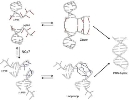
Part 2. Dynamics of ()/(+)PBS annealing reaction and its promotion by the HIV-1 nucleocapsid protein using fluorescent isomorphic and environment-sensitive nucleoside analogs ... 123
3.2.1. Full mechanism of ()/(+)PBS annealing revealed by site-selective labeling of ()PBS by thienyl-3-hydroxychromone nucleoside analog ... 123
Publication 2. Full mechanism of ()/(+)PBS annealing and its promotion by the HIV-1 nucleocapsid protein revealed by site-selective fluorescence labeling ... 131
3.2.2. Photophysical characterization of the isomorphic guanosine surrogate thienodeoxyguanosine as a potential G7 mimic in ()PBS ... 133
Publication 3. Tautomers of a fluorescent G surrogate and their distinct photophysics provide additional information channels ... 137
3.2.3. Monitoring the dynamics and conformations of the G residue in ()PBS stem-loop and ()/(+)PBS duplex substituted by thienodeoxyguanosine and their side-by-side comparison with 2-aminopurine ... 139
Publication 4. Conquering 2-aminopurine’s deficiencies: highly emissive isomorphic guanosine surrogate faithfully monitors guanosine conformation and dynamics in DNA .... 145
Chapter 4. Conclusions and Perspectives ... 147
Résumé ... 151
4
This thesis work is an important part of my life, a real adventure and challenge that will leave many-many bright memories. It would never be possible without the answer of Professor Yves Mély one day to my naive email sent from Ukraine. I am sincerely grateful to you for supervising my work, your continuous guidance and encouragement given to me along the whole period in the Lab, and for the motivation boosted hundred times after each our meeting. Thank you for teaching me, dedicating your precious time and your understanding that finally conducted us to reaching the objectives of the work. I’m grateful to my co-supervisor Professor Olga Zaporozhets for your wisdom and warm support all over the PhD period.
I’d like to thank the jury members Dr. Carine Tisné, Dr. Marie-Paule Teulade-Fichou, Dr. Marc Ruff, and Dr. Eric Deprez for accepting my request to evaluate this work.
I’m deeply thankful to Dr. Guy Duportail for your care and advices, to Dr. Andrey Klymchenko for your help and support, to Dr. Hugues de Rocquigny, Dr. Manuel Boutant and Dr. Eléonore Real for consulting me in complex biological issues, to Dr. Christian Boudier, Dr. Nicolas Humbert, Dr. Ludovic Richert, Dr. Julien Godet, Dr. Frederic Przybilla, Dr. Pascal Didier, Dr. Youri Arntz, Dr. Halina Anton, Dr. Andreas Reisch and Dr. Mayeul Collot for scientific discussions and help. Moreover, I’d like to thank Dr. Jean-Luc Darlix for careful reading of my manuscript introduction. I have my thanks to Marlyse Wernert, Ingrid Barthel, Ludovic Four and Michel Morciano for your availability and help.
I thank all my colleagues from the Laboratory of Biophotonics and Pharmacology for a friendly atmosphere in the Lab, useful discussions and happy moments. I highly appreciate the time spent with Lesia, Iryna, Katya, Liliana, Redouane, Rajhans, Sasha, Bogdan, Manu, Kamal, Yosuke, Krishna, Vika, Alex, Taras, Evgen, Natalia, Avisek, Sarwat, Nedal, Hassan, Wassem and others. Thank you guys for creating pleasant atmosphere and good memories of the PhD life!
Many thanks to our numerous collaborators, namely Professor Vasyl Pivovarenko for your “parental” attitude to me and our fruitful work together, to Dr. Dmytro Dziuba, Dr. Nicolas Barthes, Dr. Benoit Michel and Dr. Alain Burger from Nice, to Dr. Dongwon Shin and Professor Yitzhak Tor from the US, to Dr. Mattia Mori and Dr. Roberto Improta from
5
Strasbourg. It was a pleasure to work together on different projects and to learn a lot from it. Thank you Iuliia for being my neighbor during the most part of the PhD period, for your encouragement, scientific discussions and active leisure time together. I highly appreciate the support and care of my friends here and in Ukraine. Thank you for being next to me in more or less good moments.
Finally, I’m grateful to my husband Sergii and my beloved family in Kyiv for your patience, enormous support, and your love that I’m feeling all the time.
Last but not the least, I want to acknowledge Prof. Serge Potier, Mélanie Muser and Ecole Doctorale des Sciences de la Vie et de la Santé for providing me the financial support; the Dean of the Faculty of Pharmacy Professor Jean-Pierre Gies, Professor Eric Marchioni, the Faculty of Pharmacy and the University of Strasbourg for giving me an opportunity to gain the teaching experience and finalize my thesis manuscript in parallel.
Thank you very much! Marianna Sholokh
6 AIDS acquired immunodeficiency syndrome ALADAN/DANA
6-(2-dimethylaminonaphthoyl) alanine ANS 1,8-anilinonaphtalene sulfonate 2Ap 2-aminopurine
AZT azidothymine
BDF base discriminating fluorescent probe BMFC 6-bromomethyl-2-(2-furanyl)-3-hydroxychromone CA capsid protein CTD C-terminal domain dG deoxyguanosine 4-DAPA 4-(N,N-dimethylamino)-phtalimide propionic acid
DIS dimerization initiation sequence DLS dimer linkage structure
4-DMAP 4-N,N-dimethylaminophtalimide 4-DMN
4-N,N-dimethylamino-1,8-naphtalimide
6-DMN 6-N,N-dimethylamino-2,3-naphtalimide
DNA deoxyribonucleic acid dNTP deoxynucleotide triphospate
ESCRT endosomal sorting complex required for transport
ESIPT excited-stated intramolecular proton transfer
FCS fluorescence correlation spectroscopy FITC fluorescein isothiocyanate
FLIM fluorescence lifetime imaging microscopy
FRET Förster resonance energy transfer G guanine
GFP green fluorescent protein gRNA genomic ribonucleic acid
HAART highly active antiretroviral therapy 3HC 3-hydroxychromone
3HCaa 2-furyl-3-hydroxychromone amino acid analog
3HCnt 2-thienyl-3-hydroxychromone nucleoside analog
3HF 3-hydroxyflavone
HIV-1 human immunodeficiency virus type 1 HTLV human T-cell lymphotropic virus IC internal conversion
ICS intersystem crossing IN integrase
IRES internal ribosome entry segment
MA matrix protein
3MI 3-methyl-isoxanthopterin 6MI 6-methyl-isoxanthopterin mRNA messenger RNA Myr myristic acid moiety NA nucleic acid
NC nucleocapsid protein
NMR nuclear magnetic resonance NPC nuclear pore complex NTD N-terminal domain ODN oligonucleotide
PAH polycyclic aromatic hydrocarbons PAS primer activation signal
PBS primer binding site Phe phenylalanin
PIC pre-integration complex
PIP2 phosphatidylinositol-(4,5)-biphosphate phospholipids PPT polypurine tract PRODAN 6-propionyl-2-(dimethylamino)naphthalene PR protease 2PyG 8-(2-pyridyl)-2ʹ-deoxyguanosine QD quantum dot
QY fluorescence quantum yield RNA ribonucleic acid
RRE Rev response element RT reverse transcriptase
RTC reverse transcription complex SD splice donor site
SL stem-loop SP spacer peptide
()/(+)ssDNA minus/plus strong-stop DNA SU surface protein
TAMRA carboxytetramethylrhodamine TAR transactivation response element Tat transcription trans-activator
th G thienoguanosine tz G isothiazologuanosine TM transmembrane protein TMR tetramethylrhodamine Trp tryptophan
tRNA transfer RNA UTR untranslated region
8VdG 8-vinyl-2ʹ-deoxyguanosine Vif viral infectivity factor
Vpr virus protein R ZF zinc finger
8
Part 1. Overview on the human immunodeficiency virus type 1 (HIV-1)
1.1.1. Discovery of HIV-1
The story of HIV began more than 35 years ago, when the first clinical observations of a new alarming epidemic were made in the United States in 1981. The only known human retrovirus at that time was the Human T-cell lymphotropic virus (HTLV) that causes transformation of the T cells. However, the HTLV was not assumed as an etiological agent causing AIDS (Acquired Immunodeficiency Syndrome), and in 1983 the group of Luc Montagnier, Françoise Barré-Sinoussi and Jean-Claude Chermann from the Institut Pasteur in Paris first reported the isolation of a new human retrovirus (at that time known as LAV, lymphoadenopathy virus or HTLV-III, human T-cell lymphotropic virus type III) (Barré-Sinoussi, Chermann et al. 1983; Popovic, Sarin et al. 1983) possibly responsible for the AIDS disease. In 1984 Gallo et al (Gallo, Salahuddin et al. 1984) and Levy et al (Levy, Hoffman et al. 1984) published reports that confirmed the identification of the causative agent of AIDS. Françoise Barré-Sinoussi and Luc Montagnier in 2008 were awarded the Nobel Prize in Physiology and Medicine for the discovery of the HIV.
HIV is a lentivirus, a member of the large family of retroviruses. CD4+ T lymphocytes and cells of the monocyte/macrophage lineage are the predominant targets of the virus (Dalgleish, Beverley et al. 1984; Klatzmann, Champagne et al. 1984; Silvin and Manel 2015). Less abundant cell populations such as dendritic cells can also be infected by HIV-1 (Altfeld, Fadda et al. 2011). Macrophages and TCD4+ cells constitute the major reservoirs for HIV-1 virus and are involved in various aspects of the AIDS disease (Venzke and Keppler 2006; Pan, Baldauf et al. 2013). When the TCD4+ cells number declines below a critical level, the cell-mediated immunity is lost, and infected persons become progressively susceptible to opportunistic infections. HIV-1 is transmitted by sexual contacts, drug addiction, through blood and from infected mother to child.
HIV-1 is characterized by extensive and dynamic genetic diversity, generating several molecular subtypes as well as recombinant forms. HIV-1 phylogenetic classifications are currently based either on nucleotide sequences derived from multiple subgenomic regions (gag, pol or env) of the same isolates or on the full length genome sequence analysis. Three major phylogenetic groups of the HIV-1 can be distinguished: group M (main), group O (outlier), and group N (non-M/non-O). The majority of infections worldwide belongs to the
9
group M and can be further subdivided into 10 subtypes (from A to K). Among them, the subtypes A (12% of infections), B (10% of infections), and C (50% of infections) are the most prevalent HIV-1 genetic forms worldwide. There is a specific geographic distribution for the subtypes, except sub-Saharan Africa, where almost all subtypes and recombinant forms have been detected. For instance, viruses of the subtype A are spread in central and eastern Africa as well as in eastern Europe; subtype B has been found in central and western Europe, North and South America, Australia, Southeast Asia, northern Africa, and Middle East. The subtype C is responsible for the HIV-1 infections mainly in southern Africa and India (Buonaguro, Tornesello et al. 2007).
The origin of HIV-1 was reported to be the cross-species transmission of the simian immunodeficiency virus SIVcpz from the chimpanzee subspecies Pan troglodytes troglodytes to humans likely due to direct exposure to animal blood upon hunting, butchering or consumption of raw meat (Gao, Bailes et al. 1999; Keele, Van Heuverswyn et al. 2006; Buonaguro, Tornesello et al. 2007).
In 1986 the second causative agent of AIDS, called HIV-2, was isolated by Luc Montagnier’s group (Clavel, Guetard et al. 1986; Guyader, Emerman et al. 1987). HIV-1 and HIV-2 have similar genomic organization, replication cycle, modes of transmission and both results in AIDS. However, HIV-2 is characterized by a much lower transmissibility and reduced progression of AIDS. Geographically, HIV-2 is predominantly present in western Africa and more recently in southern India (Nyamweya, Hegedus et al. 2013).
According to the recent data of UNAIDS, in 2014 36.9 million people have been living with HIV-1, about 2 million people have been infected and further 1.2 million people died of AIDS. These statistics demonstrate that HIV/AIDS still remain a crucial global health issue. However, the new HIV infections has fallen by 35% since 2000, and AIDS-related deaths decreased by 42% since the peak in 2004 mainly due to the development of efficient therapies, and early diagnostics. These achievements further encourage scientists and medicine workers all over the world to consolidate their research and finally eradicate the HIV.
1.1.2. Structure of the HIV-1 viral particle
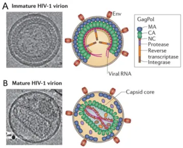
The mature HIV-1 viral particle has a spherical shape of 110 – 130 nm in diameter (Figure 1.1.1). The internal part of the viral particle is composed of two copies of homologous
10
single-stranded genomic RNA (gRNA) extensively coated by the nucleocapsid protein (NC). About 1500-2000 NC molecules are present in the viral core and interact with the gRNA via electrostatic, H-bonding and π-stacking interactions (Darlix, Lapadat-Tapolsky et al. 1995; Muriaux and Darlix 2010). The viral enzymes, namely reverse transcriptase (RT), protease (PR) and integrase (IN) are also enrolled in the viral core called capsid. Additionally, three accessory proteins Vpr (100-200 molecules), Vif and Nef are found in the viral particle (Darlix, Lapadat-Tapolsky et al. 1995; Darlix, Garrido et al. 2007; Muriaux and Darlix 2010). The capsid is composed of the capsid protein (CAp24) surrounded by a viral shell of the matrix protein (MAp17). The latter is bound to the outer phospholipid envelope originating from the host cell membrane. In the envelope a small number of viral glycoproteins in trimeric form are anchored, namely the surface SUgp120 and the transmembrane TMgp41 proteins (Ganser-Pornillos, Yeager et al. 2008; Muriaux and Darlix 2010; Sundquist and Krausslich 2012; Campbell and Hope 2015).
Figure 1.1.1. Schematic illustration of the mature HIV-1 virion (Adapted from Godet 2010). The viral capsid formed of the capsid protein CAp24 (red disks) enrolls two copies of viral RNA coated by the nucleocapsid protein NC and condensed with the viral enzymes. The conical capsid is surrounded by the matrix protein MAp17 (light blue spheres), bound to the envelope anchored with viral glycoproteins SUgp120 and TMgp41 (in orange).
1.1.3. Genetic organization of the HIV-1 genome
Viral RNA
The HIV-1 genome is packaged in the viral particle in the form of a 60S complex containing two tightly associated 9200 nucleotide-long molecules of the full length
single-11
stranded viral RNA with a 5' cap and 3' polyadenylation signal (Yedavalli and Jeang 2010). The dimeric RNA results from non-covalent interactions involving the 5ʹ dimer linkage structure (DLS), the dimerization initiation sequence (DIS) as well as many other contacts. During the reverse transcription step, the gRNA serves as a template for reverse transcriptase to generate a linear viral DNA duplex flanked with terminal redundant long terminal repeats (LTR).
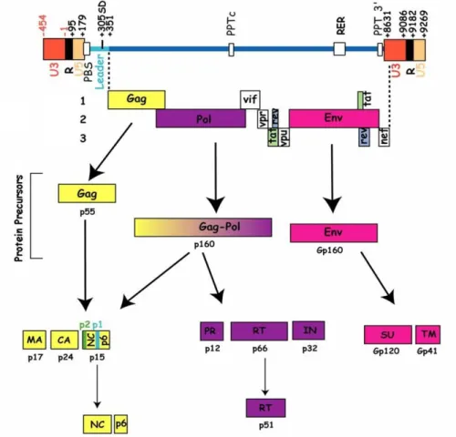
The HIV-1 genome contains two types of genetic elements: coding and non-coding. The coding elements are three genes, namely gag, pol and env responsible for structural, enzymatic and envelope proteins, and the genes responsible for the regulatory (Tat, Rev) and accessory proteins (Nef, Vif, Vpr and Vpu) (Figure 1.1.2).
Figure 1.1.2. HIV-1 genetic organization. Three major genes: gag, pol and env, and genes encoding regulatory and accessory proteins are present in the viral RNA. Adapted from Goldschmidt 2004.
The 5ʹ-untranslated region (UTR) is highly conserved in the HIV-1 RNA genome. It adopts complex structures involved in the key steps of the viral replication process. It is composed of the R, U5, PBS, and the leader regions. Recent NMR studies show that the 5ʹ-UTR exists in equilibrium between two structures: one that promotes translation of the gag
12
gene by the internal ribosome entry segment (IRES) mechanism (Brasey, Lopez-Lastra et al. 2003), and the second that promotes dimerization of the gRNA (Berkhout and Van Wamel 2000; Berkhout, Ooms et al. 2002; Abbink and Berkhout 2003; Lu, Heng et al. 2011; Deforges, Chamond et al. 2012; Heng, Kharytonchyk et al. 2012) (Figure 1.1.3). The conformational change allows switching between these two functions during virus assembly.
Figure 1.1.3. Secondary structures of the 5ʹ-UTR of the HIV-1 gRNA (NL4.3 strain). (A) Secondary structure that promotes translation, where the unique 5ʹ-end (U5) region base pairs with the dimerization initiation site (DIS) sequence; (B) Secondary structure associated with U5:AUG base pairing that exposes the DIS and high-affinity NC binding sites in order to promote the gRNA dimerization and packaging. The primer binding site (PBS) sequence is represented in blue, the primer activation signal (PAS) in red, the A-rich region in green, and the C-rich region in purple. The polyadenylation signal, the nucleotides involved in the dimerization signal (SL1), and the starting codon for the translation of gag are in orange. Adapted from Sleiman, Goldschmidt et al. 2012.
The region R (redundant/repeat) counts over a hundred nucleotides and is present as two identical copies at the 5ʹ and 3ʹ ends of the viral RNA. It contains two stem-loops: the transactivation response element (TAR) involved in regulation of the synthesis of the proviral DNA via the Tat protein (Peterlin, Luciw et al. 1986), and a polyA hairpin containing the AAUAAA polyadenylation signal (Feng and Holland 1988; Berkhout, Klaver et al. 1995). This motif is functional only at the 3ʹ-end of the gRNA where it allows cleaving the viral RNA and addition of the poly-A tail (Klasens, Thiesen et al. 1999).
13
The unique U5 region is located at the 5ʹ-end of the gRNA and is the first part of the genome to be reverse-transcribed. U5 as well as the U3 unique region contain sequences involved in integration of the proviral DNA into the host cell DNA.
The primer binding site (PBS) region, immediately following the U5 region, is an 18 nucleotide-long sequence strictly complementary to the 18 3ʹ-terminal nucleotides of tRNALys3 (G59-A76). PBS acts as a primer for the HIV-1 reverse transcription process.
The leader region is located between PBS and the starting codon for gag translation and is composed of four stem-loops (SL):
SL1 contains the primary dimerization initiation site (DIS) that promotes gRNA dimerization (Paillart, Marquet et al. 1994; Skripkin, Paillart et al. 1994; Gotte, Li et al. 1999) and may be linked to the gRNA packaging (Russell, Liang et al. 2004). It interacts with the U5 region of the gRNA and promotes translation of the gRNA (Figure 1.1.3A);
SL2 contains the splice donor site (SD) critical for splicing of the viral RNA for the synthesis of the viral accessory proteins (Purcell and Martin 1993; Oreilly, Mcnally et al. 1995);
SL3 is one of the main determinants of viral RNA encapsidation ( encapsidation signal), a critical step of HIV-1 assembly (Paillart, Shehu-Xhilaga et al. 2004; Lu, Heng et al. 2011);
SL4 contains an AUG initiation codon of translation. The AUG base pairs with the residues of the U5 region that promotes packaging of gRNA (Figure 1.1.3B).
The non-coding polypurine tracts 3ʹ PPT and cPPT are purine-rich sequences resistant to RNase H activity of RT that serve as primers for synthesis of the plus-strand DNA [(+)ssDNA] towards the 3ʹ end of gRNA.
The Rev response element (RRE) is a ~ 350 nucleotide-long highly structured element embedded in the env gene in unspliced and singly spliced viral RNA transcripts. RRE is the binding site of the HIV-1 Rev protein, a ~ 13 kDa accessory protein expressed during the early stage of the virus replication. After translation, Rev enters the nucleus, binds the RRE, and recruits cellular proteins to form larger complexes that are exported from the nucleus. Once in the cytoplasm, the complexes dissociate giving the unspliced and singly-spliced viral
14
RNAs that are packaged into the new virions or translated into the viral structural proteins and enzymes, respectively (Rausch and Le Grice 2015).
Proviral DNA
The HIV-1 proviral DNA encodes for three polyprotein precursors: Gag (Pr55gag), GagPol and Env, whose cleavage during the maturation step results in mature infectious virions. Gag and GagPol are cleaved by viral PR after virion assembly, while Env is cleaved by the cellular protease during its trafficking from endoplasmic reticulum to plasma membrane (Adamson and Freed 2007). The viral DNA contains also two LTRs, the non-coding regions involved in the integration of viral DNA and transcription of viral RNA.
Gag gene encodes the 55 kDa polyprotein precursor Pr55Gag that forms the viral particle. It contains the matrix protein p17 (MA), capsid protein p24 (CA), nucleocapsid protein p7 (NC), p6 domain, and two spacer peptides, SP1 and SP2 (Freed 2015) (Figure 1.1.4).
Figure 1.1.4. Structure and major functions of Pr55Gag polyprotein precursor: N-terminal myristic acid moiety (Myr) is covalently attached to the matrix domain (MA), capsid domain (CA), spacer protein 1 (SP1), nucleocapsid (NC), spacer protein 2 (SP2) and p6 domain. Adapted from Freed 2015.
The MA is folded into a highly globular structure composed of five α-helices and a three-stranded mixed β-sheet (Hill, Worthylake et al. 1996; Massiah, Starich et al. 1994). A number of basic residues at the top of MA allow the protein to interact electrostatically with the negatively charged phospholipids of the inner leaflet of the plasma membrane. Additionally, the myristic acid moiety (Myr) is inserted into the lipid bilayer in order to stabilize the Gag-membrane attachment during the assembly step. Moreover, MA assists the incorporation of Env glycoproteins into the forming virions (Alfadhli and Barklis 2014; Freed 2015).
15
The CA consists of two domains: a 150 amino acid-long N-terminal domain (CANTD)
and an 80 amino acid-long C-terminal domain (CACTD) connected by a flexible linker
(Campbell and Hope 2015). When assembled, CANTD is located on the outer surface of the
capsid core and CACTD is oriented towards the interior of the capsid. A proline-rich loop of
the CANTD is involved in binding of the host protein cyclophilin A (CYPA) that may help to
protect viral capsid against the antiviral innate immune defenses during virus entry (Gamble, Vajdos et al. 1996; Freed 2015). The CACTD promotes Gag-Gag multimerization as a result of
CA-CA interactions during viral assembly (Briggs and Krausslich 2011), and contains a highly conserved major homology region (MHR) important for stabilization of Gag oligomers associated to plasma membrane (Tanaka, Robinson et al. 2015).
The NC is a 55 amino acid protein with two conserved zinc fingers. NC serves as a nucleic acid (NA) chaperone, notably during the gRNA reverse transcription. As a part of Pr55Gag, NC is critical for recognition and selection of the viral RNA during viral assembly and participates in RNA encapsidation. The structure and functions of NC will be discussed in details below.
The spacer proteins SP are located upstream of NC domain in Gag (SP1) and between NC and p6 (SP2) (Figure 1.1.4). The SP1 is involved in Gag assembly (Datta, Temeselew et al. 2011), while the importance of SP2 is less defined. It is believed to be dispensable for virus maturation and viral infectivity (de Marco, Heuser et al. 2012).
The p6 domain is largely unstructured (Fossen, Wray et al. 2005) and was shown to recruit cellular ESCRT (endosomal sorting complex required for transport) machinery to facilitate viral budding (Votteler and Sundquist 2013).
Pol gene encodes a 160 kDa polyprotein Pr160GagPol, synthesized as a result of a programmed frameshifting event during the translation of Gag-encoding viral mRNA that brings pol sequence in the same reading frame as gag (Jacks, Power et al. 1988). It is an interesting fact that during HIV-1 replication, a large number of Gag molecules must be generated to serve as precursors to the structural proteins of the virions, while the enzymes encoded by pol gene are needed in smaller amount. The ribosomal frameshifting event occurs at a frequency of 5-10% during translation of Gag or Gag-Pol mRNA resulting in a Gag/Gag-Pol polyprotein ratio of around 20:1 in cell. Therefore, to express Gag protein at high levels in comparison with the pol-encoded proteins, the virus uses the same initiation codon in the same mRNA to express gag and pol genes. Translation of these RNAs leads to the synthesis
16
of a fusion protein that is usually called Gag-Pol precursor (Swanstrom and Wills 1997). Thus, Pr160GagPol polyprotein contains MA, CA, NC domains from Gag, and three viral enzymes, namely RT, PR and IN.
RT is a heterodimer composed of the two polypeptide chains p66 (66 kDa, 560 amino acids) and p51 (51 kDa, 440 amino acids). The p66 contains catalytically active N-terminal DNA polymerase domain and C-terminal RNase H domain. When two p66 chains dimerize, viral protease cleaves the RNase H moiety of one chain to produce a stable p66/p51 heterodimeric RT (Das and Arnold 2013). RT uses viral gRNA as a template and a host-cell tRNA as a primer to synthesize the minus-strand DNA, producing an RNA-DNA hybrid. This complex becomes a substrate of RNase H domain of RT, which cleaves the RNA strand at numerous points producing short RNA segments hybridized to the nascent DNA. Among these RNAs, two specific purine-rich sequences PPTs play roles of primers that initiate synthesis of the plus-strand DNA, ultimately synthesizing double-stranded DNA viral genome. Then RNase H removes the tRNA and PPT primers from minus- and plus-strand DNAs, respectively.
PR is a 99 amino acid aspartyl protease that functions as a dimer with an active site located at the dimer interface (Sundquist and Krausslich 2012). PR triggers virus maturation by cleaving Gag and GagPol polyproteins at specific sites to produce functional proteins. Protease-mediated Gag and GagPol cleavage is activated during virus assembly that is accompanied by significant changes in the virion morphology (Figure 1.1.5). In the immature virion, Gag molecules are assembled in a radial way. Following protease cleavage, the newly processed proteins, namely MA, CA, SP1, NC, SP2 and p6 reassemble to form the distinct layers of the mature virion. MA remains bound to the inner viral membrane, NC coats viral RNA, and CA assembles into a conical capsid to envelope the ribonucleoprotein complex.
17
Figure 1.1.5. HIV-1 immature (A) and mature (B) virions. Representative central sections from the electron cryotomograms together with the schematic illustrations of an immature and a mature
HIV-1 particles. Adapted from Freed 2015.
IN is a 32 kDa protein, encoded at the 3’ end of pol gene, composed of three structurally and functionally different domains: (1-51) dimeric N-terminal domain containing an HH-CC Zn-binding motif (Cai, Zheng et al. 1997); (52-212) catalytic core domain containing endonuclease and polynucleotidyl transferase sites; (220-288) C-terminal domain responsible for DNA binding and oligomerization during the integration process (Zheng, Jenkins et al. 1996; Chen, Krucinski et al. 2000; Li, Xuan et al. 2015). IN catalyzes the 3′- processing step (cleavage of two nucleotides from the two 3′ ends of the double-stranded viral DNA) and the strand transfer reaction resulting in the insertion of the proviral DNA into the host cell DNA.
Env gene encodes for a polyprotein precursor Pr160Env, whose cleavage product is the Env trimer. It is composed of three copies of non-covalently associated heterodimers of gp120 surface protein (SU) that interact with cellular receptors, and gp41 transmembrane protein (TM) necessary for fusion between the viral and cellular membranes.
The proviral DNA also encodes regulatory (Tat and Rev) and accessory (Nef, Vif, Vpr, Vpu) proteins responsible for viral pathology (Frankel and Young 1998; Malim and Emerman 2008).
18
1.1.4. Virus replication cycle
The HIV-1 viral life cycle can be divided into two main phases: from entry to proviral DNA integration (early) phase and post-integration (late) phase.
The early phase of the HIV-1 infection is initiated by binding of the viral envelope glycoproteins to the CD4 receptor and chemokine co-receptor 5 (CCR5) or C-X-C chemokine receptor 4 (CXCR4) on the host cell surface (Figure 1.1.6). Following viral attachment, fusion between the viral and host cell membranes occurs and results in a release of the HIV-1 conical capsid into the cytoplasm of the infected cell. There, it is believed to be present as a large ribonucleoprotein structure called the reverse transcription complex (RTC), where reverse transcription occurs resulting in synthesis of the proviral DNA (Mougel, Houzet et al. 2009). The RTC is thought to contain gRNA coated with NC and other components such as MA, CA, Vpr as well as viral enzymes RT and IN. The RTC enrolled by the capsid core uses the microtubule network of the host cell to traffic towards the nucleus. At this stage the capsid core disassembly may occur, though its exact timing and location in the cell are debated (Hulme, Perez et al. 2011; Ambrose and Aiken 2014). Then, the proviral DNA crosses the nuclear envelope and enters the nucleus in the form of the pre-integration complex (PIC). The PIC enters into the nucleus through the nuclear pore complex (NPC) which is composed by multiple copies of the nucleoporins embedded in the double layer of the nuclear membrane (Di Nunzio 2013). The viral partners involved in the nuclear entry are currently under investigation but the HIV-1 CA was evoked as one of the main viral components in this step (Campbell and Hope 2015). Following nuclear import, the proviral double-stranded DNA is inserted into the host cell chromosome.
The integration process is catalyzed by IN and consists of two steps. First, IN binds to a short sequence at each end of the LTR of the proviral DNA and catalyzes the endonucleotide cleavage called 3ʹ-processing reaction leading to a removal of two nucleotides from each of the 3ʹ ends of LTR. This process is conducted by IN in dimeric form and takes place in the cytoplasm of the infected cells (Guiot, Carayon et al. 2006). Next, the processed DNA is used for the strand transfer reaction leading to the covalent insertion of the viral DNA into the host genome. The strand transfer is a series of phosphodiester transesterification reactions catalyzed by integrase, in which the 3′ OH-groups of the processed viral DNA ends attack a pair of phosphodiester bonds in the target DNA. The strand transfer occurs in the nuclei and is believed to require IN in its tetrameric form (Li, Mizuuchi et al. 2006). The integrated
19
provirus is responsible for the expression of viral proteins and gRNA necessary for the synthesis of the new infectious virions.
Figure 1.1.6. Early phase of the HIV-1 replication cycle. It includes binding of the viral envelope to cell receptors, fusion of the viral and cellular membranes, reverse transcription of the viral genome, nuclear import and integration of the proviral DNA. Env, envelope glycoprotein; MA, matrix protein; CA, capsid protein ; NC, nucleocapsid protein (Campbell and Hope 2015).
During the late phase of HIV-1 replication (Figure 1.1.7), viral RNAs are synthesized and exported from the nucleus to the cytoplasm where the synthesis of viral proteins occurs. Translation of viral RNAs produces Gag and GagPol polyprotein precursors, Env glycoproteins, as well as regulatory and accessory viral proteins.
Assembly of the HIV-1 particles is initiated by the trafficking of Gag to the site of assembly – the inner leaflet of plasma membrane. During this trafficking, molecules of Gag interact with viral RNA acting as a scaffold for Gag oligomerization (Chen, Nikolaitchik et al. 2009; Briggs and Krausslich 2011; Lu, Heng et al. 2011; Kuzembayeva, Dilley et al. 2014).
20
The process of Gag oligomerization is a series of homotypic interactions involving CA, MA and SP1 regions (Hill, Worthylake et al. 1996; Datta, Temeselew et al. 2011; de Rocquigny, El Meshri et al. 2014; Datta, Clark et al. 2016). Recently, it has been established that Gag-Gag oligomerization takes place in the cytoplasm and that NC domain plays a key role in this process (El Meshri, Dujardin et al. 2015). As deletion of NC strongly reduces binding of Gag to NAs, its role is likely related to its ability to form with NAs a scaffold for further Gag oligomerization. This scaffolding results in close packaging of Gag proteins that will next bind the plasma membrane. When oligomers progressively assemble, they likely grow in size during the trafficking from the cytoplasm toward the membrane. Other investigations suggest that a relatively small number of Gag molecules may contact gRNA in the cytosol, then move to the membrane, where several thousand additional Gag molecules localize (Jouvenet, Simon et al. 2009; Kutluay and Bieniasz 2010; Lu, Heng et al. 2011).
At the plasma membrane Gag – gRNA oligomers bind to the negatively-charged phosphatidylinositol-(4,5)-biphosphate (PIP2) phospholipids of the inner leaflet of plasma
membrane through its MA domain. This binding is ensured specifically by the myristic acid moiety of Gag that stabilizes Gag-membrane association (Ono, Ablan et al. 2004; Saad, Miller et al. 2006). Following their localization at the plasma membrane, Gag, full-length viral RNA and GagPol assemble into immature viral particles. Before packaging into virions, gRNA undergoes the process of dimerization. Previously, it has been hypothesized that the two copies of viral RNA dimerize in the cytoplasm, then the dimers in complex with Gag move to plasma membrane (D'Souza and Summers 2005; Jouvenet, Simon et al. 2009; Moore, Nikolaitchik et al. 2009; Jouvenet, Laine et al. 2011; Lu, Heng et al. 2011; Keane, Heng et al. 2015). Recently, it has been suggested that gRNA dimerizes at the plasma membrane and Gag plays a major role in this process (Chen, Rahman et al. 2016).
After incorporation of the Env glycoproteins into the assembling particles, the p6 domain of Gag recruits the cellular ESCRT machinery required for particle release. The PTAP domain of p6 binds to the tumor susceptibility gene 101 (GST101) of the ESCRT-I multiprotein complex. The LYPX domain of p6 binds to the ALG2-interacting protein X (ALIX), while the ESCRT-III and vacuolar protein sorting 4 (VPS4) complexes also participate to the virion release.
The immature particles are produced by budding during which a cleavage of Gag and GagPol polyproteins by viral PR occurs (Briggs, Simon et al. 2004; Pettit, Lindquist et al.
21
2005; Mougel, Houzet et al. 2009). Activation of PR may occur already during the assembly step, as evidenced by the presence of mature CA, MA and NC proteins in the cytoplasmic extracts of infected cells (Mougel, Houzet et al. 2009) and proofs of reverse transcription in budding virions (Trono 1992; Chamontin, Rassam et al. 2015).
Following budding, new virions are released and undergo the maturation step, where CA spontaneously assembles into a cone, which contains the HIV-1 genome, NC, viral replicative enzymes and several accessory proteins. The maturation process leads to the formation of an infectious virus.
Figure 1.1.7. Late phase of the HIV-1 virus life cycle. It includes synthesis of viral RNAs, their export from the nucleus to the cytoplasm; translation of viral RNAs and production of Gag (that contains matrix MA, capsid CA, nucleocapsid NC and p6 domains), GagPol (that contains MA, CA, NC, PR, RT and IN domains), envelope Env glycoproteins (gp120 and gp41), regulatory and accessory
22
proteins; trafficking of Gag, GagPol and Env to plasma membrane; assembly of Gag and GagPol at the plasma membrane; encapsidation of viral RNA; incorporation of viral Env glycoproteins; ESCRT recruitment and budding of the new virions; release and virus particle maturation (Freed 2015).
1.1.5. Antiretroviral therapies
The extension of knowledge about the HIV-1 has led to an enormous progress in the development of antiretroviral drugs. Historically the first anti-HIV molecule was azidothymidine (AZT) (Figure 1.1.8), which was developed on the basis of the characterization of the viral RT activity (Broder, Yarchoan et al. 1985). It is a thymidine nucleoside analog acting as a RT inhibitor. Cellular kinases add three phosphate groups to its sugar moiety in order to convert AZT into an active metabolite zidovudine triphosphate. This active metabolite acts as a competitive substrate to dATP, dGTP, dCTP or dTTP and leads to termination of the chain elongation during the synthesis of proviral DNA. AZT was also a first therapeutic approach to prevent mother-to-child transmission of HIV.
Figure 1.1.8. Structure of azidothymidine (AZT), RT nucleoside inhibitor.
Due to virus mutations, resistance to the monotherapy was quickly observed. This led to the development in the early 90s of the highly active antiretroviral therapy (HAART) based on a combination of several antiviral drugs that target viral enzymes RT, PR and more recently IN. The HAART in the clinic is mostly based on RT and PR inhibitors and in some instances on IN inhibitors. Around 25 anti-HIV drugs have been approved (De Clercq 2010) by the US food and drug administration (FDA). Based on the target, these compounds can be divided into 7 categories: (1) NRTIs (nucleoside reverse transcriptase inhibitors); (2) NtRTIs (nucleotide reverse transcriptase inhibitors); (3) NNRTIs (non-nucleoside reverse transcriptase inhibitors); (4) PIs (protease inhibitors); (5) FIs (fusion inhibitors); (6) CRIs (co-receptor inhibitors); and (7) INIs (integrase inhibitors) (Scheme 1.1.1).
23 Scheme 1.1.1. Approved HIV-1 inhibitors.
Over the decade, HAART has gradually evolved from drug regiments with more than 20 pills daily in 1996, to 3 pills daily in 2003, to 2 pills daily in 2004 (Truvada®), and finally to 1 pill daily in 2006 (Atripla®) (De Clercq 2007). Atripla is the first anti-HIV drug that contains three active ingredients belonging to three different classes of the HIV inhibitors (NRTIs, NtRTIs and NNRTIs). The INIs represent the most recent advance in the search for effective and selective anti-HIV agents. Combination of several anti-HIV drugs has drastically altered AIDS from an almost uniformly fatal disease to a chronic manageable one, but however does not cure the HIV infection.
Despite the immense benefits of the HAART treatment, many issues still need to be improved. Life-long treatment is associated with emerging drug-resistance, metabolic disorders and cancers. Currently many areas of the HIV investigation require fundamental research. Further insight into the very early stages of HIV infection, the establishment of viral reservoirs and the immune responses is crucial for the development of novel therapeutic and vaccine strategies.
24
Part 2.
Nucleocapsid protein as a potential target for anti-HIV therapy
1.2.1. Structural characteristics of the nucleocapsid protein (NC)
The nucleocapsid protein is a 55 amino acid peptide resulting from the PR-mediated cleavage of the HIV-1 Gag polyprotein. During virus maturation, the initial PR cleavage liberates NCp15, a polyprotein containing NCp7, SP2 and p6 (Shehu-Xhilaga, Kraeusslich et al. 2001) (Figure 1.2.1). The subsequent cleavage results in NCp9 (71 amino acids) containing NCp7 and SP2. In fully mature viral particles, NCp7 (55 amino acids) and NCp9 forms are found in freshly collected highly infectious viral particles (Henderson, Bowers et al. 1992; Thomas and Gorelick 2008; Muriaux and Darlix 2010).
Figure1. 2.1. Proteolytic processing of HIV-1 Gag by PR. During proteolysis, NC exists in two intermediates: NCp15 and NCp9 further cleaved into the final form NCp7 (Thomas and Gorelick 2008).
NCp7 (NC) is a structural protein of HIV-1 which coats gRNA in its dimeric form. One of the functions of NC is to protect gRNA from nucleases through its NA binding and condensing properties (Tanchou, Gabus et al. 1995; Krishnamoorthy, Roques et al. 2003; Darlix, Godet et al. 2011). NC is characterized by two highly conserved CCHC zinc fingers (ZFs) connected by a short linker rich in basic residues and two unfolded N- and C-terminal parts (Figure 1.2.2A). The CCHC motifs and the basic linker are invariant in diverse HIV-1 subtypes and drug-resistant viruses (Figure 1.2.3). Sequence variations are mainly observed in residues of the N-terminal domain while many residues of the ZFs, and notably the Phe16 in ZF1 and Trp37 in ZF2, important for NA recognition and chaperone properties of the HIV-1 NC are highly invariant (Darlix, Godet et al. 2011).
25
Figure 1.2.2. Sequence and 3D structure of NC protein of HIV-1. (A) Sequence of the HIV-1 nucleocapsid protein and its structural components. Positively charged amino acids are in blue, amino acids of the hydrophobic plateau are in red, CCHC motifs are in bold. (B) 3D structure of the hydrophobic plateau at the top of NC zinc finger domain, zinc ions are in red.
Figure 1.2.3. Sequence of NC amino acids in different HIV-1 subtypes as well as in viral isolates obtained from antiretroviral naïve (top panel) and treated (bottom panel) individuals. Top panel: NC sequences from B (594 sequences) and non-B subtypes (4938 sequences) as well as the HXB2
26
molecular clone, which is often considered as a representative subtype B. Bottom panel: NC sequences from B (7351 sequences), non-B (14286 sequences), and HXB2 subtypes. Grey circles on amino acids indicate non conservative amino acids, whereas grey blocks indicate conservative amino acids. Nucleocapsid variability index graphs show the variability at each position of NC (higher value corresponds to higher amino acid variability). Black lines represent the sequences of the B subtype, whereas the grey dashed lines are the non-B subtypes (Mori, Kovalenko et al. 2015).
The central ZF domain of the NC protein is folded into a tight structure, while flanking N- and C-terminal parts are flexible and independent from the central part. Functionally, the basic N-terminal domain is mainly responsible for the NA aggregation activity of NC (Stoylov, Vuilleumier et al. 1997). In many biophysical studies, the NC(11-55) sequence, lacking the N-terminal part, is used in order to limit aggregation of the NAs. Each ZF coordinates one zinc ion through three Cys and a His residue with high affinity of 1013 – 1014 M-1 (Berg 1986; Mely, Cornille et al. 1991; Mely, de Rocquigny et al. 1996). The two zinc fingers are spatially close and show similar folding patterns (Morellet, Jullian et al. 1992; Summers, Henderson et al. 1992; Morellet, de Rocquigny et al. 1994). However, they are not functionally equivalent (Fisher, Rein et al. 1998; Guo, Wu et al. 2000; Guo, Wu et al. 2002; Fisher, Fivash et al. 2006; Zargarian, Tisne et al. 2014), and the substitution of either of the ZFs by an identical counterpart leads to a loss of the NC functions (Gorelick, Chabot et al. 1993). The 7 amino acid-long linker RAPRKKG located downstream of the ZF1 is
responsible for the proximity of the ZFs and for their transient globular structure (Mely, Jullian et al. 1994; Morellet, de Rocquigny et al. 1994; Ottmann, Gabus et al. 1995; Lee, De Guzman et al. 1998). This relative orientation is additionally stabilized by the hydrophobic and aromatic residues of the proximal (Val13, Phe16, Thr24, Ala25) and distal (Trp37, Gln45, Met46) ZFs forming a hydrophobic plateau (Figure 1.2.2B). This hydrophobic plateau is of key importance for NC functions, since single point mutations in this plateau induce a loss of viral infectivity (Demene, Dong et al. 1994; Wu, Mitra et al. 2013). Similarly, mutations of the zinc-binding CCHC residues also lead to a loss of viral infectivity through preventing the appropriate folding of the ZFs and, thus, hindering the formation of the hydrophobic plateau (Aldovini and Young 1990; Gorelick, Nigida et al. 1990; Dorfman, Luban et al. 1993; Stote, Kellenberger et al. 2004). Folded ZFs and the hydrophobic plateau at their top play a critical role in recognition, destabilization, binding and dynamic rearrangements of NAs (Tisne, Roques et al. 2003; Levin, Guo et al. 2005; Cruceanu, Urbaneja et al. 2006; Darlix, Garrido et al. 2007; Thomas and Gorelick 2008; Levin, Mitra et al. 2010; Darlix, Godet et al. 2011).
27
1.2.2. Binding of NC to nucleic acids
The role of NC in the HIV-1 virus life cycle is mainly associated with interactions with NAs. NC can bind in a non-specific or specific manner to nearly any NA sequence of 5-8 nt in length (Darlix, Godet et al. 2011). However, the affinity of such binding may vary by several orders of magnitude and depends on the nature, sequence and folding of the NAs. NC protein binds to both RNA and DNA sequences with a preference for single-stranded oligonucleotides (ODNs) (Mirambeau, Lyonnais et al. 2006; Darlix, Garrido et al. 2007). The affinity of NC for RNAs follows the order: retroviral RNA > mRNA > rRNA> poly (rA) (Karpel, Henderson et al. 1987; Coffin 1995). The relative affinity of NC for retroviral RNA, DNA and ODNs follows the order: retroviral RNA > ssDNA, dsDNA > ODNs (Darlix, Lapadat-Tapolsky et al. 1995). Non-specific and low-affinity binding of NC to NAs is the dominant binding mode that occurs through electrostatic interactions between the positively-charged NC and negatively-positively-charged NAs. At saturating concentrations, NAs are coated by NC molecules resulting in their protection from the nuclease degradation (Lapadat-Tapolsky, de Rocquigny et al. 1993; Krishnamoorthy, Roques et al. 2003).
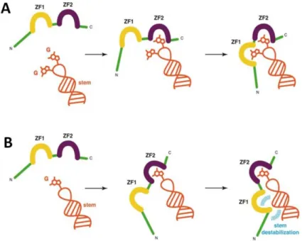
Studies performed with RNA and DNA ODNs reveal that NC binds with high affinity to sequences containing unpaired guanine residues like TG, GXG and TXG (Vuilleumier, Bombarda et al. 1999; Avilov, Piemont et al. 2008; Avilov, Godet et al. 2009). This efficient binding is ensured by the interactions of the hydrophobic plateau of NC with the nucleic bases and NA phosphate backbone (de Guzman, Wu et al. 1998; Morellet, Déméné et al. 1998; Amarasinghe, De Guzman et al. 2000; Bourbigot, Ramalanjaona et al. 2008; Spriggs, Garyu et al. 2008; Bazzi, Zargarian et al. 2011). Within the hydrophobic plateau, the highly conserved aromatic Trp37 residue plays a critical role through its π-stacking interactions with the nucleobases, notably with guanosines as was shown by 3D structures of NC/ODN complexes (de Guzman, Wu et al. 1998; Amarasinghe, De Guzman et al. 2000; Bourbigot, Ramalanjaona et al. 2008; Bazzi, Zargarian et al. 2011). Careful examination of these 3D structures revealed that each ZF is specialized in one function. ZF2 is responsible for binding
of the accessible and flexible guanines, while ZF1 either binds a second G (in case of GXG
sequence) or destabilizes the stem through its hydrophobic residues (in case of TG or TXG sequences) (Zargarian, Tisne et al. 2014) (Figure 1.2.4).
28
Figure 1.2.4. Schematic representation of the task specialization of each ZF upon binding to NAs. The NA fragment is in orange, NC zinc fingers are in yellow (ZF1) and purple (ZF2), and the rest
of the protein is in green. (A) Presence of two accessible guanines. ZF2 binds first to an accessible
guanine, while ZF1 binds the remaining guanine. The stem is not destabilized. (B) Presence of only
one accessible guanine. ZF2 binds first to this accessible guanine, while ZF1 remains free to contact the
stem via its hydrophobic platform, leading to the destabilization of the stem (in blue). Adapted from Zargarian, Tisne et al. 2014.
In addition, it was shown that NC binds to DNA and RNA sequences with opposite polarities (Bazzi, Zargarian et al. 2011). NC complexes with DNA sequences like ACGCC pentanucleotide (Morellet, Déméné et al. 1998) and ΔP()PBS (Bourbigot, Ramalanjaona et al. 2008), and RNA sequences like SL2 (Amarasinghe, De Guzman et al. 2000) and SL3 (de Guzman, Wu et al. 1998) show two distinct modes of NC binding. In complexes with DNAs, ZF2 interacts with a G residue of the ODN sequence while ZF1 contacts the residue (C or T)
upstream to G (Figure 1.2.5). In complexes with RNAs, ZF1 and ZF2 interact with two G
residues separated by a third residue (GXG). The Trp37 residue of ZF2 stacks with the G
residue located at the 3ʹ end of the GXG sequence. This different binding polarity is likely due the sugar moieties. The riboses possess an additional –OH group at the C2ʹ position as compared to deoxyriboses, which creates additional hydrophobic contacts with NC. The different binding polarities of NC to RNAs and DNAs may play a role during reverse transcription and NA recombination, since a similar behavior has been observed for a family of proteins that possess an oligosaccharide/ODN binding fold and are involved in DNA
29
recombination (Yang, Jeffrey et al. 2002; Bochkarev and Bochkareva 2004; Bazzi, Zargarian et al. 2011).
Figure 1.2.5. Schematic illustration of the two modes of NC binding to DNA and RNA sequences.
The specific binding properties of NC play a critical role in gRNA recognition and the assembly step of the viral life cycle. NC as a domain of the Gag polyprotein recognizes the encapsidation signal of gRNA among a large excess of non-viral mRNAs and spliced viral mRNAs (Aldovini and Young 1990; Cimarelli and Darlix 2002; Muriaux, Darlix et al. 2004; Muriaux and Darlix 2010). This specific recognition is required for the preferential incorporation of the viral gRNA in virions (Berkowitz and Goff 1994; Clever, Sassetti et al. 1995; McBride and Panganiban 1997; de Guzman, Wu et al. 1998; Amarasinghe, de Guzman et al. 2000; Amarasinghe, Zhou et al. 2001; Mark-Danieli, Laham et al. 2005). The encapsidation signal (Figure 1.1.3) is a ~120 nucleotide-long sequence composed of four stem-loops (SL1-SL4) located near the 5ʹ end of the genome (Lever, Gottlinger et al. 1989; Aldovini and Young 1990; Clavel and Orenstein 1990). Among these four stem-loops, SL3 is the most highly conserved sequence of the RNA site (Hayashi, Ueno et al. 1993). SL2 and SL3 are thought to be the primary determinants for the interaction with NC as they show high binding affinities to NC (de Guzman, Wu et al. 1998; Amarasinghe, De Guzman et al. 2000). According to the 3D structures of NC/SL2 and NC/SL3 complexes, the protein binds preferentially to the GXG motif of SL2 and SL3 loops. The residues of the hydrophobic plateau, notably Val13, Phe16, Thr24 (Ile24), and Ala25 interact with the 10-UG-11 residues of the 8-GGUG-11 loop of SL2, and with the 8-AG-9 residues of the 6-GGAG-9 loop of SL3 (Figure 1.2.6). The G9 residue in SL2 and the G7 residue in SL3 are involved in stacking and hydrophobic interactions with the Trp37, Gln45 and Met46 residues of the hydrophobic plateau. This mode of binding corresponds to the situation with two accessible guanines where ZF2 binds firstly to an accessible guanine, and ZF1 binds to the remaining guanine
(Figure 1.2.4A). The stems of SL2/SL3 were not shown to be destabilized. The binding constants of NC(1-55) to SL2 and SL3 were found to be similar (Kd = 110 (±50) nM and 170
30
during assembly stage of HIV-1 life cycle. Moreover, the interaction of NC with multiple stem-loop packaging elements further illustrates the ability of NC to bind various NA targets (Darlix, Godet et al. 2011; Godet, Kenfack et al. 2013).
Figure. 1.2.6. Ribbon representation of the (A) NC(1-55)/SL2 complex and (B) NC(1-55)/SL3 complex. Phe16 and Trp37 are in blue. The ODNs are in yellow. Nucleobases involved in the interaction are in green: 9-GUG-11 for SL2 and 10-GAG-12 for SL3. The remaining bases from the
loops are in red. Adapted from Bourbigot, Ramalanjaona et al. 2008.
1.2.3. Chaperone properties of NC
The NA molecules (mainly RNAs) can adopt a large number of conformations while only one is functional. The inactive conformations of NAs can be very stable and persistent. This key biological issue appears to be solved by a class of proteins called NA chaperones (Herschlag 1995). NA chaperones are non-specific NA binding proteins that rescue NAs trapped in unproductive folding states (Cristofari and Darlix 2002; Schroeder, Barta et al. 2004; Zuniga, Sola et al. 2009). NC is a member of this abundant class of proteins due to its ability to promote the rearrangement of NAs into their most stable conformations (Rein, Henderson et al. 1998; Godet and Mely 2010; Levin, Mitra et al. 2010). The chaperone activity of NC relies on three main components. The first one consists in the destabilization of the NA secondary structure mainly driven by the hydrophobic plateau at the top of the folded ZFs of NC (Bernacchi, Stoylov et al. 2002; Azoulay, Clamme et al. 2003; Egele, Schaub et al. 2004; Beltz, Clauss et al. 2005; Cosa, Zeng et al. 2006; Levin, Mitra et al. 2010; Wu, Rouzina et al. 2010). The destabilizing activity of NC strongly depends on the NA stability and
31
structure. The presence of destabilizing motifs along the NA sequence like bulges, mismatches, internal loops and terminal base pairs favors the destabilization and likely ensures a selective recognition of a limited number of NA sequences by NC during the viral life cycle (Godet and Mely 2010). The destabilization is further accompanied by an exposure and restricted mobility of the bases of the NA sequences bound to NC (Avilov, Piemont et al. 2008; Bourbigot, Ramalanjaona et al. 2008; Godet, Ramalanjaona et al. 2011; Godet, Kenfack et al. 2013). This is thought to be critical for the annealing of the complementary sequences. The second component of the NC chaperone activity consists in the promotion of the annealing of complementary sequences (Darlix, Vincent et al. 1993; You and McHenry 1994; Hargittai, Gorelick et al. 2004; Godet, de Rocquigny et al. 2006; Liu, Zeng et al. 2007; Ramalanjaona, de Rocquigny et al. 2007; Vo, Barany et al. 2009). Here, the N-terminal domain of NC rich in basic residues possesses the NA aggregation ability and is, thus, actively involved in the annealing process. The hydrophobic plateau and the two ZFs also play a role in the annealing process through activation of specific pathways (like loop-loop kissing complexes). These specific pathways are required to promote faithfully, timely and specifically the hybridization of the two complementary sequences (Godet, Ramalanjaona et al. 2011; Godet, Kenfack et al. 2013).
The third component of the NC chaperone activity consists in the fast binding and dissociation from NAs. Indirect evidence of this property has been demonstrated using single molecule stretching experiments to explore the chaperone properties of NC (Cruceanu, Gorelick et al. 2006). The efficiency in strand annealing was limited when the retroviral NC performed slow on/off binding kinetics, though it may still be capable to bind NAs with high affinity, destabilize duplexes and aggregate NAs. For example, HIV-1 NC exhibits fast on/off kinetics and is, thus, one of the most efficient NC chaperones in comparison with NCs from the Rous sarcoma virus, Moloney murine leukemia virus or HTLV-1 (Stewart-Maynard, Cruceanu et al. 2008). In contrast, the HTLV-1 NC performs poor chaperone properties, despite its strong duplex destabilizing activity, mainly due to very slow NA dissociation kinetics. The on/off binding kinetics rate is found dependent on the ZFs of NC, since mutations in the ZF domains lead to a decrease in the protein dissociation rate (Cruceanu, Gorelick et al. 2006).
Taken together, in the HIV-1 virus life cycle the chaperone activity of NC is indispensable during viral replication and assembly steps. In the present work we focused mainly on the viral replication step, so that this process will be discussed in details below.
32
1.2.4. Role of NC in HIV-1 reverse transcription
Reverse transcription process
The reverse transcription step of the HIV-1 is a complex process resulting in synthesis of a double-stranded linear proviral DNA from the single-stranded viral RNA (Arts and Wainberg 1996; Telesnitsky and Goff 1997). This process is controlled by RT enzyme and involves two obligatory strand transfers (Basu, Song et al. 2008). Schematically, it may be divided into the following steps (Figure 1.2.7).
Figure 1.2.7. Schematic representation of HIV-1 reverse transcription process (Levin, Mitra et al. 2010).
Step 1. Initiation of reverse transcription occurs when 18-nt of the cellular tRNALys3 primer (orange line) (Figure 1.2.7) hybridize to the 18-nt sequence PBS (red solid line) located at the 5ʹ end of the genome. This reaction is called the tRNA primer placement.
33
Step 2. RT enzyme promotes extension of the primer in order to synthesize the minus strong-stop DNA [()ssDNA] (blue solid line) that contains copies of the unique 5ʹ genomic sequence (U5) and repeat R regions. As the primer is extended, the RNase H activity of RT cleaves gRNA (fragments represented by short red solid lines) that has already been read. Step 3. To continue the minus-strand, the ()ssDNA is transferred to the 3ʹ end of gRNA where annealing of the complementary R sequences at the 3ʹ ends of gRNA and ()ssDNA occurs. This step is called minus-strand transfer.
Step 4. Following minus-strand transfer, elongation of ()ssDNA proceeds to reach the PBS sequence of the 5ʹ end, while RNase H degradation continue. The polypurine tracts PPT and cPPT located near the U3 are resistant to RNase H activity and initiate the synthesis of the plus-strand DNA [(+)ssDNA] towards the 3ʹ end of gRNA using newly synthesized ()ssDNA as a template.
Steps 5 and 6. When the PBS region of the plus strand is reconstituted, the formed sequence is called (+)ss DNA. RNase H removes the tRNA and PPT primers from minus- and plus-strand DNAs, respectively. A plus-strand transfer is required to complete the synthesis of viral DNA.
Step 7. Annealing of the two complementary PBS sequences at 3ʹ ends of (+)ssDNA and minus-strand DNA results in a circular DNA intermediate.
Step 8. Complete minus- and plus-strand DNAs are elongated into a linear double-stranded DNA with an LTR at each end. This proviral DNA is used for further integration step.
The NC protein assists RT during the whole reverse transcription process (Levin, Guo et al. 2005; Thomas and Gorelick 2008; Levin, Mitra et al. 2010; Darlix, Godet et al. 2011). It directs annealing of the tRNALys3 primer with the PBS, chaperones both strand transfers, ensures fidelity of the plus-strand DNA priming at two polypurine tracts (Hergott, Mitra et al. 2013), and facilitates removal of tRNALys3 primer from the 5ʹ end of the minus strand DNA (Levin 2010). Also NC assists RT in the fidelity of vDNA synthesis by providing nucleotide excision repair (Bampi, Bibillo et al. 2006).
34
tRNALys3 PBS interaction
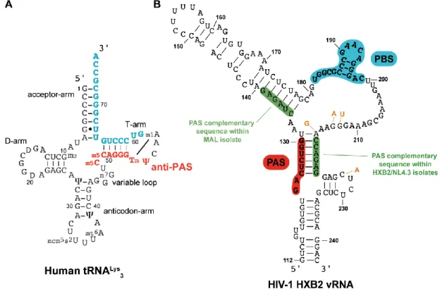
The HIV-1 reverse transcription initiation consisting in base-pairing of tRNALys3 to PBS RNA requires NC chaperone activity (Levin, Mitra et al. 2010; Darlix, Godet et al. 2011; Sleiman, Goldschmidt et al. 2012). NMR studies revealed that both basic and ZF domains are involved in the formation of the tRNA-RNA complex (Tisne, Roques et al. 2004; Barraud, Gaudin et al. 2007). The role of basic residues consists in destabilization of a few base pairs of the tRNA, while the ZFs affect the ternary interactions within the tRNA molecule. As a precondition for reverse transcription, tRNA primer, viral RNA template and RT form a productive ternary ribonucleoprotein complex under the action of NC.
Interestingly, exploration of the tRNALys3 specificity as a primer of HIV-1 identified an 8-nucleotide sequence (nucleotides 123-130, Figure 1.2.8) in the U5 region of viral RNA complementary to 48-55 nucleotides of tRNALys primer (Beerens, Groot et al. 2001; Beerens and Berkhout 2002). This sequence of the viral RNA is called the primer activation signal (PAS) and is involved in annealing with the so-called anti-PAS region of tRNALys3. Using single- molecule FRET assays it was shown that this interaction appears to be dynamic and stimulated by NC (Beerens, Jepsen et al. 2013). Therefore, the PAS/anti-PAS interaction is an additional illustration of NC chaperone activity (Sleiman, Barraud et al. 2013).
Figure 1.2.8. Secondary structures of (A) the human tRNALys3, in blue is the sequence complementary to the PBS and in red is the anti-PAS sequence; (B) PBS domain within the HIV-1 HXB2 isolate, in