HAL Id: tel-02322688
https://hal.archives-ouvertes.fr/tel-02322688
Submitted on 21 Oct 2019
HAL is a multi-disciplinary open access archive for the deposit and dissemination of sci-entific research documents, whether they are pub-lished or not. The documents may come from
L’archive ouverte pluridisciplinaire HAL, est destinée au dépôt et à la diffusion de documents scientifiques de niveau recherche, publiés ou non, émanant des établissements d’enseignement et de
FROM VISUAL MOTION PERCEPTION TO
SPATIAL COGNITION: Study of behavior and brain
activity
Anne-Lise Paradis
To cite this version:
Anne-Lise Paradis. FROM VISUAL MOTION PERCEPTION TO SPATIAL COGNITION: Study of behavior and brain activity. Neuroscience. Sorbonne Université, UPMC, 2017. �tel-02322688�
Habilitation à diriger les recherches
présentée à
L’UNIVERSITÉ PIERRE ET MARIE CURIE
Par Anne-Lise PARADIS
SPÉCIALITÉ : Biologie / Neurosciences Cognitives
FROM VISUAL MOTION PERCEPTION TO SPATIAL COGNITION
Study of behavior and brain activity
Date de soutenance : 11 mai 2017
Devant la commission d’examen formée de :
M. Olivier BERTRAND DR Inserm, Centre de Recherche en Neurosciences de Lyon Mme Nathalie GEORGE DR CNRS, Social and Affective Neuroscience Laboratory, ICM, Paris Mme Anne GIERSCH DR Inserm, CHRU Strasbourg – Rapporteur
M. Pascal MAMASSIAN DR CNRS, Laboratoire des systèmes perceptifs, ENS Paris – Rapporteur Mme Marie VIDAILHET PU-PH, UPMC - CHU Pitié-Salpêtrière Paris
M. Thomas WOLBERS Professor, German Center for Neurodegenerative Diseases (DZNE) – Rapporteur
R
EMERCIEMENTSJe souhaite tout d’abord remercier les étudiants, doctorants, jeunes chercheurs : Giorgia Committeri, Agnieszka Miskiewicz, Frédéric Benmussa, Ying Liu, Kinga Igloi, Bénédicte Babayan, Daphné Sylvestre, Jean-Etienne Bergemer, Amélie Greiner, Nicolas Traut, Bérenger Thomas, Nadine Francis, ainsi que mes collègues Christophe Gitton, Laurent Hugueville, Lydia Yahia Cherif, Jean-Didier Lemaréchal, Denis Schwartz, Romain Valabrègue, Christine Tobin, Christelle Rochefort, Grégory Sedes, Aurélie Watilliaux, et j’en oublie… dont le travail a alimenté ce document. Certain(e)s parmi eux m’ont encouragée avec conviction à soutenir cette habilitation et je les en remercie particulièrement.
Je remercie également de tout cœur les collègues avec qui j’ai co-encadré les études rapportées ici : Jean Lorenceau tout d’abord, qui m’a accueillie dans son équipe et permis de profiter de son expérience avec enthousiasme et générosité, ces années de collaboration ont été particulièrement enrichissantes ; Catherine Tallon-Baudry qui m’a montré l’exemple à plus d’un titre ; Laure Rondi-Reig enfin, qui a su me convaincre de chercher à m’amuser sérieusement, et vice-versa…
J’ai une pensée particulière pour Line Garnero qui, en m’ouvrant la porte de son laboratoire, m’a impulsé un nouvel élan : notre rencontre a été trop courte.
Last but not least, many thanks to Olivier Bertrand, Nathalie George and Marie Vidailhet for agreeing to be members of my jury, and to Anne Giersch, Pascal Mamassian and Thomas Wolbers for additionally taking on the burden of reviewing my manuscript. I hope this will provide all of us with the opportunity for fruitful discussions.
T
ABLE OF CONTENTSFOREWORDS ... 7
3D MOTION AND 3D FORM FROM 2D MOTION ... 9
Visual processing vs. attention selection ... 11
Protocol and results ... 11
Discussion ... 12
Additional unpublished data: 3D motion and form attributes in passive viewing ... 13
Interaction between form and motion attributes in 3D structure-from-motion perception ... 15
Protocol and results ... 15
Discussion ... 17
Timing of stimulus- and task-related processes ... 19
Protocol and results at sensors’ level ... 19
Time course of the sources of activity ... 20
Stimulus-driven vs. task-related activity ... 22
Further into shape perception with the use of familiar objects ... 23
Protocol and results ... 23
Anterior Temporal Pole ... 25
Study comparison ... 25
INTEGRATION AND SELECTION ... 27
Feature selection and conscious perception ... 28
Protocol and results ... 28
Intra-parietal sulcus ... 29
Feature selection ... 29
Selection vs. binding ... 31
Protocol and results ... 31
Discussion ... 32
When motion perception tells about contour integration and shape processing ... 34
Working hypotheses and protocol ... 34
Results and interpretation ... 35
NAVIGATION:SELF-MOTION,SPATIO-TEMPORAL MEMORY AND STRATEGIES ... 36
Possible role of the cerebellum in navigation ... 38
Multimodal integration ... 38
Sensory prediction ... 38
Interaction with space-coding structures ... 39
The cerebellum in hippocampus-dependent navigation strategies ... 41
Sequence-based vs. place-based navigation ... 41
Sequence learning from self-motion ... 43
Reference frames ... 48
Posterior parietal cortex ... 48
Medial occipito-temporal cortex ... 49
Medial parietal cortex: retrosplenial cortex and precuneus ... 50
ELECTROPHYSIOLOGICAL INVESTIGATION OF CEREBELLAR-FOREBRAIN COUPLING (PROJECTS) ... 51
Cerebellar-hippocampus coherence in spontaneous sequence behavior ... 52
Navigation beyond the hippocampus ... 53
Develop electrophysiological biomarkers of the healthy navigation... 54
TESTING NAVIGATION TO DIAGNOSE POSSIBLE COGNITIVE DEFICITS (PROJECTS) ... 55
Cerebellar volume in autism ... 56
Results per cerebellar sub-region ... 56
Stratification of ASD patients based on navigation abilities and cerebellar anatomy ... 59
Health record of spatial cognition ... 60
Virtual Starmaze and Navigation Analysis Tool for humans (NAT-h)... 60
Scoring assessment and development of new navigation scores ... 60
Memory and executive functions in Alzheimer’s disease ... 62
BIBLIOGRAPHY ... 63
CURRICULUM VITAE MARCH 2017 ... 68
SUPERVISED MASTER AND PHD WORK ... 70
Publications from supervised or co-supervised work ... 70
Conference abstracts from supervised or co-supervised work ... 70
PUBLICATION LIST ... 72
Peer-reviewed papers ... 72
Peer-reviewed conference papers ... 73
Invited papers ... 73
ANNEXES... 74
F
OREWORDSOpen your eyes! By simultaneously processing numerous visual cues, your brain allows you to locate yourself in the environment, recognize the surrounding objects and estimate their distance, identify people and anticipate their movements. Take a look to the left, turn your head and step forward: The visual signal rapidly changes on your retina, but the world appears stationary.
Visual motion is an amazing cue. Obviously, it gives us access to the movement of the surrounding objects, allowing us to anticipate their future position and interact with them efficiently: avoid a bicycle on the road, catch a ball, or pour tea into a cup without overflowing. Less obviously, visual motion also informs us about our own movements: the movement of our gaze in the visual scene, the movement of our body in space. Paradoxically, it is likely that the more accurately this information is processed, the more the motion sensation is suppressed, so the less we perceive it. Last but not least, visual motion also provides us with information about the 3-D layout of our environment. This can be easily realized when looking through a train window: the nearby elements of the landscape rush by us, while the more distant ones appear motionless. Those relative visual speeds are the cue revealing the underlying in-depth structure of the scene.
Since my PhD Thesis, my first interest was to understand by which processes the brain could extract depth information from visual motion, and how it could, from one same visual flow, distinguish between the motion related to the 3-D structure of the object and that related to its movement. To that end, I used stimuli devoid of shape and contour information and explored the brain regions involved in building 3-D shape information from visual motion only.
The apparent cross talk between visual motion and form information led me to question the possible interactions between the two visual features. Is there any evidence of the motion feature influencing the shape feature, or reciprocally? Using behavioural testing and brain imaging in the framework of different collaborations, I thus examined the relationships between motion and shape processing. This work has been conducted from 2001 to 2005 in the group of Jacques Droulez at the ‘Laboratoire de Physiologie de la Perception et de l'Action’ (LPPA), headed by Pr. Alain Berthoz; from 2006 to 2008, in the group of Jean Lorenceau at the ‘Laboratoire de Neurosciences Cognitives & Imagerie Cérébrale’ (LENA), headed by Dr. Line Garnero, and from to 2009 to 2012, at the Brain and Spine Institute (ICM).
In 2012, I joined Laure Rondi-Reig’s lab to explore new topics: cerebellum, navigation and memory, or how brain structures interact during self-motion to implement space cognition. I thus discovered that the cerebellum was not just a little pernicious structure aimed at masking the ventral surface of the visual cortex in fMRI and in turn I had a chance to convince my new lab partners that the visual cortex was not just a big fat structure intending to crush the cerebellum…
Given the various types of information associated with ‘visual motion’ in this document, I have tried to use specific words to disentangle the different possible meanings and better characterize the studied processes. In the following, I will thus specify 2D visual motion as a synonym for optic flow, which is the change of local contrasts on the retina due to relative movements between the eyeball and the visual scene or its elements. Moreover, I will distinguish this 2D component of the physical motion projected on the retina of the observer from the 3D motion perceived by the observer once the retinal signal has been decoded by the brain.
In the first part of this manuscript, I address how the brain reconstructs the 3D features of visual objects (3D motion and 3D shape) from the 2D visual motion projected on the retina. In this part, we consider static observers and make the assumption that visual motion is related to the surrounding visual objects only. To further isolate the activity related to the structure from motion process, we used impoverished visual stimuli providing structure information from motion inputs
only. We then studied the possible interactions arising between the different perceptive attributes extracted from this unique visual input, and further explored whether they could be mediated by stimulus-driven or task-related processing.
In a second part, I address more widely the question of feature integration and selection through some collaborative studies. In particular, I consider structure perception from the perspective of ‘visual binding’, and feature interactions from the perspective of object-based attention.
In the third part, I address the neural bases of spatial cognition and specifically how the cerebellum may contribute to process sensory inputs during navigation to disentangle self-motion information from external cues.
The last two parts shortly present the on-going projects in which I participate. They are organized in two topics: the electrophysiological investigation of cerebellum-forebrain coupling and the use of navigation tasks as a translational tool to explore cognitive deficits, with application in autistic spectrum disorders (ASD) and Alzheimer’s disease.
G
LOSSARYMT: middle temporal area; area processing visual motion in monkeys, also called V5 hMT: human MT, also called V5 or hMT/V5
hMT+/V5+: stands for hMT/V5 along with its satellite areas also sensitive to visual motion fMRI: functional Magnetic resonance Imaging
MEG: Magneto-Encephalography ASD: Autistic Spectrum Disorder
3D
MOTION AND3D
FORM FROM2D
MOTIONWith one eye shut, depth perception is severely impaired and it proves difficult to determine which pen is the closest in the pencil pot for example. However, it is possible to gain depth information again just by moving one’s head back and forth or left and right, or by rotating the pot in one’s hand. This perception of the structure from motion has a twofold interest. First, shape information is extracted from a motion signal, which reveals a link between features often considered independent. Second, the process of extracting structure from motion also leads to 3D motion information, so that two distinct perceptual features –motion and form– are extracted from a unique sensory input, which is retinal speed distribution.
The extraction of 3D structure information from 2D motion has been theoretically described as a hierarchical process divided in two main stages (Hildreth and Koch, 1987):
- optic flow characterization, through the measurement of the retinal speeds and their gradients; - recovery of the 3D layout of the scene, from a depth map relative to the observer to a 3D representation independent of the observer’s viewpoint.
By studying motion and shape perception from retinal speed distribution, it becomes possible to disentangle these two stages of perception: the analysis of the 2D visual input on one hand, and the coding of the reconstructed perceptual attributes, namely 3D motion and shape, on the other hand.
The issue is to fit this theoretical model with a neurophysiological substrate. The human retina encodes spatial and temporal variations of luminance but does not provide an actual speed distribution. In V1, most motion-sensitive cells encode ambiguous speed directions, because only motion orthogonal to a contrast edge is extracted. In area MT/V5 however, cells are able to encode 2D motion information. In addition, it was shown in monkeys that, thanks to the specific organization of their receptive fields, some MT cells are selective for the orientation in depth of a surface defined through visual motion (Xiao et al., 1997). Those specificities thus make MT a likely candidate to implement the first stage of structure-from-motion extraction.
To study the fMRI activity induced by depth perception from visual motion cues, independently of any other static cue, we used random dot stimulation. With a random distribution on the screen, dot arrangement does not provide any structure or shape information as long as the view is static. Yet, the movement of each dot is computed as if they belonged to a 3D rigid surface oscillating in depth, so that the shape of the underlying surface can be recognized through the succession of views (see Figure 1).
(A) (B)
Figure 1. Distribution of speeds and 3D perception generated by this distribution (A). Snapshot of the random dot
distribution on the screen (B).
In humans, functional brain imaging studies revealed a large ‘visual motion’ network of occipito-parietal and posterior temporal areas (including MT) sustaining the perception of coherent and/or structured motion (Braddick et al., 2000; Kriegeskorte et al., 2003; Murray et al., 2003; Orban et al., 1999; Paradis et al., 2000; Peuskens et al., 2004). Human electrophysiology further highlighted that perceiving objects defined by motion induced sequential activity, with early activation of the occipital visual cortex and later activation of the lateral occipital and temporal areas involved in shape recognition (Jiang et al., 2008).
The number of different visual areas highlighted by those studies raises questions about the specific role of each one in the analysis of those coherent motion stimuli. We hypothesize that part of those
regions are involved in the early processing of optic flow, while others contribute to the subjective perception of the object motion and form – both simultaneously available when perceiving structure from motion. Furthermore, it is also likely that some are involved in attentional and decisional processes related to the task performed by the observer. In the studies presented below, we used brain imaging and behavioral exploration to better understand the different processes and brain areas at stake when perceiving 3D motion and 3D form from 2D retinal motion.
Visual processing vs. attention selection
Publication: Paradis AL, Droulez J, Cornilleau-Peres V, Poline JB (2008) Processing 3D form and 3D motion: Respective contributions of attention-based and stimulus-driven activity. Neuroimage, 43: 736-47 (attached in Annexes)
http://dx.doi.org/10.1016/j.neuroimage.2008.08.027
The aim of this first study was to segregate the brain activity related to the perception of the 3D shape from that related to the perception of the 3D motion, while disentangling the analysis of the visual inputs from the influence of the attentional selection.
Protocol and results
Because we cannot just selectively suppress motion or shape information in the structure-from-motion stimulus to highlight the activity related to the processing of each attribute, we introduced independent variations of motion and shape along time. To further dissociate the activity related to the analysis of the visual transitions from the attention directed to the perceived attributes, we instructed the participants to alternately detect motion, shape or color changes. We thus disposed of two experimental factors (visual transitions and attentional selection through the task) to target the brain activity related to each perceptual attribute (see Figure 2).
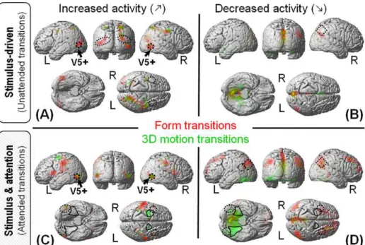
Figure 2. Experimental design (left) and main results (right). Adapted from Paradis et al., 2008.
Following the study by Corbetta et al. (Corbetta et al., 1990) with color, speed and shapes defined by contours, our assumption was that attention to the 3D perceptual attributes of an object would enhance the activity in the areas processing these attributes, and that these areas would be the same as those activated when passively presenting the perceptual changes, supposedly the above described ‘visual motion’ network.
As a first result, the segregation of activity between 3D motion and form very nicely fitted the classical dichotomy of the visual system into two pathways. Whatever the contrast (unattended transitions, task modulation only or attended transitions), 3D motion was associated with activity in the dorsal pathway and form with activity of the ventral pathway.
However, contrary to our initial expectations, the activity involving attention selection (contrasts 2 and 3 in Figure 2) was not just an enhanced version of the activity elicited by the unattended transitions (contrasts 1). In fact, the main foci of activity for each attribute did not overlap between the two contrasts (compare dotted and solid lines for each contrast). Furthermore, the observed activity was not restricted to the classical ‘visual motion’ network, especially for 3D motion and/or attended transitions. Thus, it seemed that paying attention to the 3D motion or form of the objects did not just consist in modulating the activity of areas otherwise involved in the automatic processing of those visual features.
Further analysis revealed that both the motion and form unattended transitions induced enhanced activity in the network classically associated with the perception of coherent visual motion (Figure 3.A), as well as possible decreased activity in the default mode network (Raichle et al., 2001) (Figure 3.B). In contrast, attended transitions induced little activity increase (Figure 3.C), but extended activity decrease in brain regions located downstream the above-mentioned network (Figure 3.D). Of note, this activity decrease appears in the ‘competing’ pathway compared to the attended attribute, i.e. decreased activity in the dorsal pathway for attended transitions of form (in red) and decreased activity in the ventral pathway for attended transitions of 3D motion (in green).
Figure 3. Activity with respect to a ‘low level baseline’ without 3D structure nor visual motion: increased/decrease
activity (Left/right) for unattended /attended transitions (Top/Bottom). Figure from Paradis et al., 2008.
Discussion
In summary, the activity in the classical ‘visual motion’ network including MT/V5 and intraparietal areas appears enhanced by all –attended or unattended, 3D motion and form– transitions (see Figure 3) without significant selectivity for one perceptive feature compared to the other. Accordingly, this network likely sustains the analysis of the visual inputs (2D speed distribution) rather than the perception of 3D motion and form per se. In contrast, differential activity between 3D motion and form clearly highlights the segregation into two pathways: dorsal for 3D motion and ventral for form. However, this attribute-specific activity appears to depend on whether the feature is attended or not. Then, processing the 3D perceptual attribute may depend on different mechanisms –possibly excitatory versus inhibitory– depending on the task performed by the observer (automatic perception versus active detection). Although difficult to interpret if we do not distinguish 2D motion processing from 3D motion perception, those results are fully compatible with two stages of the computation and different effects of the attentional selection at each stage. Figure 4 illustrates a possible mechanism of attentional selection accounting for those results.
Figure 4. Proposed mechanism for the attentional selection of the 3D features: 3D motion (left) and form (right).
Selection per se might arise through down modulation (left, −; right, −) in the pathway processing the concurrent
feature (i.e. ventral areas for attention to 3D motion vs. dorsal areas for attention to form). Attention directed toward either feature however induces a non-specific enhancement (+) in MT/V5 likely helping to process the 2D visual input from which both features are extracted.
Note that the observers were never passive in this experiment. During the unattended transitions, they were in fact performing a visual task related to color. Although irrelevant and independent from motion processing, this task was still likely to orient subjects’ attention toward the visual modality, and may have globally induced non-specific activity enhancement. To further characterize the activity of the regions found selective for 3D motion and form in different perceptual contexts, I now wish to present additional unpublished data recorded in actual passive viewing.
Additional unpublished data: 3D motion and form attributes in passive viewing
https://www.researchgate.net/publication/311809839_Specificity_of_the_responses_to_3D_motion_and_form_in_passive_v iewing
In this experimental condition, I targeted the areas highlighted in the previous study (Figure 5A) and tested their response to changes of color, 3D motion or form viewed passively (Figure 5B). For the motion and form attributes in particular, I wished to check whether the activity evoked by unattended transitions (i.e. occurring during the color task: Figure 5A, Unatt and Unatt) was similar to the activity evoked by passively viewed transitions (Figure 5B, F and M).
In the occipito-temporal regions of the ventral pathway, there was significant activity related to passively viewed form transitions, consistent with the previously established selectivity of those regions (Figure 5A). In contrast, no significant activity was found in the dorsal regions whatever the modulated attribute (Figure 6B). This absence of significant activity could be due to a decrease of signal and statistical power in passive viewing compared to active conditions. In line with this, the two ventral areas V3A and LO keeping significant activity in passive viewing also presented greater activity than all other areas in the active conditions. However, the dorsal regions not only show reduced activity: most of them also appear to lose their selectivity for the 3D motion feature, and the activity of medial FEF and cuneus could even be suggestive of a selectivity for form in passive viewing conditions
Overall, these results suggest that the dorsal areas considered here are selective for 3D motion and activated by 3D motion transitions in a task context only. This is consistent with the dorsal pathway associated with perception for action (Goodale and Milner, 1992): dorsal areas would be less active in the absence of a task requiring action after perception. It is also reminiscent of how task ‘usefulness’ may affect perception in transparent motion stimuli (Chopin and Mamassian, 2011). Thus, we can hypothesize that dorsal areas may support perceptual decision about motion depending on the task. This however
questions whether there is an automatic processing of objects’ 3D motion or whether accessing this feature systematically requires attention.
Figure 5. Compared pattern of activity in active and passive viewing conditions. Regions of interest are defined
and colored depending on their activity in the active conditions reported in Paradis et al., 2008. Green ROI were more activated by 3D motion than form in either unattended (•) or attended (•) transitions. Red ROI were more activated by form than 3D motion in unattended (•) or attended (•) transitions. For each ROI, the activity profile plots the mean amplitude of response (SPM beta ± one standard error) to each type of transition. (A) Activity observed in the active conditions for form/3D-motion transitions during the color task (Unatt/Unatt); for color transitions during the form/3D-motion task (Task/Task); for form/3D-motion transitions in the form/3D-motion task (Att/Att). (B) Activity induced by passively viewed transitions of 2D motion (2D), from 2D motion to a 3D object (3D), Color (C), Form (F) and 3D motion (M).
Interaction between form and motion attributes in 3D structure-from-motion perception
Publication: Miskiewicz A, Buffat S, Paradis A-L & Lorenceau J (2008) Shape and motion interactions at perceptual and attentional levels during processing of structure from motion stimuli. Journal of Vision, 8(16):17, 1-14
http://dx.doi.org/10.1167/8.16.17 (attached in Annexes)
This study was part of Agnieszka Miskiewicz’ PhD work. A Miskiewicz was co-supervised with Jean Lorenceau (ICM), in the framework of a collaboration with S. Buffat (IRBA, ex-IMASSA).
With 3D structure defined from motion, the 3D motion and form attributes are both extracted from the retinal distribution speed. We then wondered whether these attributes could actually be processed independently or whether their common source could induce interaction between them.
Protocol and results
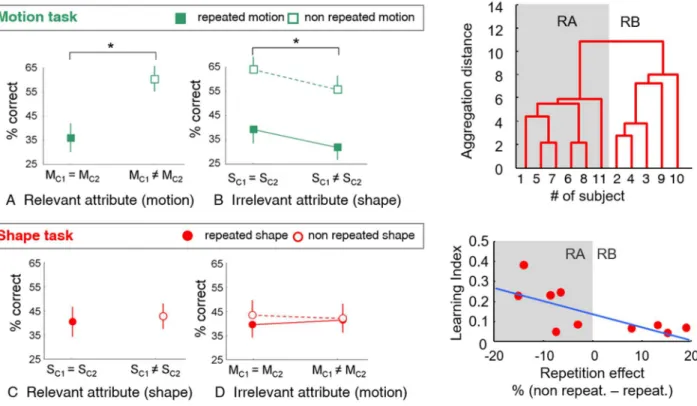
To answer this question, we used a rapid visual serial presentation (RSVP) protocol (Figure 6). After being presented a rapid sequence of three 3D-from-motion stimuli, participants were asked to report the number of times they had perceived a specific form or a specific motion direction. To perform this task, the participants not only have to perceive and identify the form and/or motion direction (i.e. type) of the stimulus, they also have to attribute a ‘token’ to their perceptions in order to individuate and count their occurrences (Kahneman et al., 1992; Kanwisher, 2001, 1987). This protocol thus involves two stages of processing at least, likely related to feature perception on one hand and episodic memory on the other hand. To reveal the possible interaction between motion and form at the two stages, we evaluated the observers’ performance in both motion- and form-related tasks, depending on whether the C1 and C2 items (see Figure 6) were different or shared similar motion and/or shape features. The overall percentage of correct responses was used to evaluate type identification, while the repetition effect was assumed to evaluate token attribution.
Figure 6. RSVP protocol (left) and working hypotheses (right). Depending on the task, observers had to report either
the shapes (S) or the motion directions (M) of the three structure-from-motion (SFM) stimuli. C1 and C2 could have identical or different features (repeated vs. non repeated condition with respect to the feature). D was always different from C1 and C2. Correct responses corresponded to both C1 and C2 being reported correctly, i.e. feature reported twice if repeated or C1 and C2 features reported once only if different. D could be reported once or omitted. The repetition effect is the difference of correct responses between repeated and non-repeated conditions. Adapted from Miskiewicz et al., 2008.
Overall, performances were higher when the task concerned the motion direction rather than the form. For the motion task, performances were poorer when motion direction was repeated, revealing repetition blindness (Figure 7A). In contrast, performances were increased when the form, irrelevant to the task, was repeated, although the rate of repetition blindness to motion direction was not modified (See Figure 7B).
For the form-related task, we did not find any repetition blindness at the group level, i.e. no effect of repeating the form on the subject’s ability to report the perceived shapes and their number of occurrences (Figure 7C). In addition, repeating the motion direction, irrelevant to the task, did not impact the form-related performance either (Figure 7D).
Figure 7. Results of the RSVP protocol. (Left) Performances in the motion and form tasks, depending on whether
the attributes are repeated (M/Sc1 = M/Sc2) or not (M/Sc1 ≠ M/Sc2). (Upper right) Subjects can be segregated into two statistically different groups showing either repetition blindness (RB) or repetition advantage (RA) in the form task. (Lower right) The repetition effect appears correlated to the learning index, which is inversely related to the initial ability of the subject of perceiving 3D shape from motion at the beginning of the training. Adapted from Miskiewicz et al., 2008.
Because repetition blindness is classically found when repeating static shapes, the absence of repetition blindness for the form task was rather surprising. Why should structure-from-motion stimuli suppress this effect? A closer look at the results of the form task showed that the participants could in fact be segregated into two statistically distinct groups (Figure 7 upper right). Five participants then revealed a clear repetition blindness effect, while the six other appeared to experience repetition advantage (RA). This behavioral difference was found related to the performance of the participants in the training phase. Precisely, the repetition effect was correlated with the evolution of participants’ performances in the shape task during the training phase (learning index, Figure 7 lower right). As the training phase aimed to bring all participants at a similar level of performance (80% correct identification), the greater the learning index, the poorer the performance at the beginning of the experiment. In other words, repetition blindness was only observed in subjects who reached the required performance right from the beginning of the training. We specifically checked that the two groups showed no performance difference in the motion task, and as a reminder, both groups showed repetition blindness for motion direction. Consistently in this task, both groups were also able to reach the required performance of 80% correct from the beginning of the training phase.
Discussion
Overall, this study provided two new and paradoxical results:
1) Although we used structure-from-motion stimuli, the repetition of motion direction had no impact on shape perception, but shape repetition facilitated motion perception (Figure 8);
2) Repetition blindness appeared associated with a ‘spontaneous’ ability to identify the type of the stimulus (in practice, ability to reach 80% correct response with minimal training for the motion and/or the shape task), while the participants who necessitated more than four training sessions to reach 80% correct response in the shape task experienced repetition advantage.
Figure 8. Interactions between motion and form observed in the shape and motion tasks.
Motion repetition impacted neither identification nor token attribution in the shape task (left). Shape repetition facilitated motion direction identification (right). Adapted from Miskiewicz et al., 2008.
This repetition blindness vs. repetition advantage is reminiscent of the suppressive masking effects observed for suprathreshold stimuli on one hand (e.g. attention blindness) as opposed to the facilitatory priming effects observed for subliminal stimuli on the other hand. It is also consistent with a RSVP study showing repetition blindness for words but repetition advantage for non-words (Coltheart and Langdon, 2003). In this study, the authors proposed that repetition blindness is limited to items with pre-existing orthographic or lexical representations. Here this could correspond to all participants having a pre-existing representation of motion direction, while only a sub-group having a pre-existing representation of the 3D structure from motion. Yet, which mechanism should make a stimulus both easier to identify and store in memory, but harder to individuate?
Repetition blindness has been attributed to a failure of token attribution (Kanwisher, 1991) and especially a failure of the contextualization process, which is the assignment of the stimulus instance to a specific temporal and spatial context. Alternately, repetition blindness could also be considered as a success to merge interrupted visual inputs (between blinks or saccades for instance), and interpret them as one stable perception of the same object. This can explain why repetition blindness is reduced when a static object is repeated with two very different orientations (Harris and Dux, 2005). Such changes are barely compatible with the stability expected from a single object and may rather be interpreted as two different objects. Then, existing internal representations of the stimulus may help to maintain the ‘object file’ (Kahneman et al., 1992) open, and thus stabilize the perception in the absence of continuous or consistent inputs. This is also in line with more recent results showing that the level of semantic awareness associated with faces affected the recognition of those faces in an episodic memory task (La Corte et al., 2012).
Let us now come back to the observation that motion direction had no impact on form perception. This suggests that object identification was independent of motion direction. Such conclusion is consistent with our postulate that perceiving the 3D motion is not an intermediate stage of structure-from-motion (SFM) processing, but rather an end product of this processing just like 3D shape.
The reverse observation that form repetition influenced motion identification is more puzzling because it suggests that 3D motion processing could depend on shape perception. This direction of interaction is at odds with the observation that motion identification was usually faster than shape identification. However, the delayed response at the end of the trial also allowed interactions to occur after the initial extraction/perception/encoding of motion and form. Hence, the observed influence of (slow) shape on (fast) motion is likely related to a later process, e.g. recall or storing during the delay. In addition, our previous results suggested that form-related activity might be more robust and independent of the on-going task than 3D-motion-related activity. The observation that the encoded shape can influence the motion task but not the other way round is consistent with this assumption: motion information might not be ‘maintained’ enough to influence another percept when it is not the focus of the task.
In summary, although 2D motion processing is a prerequisite for 3D structure perception, we found the 3D structure has an influence on 3D motion perception, but we did not find any effect of motion direction on 3D structure identification. Moreover, motion and form features did not influence each other at the individuation level. The next study hence questioned the timing of motion and shape processing: can we determine a cascade of activity compatible with such behavioral observations?
Timing of stimulus- and task-related processes
Publication: Miskiewicz A, Buffat S, Lorenceau J, Paradis A-L (2010) Temporal dissection of stimulus-driven and task-driven processes during perceptual decision about 3D SFM stimuli. IFBME Proceedings Series http://dx.doi.org/10.1007/978-3-642-12197-5_76 (attached in Annexes)
In this study, we used magneto-encephalography (MEG) to highlight the cascade of processing at stake when performing a perceptual task on a structure-from-motion stimulus. In usual conditions, processing stages such as sensory processing, attentional selection and perceptual decision likely overlap in time. We hence designed an experimental protocol with specific time constraints in order to distinguish the brain signals related to different stages.
Protocol and results at sensors’ level
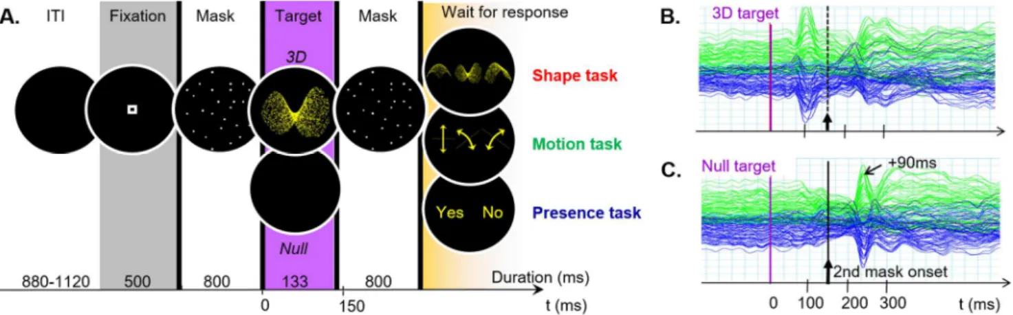
The stimulation sequence consisted of a 3D-structure-from-motion target presented shortly and flanked by two preceding and following masking stimuli. The masking stimuli were aimed at delimiting the processing duration for the 3D target (see Figure 10A). Figure 10B shows the activity evoked by a stimulation sequence with a 3D target. Figure 10C illustrates the MEG signal evoked by a stimulation sequence in the absence of a 3D target: the activity related to the second mask (at +90ms after the mask onset) delimits the time windows during which the 3D target can be visually integrated.
Figure 9. Activity related to a 3D-structure-from-motion stimulus in a time-constrained protocol. (A) Time course
of the stimulation sequence. ITI: Inter-stimulus interval. (B) MEG signal evoked by a stimulation sequence including a 3D target. (C) MEG signal evoked by the stimulation sequence in the absence of 3D target. Adapted from Miskiewicz et al., 2010.
MEG activity has been recorded in the context of three different perceptual tasks. During the runs of Presence task, participants had to detect whether a 3D stimulus was presented or not in the stimulation sequence. For the Motion task and Shape task runs, instructions were to identify the motion direction or 3D shape of the presented target among three possible answers. The spatial organization of the response screen was randomized so that the participants could not choose their manual response in advance, in order to minimize the temporal overlap between perceptual and motor activity. That the response be delayed also gave similar conditions to that of the RSVP protocol. Yet, the observers knew which feature they had to attend before seeing the target so that we could assess the effect of the task on the target processing.
The analysis of the evoked magnetic fields first showed separate effects in time for stimulus and task, with significant stimulus effect (presence vs. absence) between 100 and 200ms around the occipital sensors, and significant task effect (Presence/Shape/Motion) between 300 and 320ms around the parietal and temporal sensors (Figure 10). Although later than the stimulus effect, this task-related effect still occurs during the presentation of the second mask, long before the response screen is displayed and a
motor preparation is possible. With the stimulus- and task-related effects non-overlapping in time, we do not have any evidence of a direct effect of the task on the early stimulus processing. However, the precedence of the stimulus effect −between 100 and 200ms− with respect to the task-related effect −between 300 and 320ms− is compatible with one feature of the stimulus being able to influence the later response to the task.
Figure 10. Time-separated effects of stimulus and task. The effect of the target (3D/Null) and the task
(Shape/Motion/Presence) was tested along with groups of sensors (Top right). Significant target effect and target x sensors interaction were found between 100ms and 200ms, while significant task x sensor interaction was found between 300 and 320ms. The interaction with the sensors reveals that the target (resp. the task) does not only vary the amplitude but also modify the topography of activity, adapted from Miśkiewicz, 2009.
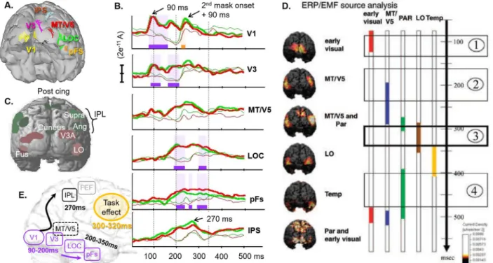
Time course of the sources of activity
After source reconstruction, the brain areas activated by the 3D target (Figure 11A) displayed notably different time courses, with peak of activity spread over a period from 90 to 270ms (Figure 11B). These time courses were globally consistent with the chronology described by Jiang et al., 2008. Thus, we found similar early activity arising before 100ms in the occipital areas (V1) and a peak of activity in the medial part of the parietal cortex just before 300ms. We also found LOC activity temporally overlapping with parietal activity and sustained in time beyond 300ms, although the present LOC activity arose much earlier, as soon as 200ms (Figure 11, B vs. D).
The main differences were observed in late activity. Indeed, Jiang et al. (2008) described temporal activation between 350 and 400ms and re-activation of parietal and early visual cortex after 400ms, which were not observed in the present study. We assume this discrepancy is due to our masking stimuli, which seemingly suppressed differential activity between null target and 3D target beyond 350ms. Since the response mapping in Jiang et al. was not randomized, another possibility is that part of their late activity be related to the preparation of the motor response.
were partly confounded, perhaps because of a lack of spatial selectivity at the source level. However, this interpretation does not hold when we consider the time course of activity evoked by the stimulation sequence in absence of 3D target. In this case indeed, a peak of activity −consistent with the quick response of MT to motion onset− is observed in MT/V5 around 90 ms after the 2nd mask onset. In contrast, the corresponding peak is delayed around 270 ms in LOC (onset plus 140 ms), which does not support the interpretation that both sources were confounded. Instead, we suggest the specific time course of activity observed here in MT/V5 could be due to the presence of our dynamic masks. Because they maintain a continuous 2D motion input before and after the 3D target, they likely suppress the response evoked by the onset of 2D motion which occurs when presenting a 3D dynamic stimulus after a static baseline. This modification of activity in MT/V5 signal with the motion masks further supports the interpretation that MT/V5 is involved in processing 2D motion rather than coding 3D object features.
Figure 11. Time course of activity. Selected sources of MEG signal activated by the 3D target in the presence task
(A), and their reconstructed time course of activity in the other two tasks (B). Bold lines correspond to the activity for the 3D target; Red and green code for the Shape and Motion tasks, respectively; periods of significant stimulus (resp. task) effect are highlighted in purple (resp. yellow). For comparison, regions of interest from the fMRI study (C), and the activation sequence described by Jiang et al. (2008) for the perception of 3D dynamic shapes in a protocol without time constraints (D). (E) Summary of the observed sources and their timing of activity.
Eventually, we observed a very particular pattern of activity in the intraparietal sulcus (IPs). The signal increased slowly with LOC activity and decreased rapidly after 270ms, long before the possible preparation of the motor response. The timing of the decrease for the 3D target is also coincident with the peak of activity visible in null target condition and likely evoked by the onset of the 2nd mask. A possible interpretation is that IPS activity increases until the information related to the 3D target is replaced/interrupted by information related to the 2nd mask. Interestingly, this pattern is consistent with cellular recording in monkeys suggesting that IPS accumulates information about moving visual stimuli as long as more information about the target is required and available (Huk and Shadlen, 2005). Given the complex relationship between cellular recording in monkeys on one hand and macroscopic MEG recording in humans on the other hand, additional data would be required to elucidate the questions put forward by this particular pattern of activity: does it actually reflect accumulating information? Does the IPS region process motion information only or does it integrate information from shape-processing areas as well?
Stimulus-driven vs. task-related activity
When directly assessing the source signal, the significant stimulus effect detected at sensor level appeared concomitant with the activity of the earliest visual areas (V1, V3). In contrast, the task effect was concomitant with stimulus-related activity of the later areas (LOC, pFS). However, we did not find any significant effect of the task in those areas (Figure 11B). Overall, none of the regions of interest involved in processing the structure-from-motion stimulus (Figure 11A) appeared consistently modulated by the type of feature targeted by the task. This negative result is in line with our previous fMRI results showing that the attentional selection of 3D motion and shape involved different areas from stimulus-driven activity. Here, the task effect observed at the sensor level rather seemed to arise from more anterior visual brain regions as well as right fronto-polar areas. We interpreted this frontal involvement as a possible effect of task difficulty (Mangina et al., 2009) and/or prospective memory required by the delayed response (Burgess et al., 2003). The following unpublished data may bring another insight about the possible involvement of anterior temporal areas as well.
Further into shape perception with the use of familiar objects
Posters: Benmussa F, Dornbierer J-G, Buffat S, Paradis A-L & Lorenceau J (2012). Looking for the LOC with MEG using frequency-tagged natural objects. JoV August 2012, Vol.12, 511 (VSS) http://dx.doi.org/10.1167/12.9.511;
https://f1000research.com/posters/2249. PhD work of F. Benmussa, co-supervised with J. Lorenceau, in collaboration with S. Buffat (IRBA). The project was supported by a doctoral contract from DGA to FB and REI grant n°2006 34 059 to JL.
The main objective of this study was to set up an MEG ‘localizer’ protocol aimed at delineating the brain areas involved in object recognition. We thus used clouds of points obtained from 3D scans of familiar objects (Buffat et al., 2014) as well as their scrambled versions (see Figure 12). To optimize the detection of MEG activity related to object perception, we further decided to “tag” the signal related to the stimuli with a specific frequency. Frequency tagging is associated with powerful signal at a known frequency, which allows to precisely identify the signal of interest. In that context, we can also analyze the magnetic signal “evoked” by the stimulus by considering each stimulation period as an event.
Protocol and results
Several visual features can be used to tag a stimulus. In this study, we compared three types of tagging related either to luminance or object identity (Figure 12).
Figure 12. Frequency tagging protocol used to spot the activity related to object perception with MEG.
The first tagging condition, here called “Pure Between Object Tagging”, consisted in switching object shapes (resp. scrambles) every 400ms (i.e. 2.5Hz) or 83ms (12Hz). The second condition (“Mix”) alternated objects with scrambles. The third condition consisted in replacing dots of the cloud, thus inducing luminance tagging without changing the shape of the stimulus (“Within Object Tagging”). Preliminary analyses revealed that this within object tagging was poor at activating brain areas involved in object recognition. Also, 12Hz tagging conditions only induced low power activity. This later observation was consistent with a masking effect of each new object (resp. scramble) on the previous one, likely interrupting the later component of object processing, as suggested by the results of the above study.
We thus focused our analysis on the data recorded in the between-object-tagging conditions at 2.5Hz. We then computed the signal evoked by the stimulus transitions –i.e. the onset of a new object or a new scramble− on a time window of 400ms, and compared the four possible types of transitions (Figure 13A) in a two-factor ANOVA. A main effect of the stimulus appearing after transition (Object/Scramble) aroused around 170-185ms (Figure 13B, green). The associated sources of activity were located in the left lateral occipital cortex (LOC) at 170ms and bilaterally in the anterior temporal pole at 185ms. The mix/pure condition did not induce any significant effect per se. However there was an interaction between the appearing stimulus and the mix/pure condition, which can also be interpreted as a main effect of the stimulus preceding the transition (Object or Scramble). Although later and more spread in time than the activity evoked by the appearing stimulus, this activity related to the disappearing stimulus was found at the same location and reproduced the same temporal order: peak of activity at 190ms in the LOC, followed by anterior temporal activity around 230ms.
Figure 13. Two-way ANOVA. (A) Averaged MEG signal triggered by stimulus transition from scramble to object
(Object mix); from object to scramble (Scramble mix); between objects (Object pure); between scrambles (scramble pure). (B) Effects of stimulation conditions on the evoked MEG signal.
The parametrical tests carried out on all sensors and time samples were supplemented by a non-parametric “clustering” analysis (Maris and Oostenveld, 2007), which allowed to assess the statistical significance of the observed effects while overcoming the issue of multiple comparisons raised by the point-by-point analysis. In practice, all signal samples with uncorrected p<0.05 were clustered based on time and space adjacency (ie. consecutive time samples and sensors less than 4cm away). Each cluster was then assigned the sum of T-values of all its constitutive samples. The same clustering procedure was then repeated a thousand times with the condition labels randomly reassigned to the original dataset, in order to model the null hypothesis. For each randomization we selected the maximal “sum of T” and used it to build an estimated distribution of this statistical value under the null hypothesis. Because this method uses the maximum statistics, it intrinsically controls for multiple comparisons. We then compared the value of each original cluster with this distribution: clusters with values greater than 99% of the distribution were considered significantly activated with a probability of false positive smaller than 0.01. This approach confirmed there were two significant clusters associated with the Object minus Scramble contrast: one including left occipito-temporal sensors and lasting from 159ms to 273ms; and its symmetrical counterpart including right occipito-temporal sensors and lasting from 152ms to 286ms (Figure 14A). The mid-temporal sensors, which likely reveal LOC activity, are those involved in the Object minus Scramble effect for the longest duration. However, we also observed a sustained effect in the anterior temporal sensors, which corresponded to a powerful activity located in the anterior temporal lobe and Orbito-Frontal areas after source reconstruction, and maximal between 180ms and 220ms (see Figure 14B and C).
Figure 14. Object minus Scramble effect. Significant clusters of activity (p<0.01, N=16) determining the time window
of the effect between 150 and 290ms. (B) Evolution of the activity topography across the time window of interest, and corresponding source reconstructions (C).
Anterior Temporal Pole
We hardly found fMRI reports of anterior temporal pole activity in the context of visual object recognition. This is not so surprising since strong magnetic susceptibility artifacts tend to dramatically lower the signal recorded with fMRI in this region, and studies designed for visual object recognition are likely to focus on occipital and posterior temporal areas rather than tune the acquisition sequence to record the anterior temporal pole. The anterior temporal poles were rather found associated with semantic memory (Martin and Chao, 2001) as well as social and emotional processing (Olson et al., 2007). Previous neuropsychological and PET data (Damasio et al., 1996) however suggest that these regions could be involved in processing lexical information. Some TMS studies (Lambon Ralph et al., 2009; Pobric et al., 2007) and a meta-analysis (Visser et al., 2010) suggest that the anterior temporal pole is implicated in semantic tasks across various input –words, pictures, objects, sounds, smells − and output –written and spoken− modalities. They further propose that this region processes and stores amodal semantic representations associated to sensory stimuli, and acts as a ‘hub’ allowing conceptual manipulation and information exchange between different modalities. Globally, its involvement in the present study would be consistent with the fact that our stimuli were familiar objects belonging to different semantic categories (namely fruits, vehicles and tools), and that participants were instructed to report a maximum number of seen objects after recording, thus requiring lexical storage.
Study comparison
With 400ms duration for each stimulus, this study imposed much less time constraint on object processing than the previous study. It is then likely that the evoked magnetic wave could fully develop before the activity was disrupted by the onset of a new stimulus onset. The time issue however is unlikely to account for the absence of activity in the anterior temporal lobe with 3D structure from motion stimuli. Indeed, the significant cluster of activity including the ATL was spread over a 160-280ms interval, which perfectly overlaps with the time interval of significant activity in the 3D structure from motion study (see Figure 15A and B). Then, it is more likely that the difference comes from the nature of the stimuli, especially the use of familiar and nameable objects vs. 3D geometrical surfaces.
Figure 15. Comparative summary of the previous MEG results. Localization and timing of the stimulus effect when
comparing (A) the presentation of 3D structure from motion with respect to a null target (Miskiewicz et al., 2010) or (B and C) Static familiar objects with respect to their scrambled counterparts at 2.5 Hz and 12Hz respectively
(Benmussa, 2013).
It is noteworthy, that activity related to the 3D structure-from-motion stimuli could still be observed at the source level beyond the timing of the response to the second mask in the null target condition (see Figure 11B in LOC and pFs). In contrast with this, the tagging protocol did not allow us to identify activity beyond the onset of the next stimulus i.e. after 400ms at 2.5Hz and 83ms at 12 Hz. There was however a risk of temporal aliasing, meaning that late components of the response could be folded in the observation period and thus misinterpreted as early components. The risk was especially high for the 12Hz presentation frequency since the observation window was limited to 83ms. However, the activity evoked by the tagging conditions at 12Hz, which did not develop beyond occipital regions and was much weaker than the activity at 2.5Hz, rather suggests that late components were actually suppressed. This interpretation is also consistent with a subsequent behavioral study in which participants had to identify a target among a rapid visual serial presentation at different presentation frequencies: participants were not only worse at identifying a target object when the presentation frequency was higher, they also revealed much less confident in their responses, which suggested an absence of conscious perception.
I
NTEGRATION AND SELECTIONWith separate timings of activity for stimulus and task, the previous results were in favor of independent task- and stimulus-related activity. However, we also found that repeating the form feature had an effect on the motion perception task, which supports that processing a non-attended feature of a stimulus can interfere with the task performed on another feature of the same stimulus. Then, I wished to further investigate the links between feature perception, attentional selection and task, and their neural correlates.
Our RSVP results further suggested that form repetition proceeded like a priming effect on the perception of motion. However, contrarily to the usual priming conditions, the primed feature (i.e. shape) was not the one targeted by the task (i.e. motion), so the following questions arose: Could an attentional priming directed at one feature enhance the perception of another feature of the same object? This question was in line with the more general theory proposing that conscious visual perception depends on attention. Indeed, we could imagine that repeating the form may enhance the attention paid to the overall stimulus, and thus facilitates the conscious perception of its motion in the motion task.
Feature selection and conscious perception
Publication: Liu Y, Paradis A-L, Yahia Cherif L & Tallon-Baudry C. (2012) Activity in the lateral occipital cortex between 200 and 300 ms distinguishes between physically identical seen and unseen stimuli. Front Hum Neurosci 6: 211.
http://dx.doi.org/210.3389/fnhum.2012.00211
Although the role of attention in conscious perception had been questioned (Koch and Tsuchiya, 2007) and even challenged by evidence that the neural bases for conscious perception and spatial attention are dissociated (Wyart and Tallon-Baudry, 2008), the status of feature-based attention remained to be explored. Here, we investigated the possible relationships between feature-based priming and conscious perception using color and static orientation. These two features do not depend on motion processing and are not likely to interfere because of a common early extraction process. If an interaction was found between those features, it would rather be due to attentional mechanisms operating at the object level.
Protocol and results
This work has been performed by Ying Liu during her PhD, co-supervised with C. Tallon-Baudry. In this study, we recorded MEG activity while testing the effect of color priming on static orientation perception. To this end, we used a protocol previously designed to assess interactions between spatial attention and perceptual consciousness (Wyart and Tallon-Baudry, 2008). The previous spatial priming was replaced with color priming (Figure 16).
Figure 16. Protocol to assess possible interaction between color priming and orientation perception during MEG recording. (A) Time course of a typical trial, and (B) the four types of ‘Stimulus’ used in the protocol. The
grating-present stimulus were used in 87% of the trials, and the color of the Stimulus was identical to that of the Cue (i.e. Congruent) in 65 % of the trials. We thus manipulated color congruency between the target stimulus and a prior visual cue irrelevant to the task. Two questions were asked at the end of the trial; by gathering both objective and subjective answers, we could determine whether the observers consciously perceived the grating in the colored ring. Figure from Liu et al., 2012.
One objective of this study was to determine whether the neural correlates of consciousness could depend on feature-based attentional selection, with the assumption that the color cue presentation would induce enhanced attention to a target object with the same color. Since the behavioral results did
By comparing seen and unseen present stimuli, we found correlates of conscious perception between 200 and 300ms. We could further localize the main sources of this activity bilaterally in the lateral occipital complex (rLO et lLO, Figure 17A). Just as color congruency did not affect the behavioral response to the grating, it did not affect the LO activity related to conscious perception either, further suggesting that the co-localized color and orientation features did not influence each other and were processed independently.
Figure 17. Localization and timing of MEG activity evoked by consciously seen stimuli (A,C) and color congruency (B,D). Significant seen-unseen difference in bilateral lateral occipital areas and right inferior-temporal cortex, from
190 to 350ms after stimulus onset; significant incongruent-congruent difference in left IPS from 150 to 250ms, for grating-present stimuli only; adapted from Liu et al., 2012.
Compared to congruent colors, incongruent colors evoked more electrophysiological activity in IPS at 150-250ms. As stated above, this color (in)congruence effect did not depend on whether the presented grating was consciously perceived or not. However, it depended on whether the grating was physically present or not.
Intra-parietal sulcus
In the literature, left IPS activity has been related to the selection of low saliency stimuli (Mevorach et al., 2009), in particular when more salient distractors are present. Here, the incongruent color could represent a distractor and the grating the low saliency stimulus to select.
Thus the present IPS activity could be specifically related to the processing of a feature irrelevant to the task. This does not support the existence of an active contamination of one feature by another through a possible attention effect at the level of the object, nor an active binding of the features belonging to the same object. Instead, the present results are rather in favor of an active selection mechanism occurring between competing co-localized and/or co-occurring features to allow the independent processing of each feature.
Feature selection
Since color repetition did not prove to enhance attention to the stimulus target, we cannot conclude that the effect of shape repetition observed in the RSVP protocol was due to an attentional enhancement of all perceptive features associated with the repeated object.
On the contrary, the present results suggest that an active processing is required to be able to selectively analyze the feature relevant to the task among two co-localize features. This seemed to occur especially when the irrelevant feature was incongruent and may have acted as a distractor with respect to the main task. With this perspective, a shape transition occurring during the motion task of the RSVP
protocol is likely to induce extra-processing load in order to select the motion feature relevant to the task. The advantage related to shape repetition could then be interpreted as an absence of detrimental effect related to shape transition.
This feature-selection interpretation further questions the asymmetry observed between motion and shape. Mevorach and colleagues suggested that two different selection processes occur depending on whether the relevant stimulus is more or less salient than the distractor (Mevorach et al., 2009). In the case of shape and motion, does it mean that one feature is more salient than the other? The relatively low activity related to 3D motion in passive viewing might be interpreted as the motion feature being less salient. Nevertheless, the fMRI results obtained in active conditions did not reveal any consistent left/right parietal asymmetry for motion vs. form that could support this view.
Selection vs. binding
Publication: Caclin A, Paradis A-L, Lamirel C, Thirion B, Artiges E, Poline J-B, Lorenceau J (2012). Perceptual alternations between unbound moving contours and a bound shape motion engage a ventral/dorsal interplay. Journal of Vision.
http://dx.doi.org/10.1167/12.7.11
We have seen that motion and shape information were processed by distinct cerebral areas. However, both features have to be combined to produce the unified and coherent percept of a moving object. We have also seen that the areas involved in perceiving the 3D motion of an object are different from the areas usually associated with (2D) motion processing. It is thus important to dissociate the neural activity related to the conscious end percept from that related to the physical specificities of the stimulus, and better understand how the visual system translates the analysis of a visual input into the representation of a perceptive attribute. The objective of the fMRI study presented here was thus to identify the brain areas sustaining the binding of distinct visual elements into the unified percept of one moving shape.
Protocol and results
In this study, stimuli were line segments arranged into a diamond shape (Figure 18), with each segment following a vertical oscillatory movement. This stimulus is called “bistable” because it is compatible with two visual interpretations: the observer can either perceive several unbound elements moving back and forth, or a rigid diamond following a circular trajectory (Figure 18A). The observer can spontaneously jump from one perceptive interpretation to the other. However, some physical characteristics of the stimulus can favor one interpretation or the other. For instance, segments with low-contrast ends (Figure 18B) contribute to spatial integration and promote the perception of a diamond. In contrast, segments made of dots are more easily perceived as independent elements. Yet, if the dots constituting the segments are moving along the segment orientation thus blurring the information about their relative positions, the bound interpretation returns to the fore (Figure 18C). Finally, compared to the arrangement into a closed shape like the diamond, an arrangement in chevron promotes the unbound interpretation (Figure 18D). Since it is possible to progressively modify those contrast, motion or configuration characteristics in time, it was also possible to drive changes of perceptual interpretation (Figure 18E).
We analyzed the fMRI activity related to the bound and unbound percepts, when perception changes were either spontaneous or evoked by physical changes of the stimuli. Comparing bound and unbound perception into previously defined regions of interest revealed enhanced activity for the bound percept in the ventral areas and greater activity for the unbound percept in hMT.
Supplementary analyses revealed that ventral and dorsal areas were similarly activated whether the stimuli were physically modulated in time or not, so that the activity of those regions appears to mainly depend on the subjective perception. This observation supports the idea that the brain areas sustaining subjective perception are not necessarily processing the physical characteristics of the stimulus. This further suggests that the perception-related areas described here could be part of a generic network involved in involved in spatially binding sparse visual elements into a unique moving shape.
With this perspective, the mirroring responses observed between the ventral fusiform area and the dorsal hMT area could reflect a dynamic weighting of the evidence in favor of one interpretation or the other (e.g. more activity in MT in favor of several moving elements, more activity in the fusiform in favor of a coherent shape).
Figure 18. Neural bases of perceptual binding. (A) Bistable perception switching between bound and unbound
percepts. (B,C,D) Three types of stimulus modulation used to induce perception switches –segment contrast, local motion and shape arrangement. (E) There were two types of perceptual switches –those evoked by the physical variations of the stimulus and spontaneous switches occurring without stimulus changes for an intermediate value of the visual parameters. (F) Activity evoked by the transitions toward bound perception in red (resp. unbound in green) in previously determined regions of interest. The gray line displays the difference between the two responses, and the gray bar indicates the first time of significant difference between the responses to the two percepts, adapted from Caclin et al., 2012.
Discussion
The pattern of activity described here is somehow reminiscent of the competing activity observed between the dorsal and ventral pathways when comparing the activity related to 3D motion and form (Figure 3, p12), but with notable differences however. Here, the dorsal activity did not extend beyond hMT/V5. By contrast with the 3D features, which both required to analyze the 2D motion distribution at the level of hMT and were perceptually segregated through a competition engaging downstream areas of the dorsal pathway, the present bistable stimulus seemed to engage the early stage of 2D motion analysis in the competition.
In addition, a closer look at the time course of activity revealed that the balance of activity between pFs and hMT was not symmetrical. Although, the activity evoked by perceptual transitions was always positive in the fusiform region, it was either positive −toward the unbound percept− or negative −toward the bound percept− in the hMT+ region (see Figure 18F). This asymmetry may reveal a predominant role