HAL Id: cea-02474767
https://hal-cea.archives-ouvertes.fr/cea-02474767
Submitted on 11 Feb 2020HAL is a multi-disciplinary open access archive for the deposit and dissemination of sci-entific research documents, whether they are pub-lished or not. The documents may come from teaching and research institutions in France or abroad, or from public or private research centers.
L’archive ouverte pluridisciplinaire HAL, est destinée au dépôt et à la diffusion de documents scientifiques de niveau recherche, publiés ou non, émanant des établissements d’enseignement et de recherche français ou étrangers, des laboratoires publics ou privés.
Molecular and biochemical analysis of Saccharomyces
cerevisiae cox1 mutants
C. Lemaire, S. Robineau, P. Netter
To cite this version:
C. Lemaire, S. Robineau, P. Netter. Molecular and biochemical analysis of Saccharomyces cerevisiae
cox1 mutants. Current Genetics, Springer Verlag, 1998, 34, pp.138-145. �10.1007/s002940050378�.
Abstract We report on the molecular and biochemical analysis of a set of 13 respiratory deficient mutants of Saccharomyces cerevisiae which are specifically altered in COX1, the gene encoding the subunit Cox1p of cytochrome c oxidase. DNA sequence analysis shows that three are due to frameshift mutations, two to nonsense mutations, and eight to missense mutations. All, except the missense mu-tant S157L, have impaired electron transfer and respira-tory activity. Analysis of the mitochondrial translation products shows that when Cox1p is absent, Cox2p and Cox3p are still synthesized. In the missense mutants, the steady state levels in the mitochondrial membranes of the three mitochondrially encoded subunits Cox1p, Cox2p and Cox3p and the nuclear-encoded subunit Cox4p are re-duced. In the frameshift and nonsense mutants, Cox1p is absent and Cox2p, Cox3p and Cox4p are considerably de-creased or undetectable. A comparison of the steady state levels of Cox1p through Cox4p in the COX1, COX2, COX3 and COX4 mutants shows the interdependance of the ac-cumulation of these four subunits in the mitochondrial membranes.
Key words Cytochrome c oxidase ·
Saccharomyces cerevisiae · Complex assembly
Introduction
Cytochrome c oxidase, the terminal enzyme of the mito-chondrial respiratory chain, catalyzes oxygen reduction
and connects this reaction to the generation of a proton gra-dient across the inner mitochondrial membrane. Recently, two different three-dimensional structures of heme copper oxidases have been reported almost simultaneously: the bacterial cytochrome aa3 of Paracoccus denitrificans which consists of four subunits (Iwata et al. 1995) and the eucaryotic cytochrome aa3 of beef heart mitochondria which contains 13 subunits (Tsukihara et al. 1995, 1996). These structures present the detailed organization of the metal centers. In the case of the beef heart enzyme, in-volvement of some amino acids in proton-, water- and bi-molecular oxygen-channels has been discussed (Tsukihara et al. 1996). In P. denitrificans, the structure has led the au-thors to propose a mechanism of proton pumping that can be tested by site-directed mutagenesis (Iwata et al. 1995). In eucaryotes, the possibility of isolating mutants is lim-ited and is more laborious than in bacteria. However, the facultative aerobe Saccharomyces cerevisiae is a powerful tool for isolating respiratory deficient mutants which would be lethal in strictly aerobic eucaryotes. In S. cerev-isiae, cytochrome c oxidase consists of 11 subunits (Geier et al. 1995). Cox1p, Cox2p and Cox3p are encoded in the mitochondrial DNA (Rubin and Tzagoloff 1973) while the other subunits are of nuclear origin (for review, see Poy-ton and McEwen 1996). The gene coding for Cox1p is split and contains eight exons (A1 to A8) and seven introns (aI1 to aI7) (Bonitz et al. 1980; Hensgens et al. 1983). S. cerev-isiae Cox1p is 50% identical (and 69% similar) to that of P. denitrificans, and 58% identical (and 77% similar) to that of beef heart mitochondria. Studies of missense mu-tants of S. cerevisiae could be useful in elucidating the re-action mechanism of the cytochrome c oxidase complex. On the other hand, analysis of cytochrome c oxidase sub-units in strains carrying nonsense mutations may give in-formation on the mode of assembly of the complex (for a review see Capaldi 1990).
In the present study, we report the molecular and bio-chemical characteristics of a set of exonic cox1 mutants in S. cerevisiae which have lost their ability to grow on res-piratory substrates. The accumulation of Cox1p to Cox4p in the mitochondrial membranes of the cox1 mutants
com-Received: 20 January / 23 June 1998
Claire Lemaire · Sylviane Robineau · Pierre Netter
Molecular and biochemical analysis
of Saccharomyces cerevisiae cox1 mutants
C. Lemaire (½) · S. Robineau · P. Netter1
Centre de Génétique Moléculaire, Laboratoire propre du CNRS associé à l’Université Pierre et Marie Curie, F-91198 Gif-sur-Yvette cedex, France
e-mail: Lemaire@cgm.cnrs-gif.fr Fax: +33-0-169075539
Present address:
1Institut Jacques Monod, 2 place Jussieu, Tour 43, F-75251 Paris cedex 05, France
Communicated by K. Wolf O R I G I N A L PA P E R
pared to cox2, cox3 and cox4 mutants provides some in-sight into the stability of these subunits in these various cytochrome c oxidase-defective mutants.
Materials and methods
Strains and media. All the strains are listed in Table 1. The mit– mu-tants were isolated from the haploid strain 777-3A (op1), mapped to the COX1, COX2 or COX3 genes (Kotylak and Slonimski 1977) and were localized more precisely, for the cox1 mutants, by deletion map-ping (Netter et al. 1982, 1995). The cox2 mutant (V44) and the cox3 mutant (V206) have been previously described as defective in the synthesis of Cox2p and Cox3p respectively (Kruszewska et al. 1980; Baranowska et al. 1983). Growth analyses were performed on dip-loid strains constructed by crossing the various mutants with the strain KL14-4A/60. All the other experiments have been performed on haploid strains. The media N3, NE and NL were as described in Dujardin et al. (1980). YPGAL medium contained 2% galactose, 1% bactopeptone and 1% yeast extract. YPGALa was the same as YPGAL but with the addition of 2µg/ml of adenine.
Sequence analysis of the cox1 mutations. For analysis of the
muta-tions G1099, G1547 and G2508, single-stranded DNA was se-quenced according to Sanger et al. (1977) after cloning into M13/ mp18 or M13/mp19. To identify mutations G2567, G291, G450, W164, G1979, G2394 and G2276, double-stranded PCR products were cloned into pBluescript KS+ (Stratagene) and two clones per mutation were sequenced.
Pulse-labelling experiments and preparation of mitochondrial mem-branes. Pulse-labelling experiments were carried out according to
Claisse et al. (1980) except that cells were re-suspended in 0.4 M mannitol, 50 mM Tris/H2SO4pH 7.4, 2 mM EDTA, 1 mM phenyl methyl sulfonyl fluoride, 10µg/ml leupeptin, 10µg/ml antipaïn, 10µg/ml chymostatin and 1µg/ml pepstatin. Mitochondrial mem-branes were then prepared according to LaMarche et al. (1992). Pro-tein concentrations were determined using Bio-Rad proPro-tein assay.
SDS-polyacrylamide gel electrophoresis. Electrophoresis was
per-formed using the Laemmli system (Laemmli 1970). The molecular weights of the polypeptides were estimated using a low-molecular-weight calibration kit from Bio-Rad.
Immunoblots experiments. Electrophoretic transfers were carried out
according to Towbin et al. (1979). Immunodetection was carried out using an enhanced chemiluminescence (ECL) method according to the manufacturer’s instructions (Amersham International). The monoclonal antibodies anti-yeast cytochrome c oxidase Cox1p, Cox2p, Cox3p and Cox4p were purchased from Molecular Probes and have been described in Taanman and Capaldi (1993). The
poly-clonal antibody anti-yeast cytochrome c oxidase Cox6p was kindly provided by R. O. Poyton.
Activity and cytochrome spectra measurements. Oxygen
consump-tion was measured with a Clark electrode (Gilson oxygraph), at 28 °C, using 2% ethanol as the substrate. The effect of the uncoupler was tested by the addition of 25µM of Carbonyl-cyanide-p-trichlorophe-nylhydrazone and cyanide-sensitive respiration was measured in the presence of 0.5 mM KCN. Respiratory rates were expressed in nmoles O2consumed/min per 5×10
8
cells, taking into account that the solubility of O2is 243.75µmoles/l of H2O at 28 °C. Cytochrome
c oxidation measurements were performed spectrophotometrically
as described in Pajot et al. (1976). Cytochrome absorption spectra were recorded on whole-cell pastes at liquid nitrogen temperature, after dithionite reduction, according to Claisse et al. (1970).
Results
Sequences of the cox1 mutations
The mutations had been previously localized by deletion mapping in the mitochondrial COX1 gene (Netter et al. 1982, 1995). Sequencing revealed that the mutations are in positions consistent with their location determined by deletion mapping. All the results are listed in Table 2. Amongst the 13 mutants studied, two display a nonsense mutation: G2508 (S219ochre) and G291 (Y245ochre), and three a frameshift mutation: G1099 (N99ochre), G2567 (L109amber) and G1547 (L212amber), leading to a non-sense mutation downstream (see Table 3). Eight exhibit a single missense mutation, except for G2394 which is a double mutant in which the second mutation is silent (C12583T→Y394Y). The six missense mutations are lo-cated mainly in exons A4 and A5, while G1979 (G330D) and G2394 (G384D) are localized in exons A6 and A8 re-spectively. The implications of the amino-acid changes will be considered in the Discussion section.
Respiratory growth and activities of the cox1 mutants All the mutants have been previously selected for their inability to grow on glycerol medium (N3) (Kotylak and Slonimski 1977). We completed this analysis by testing the
139 Table 1 List of strains
Name Nuclear genotype Mitochondrial Origin
genotype
777-3A MATαade1 op1 rho+mit+ Kotylak and Slonimski (1977)
777-3A/G291 to G2394 MATαade1 op1 rho+mit– Kotylak and Slonimski (1977) 777-3A/V44-V206
BGT1 MATa ade2 ura3-1 trp1-1 rho+mit+ Kindly provided by B. Guiard
his3-11,15 leu2-3,112 can1-100 COXIV::TRP1
KM612-2D MATαade1 OP rho+mit– Dujardin (1983)
KM612-2D/60 MATαade1 OP rho° Ethidium bromide mutagenesis
of KM612-2D (this work) KL14-4A/60 MATa his1 trp2 OP rho° Groudinsky et al. (1981)
thermosensitivity or cryosensitivity of these strains and their growth on other non-fermentable media. Strains car-rying the various mitochondrial mutations were incubated on N3 medium at different temperatures, 20 °C, 28 °C and 36 °C, and were tested on NL (lactate) and NE (ethanol) media at 28 °C. After 3 days of incubation, no growth was observed under any condition. Extending the time of incu-bation leads to the appearance of rare revertants.
As has been previously shown for the cytochrome bc1 complex (Oudshoorn et al. 1987; Tron and Lemesle-Meu-nier 1990), there is no direct relationship between growth on non-fermentable media of a strain altered in a given complex and the activity of this complex. Thus, measure-ments of the respiratory activity of the different cox1
mu-tants were made in vivo by following the rate of O2 con-sumption in intact cells. In all but one of the cox1 mutants tested, the rate of oxygen uptake is not significantly dif-ferent from a rho° strain lacking mitochondrial DNA (KM612-2D/60) or a mit–strain with a mutation located in the intron aI2, which leads to a lack of COX1 mRNA (W165) (Carignani et al. 1986). Surprisingly, even though it does not grow on non-fermentable media, the mutant S157L displays a residual respiratory activity of 38 nmoles O2consumed/min/5×10
8
cells (about 15% compared to the wild-type (wt) strain). This activity is cyanide-sensitive.
Activity measurements were also performed in vitro, estimating the efficiency of the electron transfer chain by the ability of purified mitochondrial membranes to oxidize
Mutant Mutation Nucleotide Exon Amino-acid Amino-acid COX1 mRNA
change localization change conservationb synthesis
777-3A/G1099 Frameshift T6896TT A4 See Table 3 na +
777-3A/G2567 Frameshift T6910∆ A4 See Table 3 na +
777-3A/G1547 Frameshift G7219∆ A4 See Table 3 na +
777-3A/G2508 Nonsense C7259A A4 S219ochre na +
777-3A/G291 Nonsense T8348A A5 Y245ochre na nd
777-3A/G2276 Missense C7073T A4 S157L Conserved (29/31) +
777-3A/G450 Missense G8340A A5 E243K Invariant (31/31) +
777-3A/G3015a Missense G8343A A5 V244M Invariant (31/31) +
777-3A/W113a Missense G8364A A5 G251R Conserved (22/31) +
777-3A/W201a Missense A8407T A5 K265M Conserved (27/31) nd
777-3A/W164 Missense C8481T A5 H290Y Invariant (31/31) +
777-3A/G1979 Missense G9967A A6 G330D Conserved (24/31) +
777-3A/G2394 Missense G12552A + C12583T A8 G384D + Y394Y Conserved (29/31) + na = not applicable; nd = not done; ∆: deletion
a Sequenced by Lemarre et al. (1994) b
The amino-acid conservation has been considered in terms of the alignment of 31 sequences of Cox1p including bacteria, fungi, plants and animals
Table 2 Nature and localization of the COX1 mutations and analy-sis of COX1 mRNA syntheanaly-sis. The nucleotides are numbered from 1 to 13 003, starting at position –126 of the short strain (Bonitz
et al. 1980) and interrupted for exons A5 to A7 by 3060 nucleotides of the long strain (Hensgens et al. 1983)
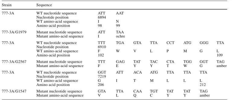
Table 3 Localization of the frameshift mutations and positions of the nonsense codons generated
Strain Sequence
777-3A WT nucleotide sequence ATT AAT
Nucleotide position 6894
WT amino-acid sequence I N
Amino-acid position 98 99
777-3A/G1979 Mutant nucleotide sequence ATT TAA Mutant amino-acid sequence I ochre
777-3A WT nucleotide sequence TTT TGA GTA TTA CCT ATG GGG TTA
Nucleotide position 6910
WT amino-acid sequence F W V L P M G L
Amino-acid position 102 109
777-3A/G2567 Mutant nucleotide sequence TTT GAG TAT TAC CTA TGG GGT TAG
Mutant amino-acid sequence F E Y Y T W G amber
777-3A WT nucleotide sequence GGT ATT ACA ATG TTA TTA TTA
Nucleotide position 7219
WT amino-acid sequence G I T M L L L
Amino acid position 206 212
777-3A/G1547 Mutant nucleotide sequence GTA TTA CAA TGT TAT TAT TAG
an exogenous electron donor, cytochrome c. The initial rate of the cyanide-sensitive cytochrome c oxidation of the wt strain is 650 nmoles of oxidized cytochrome c/ min per mg of protein. For the cox1 mutants, the activities are not sig-nificatively different from the negative control (W165) ex-cept for the strain S157L which displays an activity of about 20% of that observed in wt and which is also cya-nide-sensitive.
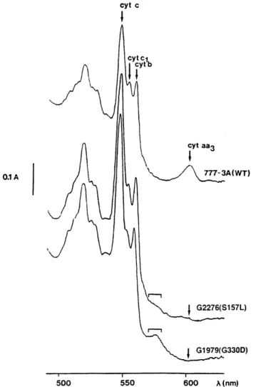
Cytochrome spectra of cox1 mutants
Cytochrome spectra of the wt strain and mutants G330D (representative of the mutants lacking of any respiratory activity) and S157L are shown in Fig. 1. As expected, the wt strain presents a significant peak at 602 nm correspond-ing to the αband of cytochromes a and a3. S157L shows a slight shoulder at this position and its cytochrome aa3/ cytochrome c ratio is estimated to be about 7% of wt. In
G330D, as in the 11 other mutants, no cytochromes aa3 can be detected. In all mutants, the cytochrome b/cyto-chrome c ratio and the cytob/cyto-chrome c1/cytob/cyto-chrome c ratio are decreased (by about 20–40%) indicating a deficiency in the accumulation of cytochromes bc1. Such a decrease in all the cytochromes has already been reported (Leme-sle-Meunier et al. 1993).
Analysis of the translation products in the cox1 mutants Since all the tested exonic cox1 mutants displayed a sig-nificant level of cox1 mRNA (data not shown), we exam-ined whether the respiratory deficiency of the mutants was due to an alteration either at the translational or at the post-translational level. The mitochondrial translation products are shown in Fig. 2. The band corresponding to Cox1p (39.5 kDa) was lacking in the frameshift and nonsense mutants (represented by G1547), as well as in the missense mutant G384D, and was present, but reduced, in the other missense mutants (represented by G330D). In two mutants
141
Fig. 1 Cytochrome spectra of whole cells from the wt strain 777-3A, mutants G1979 (G330D) and G2276 (S157L). Cells were grown for 2 days at 28 °C on solid YPGALa medium. Spectra were per-formed at –180 °C on dithionite-reduced whole-cell pastes (1-mm thick). In G1979 (G330D) and G2276 (S157L), the square bracket indicates the presence of a shoulder between 570 and 580 nm
Fig. 2 Autoradiograms of mitochondrial translation products. Cells were pulse-labelled for 60 min with 35SO
4in the presence of cyclo-heximide and mitochondrial membranes were purified and analyzed by an SDS polyacrylamide gel as described in the Materials and meth-ods. FS frameshift; NS nonsense; MS missense. G1547 is represen-tative of all the frameshift and nonsense mutants except G2508, while G1979 (G330D) is representative of all the missense mutants except G2394 (G384D). Note the presence of novel polypeptides (indicat-ed by J) in the strains G2508 and G2394 (G384D)
devoid of Cox1p, novel polypeptides were observed: in G384D we noted the presence of a 45-kDa polypeptide, which is not recognized by the monoclonal Cox1p anti-body we used (data not shown), and in G2508 the addi-tional polypeptide displays an apparent molecular weight of 20.5 kDa which is compatible with the predicted trun-cated polypeptide. In all the mutants, Cox2p and Cox3p, corresponding respectively to the 30.5-kDa and 23.5-kDa polypeptides, were synthesized.
Immunoblotting experiments with cox mutants
Using immunoblotting, we examined the stability of Cox1p, Cox2p and Cox3p and of the nuclear-encoded Cox4p, all of which are closely associated (Tsukihara et al. 1996), in the presence of mutations in each of the corre-sponding genes (cox1, cox2, cox3 or cox4 mutants). The same analysis was also performed on a rho° strain which lacks all three mitochondrial subunits. As a control, the stability of the nuclear-encoded Cox6p, which has no di-rect contact with Cox1p through Cox4p (Tsukihara et al. 1996), was also determined.
In the missense cox1 mutants, the levels of the three mitochondrially encoded subunits and of Cox4p are re-duced. This is illustrated in Fig. 3 with the mutant G330D (lane 2), which is devoid of any cytochrome c oxidase ac-tivity, and the mutant S157L (lane 3), which displays a re-sidual cytochrome c oxidase activity of about 20%. In the frameshift and nonsense cox1 mutants (represented by G1547, lane 4), Cox1p is absent and the levels of Cox2p, Cox3p and Cox4p are considerably decreased or else un-detectable. In the cox2 mutant (V44, lane 5) Cox1p is
present in small amounts and Cox3p and Cox4p are barely detectable, whereas in the cox3 mutant (V206, lane 6) sig-nificant amounts of Cox1p and Cox2p are observed and Cox4p accumulates to the wt level. In the absence of Cox4p (BGT1, lane 7) the levels of Cox1p and Cox2p are low, whereas Cox3p is even higher than in the wt strain. In the absence of the three mitochondrially encoded subunits (rho° strain, lane 8) Cox4p is absent. In all the mutants studied Cox6p accumulates as well as in the wt.
Discussion
Analysis of the amino-acid substitutions in the missense mutants
The analysis of the missense mutants reveals that, with the exception of S157L, the substitution of one amino acid has a drastic effect abolishing any accumulation of the cyto-chromes a and a3 and any cytochrome c oxidase activity. These observations are correlated with a decrease in Cox1p translation and in the level of Cox1p, Cox2p, Cox3p and Cox4p accumulated in the membranes. All these observa-tions led us to conclude that these mutants are altered in the assembly of the cytochrome c oxidase complex as a consequence of the instability of Cox1p. According to the crystal structure of bovine heart cytochrome c oxidase (Tsukihara et al. 1996) (see Fig. 4), five of the missense mutations are located in the trans-membrane helices: he-lix 4 (S157L), hehe-lix 6 (V244 M, E243 K and G251R) and helix 10 (G384D). Three mutations are in extra-membrane regions: K265 M and G330D in regions H6–7 and H8–9 respectively, orientated towards to the matrix side of the inner mitochondrial membrane, and H290Y in the H7–8 region on the periplasmic side.
Four mutations affect positions which are essential in the published structures (Iwata et al. 1995; Tsukihara et al. 1995, 1996). Our results are in good agreement with these data. The mutation H290Y alters an invariant histidine res-idue which is proposed to be one of the CuB ligands (Iwata et al. 1995; Tsukihara et al. 1995). In addition, Iwata et al. (1995) suggest that this residue could be involved in a his-tidine cycle/shuttle mechanism for the coupling of proton pumping to oxygen reduction. Three mutants (E243K, S157L and K265M) are altered in the positions of invari-ant or conserved amino acids (see Table 2) which have been proposed to be involved in proton channels either in the pathway for consumed protons (E243 and K265) (Iwata et al. 1995) or in the pathway for pumped protons (S157) (Tsukihara et al. 1996). S157L carries the only missense mutation, which led to a partial accumulation of an active complex in the mitochondrial membranes.
The three other mutants, G330D, G384D and G251R, do not affect invariant residues. However, we show here that they have important roles since they alter the level of Cox1p, Cox2p, Cox3p and Cox4p accumulated in the mem-branes and lead to an absence of detectable cytochrome c oxidase activity. All of them change a glycine residue. A
Fig. 3 Immunoblots of mitochondrial membranes from the wt strain, cox1, cox2, cox3 and cox4 mutants and the rho° strain. The loading corresponds to 40µg of mitochondrial membranes except when specified by * which corresponds to 20µg of mitochondrial membranes. Lanes 1 and 1* WT strain, 777-3A. Lane 2 G1979 (G330D), cox1 missense mutant defective in cytochrome c oxidase activity. Lane 3 G2276 (S157L), cox1 missense mutant displaying 20% of cytochrome c oxidase activity. Lane 4 G1547, frameshift
cox1 mutant. Lane 5 V44, cox2 mutant. Lane 6 V206, cox3 mutant. Lane 7 BGT1, cox4 mutant. Lane 8 rho° strain
similar substitution (G352 V) has been studied by Ortwein et al. (1997) but this resulted in a well-assembled but non-functional cytochrome c oxidase complex. In our case, the three amino-acid substitutions result in the appearance of a charge either in a trans-membrane region (TM6 for G251R and TM10 for G384D) or in a loop orientated to the matrix (H8–9 for G330D) (see Fig. 4). Consequently, we hypothesize that it is the presence of the charge which induces a change in the conformation of Cox1p, thereby altering its stability in the membranes.
Analysis of the stability of Cox1p, Cox2p, Cox3p, Cox4p and Cox6p in the mitochondrial membranes of mutants lacking one subunit
The comparison of the subunit content in the various mu-tants lacking either Cox1p, Cox2p, Cox3p or Cox4p shows that Cox1p, Cox2p, Cox3p and Cox4p accumulate in an inter-dependant way, whereas the accumulation of Cox6p appears to be independant from the four others. Indeed, Cox4p is not detectable in the rho° strain while Cox6p ac-cumulates as well as in the wt cells. Cox4p is also absent in cox1 and cox2 mutants, but not in cox3 mutants. Thus, the accumulation of Cox4p in the membranes appears to be dependent on the presence of Cox1p and Cox2p. By contrast, in all the mutants studied, Cox6p accumulates as well as in the wt strain. Similar observations have been made by Taanman et al. (1996) in the human cell line 143B206 (which is devoid of mitochondrial DNA), where the human homolog for Cox4p is reduced while the homo-log for Cox6p is present in amounts close to the mtDNA-containing parental cell line. Recently, Glerum and Tzag-oloff (1997) have reported that Cox4p is accumulated in
comparable amounts in a nuclear mutant altered in the mat-uration/stability of COX1 mRNA and in wt mitochondria, but in the mutant the protein is recovered almost entirely in the soluble fraction.
Taken together, these results confirm the interdepen-dance of the assembly of Cox4p and Cox1p in the mem-branes. The difference in the accumulation of the two nu-clearly encoded subunits Cox4p and Cox6p could be due to a distinct assembly pathway. This is supported by the crystallographic data (Tsukihara et al. 1997) showing that Cox4p and Cox6p are both extra-membrane subunits, but whereas Cox4p tightly interacts with Cox1p and Cox3p, Cox6p is without any direct contact with Cox1p.
Mitochondrial proteins of the inner membrane in yeast are rapidly degraded when not assembled. In particular, this has been shown by Nakai et al. (1994) for Cox2p and Cox3p which were degraded rapidly in a mutant lacking Cox4p. Several mitochondrial proteases have been identi-fied like the hetero-oligomeric Afg3p/Rca1p complex which degrade Cox1p and Cox3p, or Yme1p which is in-volved in the degradation of Cox2p (for review see Langer
143 Fig. 4 Two-dimensional
or-ganization of the cytochrome c oxidase subunit I of S.
cerevi-siae. The distribution of the
trans-membrane helices and of the two helices parallel to the membrane has been deduced from the structure of subunit I of cytochrome oxidase from beef heart (Tsukihara et al. 1996) and after alignment of its sequence with that of S.
cerevi-siae. Rectangles in the
N-termi-nal and H9–10 regions are α helices found in the extra-mem-brane region. The localization of the eight missense mutations and the amino-acid changes ob-served in these mutants are in-dicated. The extra-membrane regions are numbered from H1–2 to H11–12, and the trans-membrane helices from TM1 to TM12 according to the nomen-clature of Iwata et al. (1995)
Fig. 5 Hypothetical model for the sequential assembly of the three mitochondrially encoded subunits (Cox1p, Cox2p and Cox3p) and the nuclear-encoded subunit (Cox4p) of cytochrome c oxidase into the mitochondrial membranes. IMS inter-membrane space; M matrix
and Neupert 1996; Suzuki et al. 1997). However, Glerum and Tzagoloff (1997) have shown that in a cox6 back-ground, the steady state amount of Cox1p is not regulated by Afg3p/Rca1p and these authors have suggested that other proteases could exist. It would be interesting to de-termine which protease(s) is (are) involved in the subunit degradation in our cytochrome c oxidase mutants.
In conclusion, the deleterious effect produced by the absence of Cox1p on Cox2p, Cox3p and Cox4p suggests that Cox1p is necessary for the assembly of these sub-units. In the cox2 mutant, Cox3p and Cox4p were also absent indicating that their accumulation requires the presence of Cox2p. In the cox3 mutant, Cox1p, Cox2p and Cox4p accumulate in a significant way and a slight peak of cytochrome aa3 is observable in this strain, whereas no cytochrome c oxidase activity is detectable (data not shown). All these observations suggest that the assembly of Cox1p through Cox4p follows a sequential mode as schematized in Fig. 5. The first step could be the association of Cox2p with Cox1p, then Cox4p would as-sociate with Cox1p, and finally Cox3p would stabilize the three other subunits. We will test this model by im-munoprecipitation experiments. Also further experi-ments on other mutants altered in the seven remaining subunits will be necessary to better understand the se-quence of assembly of the cytochrome c oxidase complex into the mitochondrial membranes.
Acknowledgements This work was supported by a European Com-munity Stimulation Contract (CT90-0476) C. L. thanks Drs. M. Ger-vais, J. Daniel and M. Claisse for stimulating discussions. We also thank D. Menay for the synthesis of oligonucleotides. We are grate-ful to Drs. G. Dujardin, O. Groudinsky and to Dr. C. J. Herbert for critical reading of the manuscript and to C. J. H. for looking over the English.
References
Baranowska H, Szcesniak B, Ejchart A, Kruszewska A, Claisse M (1983) Recombinational analysis of oxi2 mutants and prelimi-nary analysis of their translation products in S. cerevisiae. Curr Genet 7: 225–233
Bonitz S, Corruzi G, Thalenfeld B, Tzagoloff A, Macino G (1980) Assembly of the mitochondrial membrane system – structure and nucleotide sequence of the gene coding for subunit I of yeast cy-tochrome oxidase. J Biol Chem 255: 11927–11941
Capaldi RA (1990) Structure and assembly of cytochrome c oxidase. Arch Biochem Biophys 280: 252–262
Carignani G, Netter P, Bergantino E, Robineau S (1986) Expression of the mitochondrial split gene coding for cytochrome oxidase subunit I in S. cerevisiae: RNA splicing pathway. Curr Genet 11: 55–63
Claisse M, Pere-Aubert G, Clavilier L, Slonimski PP (1970) Méthode d’estimation de la concentration des cytochromes dans les cel-lules entières de levure. Eur J Biochem 16: 430–438
Claisse ML, Slonimski PP, Johnson J, Mahler HR (1980) Mutations within an intron and its flanking sites: patterns of novel polypep-tides generated by mutants in one segment of the cob-box region of yeast mitochondrial DNA. Mol Gen Genet 177: 375–387 Dujardin G, Pajot P, Groudinsky O, Slonimski PP (1980) Long-range
control circuits within mitochondria and between nucleus and mi-tochondria. I. Methodology and Phenomenology of Suppressors. Mol Gen Genet 179: 469–482
Dujardin G (1983) Thèse de Doctorat d’Etat. Université Paris 6 Geier BM, Schagger H, Ortwein C, Link TA, Hagen WR, Brandt U
(1995) Kinetic properties and ligand binding of the 11-subunit cytochrome c oxidase from Saccharomyces cerevisiae isolated with a novel large-scale purification method. Eur J Biochem 227: 296–302
Glerum DM, Tzagoloff A (1997) Sub-mitochondrial distributions and stabilities of subunits 4, 5 and 6 of yeast cytochrome oxidase in assembly defective mutants. FEBS Lett 412: 410–414 Groudinsky O, Dujardin G, Slonimski PP (1981) Long-range
con-trol circuits within mitochondria and between nucleus and mito-chondria. II. Genetic and biochemical analyses of suppressors which selectively alleviate the mitochondrial intron mutations. Mol Gen Genet 184: 493–503
Hensgens LAM, Bonen L, de Haan M, Horst G, Grivell LA (1983) Two intron sequences in the yeast mitochondrial COX I gene: ho-mology among URF-containing introns and strain-dependent variation in flanking exons. Cell 32: 379–389
Iwata S, Ostermeier C, Ludwig B, Michel H (1995) Structure at 2.8 A resolution of cytochrome c oxidase from Paracoccus
denitrifi-cans. Nature 376: 660–669
Kotylak Z, Slonimski PP (1977) Mitochondrial mutants isolated by a new screening method based upon the use of the nuclear muta-tion op1. In: Bandlow W, Schweyen RJ, Wolf K, Kaudewitz F (eds) Mitochondria (1977) Genetics and biogenesis of mitochon-dria. de Gruyter, Berlin New York, pp 83–89
Kruszewska A, Szcesniak B, Claisse M (1980) Recombinational analysis of OXI1 and preliminary analysis of their translation products in S. cerevisiae. Curr Genet 2: 45–51
Laemmli UK (1970) Cleavage of structural proteins during the assembly of the head of bacteriophage T4. Nature 227: 680–685
Langer T, Neupert W (1996) Regulated protein degradation in mito-chondria. Experientia 52: 1069–1076
LaMarche AEP, Abate MI, Chan SHP, Trumpower BL (1992) Isola-tion and characterizaIsola-tion of COX12, the nuclear gene for a pre-viously unrecognized subunit of S. cerevisiae cytochrome c ox-idase. J Biol Chem 267: 22473–22480
Lemarre P, Robineau S, Colson A-M, Netter P (1994) Sequence anal-ysis of three deficient mutants of cytochrome oxidase subunit I of Saccharomyces cerevisiae and their revertants. Curr Genet 26: 546–552
Lemesle-Meunier D, Brivet-Chevillotte P, di Rago J-P, Slonimski PP, Bruel C, Tron T, Forget N (1993) Cytochrome b-deficient mutants of the ubiquinol-cytochrome c oxidoreductase in
Saccharomyces cerevisiae. Consequence for the functional and
structural characteristics of the complex. J Biol Chem 268: 15626–15632
Nakai T, Mera Y, Yasuhara T, Ohashi A (1994) Divalent metal ion-dependent mitochondrial degradation of unassembled subunits 2 and 3 of cytochrome c oxidase. J Biochem 116: 752–758 Netter P, Carignani G, Jacq C, Groudinsky O, Clavilier L,
Slonim-ski PP (1982) The cytochrome oxidase subunit I split gene in
S. cerevisiae: genetic and physical studies of the mtDNA
seg-ment encompassing the ‘cytochrome b-homologous’ intron. Mol Gen Genet 188: 51–59
Netter P, Robineau S, Lemaire C (1995) Mutations in the mitochon-drial split gene COXI are preferentially located in exons: a map-ping study of 172 mutants. Mol Gen Genet 246: 445–454 Ortwein C, Link TA, Meunier B, Colson-Corbisier A-M, Rich PR,
Brandt U (1997) Structural and functional analysis of deficient mutants in subunit I of cytochrome c oxidase from
Saccharomyc-es cerevisiae. Biochim Biophys Acta 1321: 79–92
Oudshoorn P, Van Steeg H, Swinkels BW, Schoppink P, Grivell LA (1987) Subunit II of yeast QH2: cytochrome-c oxidoreductase. Nucleotide sequence of the gene and features of the protein. Eur J Biochem 163: 97–103
Pajot P, Wambier-Kluppel ML, Kotylak Z, Slonimski PP (1976) Genetics and biogenesis of chloroplasts and mitochondria. Else-vier North Holland Biomedical Press, Amsterdam, pp 443–451 Poyton RO, Mc Ewen JE (1996) Cross-talk between nuclear and
Rubin MS, Tzagoloff A (1973) Assembly of the mitochondrial mem-brane system. X. Mitochondrial synthesis of three of the subunit proteins of yeast cytochrome oxidase. J Biol Chem 249: 4269– 4274
Sanger F, Nicklen S, Coulson AR (1977) DNA sequencing with chain-terminating inhibitors. Proc Natl Acad Sci USA 74: 5463– 5467
Suzuki CK, Rep M, van Dijl JM, Suda K, Grivell LA, Schatz G (1997) ATP-dependent proteases that also chaperone protein biogenesis. Trends Biochem Sci 22: 118–123
Taanman JW, Capaldi RA (1993) Subunit VIa of yeast cytochrome
c oxidase is not necessary for assembly of the enzyme complex
but modulates the enzyme activity. Isolation and characterization of the nuclear-coded gene. J Biol Chem 268: 18754–18761 Taanman J-W, Burton MD, Marusich MF, Kennaway NG, Capaldi
RA (1996) Subunit-specific monoclonal antibodies show differ-ent steady state levels of various cytochrome-c oxidase sub-units in chronic progressive external ophthalmoplegia. Biochim Biophys Acta 1315: 199–207
Towbin H, Staehelin T, Gordon J (1979) Electrophoretic transfer of proteins from polyacrylamide gels to nitrocellulose sheets: pro-cedure and applications. Proc Natl Acad Sci USA 76: 4350–4354 Tron T, Lemesle-Meunier D (1990) Two substitutions at the same position in the mitochondrial cytochrome b gene of S. cerevisiae induce a mitochondrial myxothiazol resistance and impair the respiratory growth of the mutated strains albeit maintaining a good electron transfer activity. Curr Genet 18: 413–419 Tsukihara T, Aoyama H, Yamashita E, Tomizaki T, Yamaguchi H,
Shinzawa-Itoh K, Nakashima R, Yaono R, Yoshikawa S (1995) Structures of metal sites of oxidized bovine heart cytochrome c oxidase at 2.8A. Science 269: 1069–1074
Tsukihara T, Aoyama H, Yamashita E, Tomizaki T, Yamaguchi H, Shinzawa-Itoh K, Nakashima R, Yaono R, Yoshikawa S (1996) The whole structure of the 13-subunit oxidized cytochrome c ox-idase at 2.8A. Science 272: 1136–1144