HAL Id: hal-01480146
https://hal-amu.archives-ouvertes.fr/hal-01480146
Submitted on 14 Mar 2017
HAL is a multi-disciplinary open access
archive for the deposit and dissemination of
sci-entific research documents, whether they are
pub-lished or not. The documents may come from
teaching and research institutions in France or
L’archive ouverte pluridisciplinaire HAL, est
destinée au dépôt et à la diffusion de documents
scientifiques de niveau recherche, publiés ou non,
émanant des établissements d’enseignement et de
recherche français ou étrangers, des laboratoires
diffuse gliomas in the French POLA cohort
Emeline Tabouret, Anh Tuan Nguyen, Caroline Dehais, Catherine Carpentier,
François Ducray, Ahmed Idbaih, Karima Mokhtari, Anne Jouvet, Emmanuelle
Uro-Coste, Carole Colin, et al.
To cite this version:
Emeline Tabouret, Anh Tuan Nguyen, Caroline Dehais, Catherine Carpentier, François Ducray, et
al.. Prognostic impact of the 2016 WHO classification of diffuse gliomas in the French POLA cohort.
Acta Neuropathologica, Springer Verlag, 2016, 132 (4), pp.625-634. �10.1007/s00401-016-1611-8�.
�hal-01480146�
Acta Neuropathol
DOI 10.1007/s00401-016-1611-8
ORIGINAL PAPER
Prognostic impact of the 2016 WHO classification of diffuse
gliomas in the French POLA cohort
Emeline Tabouret
1,2· Anh Tuan Nguyen
3· Caroline Dehais
4· Catherine Carpentier
5· François Ducray
6,7·
Ahmed Idbaih
4,5· Karima Mokhtari
4,5,8· Anne Jouvet
9· Emmanuelle Uro‑Coste
10,11· Carole Colin
2·
Olivier Chinot
1,2· Hugues Loiseau
12· Elisabeth Moyal
13,14,15· Claude‑Alain Maurage
16· Marc Polivka
17·
Emmanuèle Lechapt‑Zalcman
18,19· Christine Desenclos
20· David Meyronet
7,9· Jean‑Yves Delattre
4,5·
Dominique Figarella‑Branger
2,3· For POLA Network
Received: 20 July 2016 / Revised: 12 August 2016 / Accepted: 22 August 2016 © Springer-Verlag Berlin Heidelberg 2016
(range 17.1–84.4). Based on the new histomolecular
classi-fication, diagnoses included anaplastic oligodendroglioma
IDH
mutant and 1p/19q-codeleted (32.5 %), anaplastic
astrocytoma IDH mutant (IDH
mut) (11.0 %), anaplastic
astrocytoma IDH wild type (IDH
wt) (5.3 %), glioblastoma
IDH
mut(17.1 %), and glioblastoma IDH
wt(33.2 %). Ten
patients presented with a diffuse midline tumor, H3 K27M
mutant. The new WHO classification was prognostic for
progression-free survival (PFS) and overall survival (OS)
(p < 0.001). We did not find prognosis differences between
grades III and IV for IDH
mut1p/19q intact and IDH
wtglio-mas in univariate and multivariate analyses. Among
ana-plastic astrocytoma IDH
wt, cases with chromosome arm 7p
gain and 10q loss (55 %) had shorter PFS than the others
(p = 0.027). In conclusion, the new WHO histomolecular
Abstract The new WHO classification of diffuse gliomas
has been refined and now includes the 1p/19q codeletion,
IDH1/2
mutation, and histone H3-K27M mutation. Our
objective was to assess the prognostic value of the updated
2016 WHO classification in the French POLA cohort.
All cases of high-grade oligodendroglial tumors sent for
central pathological review and included into the French
nationwide POLA cohort were reclassified according to
the updated 4th WHO classification. In total, 1041 patients
were included, with a median age at diagnosis of 50.4 years
These results were presented, in part, at the 2016 ASCO annual meeting in poster discussion (abstract number 2015).
Electronic supplementary material The online version of this article (doi:10.1007/s00401-016-1611-8) contains supplementary material, which is available to authorized users.
* Dominique Figarella-Branger
dominique.figarella-branger@univ-amu.fr
1 APHM, Hôpital de la Timone, Service de Neurooncologie,
Marseille, France
2 Aix-Marseille Université, Inserm, CRO2 UMR_S 911,
Marseille, France
3 APHM, Hôpital de la Timone, Service d’Anatomie
Pathologique et de Neuropathologie, Marseille, France
4 AP-HP, Hôpitaux Universitaires La Pitié Salpêtrière, Charles
Foix, Service de Neurologie 2-Mazarin, 75013 Paris, France
5 Inserm U1127, CNRS UMR 7225, Sorbonne Universités,
UPMC Univ Paris 06 UMRS1127, Institut du Cerveau et de la Moelle épinière, ICM, 75013 Paris, France
6 Hospices Civils de Lyon, Hôpital Pierre Wertheimer, Service
de Neurooncologie, Bron, France
7 Department of Cancer Cell Plasticity, Cancer Research
Centre of Lyon, INSERM U1052, CNRS UMR5286, Lyon, France
8 AP-HP, Groupe Hospitalier Pitié-Salpêtrière, Service de
Neuropathologie Raymond Escourolle, Paris, France
9 Centre de pathologie et de neuropathologie Est, Bron, France 10 CHU Toulouse, Hôpital Rangueil, Service d’Anatomie
Pathologique et Histologie-Cytologie, Toulouse, France
11 Inserm U1037, Centre de Recherche en Cancérologie de
Toulouse, Université de Toulouse, Toulouse, France
12 CHU Bordeaux, Hôpital Pellegrin, Service de
Neurochirurgie, Bordeaux, France
13 Department of Radiation Oncology, Institut Claudius
Regaud/Institut Universitaire du Cancer de Toulouse, Oncopôle, Toulouse, France
14 Université Toulouse III Paul Sabatier, Toulouse, France 15 INSERM U1037, Centre de Recherches contre le Cancer de
Toulouse, Toulouse, France
16 CHU Lille, Pôle Pathologie Biologique, Service Anatomie
classification of diffuse gliomas presented with high
prog-nostic value. Grading was not discriminant between grade
III and IV high-grade gliomas.
Keywords Diffuse glioma · 2016 WHO classification ·
IDH1/2
mutation · 1p/19q codeletion
Background
Diffuse gliomas are the most frequent and aggressive
pri-mary brain tumors in adults, and until recently, they were
classified according to the 4th edition of the World Health
Organization (WHO) classification published in 2007 [
16
].
The former classification took into account the histological
subtype (astrocytic, oligodendrocytic, and oligoastrocytic)
and grade, ranging from grade II to grade IV
glioblas-toma, which is a highly malignant invasive and angiogenic
tumor. Inconstant reproducibility and interobserver
vari-ability were critical points of this classification, relying on
pathological criteria only [
5
]. Molecular understanding of
gliomagenesis was first improved with the identification of
the 1p/19q codeletion, associated with an oligodendroglial
phenotype and with a better prognosis [
26
]. This alteration
appeared to be a predictive marker of response to
procar-bazine, CCNU, and vincristine (PCV) [
2
,
3
]. More recently,
integrated genomic analysis of gliomas has identified IDH
mutation as the key alteration in gliomagenesis [
20
]. IDH
mutation characterizes adult grade II and III gliomas as
well as secondary glioblastoma [
28
] and is of prognostic
significance. Therefore, it was an appropriate time to
intro-duce molecular markers into the WHO classification [
17
].
Updating the 4th WHO classification of tumors of the
central nervous system (CNS) has yielded major changes
in the group of glial tumors [
18
]. The updated
classifica-tion stratifies the group of “diffuse astrocytic and
oligoden-droglial tumors” according to the occurrence of two major
genetic alterations: IDH mutation and 1p/19q codeletion. It
recognizes “diffuse midline glioma, H3 K27M mutant” as
a new entity. In addition, according to this new
classifica-tion, the diagnosis of mixed glioma, a category that was not
sharply defined until recently and was subject to high
inter-observer discordance [
1
], is strongly discouraged.
In France, since 2008, a dedicated program has been set
up for more homogeneous management of de novo adult
high-grade glioma with an oligodendroglial component
[prise en charge des oligodendrogliomes anaplasiques
(POLA network)]. The aim of the program inter alia is to
provide a pathological centralized review of the cases and
centralized molecular analysis.
The aim of this study was to reclassify the entire POLA
cohort according to the recent update of the 4th WHO
clas-sification of CNS tumors to analyze its prognostic and
dis-criminant values.
Materials and methods
Patients
All 1041 patients who were sent for a central pathological
review because of the suspicion of diffuse high-grade
glio-mas with an oligodendroglial component and included into
the French nation-wide POLA cohort on June 6, 2015 were
included in this study. For all cases, formalin-fixed,
paraf-fin-embedded (FFPE) tumor tissue was available for
patho-logical and immunohistochemical analyses. In addition,
frozen material was available in up to 974 cases. Initial
WHO 2007 diagnosis was retained after centralized review
of all cases by four national neuropathological experts. At
the time of the review process, the experts were blind of
1p19q status determination, and for cases enrolled before
2010, IDH1R132H expression was also unknown.
Patients prospectively included into the POLA cohort
provided their written consent for clinical data collection
and genetic analysis according to national and POLA
net-work policies.
Clinical characteristics of the cohort are summarized in
Table
1
.
Pathological review according to the 2007 WHO
classification and immunohistochemistry
After the initial diagnosis of high-grade glioma with an
oli-godendroglial component by local pathologists, cases were
centrally reviewed and included in the prospective POLA
cohort. In addition, automated IHC was performed on
4-µm-thick FFPE sections with an avidin–biotin–peroxidase
complex on Benchmark XT (Ventana Medical System Inc,
Tucson AZ, USA) using the Ventana Kit including DAB
rea-gent to search for the expression of IDH1 R132H (Dianova,
H09), P53 (DAKO, DO.7), and ATRX (SIGMA, polyclonal).
17 AP-HP, Hôpital Lariboisière, Service d’Anatomie et
Cytologie Pathologique, Paris, France
18 CHU Caen, Hôpital de la Côte de Nacre, Service d’anatomie
Pathologique, Caen, France
19 CNRS, UMR 6301 ISTCT, CERVOxy, GIP CYCERON,
Caen, France
20 CHU Amiens, Service de neurochirurgie, Hôpital Nord,
Acta Neuropathol
Results
Patient characteristics and pathological diagnosis
In total, 1041 cases who were addressed to a central
path-ological review because of the suspicion of diffuse
high-grade gliomas with an oligodendroglial component and
were diagnosed between September 2008 and June 2015
were included (Table
1
). The median follow-up period was
19.0 months (range 0.1–77.0 months). The median age at
diagnosis was 50.4 years (range 17.1–84.4). Only 7 % of
patients presented with an altered functional status, while
14 % had cognitive disorders at diagnosis. Half of the
patients benefited from gross total resection, and the first-line
treatment corresponded to the association of chemotherapy
and radiotherapy for more than 65 % of patients (Table
1
).
According to the 2007 WHO classification, these 1041 cases
were classified as follows: anaplastic oligodendroglioma
(31.6 %), anaplastic oligoastrocytoma (27.6 %), anaplastic
astrocytoma (7 %), glioblastoma with an oligodendroglial
component (14.9 %), and glioblastoma (18.9 %).
Molecular data and histomolecular classification
according to the 2016 WHO classification (Fig. 1)
Sixty percent of patients presented with IDH mutant
(IDH-mut
) tumors, and 34 %of patients presented with
1p/19q-codeleted tumors (34.5 %) (Supplementary Table 1).
Among the 626 patients with IDH
muttumor, 599 presented
with IDH1
mut[R132H: 562 (94 %), R132C: 15, R132G: 13,
R132S: 6, and R132L: 3] and 27 presented with IDH2
mut(R172K: 19, R172M: 5, R172I: 1, R172S: 1, and R172 W:
1). Forty-six percent of patients presented with P53
immu-nostaining in nuclei above 10 %. Immuimmu-nostaining of ATRX
was not detected in 90 % of patients with IDH
mutwithout
1p/19q-codeleted glioma, (Supplementary Table 2).
Based on the new updated WHO classification (Table
2
),
the reclassification of the POLA cases showed a substantial
change in grade (IV versus III) with an increase in
glioblas-toma diagnoses (50.3 versus 33.8 %), while the frequency
of oligodendroglioma remained stable. Thus, the 1041
cases were reclassified as anaplastic oligodendroglioma
IDH
mut1p/19q-codeleted (32.5 %), anaplastic astrocytoma
IDH
mut(10.9 %), anaplastic astrocytoma IDH
wt(5.3 %),
glioblastoma IDH
mut(17.2 %), and glioblastoma IDH
wt(33.1 %). Ten patients presented a midline tumor with the
histone H3 K27M mutation (Fig.
1
).
Mixed anaplastic oligoastrocytoma and glioblastoma
with an oligodendroglial component, which have been
removed from the new classification, were reclassified
as anaplastic oligodendroglioma IDH
mut1p/19q-code-leted (16.1 %), anaplastic astrocytoma IDH
mut(14.7 %),
DNA extraction, single‑nucleotide polymorphism (SNP)
arrays, and comparative genomic hybridization (CGH)
arrays
Following the manufacturer’s recommendations, tumor
DNA was extracted from frozen tissue, if available, or from
FFPE samples using the iPrep ChargeSwitch
®Forensic Kit.
Qualification and quantification of tumor DNA were
per-formed using a NanoVue spectrophotometer and gel
elec-trophoresis, respectively. In 974 cases, the genomic
pro-file and assessment of the 1p/19q codeletion status were
determined as described previously [
7
]. When the quantity
of DNA was insufficient to perform SNP or CGH arrays
(n = 6), microsatellite analysis was conducted, and
micro-satellite analyses (LOH) of chromosomes 1p and 19q were
assessed via PCR techniques described elsewhere [
13
].
In addition, particular attention was paid to the
follow-ing alterations: chromosome 7 gain, chromosome 10 loss,
PTEN
deletion, and EGFR amplification.
IDH1, IDH2, and TERT mutation status
When the results of IDH1R132H immunohistochemistry
were negative or unreliable, the status of IDH1 and IDH2
mutation was addressed by direct sequencing using the
Sanger method and primers, as described previously [
10
].
TERT
mutation status was also addressed in 771 cases by
direct sequencing using the Sanger method and primers, as
described previously [
14
].
Statistical analysis
SNP and CGH array analyses were performed as described
previously [
12
]. For all arrays, genomic imbalances were
classified as loss, gain, homozygous deletion, or
ampli-fication. For correlation analysis, the Chi-square test (or
Fisher’s exact test) was used to compare variables when
they were scored as positive or negative. Continuous
vari-ables were compared using the Mann–Whitney U test.
Pro-gression-free survival (PFS) was defined as the time from
the date of surgery to recurrence or death from any cause.
Overall survival (OS) was defined as the time from the date
of surgery to death from any cause. The Kaplan–Meier
method was used to estimate survival distributions.
Log-rank tests were used for univariate comparisons. Cox
pro-portional hazards models were used for multivariate
analy-ses and for estimating hazard ratios in survival regression
models. Multivariate analysis included all variables with
a p value <0.05 in univariate analyses. All statistical tests
were two-sided, and the threshold for statistical
signifi-cance was p = 0.05. Analyses were conducted using PASW
Statistics version 20 (IBM SPSS Inc., Chicago, IL, USA).
anaplastic astrocytoma IDH
wt(3.0 %), glioblastoma IDH
mut(37.1 %) glioblastoma IDH
wt(28.6 %), and midline glioma
with the histone H3 K27M mutation (0.5 %)
(Supplemen-tary Table 3; Fig.
2
).
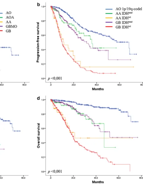
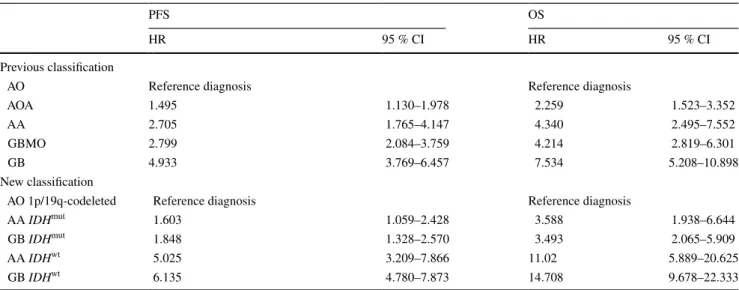
Prognostic value of WHO classifications and grading
The median PFS and OS were 23.8 months [95 %
confi-dence interval (CI): 21.1–26.5] and 62.0 months (95 % CI:
52.0–72.0), respectively. Both the 2007 WHO and the new
updated WHO classifications of gliomas were prognostic
for PFS and OS (p < 0.001) (Fig.
3
; Table
3
). Moreover,
the new WHO 2016 classification presented with high
haz-ard ratio both for PFS and OS (Table
3
), higher than those
observed for the previous 2007 classification, reinforcing
the discriminative value of this new histomolecular
clas-sification. Grading (III versus IV) for IDH
mut1p/19q intact
gliomas was not prognostic in either univariate analysis
(PFS: p = 0.505; OS: p = 0.838) (Fig.
3
b, d) or
multivari-ate analysis (adjusted by the age, type of surgery, and
first-line treatment). In addition, no prognostic difference was
observed between grade III and IV IDH
wtgliomas (Fig.
3
b,
d) in terms of PFS (p = 0.449) and OS (p = 0.335) in
uni-variate and multiuni-variate analyses (adjusted by the age, type
of surgery, and first-line treatment). Among patients with
anaplastic astrocytoma IDH
wt, cases presenting with 7p
gain and 10q loss (55 %, Supplementary Table 4) had a
worse prognosis than others in terms of PFS (p = 0.027)
but not OS. This finding suggests that anaplastic
astrocy-toma IDH
wtremains a heterogeneous subgroup.
TERT prognostic value
TERT
mutation status was available for 771 patients.
Among them, 59 % (N = 457) presented with mutated
TERT
(TERT
mut) tumor: 133 patients with the C250T
muta-tion, 321 patients with the C228T mutamuta-tion, and 3 patients
with both. A TERT
mutwas observed in 93 % (257/275),
10 % (22/222), and 65 % (176/269) of anaplastic
oligo-dendroglioma IDH
mut1p/19q-codeleted, IDH
mut1p/19q
intact gliomas, and IDH
wtgliomas, respectively. No
prog-nostic impact of TERT
mutwas observed for OS. TERT
mutwas associated with a worse PFS for glioblastoma IDH
wtpatients, but no PFS impact was observed in grade III
IDH
wtgliomas, while in this last subgroup, TERT
mutwas
associated with the 7p gain and 10q loss (p = 0.018)
(Sup-plementary Table 4).
Table 1 Patient characteristics
KPS Karnofsky performance status, PCV procarbazine–CCNU–vin-cristine, MVP microvascular proliferation; IDH1/2mut
IDH1/2-muta-tion
a Type of surgery was determined by operating report of
neurosur-geon
Factors N %
Age (median, range, years) 50.4 (17.1–84.4) Gender (men/women) 549/397 58/42 Unknown 95 KPS <60 5 1.3 60 22 5.2 70 48 11.4 80 60 14.3 90–100 285 67.8 Unknown 621
Patients with cognitive disorders at diag-nosis
115 14
Unknown 215
Type of surgerya
Gross total resection 474 51.8 Biopsy or partial resection 440 48.2
Unknown 127 First-line treatment None 26 2.9 Radiotherapy 132 14.8 PCV + radiotherapy 151 16.9 Stupp protocol 450 50.4 Chemotherapy alone 78 8.7 Other 55 6.2 Unknown 149 Pathological characteristics
Mitosis without necrosis nor MVP 234 22.4 MVP without necrosis 456 43.8 Necrosis and MVP 351 33.8 Immunostaining and molecular alterations
IDH1/2mut 626/1041 60.1 1p/19q codeletion 338/980 34.5 ATRX loss 281/772 63.5 P53 expression >10 % 484/1041 46.5 H3 K27M mutation 10 1.0 TERT mutation 457/771 59.3 7p gain and 10q loss 246/976 25.2 PTEN loss 217/965 22.5 EGFR amplification 124/966 12.8
Acta Neuropathol
Discussion
In this study, we were able to reclassify all the POLA series
according to the 2016 WHO classification of brain tumors.
Notably, the percentage of each category of diffuse, adult
high-grade gliomas recorded in this study does not reflect
the normal distribution of malignant glioma subgroups
because of the inclusion criteria in the POLA network,
i.e., adult malignant glioma with an oligodendroglial
com-ponent. Based on these results, our first observation was
a switch in grading (III versus IV) with an increased
fre-quency of glioblastoma. Although this switch was recorded
for different entities, it was more frequent for the cases
ini-tially diagnosed as grade III oligoastrocytoma, because up
to 50 % cases were reclassified as glioblastoma (IDH
mutor
IDH
wt). Our second observation was that the new updated
2016 WHO classification had a high prognostic value. Our
third observation was that grading III versus IV was
prog-nostic for neither IDH
mut1p/19q intact gliomas nor IDH
wtgliomas, which brings into question the relevance of grade
in these tumors.
In the POLA series, more than 400 cases were previously
classified according to the 2007 WHO as mixed gliomas, a
diagnostic category that should be avoided according to the
2016 classification. Of note, recommendation to limit mixed
glioma diagnosis was reinforced by the recent publications
on glioblastoma with oligodendroglial component (GBMO),
which suggested that GBMO corresponded to various and
distinct molecular entities [
6
,
9
]. We were able to reclassify
these cases on the basis of molecular characteristics. Most
diffuse high-grade gliomas are now stratified according to
IDH mutation and 1p/19q codeletion. In the present series,
Fig. 1 Molecular and immunostaining alterations and patient age and survival (in months) according to the new 2016 WHO classifica-tion of gliomas. O oligodendroglioma, A astrocytoma, GB
glioblas-toma, K27M diffuse midline glioma with histone H3 K27M mutation,
CODEL 1p/19q codeletion, IDH-MUT IDH1/2 mutant, and IDH-WT
IDH1R132H immunostaining was sufficient to assess the
IDH
status in 562 cases (94 %) showing strong IDH1R132H
expression, a frequency similar to that observed previously
[
25
]. In the remaining cases, IDH sequencing revealed
IDH1
mutation in 37 cases and IDH2 mutation in 27. Loss
of nuclear ATRX expression was almost mutually
exclu-sive from 1p/19q codeletion. We observed only three cases
that demonstrated both alterations, even after repeating
immunostaining and CGH analysis. These cases may
cor-respond to the extremely rare “true” mixed glioma [
11
].
Although most IDH
mut(1p/19q-intact) gliomas exhibited
loss of ATRX (249/276), we observed 27 cases in which
ATRX nuclear expression was retained. This rare
molecu-lar phenotype has been reported previously [
23
]. Another
rare molecular phenotype was represented by loss of nuclear
ATRX expression in IDH
wtgliomas (20 cases); among these,
a histone mutation was recorded in five of the 16 cases
stud-ied. We also observed that anaplastic oligodendroglioma
IDH
wtand 1p/19q-codeleted are exceptional (four cases).
This last result further confirmed that almost all
1p/19q-codeleted oligodendrogliomas are IDH mutants, although
some exceptions exist [
3
]. Because CGH analysis was
per-formed in all cases, in addition to the 1p/19q status, we had
information regarding the main alterations that characterize
glioblastomas, i.e., the association of chromosome arm 7p
gain and 10q loss. We observed that among the 55
anaplas-tic astrocytomas IDH
wt, 30/55 exhibited these alterations (7p
gain and 10q loss). Some authors suggested that such cases
should be classified as glioblastoma [
23
,
24
,
27
]. Reuss et al.
reported that up to 78 % of anaplastic astrocytoma IDH
wtpresented with glioblastoma molecular alterations, while
9 % were diagnosed as glioblastoma H3F3A mutated in their
series [
21
]. However, in this study, only 55 % of anaplastic
astrocytoma IDH
wtdemonstrated glioblastoma molecular
alterations, suggesting that all anaplastic IDH
wtastrocytoma
should not be classified as glioblastoma.
Interestingly, we found that the 2016 WHO
classifica-tion was highly accurate in predicting survival, confirming
the value of adding molecular characteristics. However,
this classification has some limitations. We observed that
Fig. 2 Repartition of the mixed oligoastrocytomas according to the updated 2016 WHO clas-sification
Table 2 Reclassification of the 1041 cases of the French POLA cohort according to the updated 4th WHO classification
IDHmut IDH1/2-mutant, IDHwt IDH1/2-wild type, 7p+/10q−
chro-mosome arm 7p gain with chrochro-mosome arm 10q loss
Integrated diagnoses N = 1041 %
Anaplastic oligodendroglioma. IDHmut and 1p/19q
codeletion
334 32.1 Anaplastic oligodendroglioma, IDHwt and 1p/19q
codeletion
4 0.4 Anaplastic astrocytoma, IDHmut 114 10.9
Anaplastic astrocytoma, IDHmut ATRX lost 107
Anaplastic astrocytoma, IDHmut ATRX preserved 6
Anaplastic astrocytoma, IDHmut, ATRX unknown 1
Anaplastic astrocytoma, IDHwt 55 5.3
With 7p+/10q− 30
Glioblastoma, IDHmut 178 17.2
Glioblastoma, IDHmut ATRX lost 151
Glioblastoma, IDHmut ATRX preserved 21
Glioblastoma, IDHmut, ATRX unknown 6
Glioblastoma, IDHwt 346 33.1
Acta Neuropathol
the same class of glioblastoma could refer to distinct
enti-ties, while different designations (anaplastic astrocytoma
and glioblastoma) could refer to patient groups with
simi-lar outcomes. As previously reported for grade II and III
gliomas [
4
,
27
], we observed that grade III and IV adult
diffuse gliomas can be divided into three major groups
hav-ing distinct prognoses accordhav-ing to the IDH and 1p/19q
codeletion status. The best prognosis was observed in
anaplastic oligodendroglioma IDH
mut1p/19q-codeleted,
the worst prognosis was observed in IDH
wtgliomas, and
an intermediate prognosis was observed in IDH
mut1p/19q
intact gliomas. We have also reported a distinct age
repar-tition between these three main groups (Fig.
1
).
Interest-ingly, among the groups of IDH
wtgliomas and IDH
mut1p/19q intact gliomas, grade III versus IV did not impact
survival in our series. The relevance of the grading
prog-nostic impact in the context of molecular subgroups was
previously questioned in a limited number of recent
stud-ies. If the impact of grading between grade II and III
glio-mas was already challenged by several studies [
19
,
27
], the
current data remained more contradictory between grade III
and IV gliomas [
8
,
22
], suggesting, in these publications, a
possible remaining prognostic value of grading. However,
these studies differed from ours on several points, notably
regarding the diagnosis reviewing process and the patient
treatments which were more homogenous in our series,
since the POLA network also provided treatment
recom-mendations. Taken together, grading from II to IV may be
questioned for all IDH
wtand IDH
mut1p/19q intact gliomas.
This is of high importance because the definition of
ana-plasia in IDH
wtand IDH
mut1p/19q intact gliomas relies
on the mitotic index, another parameter that is not always
reproducible. Thus, based on these results, we observed the
occurrence of three main molecular subgroups with distinct
Fig. 3 Progression-free survival and overall survival according the previous WHO classification (a, c) and updated classification (b, d).
AO anaplastic oligodendroglioma, AA anaplastic astrocytoma, AOA
anaplastic oligoastrocytoma, GB glioblastoma, GBMO glioblastoma with oligodendroglial component, IDHmut IDH1/2 mutant, and IDHwt
prognoses, which may represent a new basis for inclusion
criteria in neurooncological clinical trials [
15
].
Recent studies have emphasized the usefulness of
TERT
promoter mutation to stratify gliomas [
14
,
27
]. In
this study, we observed that TERT mutation has a bimodal
distribution: in IDHmut gliomas, it is mainly associated
with 1p19q codeletion (although some exceptions exist as
shown here and by others [
4
,
27
]), and it is also recorded
in a large number of adult IDHwt gliomas. Moreover, we
observed that TERT mutation was associated with shorter
PFS in the subgroup of glioblastoma IDH
wt. In contrast,
TERT
mutation failed to stratify the anaplastic IDH
wtglio-mas. Whether TERT promoter mutation in association with
IDH
mutation is sufficient to stratify adult gliomas requires
further investigation.
Conclusion
In conclusion, the POLA series reported in this study
shows that the 2016 WHO classification of adult diffuse
malignant gliomas provides a high prognostic value,
allow-ing the identification of three main subgroups for future
neurooncological trials. It also clarifies the limits of
grad-ing (III versus IV) in the group of IDH
mut1p/19q intact and
IDH
wtgliomas.
Acknowledgments The POLA network is funded by the French Institut National du cancer and part of the national program Carte d’Identité des Tumeurs® (CIT) (http://cit.ligue-cancer.net) funded and
developed by the Ligue Nationale contre le Cancer. Cases from Mar-seille were retrieved from the AP-HM tissue bank AC 2013-1786 and were included in the SIRIC-Marseille Glioma program (Grant INCa– DGOS–Inserm 6038). All patients were included in the French POLA network. Institut Universitaire du Cancer (IUC).
Compliance with ethical standards
Funding The POLA network is funded by the French Institut National du cancer and part of the national program Carte d’Identité des Tumeurs® (CIT) (http://cit.ligue-cancer.net) funded and developed
by the Ligue Nationale contre le Cancer.
Conflict of interest E. Tabouret has received research grants from Novartis ®. A. Idbaih has received research grants from Carthera and
honoria from Roche ® and Bristol-Meyers Squibb ®. O. Chinot has
received Honoria from Roche®. Other authors declare that they have
no conflicts of interest.
References
1. van den Bent MJ (2010) Interobserver variation of the histo-pathological diagnosis in clinical trials on glioma: a clinician’s perspective. Acta Neuropathol (Berl) 120:297–304. doi:10.1007/ s00401-010-0725-7
2. van den Bent MJ, Brandes AA, Taphoorn MJB, Kros JM, Kou-wenhoven MCM, Delattre J-Y, Bernsen HJJA, Frenay M, Tijs-sen CC, Grisold W, Sipos L, Enting RH, French PJ, Dinjens WNM, Vecht CJ, Allgeier A, Lacombe D, Gorlia T, Hoang-Xuan K (2013) Adjuvant procarbazine, lomustine, and vincristine chemotherapy in newly diagnosed anaplastic oligodendroglioma: long-term follow-up of EORTC brain tumor group study 26951. J Clin Oncol Off J Am Soc Clin Oncol 31:344–350. doi:10.1200/ JCO.2012.43.2229
Table 3 Discriminative value of the previous and new glioma WHO classification based on the hazard ratio (HR) and 95 % confidence interval (95 % CI) for each diagnosis, determined by Cox survival analysis for progression-free survival (PFS) and overall survival (OS)
AO anaplastic oligodendroglioma, AA anaplastic astrocytoma, GB glioblastoma, GBMO glioblastoma with oligodendroglial component, AOA anaplastic oligoastrocytoma, IDHmut IDH1/2-mutant, IDHwt IDH1/2-wild type
PFS OS
HR 95 % CI HR 95 % CI
Previous classification
AO Reference diagnosis Reference diagnosis
AOA 1.495 1.130–1.978 2.259 1.523–3.352
AA 2.705 1.765–4.147 4.340 2.495–7.552
GBMO 2.799 2.084–3.759 4.214 2.819–6.301
GB 4.933 3.769–6.457 7.534 5.208–10.898
New classification
AO 1p/19q-codeleted Reference diagnosis Reference diagnosis
AA IDHmut 1.603 1.059–2.428 3.588 1.938–6.644
GB IDHmut 1.848 1.328–2.570 3.493 2.065–5.909
AA IDHwt 5.025 3.209–7.866 11.02 5.889–20.625
Acta Neuropathol
3. Cairncross G, Wang M, Shaw E, Jenkins R, Brachman D, Buckner J, Fink K, Souhami L, Laperriere N, Curran W, Mehta M (2013) Phase III trial of chemoradiotherapy for anaplastic oligodendroglioma: long-term results of RTOG 9402. J Clin Oncol Off J Am Soc Clin Oncol 31:337–343. doi:10.1200/ JCO.2012.43.2674
4. Network Cancer Genome Atlas Research, Brat DJ, Verhaak RGW, Aldape KD, Yung WKA, Salama SR, Cooper LAD, Rheinbay E, Miller CR, Vitucci M, Morozova O, Robertson AG, Noushmehr H, Laird PW, Cherniack AD, Akbani R, Huse JT, Ciriello G, Poisson LM, Barnholtz-Sloan JS, Berger MS, Bren-nan C, Colen RR, Colman H, Flanders AE, Giannini C, Grifford M, Iavarone A, Jain R, Joseph I, Kim J, Kasaian K, Mikkelsen T, Murray BA, O’Neill BP, Pachter L, Parsons DW, Sougnez C, Sulman EP, Vandenberg SR, Van Meir EG, von Deimling A, Zhang H, Crain D, Lau K, Mallery D, Morris S, Paulauskis J, Penny R, Shelton T, Sherman M, Yena P, Black A, Bowen J, Dicostanzo K, Gastier-Foster J, Leraas KM, Lichtenberg TM, Pierson CR, Ramirez NC, Taylor C, Weaver S, Wise L, Zmuda E, Davidsen T, Demchok JA, Eley G, Ferguson ML, Hutter CM, Mills Shaw KR, Ozenberger BA, Sheth M, Sofia HJ, Tarnuzzer R, Wang Z, Yang L, Zenklusen JC, Ayala B, Baboud J, Chudam-ani S, Jensen MA, Liu J, Pihl T, Raman R, Wan Y, Wu Y, Ally A, Auman JT, Balasundaram M, Balu S, Baylin SB, Beroukhim R, Bootwalla MS, Bowlby R, Bristow CA, Brooks D, Butterfield Y, Carlsen R, Carter S, Chin L, Chu A, Chuah E, Cibulskis K, Clarke A, Coetzee SG, Dhalla N, Fennell T, Fisher S, Gabriel S, Getz G, Gibbs R, Guin R, Hadjipanayis A, Hayes DN, Hinoue T, Hoadley K, Holt RA, Hoyle AP, Jefferys SR, Jones S, Jones CD, Kucherlapati R, Lai PH, Lander E, Lee S, Lichtenstein L, Ma Y, Maglinte DT, Mahadeshwar HS, Marra MA, Mayo M, Meng S, Meyerson ML, Mieczkowski PA, Moore RA, Mose LE, Mungall AJ, Pantazi A, Parfenov M, Park PJ, Parker JS, Perou CM, Pro-topopov A, Ren X, Roach J, Sabedot TS, Schein J, Schumacher SE, Seidman JG, Seth S, Shen H, Simons JV, Sipahimalani P, Soloway MG, Song X, Sun H, Tabak B, Tam A, Tan D, Tang J, Thiessen N, Triche T, Van Den Berg DJ, Veluvolu U, Waring S, Weisenberger DJ, Wilkerson MD, Wong T, Wu J, Xi L, Xu AW, Yang L, Zack TI, Zhang J, Aksoy BA, Arachchi H, Benz C, Ber-nard B, Carlin D, Cho J, DiCara D, Frazer S, Fuller GN, Gao J, Gehlenborg N, Haussler D, Heiman DI, Iype L, Jacobsen A, Ju Z, Katzman S, Kim H, Knijnenburg T, Kreisberg RB, Law-rence MS, Lee W, Leinonen K, Lin P, Ling S, Liu W, Liu Y, Liu Y, Lu Y, Mills G, Ng S, Noble MS, Paull E, Rao A, Reynolds S, Saksena G, Sanborn Z, Sander C, Schultz N, Senbabaoglu Y, Shen R, Shmulevich I, Sinha R, Stuart J, Sumer SO, Sun Y, Tasman N, Taylor BS, Voet D, Weinhold N, Weinstein JN, Yang D, Yoshihara K, Zheng S, Zhang W, Zou L, Abel T, Sadeghi S, Cohen ML, Eschbacher J, Hattab EM, Raghunathan A, Schnied-erjan MJ, Aziz D, Barnett G, Barrett W, Bigner DD, Boice L, Brewer C, Calatozzolo C, Campos B, Carlotti CG, Chan TA, Cuppini L, Curley E, Cuzzubbo S, Devine K, DiMeco F, Duell R, Elder JB, Fehrenbach A, Finocchiaro G, Friedman W, Fulop J, Gardner J, Hermes B, Herold-Mende C, Jungk C, Kendler A, Lehman NL, Lipp E, Liu O, Mandt R, McGraw M, Mclendon R, McPherson C, Neder L, Nguyen P, Noss A, Nunziata R, Ostrom QT, Palmer C, Perin A, Pollo B, Potapov A, Potapova O, Rath-mell WK, Rotin D, Scarpace L, Schilero C, Senecal K, Shimmel K, Shurkhay V, Sifri S, Singh R, Sloan AE, Smolenski K, Stau-gaitis SM, Steele R, Thorne L, Tirapelli DPC, Unterberg A, Val-lurupalli M, Wang Y, Warnick R, Williams F, Wolinsky Y, Bell S, Rosenberg M, Stewart C, Huang F, Grimsby JL, Radenbaugh AJ, Zhang J (2015) Comprehensive, integrative genomic analysis of diffuse lower-grade gliomas. N Engl J Med 372:2481–2498. doi:10.1056/NEJMoa1402121
5. Coons SW, Johnson PC, Scheithauer BW, Yates AJ, Pearl DK (1997) Improving diagnostic accuracy and interobserver con-cordance in the classification and grading of primary gliomas. Cancer 79:1381–1393
6. Figarella-Branger D, Mokhtari K, Colin C, Uro-Coste E, Jou-vet A, Dehais C, Carpentier C, Villa C, Maurage C-A, Eimer S, Polivka M, Vignaud J-M, Laquerriere A, Sevestre H, Lechapt-Zalcman E, Quintin-Roué I, Aubriot-Lorton M-H, Diebold M-D, Viennet G, Adam C, Loussouarn D, Michalak S, Rigau V, Heitzmann A, Vandenbos F, Forest F, Chiforeanu D, Tortel M-C, Labrousse F, Chenard M-P, Nguyen AT, Varlet P, Kemeny JL, Levillain P-M, Cazals-Hatem D, Richard P, Delattre J-Y, POLA Network (2015) Prognostic relevance of histomolecular classifi-cation of diffuse adult high-grade gliomas with necrosis. Brain Pathol Zurich Switz 25:418–428. doi:10.1111/bpa.12227
7. Figarella-Branger D, Mokhtari K, Dehais C, Jouvet A, Uro-Coste E, Colin C, Carpentier C, Forest F, Maurage C-A, Vignaud J-M, Polivka M, Lechapt-Zalcman E, Eimer S, Viennet G, Quintin-Roué I, Aubriot-Lorton M-H, Diebold M-D, Loussouarn D, Lac-roix C, Rigau V, Laquerrière A, Vandenbos F, Michalak S, Seves-tre H, Peoch M, Labrousse F, Christov C, Kemeny J-L, Chenard M-P, Chiforeanu D, Ducray F, Idbaih A, POLA Network (2014) Mitotic index, microvascular proliferation, and necrosis define 3 groups of 1p/19q codeleted anaplastic oligodendrogliomas asso-ciated with different genomic alterations. Neuro-Oncol 16:1244– 1254. doi:10.1093/neuonc/nou047
8. Hartmann C, Hentschel B, Wick W, Capper D, Felsberg J, Simon M, Westphal M, Schackert G, Meyermann R, Pietsch T, Reifen-berger G, Weller M, Loeffler M, von Deimling A (2010) Patients with IDH1 wild type anaplastic astrocytomas exhibit worse prognosis than IDH1-mutated glioblastomas, and IDH1 mutation status accounts for the unfavorable prognostic effect of higher age: implications for classification of gliomas. Acta Neuropathol (Berl) 120:707–718. doi:10.1007/s00401-010-0781-z
9. Hinrichs BH, Newman S, Appin CL, Dunn W, Cooper L, Pauly R, Kowalski J, Rossi MR, Brat DJ (2016) Farewell to GBM-O: genomic and transcriptomic profiling of glioblastoma with oligo-dendroglioma component reveals distinct molecular subgroups. Acta Neuropathol Commun 4:4. doi:10.1186/s40478-015-0270-7
10. Houillier C, Wang X, Kaloshi G, Mokhtari K, Guillevin R, Laffaire J, Paris S, Boisselier B, Idbaih A, Laigle-Donadey F, Hoang-Xuan K, Sanson M, Delattre J-Y (2010) IDH1 or IDH2 mutations predict longer survival and response to temozolomide in low-grade gliomas. Neurology 75:1560–1566. doi:10.1212/ WNL.0b013e3181f96282
11. Huse JT, Diamond EL, Wang L, Rosenblum MK (2015) Mixed glioma with molecular features of composite oligodendroglioma and astrocytoma: a true “oligoastrocytoma”? Acta Neuropathol (Berl) 129:151–153. doi:10.1007/s00401-014-1359-y
12. Idbaih A, Ducray F, Dehais C, Courdy C, Carpentier C, de Bernard S, Uro-Coste E, Mokhtari K, Jouvet A, Honnorat J, Chinot O, Ramirez C, Beauchesne P, Benouaich-Amiel A, Godard J, Eimer S, Parker F, Lechapt-Zalcman E, Colin P, Loussouarn D, Faillot T, Dam-Hieu P, Elouadhani-Hamdi S, Bauchet L, Langlois O, Le Guerinel C, Fontaine D, Vauleon E, Menei P, Fotso MJM, Desenclos C, Verrelle P, Verelle P, Ghiringhelli F, Noel G, Labrousse F, Carpentier A, Dhermain F, Delattre J-Y, Figarella-Branger D, POLA Network (2012) SNP array analysis reveals novel genomic abnormalities including copy neutral loss of heterozygosity in anaplastic oligodendrogliomas. PLoS One 7:e45950. doi: 10.1371/jour-nal.pone.0045950
13. Kaloshi G, Benouaich-Amiel A, Diakite F, Taillibert S, Lejeune J, Laigle-Donadey F, Renard M-A, Iraqi W, Idbaih A, Paris S, Capelle L, Duffau H, Cornu P, Simon J-M,
Mokhtari K, Polivka M, Omuro A, Carpentier A, Sanson M, Delattre J-Y, Hoang-Xuan K (2007) Temozolomide for low-grade gliomas: predictive impact of 1p/19q loss on response and outcome. Neurology 68:1831–1836. doi:10.1212/01. wnl.0000262034.26310.a2
14. Killela PJ, Reitman ZJ, Jiao Y, Bettegowda C, Agrawal N, Diaz LA, Friedman AH, Friedman H, Gallia GL, Giovanella BC, Grollman AP, He T-C, He Y, Hruban RH, Jallo GI, Mandahl N, Meeker AK, Mertens F, Netto GJ, Rasheed BA, Riggins GJ, Rosenquist TA, Schiffman M, Shih I-M, Theodorescu D, Torben-son MS, Velculescu VE, Wang T-L, Wentzensen N, Wood LD, Zhang M, McLendon RE, Bigner DD, Kinzler KW, Vogelstein B, Papadopoulos N, Yan H (2013) TERT promoter mutations occur frequently in gliomas and a subset of tumors derived from cells with low rates of self-renewal. Proc Natl Acad Sci USA 110:6021–6026. doi:10.1073/pnas.1303607110
15. Labussière M, Idbaih A, Wang X-W, Marie Y, Boisselier B, Falet C, Paris S, Laffaire J, Carpentier C, Crinière E, Ducray F, El Hallani S, Mokhtari K, Hoang-Xuan K, Delattre J-Y, San-son M (2010) All the 1p19q codeleted gliomas are mutated on IDH1 or IDH2. Neurology 74:1886–1890. doi:10.1212/ WNL.0b013e3181e1cf3a
16. Louis DN, Ohgaki H, Wiestler OD, Cavenee WK, Burger PC, Jouvet A, Scheithauer BW, Kleihues P (2007) The 2007 WHO classification of tumours of the central nervous sys-tem. Acta Neuropathol (Berl) 114:97–109. doi:10.1007/ s00401-007-0243-4
17. Louis DN, Perry A, Burger P, Ellison DW, Reifenberger G, von Deimling A, Aldape K, Brat D, Collins VP, Eberhart C, Fig-arella-Branger D, Fuller GN, Giangaspero F, Giannini C, Hawk-ins C, Kleihues P, Korshunov A, Kros JM, Beatriz Lopes M, Ng H-K, Ohgaki H, Paulus W, Pietsch T, Rosenblum M, Rushing E, Soylemezoglu F, Wiestler O, Wesseling P, International Society Of Neuropathology-Haarlem (2014) International Society Of Neuropathology-Haarlem consensus guidelines for nervous sys-tem tumor classification and grading. Brain Pathol Zurich Switz 24:429–435. doi:10.1111/bpa.12171
18. Louis DN, Perry A, Reifenberger G, von Deimling A, Figarella-Branger D, Cavenee WK, Ohgaki H, Wiestler OD, Kleihues P, Ellison DW (2016) The 2016 World Health Organization classifi-cation of tumors of the central nervous system: a summary. Acta Neuropathol (Berl). doi:10.1007/s00401-016-1545-1
19. Olar A, Wani KM, Alfaro-Munoz KD, Heathcock LE, van Thuijl HF, Gilbert MR, Armstrong TS, Sulman EP, Cahill DP, Vera-Bolanos E, Yuan Y, Reijneveld JC, Ylstra B, Wesseling P, Aldape KD (2015) IDH mutation status and role of WHO grade and mitotic index in overall survival in grade II-III diffuse gli-omas. Acta Neuropathol (Berl) 129:585–596. doi:10.1007/ s00401-015-1398-z
20. Parsons DW, Jones S, Zhang X, Lin JC-H, Leary RJ, Angenendt P, Mankoo P, Carter H, Siu I-M, Gallia GL, Olivi A, McLen-don R, Rasheed BA, Keir S, Nikolskaya T, Nikolsky Y, Busam DA, Tekleab H, Diaz LA, Hartigan J, Smith DR, Strausberg RL, Marie SKN, Shinjo SMO, Yan H, Riggins GJ, Bigner DD, Karchin R, Papadopoulos N, Parmigiani G, Vogelstein B, Vel-culescu VE, Kinzler KW (2008) An integrated genomic analy-sis of human glioblastoma multiforme. Science 321:1807–1812. doi:10.1126/science.1164382
21. Reuss DE, Kratz A, Sahm F, Capper D, Schrimpf D, Koelsche C, Hovestadt V, Bewerunge-Hudler M, Jones DTW, Schittenhelm J, Mittelbronn M, Rushing E, Simon M, Westphal M, Unterberg A, Platten M, Paulus W, Reifenberger G, Tonn J-C, Aldape K, Pfister SM, Korshunov A, Weller M, Herold-Mende C, Wick W, Brand-ner S, von Deimling A (2015) Adult IDH wild type astrocytomas biologically and clinically resolve into other tumor entities. Acta Neuropathol (Berl) 130:407–417. doi:10.1007/s00401-015-1454-8
22. Reuss DE, Mamatjan Y, Schrimpf D, Capper D, Hovestadt V, Kratz A, Sahm F, Koelsche C, Korshunov A, Olar A, Hartmann C, Reijneveld JC, Wesseling P, Unterberg A, Platten M, Wick W, Herold-Mende C, Aldape K, von Deimling A (2015) IDH mutant diffuse and anaplastic astrocytomas have similar age at presentation and little difference in survival: a grading problem for WHO. Acta Neuropathol (Berl) 129:867–873. doi:10.1007/ s00401-015-1438-8
23. Reuss DE, Sahm F, Schrimpf D, Wiestler B, Capper D, Koels-che C, Schweizer L, Korshunov A, Jones DTW, Hovestadt V, Mittelbronn M, Schittenhelm J, Herold-Mende C, Unterberg A, Platten M, Weller M, Wick W, Pfister SM, von Deimling A (2015) ATRX and IDH1-R132H immunohistochemistry with subsequent copy number analysis and IDH sequencing as a basis for an “integrated” diagnostic approach for adult astrocytoma, oligodendroglioma and glioblastoma. Acta Neuropathol (Berl) 129:133–146. doi:10.1007/s00401-014-1370-3
24. Sahm F, Reuss D, Koelsche C, Capper D, Schittenhelm J, Heim S, Jones DTW, Pfister SM, Herold-Mende C, Wick W, Mueller W, Hartmann C, Paulus W, von Deimling A (2014) Farewell to oligoastrocytoma: in situ molecular genetics favor classification as either oligodendroglioma or astrocytoma. Acta Neuropathol (Berl) 128:551–559. doi:10.1007/s00401-014-1326-7
25. Sanson M, Marie Y, Paris S, Idbaih A, Laffaire J, Ducray F, El Hallani S, Boisselier B, Mokhtari K, Hoang-Xuan K, Delat-tre J-Y (2009) Isocitrate dehydrogenase 1 codon 132 muta-tion is an important prognostic biomarker in gliomas. J Clin Oncol Off J Am Soc Clin Oncol 27:4150–4154. doi:10.1200/ JCO.2009.21.9832
26. Smith JS, Perry A, Borell TJ, Lee HK, O’Fallon J, Hosek SM, Kimmel D, Yates A, Burger PC, Scheithauer BW, Jenkins RB (2000) Alterations of chromosome arms 1p and 19q as predic-tors of survival in oligodendrogliomas, astrocytomas, and mixed oligoastrocytomas. J Clin Oncol Off J Am Soc Clin Oncol 18:636–645
27. Suzuki H, Aoki K, Chiba K, Sato Y, Shiozawa Y, Shiraishi Y, Shimamura T, Niida A, Motomura K, Ohka F, Yamamoto T, Tanahashi K, Ranjit M, Wakabayashi T, Yoshizato T, Kataoka K, Yoshida K, Nagata Y, Sato-Otsubo A, Tanaka H, Sanada M, Kondo Y, Nakamura H, Mizoguchi M, Abe T, Muragaki Y, Wata-nabe R, Ito I, Miyano S, Natsume A, Ogawa S (2015) Mutational landscape and clonal architecture in grade II and III gliomas. Nat Genet 47:458–468. doi:10.1038/ng.3273
28. Yan H, Parsons DW, Jin G, McLendon R, Rasheed BA, Yuan W, Kos I, Batinic-Haberle I, Jones S, Riggins GJ, Friedman H, Friedman A, Reardon D, Herndon J, Kinzler KW, Velculescu VE, Vogelstein B, Bigner DD (2009) IDH1 and IDH2 muta-tions in gliomas. N Engl J Med 360:765–773. doi:10.1056/ NEJMoa0808710