95
DOI 10.1095/biolreprod.102.005389
Expression of Antimicrobial Defensins in the Male Reproductive Tract of Rats,
Mice, and Humans
1Emmanuelle Com,
3Fre´de´ric Bourgeon,
3Bertrand Evrard,
3Tomas Ganz,
4Daniel Colleu,
5Bernard Je´gou,
3and Charles Pineau
2,3GERM-INSERM U.435,
3Campus de Beaulieu, Universite´ de Rennes I, 35042 Rennes Cedex, Bretagne, France
Department of Medicine,
4UCLA School of Medicine, Los Angeles, California 90095-1736
Centre de Fe´condation in Vitro,
5Clinique de la Sagesse, 35000 Rennes, Bretagne, France
ABSTRACT
Defensins are antimicrobial peptides that play a major role in innate immunity. Using reverse transcriptase-polymerase chain reaction, immunochemistry, or both, we performed a search of all presently known defensins in rat testis, epididymis, and isolated testicular cells; in mouse testis and epididymis; and in human testis and ejaculates. In the rat, alla- and b-defensins except RNP-4 were expressed within the testis, whereas a-de-fensins RNP1–2, RNP-4, and b-defensins RBD-1 and RBD-2 were present within the epididymis. In the mouse, the cryptdin transcripts CRS1C, mBD-1, and mBD-2 were detected in the testis and epididymis, whereas mBD-3 and mBD-4 were ex-pressed only in the epididymis, and CRS4C was absent in both organs. In the human testis, transcripts for four known defensins were expressed with the consistent exception of HBD-2 and HBD-3. In rat interstitial tissue, resident macrophages expressed most of the defensins studied, whereas Leydig cells produced only RBD-2. In contrast, all studied defensins except RNP-4 were present in the seminiferous tubules. Within these tubules, peritubular and Sertoli cells expressed most of the studied a-andb-defensins, whereas spermatogonia displayed only a-de-fensins, but at relatively high levels. Meiotic pachytene sper-matocytes expressed only b-defensins, whereas postmeiotic spermatids and their cytoplasmic lobes displayed both types. In humans, the HBD-1 peptide was expressed mainly in the germ line from pachytene spermatocytes to late spermatids. The pep-tide was also present in ejaculated spermatozoa and seminal plasma, where multiple soluble forms were present. Finally, high salt concentration or dithiothreitol-sensitive cationic extracts from human seminal plasma were indeed found to display an-timicrobial activity. We conclude that the male reproductive tract produces defensins that most probably assume an impor-tant, innate organ defense system against pathogens.
epididymis, gamete biology, sperm, spermatogenesis
INTRODUCTION
A number of microorganisms are able to infect the
re-productive tract tissues and semen in humans and animals,
with serious consequences for reproductive and endocrine
1This work was supported by grants from INSERM, Rennes District, Re´gion
Bretagne, and Fondation Langlois. E.C. is a recipient of a fellowship from the Ligue Nationale contre le Cancer. F.B. is a recipient of a fellowship from Ministe`re de l’Education et de la Recherche.
2Correspondence. FAX: 33 2 2323 5055;
e-mail: charles.pineau@rennes.inserm.fr Received: 12 March 2002.
First decision: 8 April 2002. Accepted: 1 July 2002.
Q 2003 by the Society for the Study of Reproduction, Inc. ISSN: 0006-3363. http://www.biolreprod.org
function. Infecting microorganisms may penetrate the testis
via the blood and lymphatic vessels in the interstitial
com-partment of the testis, the rete testis, or rarely, through the
tunicae albuginea or the scrotum skin [1]. Retrograde
in-fection of the epididymis may occur from microorganisms
present in the vas deferens, or via the blood vessels
sup-plying this organ. A common result of microbial infection
of the testis and epididymis is orchitis or epididymal
in-flammation [1, 2], conditions that may lead to the
destruc-tion of the epididymal duct and transient or permanent
ste-rility [3].
Whereas certain microorganisms represent a threat to
fertility in men and semen is an essential vector in sexually
transmissible diseases [3], the ability of the testis or other
components in the male reproductive tract to react to a
mi-crobial attack has been explored only recently, when
De-jucq et al. demonstrated that the rat testis possesses an
ef-ficient interferon-inducible antiviral protein system [4–7].
To broaden our exploration of the testicular host defense
system, we have undertaken a study of a class of
antimi-crobial peptides known as defensins. These antimiantimi-crobial
peptides have been the subject of numerous studies and are
reported to participate in innate immunity of various
mul-ticellular organisms, including plants, insects, and
mam-mals [8–10]. Defensins are 3- to 6-kDa cationic peptides
containing six disulfide-paired cysteines. Their broad
spec-trum of microbicidal activity includes Gram-positive and
Gram-negative bacteria, mycobacteria, many fungi, and
several enveloped viruses such as herpes simplex virus,
ve-sicular stomatitis virus, cytomegalovirus, influenza A/
WSN, and human immunodeficiency virus [11, 12].
Defen-sins rapidly kill microorganisms by permeating the
micro-bial membranes and impairing the microorganism’s ability
to carry out metabolic processes [13]. In mammals,
defen-sins are divided into
a-defensins and b-defensins, which
differ in the placement and connectivity of their six
con-served cysteines, and in their patterns of expression.
a-De-fensins have been isolated from neutrophils and Paneth
cells of the small intestine [14–16], whereas
b-defensins
have been found principally in the epithelial cells in the
kidneys, skin, and the female reproductive tract [17].
The aim of the present work was to study the expression
of
a- and b-defensins in the male reproductive tract of rats,
mice, and humans. For this purpose, a systematic search of
all known defensins was performed using reverse
transcrip-tase-polymerase chain reaction (RT-PCR) experiments in
total testes of all three species, in isolated rat testicular
cells, in rat and mouse epididymides, and in human
ejac-ulates. The in situ immunolocalization of a particular
de-fensin, human beta-defensin-1 (HBD-1), was more
ically studied in human testicular biopsies, seminal plasma,
and ejaculated spermatozoa using immunohistochemistry
and cytochemistry. We show that the male urogenital tract
in mammals expresses a broad range of defensins,
sug-gesting the presence of innate defenses against infection or
other yet-to-be-discovered functions in the reproductive
tract.
MATERIALS AND METHODS
Animals, Human Tissues, and Reagents
Male Sprague-Dawley rats were purchased from Elevage Janvier (Le Genest Saint Isle, France). Male Swiss white mice were obtained from our in-house breeding stock. Human testes were obtained from patients un-dergoing therapeutic orchidectomy for metastatic prostate carcinoma (the protocol was approved by the Ethical Committee of the city of Rennes, Bretagne, France). Enzymes for cell preparations were purchased from Sigma-Aldrich (L’Isle d’Abeau Chesnes, France).
Cell Isolation
Sertoli cells. Sertoli cells were isolated from 20-day-old rats as
previ-ously described [18]. Sertoli cell suspensions were seeded at a density of approximately 13 106cells/ml in 75 cm2tissue culture flasks (NUNC,
Copenhagen, Denmark). The cells were then incubated at 328C in a hu-midified atmosphere of 5% CO2and 95% air in Ham F12/Dulbecco
mod-ified Eagle medium (DMEM; Life Technologies, Cergy Pontoise, France) supplemented with insulin (10 mg/ml), human transferrin (5 mg/ml), and gentamycin (10 mg/ml). The culture medium was changed daily until the end of the experiment. On the second day of culture, cells were exposed to a hypotonic treatment in order to eliminate the contaminating germ cells [19]. The purity of the isolated Sertoli cells was $98%. On Day 5 of culture, cells were recovered and frozen in liquid nitrogen and stored at
2808C until they were used for RNA extraction.
Peritubular cells. Isolation of peritubular cells from 20-day-old rats was
carried out during the Sertoli cell preparation process as previously de-scribed [20]. Cells were cultured in the same culture medium as dede-scribed for Sertoli cells above, supplemented with 10% fetal calf serum (FCS). Cells were seeded at a density of approximately 200 000 cells/ml in 75 cm2tissue culture flasks. After 4–5 days of culture the cells were removed
by a brief treatment with 0.05% trypsin and 0.5 mM EDTA in PBS, washed, and then replaced at ¼ density in 175 cm2 flasks. When the
subcultured cells had grown to confluence, they were washed with PBS buffer and incubated with culture medium without FCS three times for 2 h and then for 24 h. The cells ($99% purity) were then collected, snap-frozen in liquid nitrogen, and stored at2808C until they were used for RNA extraction.
Leydig cells and testicular macrophages. Highly enriched Leydig cells
($98% purity) and testicular resident macrophages were prepared from adult rat testes according to the multistep isolation method described by Klinefelter et al. [21]. Testicular macrophages were plated for 15 min in medium supplemented with 10% FCS. Macrophages and Leydig cells were then cultured for 24 h in Ham F12/DMEM (1:1 v/v) supplemented with gentamycin (50mg/ml), 0.1% BSA, and 10% FCS. After culture the cells were snap-frozen separately in liquid nitrogen and stored at2808C until they were used for RNA extraction.
Germ cells. Germ cells were prepared from adult rat testes as
previ-ously described [22] except that enzymatic dissociation of cells was re-placed by a mechanical dispersion. Pachytene spermatocytes, early sper-matids, and the cytoplasmic lobes from late spermatids were prepared by centrifugal elutriation with a purity of greater than 85%–90% [22]. Upon isolation, cells were snap-frozen in liquid nitrogen and stored at2808C until they were used for RNA extraction.
Spermatogonia. Testes of male Sprague-Dawley rats at 9 days
post-partum were excised and decapsulated. Seminiferous epithelial cells were dispersed by enzyme treatment and separated with a purity of greater than 90%, as previously described [23]. Briefly, after enzymatic dissociation, the cells were separated by sedimentation velocity at unit gravity at 48C using a 2%–4% BSA gradient in Ham F12/DMEM in an SP-120 chamber (STAPUT). After 2.5 h of sedimentation, 35 fractions were collected. Cell fractions 16 to 21 were pooled, washed with PBS, centrifuged, and the dry cell pellet was snap-frozen in liquid nitrogen and stored at 2808C until it was used for RNA extraction.
Semen Collection and Preparation
Semen was obtained by masturbation after 3 days of abstinence from patients with normal sperm characteristics who were undergoing in vitro fertilization protocols. After ejaculation, semen samples were allowed to liquefy at 378C for 1 h. A fraction was used to prepare smears and the remaining semen was centrifuged at 10003 g for 10 min in order to separate spermatozoa from seminal plasma. Spermatozoa and seminal plasma samples were frozen and stored at2808C until they were used for RNA extraction and cationic peptide purification, respectively.
Total RNA Isolation and RT-PCR
RT-PCR was performed to analyze the expression of defensins in the testes, epididymides, isolated rat testicular cell populations, and human ejaculated spermatozoa. Total RNA was purified from the various types of samples using the RNeasy Total RNA isolation kit (Qiagen, Courta-boeuf, France) following the manufacturer’s instructions. Two micrograms of RNA were ethanol-precipitated in the presence of 3 M sodium acetate pH 5.2 and resuspended in sterile water. A treatment with 5 U deoxyri-bonuclease I (Promega, Charbonnie`res Les Bains, France) for 1.5 h at 378C in the presence of 20 U RNasin (Promega) was undertaken to ensure that contaminating genomic DNA in the RNA template was removed, RNA was then precipitated again as described above and resuspended in sterile water, and the cDNA synthesis was performed with 2mg/ml of hexanu-cleotides and 200 U Moloney murine leukemia virus (MMLV) reverse transcriptase (Life Technologies) in the reaction medium (50 mM Tris-HCl pH 8.5, 75 mM KCl, and 3 mM MgCl2) containing 0.5 mM of each
deoxy-NTP, 10 mM dithiothreithol (DTT), and 40 U RNasin (Promega) in a final volume of 20ml. After incubation at 378C for 1.5 h, the reaction volume was brought up to 100ml. A negative control was performed at the same time using a similar reaction mixture but without MMLV reverse tran-scriptase.
RT-PCR was conducted in a 25-ml reaction volume containing Taq DNA polymerase buffer (Life Technologies), 0.2 mM of each deoxy-NTP, 1.5 or 3 mM MgCl2, 0.2 mM of each sense and antisense primer, and
0.625 U Taq DNA polymerase. Four microliters of each cDNA mixture was used as a template. The PCR primers used to screen for all known defensins in rats, mice, and humans were designed from published se-quences and are given along with the expected products sizes in Table 1. Amplification took place in a PTC-200 thermocycler (MJ Research, Wa-tertown, MA) in 35 cycles for 1 min at 948C, 1 min at the temperature chosen for annealing (Table 1), and for 2 min at 728C. All RT-PCR prod-ucts were subsequently separated by electrophoresis on 2% agarose gels containing 0.5mg/ml ethidium bromide and visualized with UV light. All amplification products detected in this study were sequenced and con-firmed to correspond to the defensins of interest.
Complementary DNA Sequencing
PCR products were extracted from agarose gels with the QIAEX II Gel Extraction Kit (Qiagen) following the manufacturer’s recommenda-tions. Extracted DNA were quantified using a TM 950 Alpha Imager (Al-pha Innotech Corporation, San Leandro, CA). Sixty nanograms of each cDNA template were sequenced on both strands using a thermal cycling method with fluorescent dye labeled-dideoxynucleotide terminators and Taq polymerase (ABI PRISM Dye Terminator Cycle Sequencing Ready Reaction Kit; Perkin Elmer, Courtaboeuf, France) in accordance with the manufacturer’s instructions. The different sequences were analyzed by an automated DNA sequencer (373A, Applied Biosystems, Courtaboeuf, France). A database search was performed for the obtained sequences us-ing the Basic Local Alignment Search Tool (BLAST) 2.0 program (avail-able at http://www.ncbi.nlm.nih.gov).
Immunohistochemistry and Cytochemistry
Human testes were fixed in Bouin-Holland fixative and embedded in paraffin wax. Thin sections (5 mm) were deparaffined, rehydrated, and microwaved in a citrate buffer (10 mM pH 6.0) for 15 min for antigen retrieval [24]. Sperm smears fixed with alcohol/ether (50:50, v/v) were permeabilized for 5 min in methanol at2208C. Testis sections and sperm smears were treated to inactivate endogenous peroxidases by incubation for 5 min in 3% hydrogen peroxide. Samples were then blocked for 10 min with 1% BSA in PBS (p/v) and subsequently incubated for 18 h at 48C with the anti-HBD-1 rabbit polyclonal antiserum [17] used at a final dilution of 1:500 in PBS containing 0.1% Tween-20 (v/v) and 1% BSA (p/v) (TPBS-BSA). In parallel, a negative control was performed using
TABLE 1. PCR primers designed for the study of defensin expression in reproductive tracts of male rats, mice, and humans. Peptide Primer sequences Temperature
Product
size (bp) GenBank accession no.
Refer-ence Rat RNP 1/2 S AS 59-GGACGCTCACTCTGCTTACC-39 59-TGGATTCTTCTTGGTCGGAG-39 608 304 U16686/U16685 [26] RNP-3 S AS 59-AAGAGCGCTGTGTCTCTTGC-39 59-CAACAGAGTCGGTAGATGCG-39 608 320 U16683 [26] RNP-4 S AS 59-TCTGCTCATCACCCTTCTCC-39 59-AACAGAGACGGTAGATGCGG-39 608 260 U16684 [26] RD-5 S AS 59-ACTTGTCCTCCTTTCTGCCC-39 59-ATCCCCATAATGCCTTCTCC-39 588 231 AF115768 [27] RBD-1 S AS 59-TACCTGGGAGTCTCACGTCC-39 59-CCCTTGCTGTCCTTTATGTC-39 608 300 AF068860 [28] RBD-2 S AS 59-ACCAGGCTTCAGTCATGAGG-39 59-CATCCCATTGGTTCTTGGTC-39 608 233 AF068861 [28] Mouse Cryptdins S AS 59-ACTCCCAGCCATGAAGACAC-39 59-CATGTTCAGCGACAGCAGAG-39 558 297 AH0053998/AH0053999 AH005400/AH005401 AH 005402 [29] CRS1-C S AS 59-ACTCCCAGCCATGAAGACAC-39 59-TTGCAATTGACGCTAAGCAC-39 558 439 M33226.1 [30] CRS4-C S AS 59-ACTCCCAGCCATGAAGACAC-39 59-GAAGCAAGAGCAATCAAGGC-39 558 429 M33227.1 [30] mBD-1 S AS 59-TCCTCTCTGCACTCTGGACC-39 59-ATCGCTCGTCCTTTATGTCC-39 608 271 AF003524/5 [31] mBD-2 S AS 59-CCTTTCTACCAGCCATGAGG-39 59-GCAACAGGGGTTCTTCTCTG-39 598 215 AJ011800 [32] mBD-3 S AS 59-CTCCACCTGCAGCTTTTAGC-39 59-GCTAGGGAGCACTTGTTTGC-39 628 263 AF092929 [33] mBD-4 S AS 59-CTCCACTTGCAGCCTTTACC-39 59-CATGGAGGAGCAAATTCTGG-39 598 202 NMp019728 [34] Human HNP 1/3 S AS 59-AGCTAGAGGATCTGTGACCC-39 59-GCAGAATGCCCAGAGTCTTC-39 588 304 M21130/M23281.1 [54] HD-5 S AS 59-TGAGGCTACAACCCAGAAGC-39 59-AGCAGAGTCTGTAGAGGCGG-39 608 197 M97925.1 [15] HD-6 S AS 59-TAGCCATGAGAACCCTCACC-39 59-TGGCAATGTATGGGACACAC-39 608 388 M98331 [16] HBD-1 S AS 59-GTCAGCTCAGCCTCCAAAGG-39 59-CTTCTGCGTCATTTCTTCTG-39 568 310 U73945 [55] HBD-2 S AS 59-TTTGGTGGTATAGGCGATCC-39 59-GAGGGAGCCCTTTCTGAATC-39 628 253 NMp004942 [56] HBD-3 S AS 59-AGCCTAGCAGCTATGAGGATC-39 59-CTTTCTTCGGCAGCATTTTC-39 608 210 AJ23673 [57] Actin S AS 59-GACTACCTCATGAAGACT-39 59-TTGCTGATCCACATCTTG-39 558 512
the preimmune serum at a final dilution of 1:500 in TPBS-BSA. After several washes in TPBS, slices and smears were incubated for 45 min with a secondary biotinylated goat anti-rabbit antibody at a final dilution of 1: 500 in TPBS-BSA. Samples were subsequently washed in TPBS and in-cubated for an additional 30 min with a streptavidin-peroxidase complex (DAKO, Trappes, France) at a dilution of 1:500 in TPBS-BSA. Immu-noreaction was revealed for 1 to 5 min with a diaminobenzidine solution (Sigma-Aldrich) or for 20 min with 3-amino-9-ethylcarbazole (AEC; DAKO). Finally, sections and cells were counterstained with Masson hem-alun.
Purification of Cationic Peptides from Seminal Plasma
and Conditioned Media from Caco-2 Cells
Cationic peptides from seminal plasma and human Caucasian colon adenocarcinoma conditioned media (Caco-2-CM) were extracted using the weak cation exchange matrix Macro-Prep CM support (Bio-Rad, Ivry-sur-Seine, France). Briefly, matrix Macro-Prep CM beads were added to the seminal plasma after its 30-fold dilution in PBS or to the Caco-2-CM at a ratio of 1:60 (v/v). Mixtures were incubated overnight at 48C with gentle stirring, then sedimented at 10003 g for 5 min, and washed three times for 5 min with 80 volumes of 25 mM ammonium acetate (pH 7.5). Pep-tides bound to the matrix beads were eluted with four matrix volumes of 10% acetic acid in water for 30 min, followed by two successive elutions with 5% acetic acid in water. Eluates were finally pooled and lyophilized for immediate use in AU-PAGE or antimicrobial activity assays.
Western Blot Analysis
Freshly lyophilized cationic extracts were resuspended in 5% acetic acid in water and fractionated by AU-PAGE [25]. Samples were subse-quently transferred onto Immobilon-PSQ(PVDF) membranes (Millipore)
for 30 min with 0.7% acetic acid and 10% methanol at 0.18 A using the Hoefer mini-VE blot module (Amersham Biosciences, Orsay, France). Fol-lowing transfer, blots were fixed for 30 min at room temperature with 0.5% glutaraldehyde in TBS, blocked for 1 h in 5% nonfat powdered milk in TBS containing 0.1% Tween-20 (TTBS), and then incubated for 18 h at 48C in a rabbit anti-HBD-1 antiserum [17] at a final dilution of 1:2000 in TTBS supplemented with 1% nonfat powdered milk. In parallel, a negative control was performed using the preimmune serum at a final dilution of 1:2000 in TTBS supplemented with 1% nonfat powdered milk. Blots were washed several times in TTBS and the peroxidase-conjugated anti-rabbit antibody was added (final dilution 1:2000) for 2 h. Finally, membranes were washed several times in TTBS and the complexes were detected using the enhanced chemiluminescence detection method ECL1according
to the manufacturer’s recommendations (Amersham Biosciences), and sub-sequent exposure of the membranes to Biomax MR film (Kodak).
Antimicrobial Activity Assay of Human Semen
Freshly lyophilized cationic extracts were resuspended in Mueller Hin-ton Browth (MHB) culture medium (AES Laboratoires, Combourg, France) for antimicrobial activity assays. Bacillus megaterium (strain MA), Micrococcus luteus (strain A270), and Escherichia coli (strains D22 and 363), kindly provided by Dr. M.H. Metz-Boutigue (INSERM U.338,
FIG. 1. RT-PCR analysis of defensins expression in rat epididymis and testis (negative view). Amplifications from total RNA of spleen (RNP1-2, RNP-3, and RNP-4), small intestine (RD-5), kidney (RBD-1), and lung (RBD-2) were performed as positive controls. Control amplification was performed with no cDNA as a template (2). The integrity of the cDNA was verified withb-actin amplification. Arrows indicate the size of each amplification product. The results presented are representative of three to five independent experiments performed with different batches of tissues.
FIG. 2. RT-PCR analysis of defensin expression in rat isolated testicular cells (negative view). Amplifications of defensin transcripts RNP1-2, RNP-3, RNP-4, RBD-1, and RBD-2 were performed with total RNA from tes-ticular macrophages (M), Leydig cells (L), peritubular cells (P), Sertoli cells (S), spermatogonia (SPG), pachytene spermatocytes (SPC), early sperma-tids (SPT), and cytoplasmic lobes (CL). Control amplification was per-formed with no cDNA as a template (2). The integrity of the cDNA was verified withb-actin amplification. Arrows indicate the size of each am-plification product. The results presented are representative of three to five independent experiments performed with different batches of cells.
bourg, France) were stored frozen in 20% glycerol at2808C. Bacteria were grown to the stationary phase overnight in MHB at 225 rpm and 378C. Exponential-phase cultures were further prepared by diluting the overnight culture at 1:100 in fresh MHB and incubating them at 225 rpm and 378C until an A620nmof 0.4 to 0.6 was reached. Then, 100ml of a
midlogarithmic phase culture of bacteria with a starting absorbance of 0.001 at 620 nm were incubated in microtiter plates in the presence of 0.01 to 10mg of crude human seminal plasma or cationic extract from human seminal plasma previously resuspended in 10ml of MHB. Micro-bial growth was assessed by the increase of A620nmafter 16 h of incubation
at 378C with gentle stirring at 190 rpm. The A620nmvalue of control
cul-tures growing in the absence of test mixcul-tures was taken as 100% growth.
RESULTS
Identification of Defensin Transcripts in Rat Testis
and Epididymis
RT-PCR analyses were performed to detect defensin
mRNAs in rat testis and epididymis. Spleen, intestine,
kid-ney, and lung were used as positive controls, respectively,
for
a-defensin rat neutrophil peptide 1–2, -3, and -4
(RNP1–2, RNP-3, and RNP-4) [26]; for enteric rat
defen-sin-5 (RD-5) [27]; and for rat
b-defensin-1 and -2 (RBD-1
and RBD-2) [28] (Fig. 1). The
a-defensin RNP1–2
tran-script was detected by an expected 304-base pair (bp) DNA
fragment with a high intensity in the spleen. Much weaker
signals were revealed in the kidney, epididymis, and testis.
Expression of the
a-defensin RNP-3 mRNA was visualized
with high intensity in the spleen and intestine by the
pres-ence of an expected 320-bp DNA fragment. A signal of
lower intensity was found in the kidney and the testis. The
presence of the
a-defensin RNP-4 transcript was evidenced
by the detection of the expected 260-bp DNA fragment,
with an intense signal in the spleen and intestine, and with
a more discrete one in the epididymis. The expected
231-bp DNA fragment corresponding to the
a-defensin RD-5
mRNA was detected in the intestine and with a lower
in-tensity in the testis. The expected 300-bp DNA fragment
corresponding to the
b-defensin RBD-1 transcript was
ev-idenced at high levels in kidney and epididymis, and much
weaker levels in spleen and the testis. Expression of the
b-defensin RBD-2 mRNA was found in lung, epididymis, and
testis by the detection of the expected 233-bp DNA
frag-ment. The distribution of the different defensins in the
non-reproductive organs used as positive controls is entirely
consistent with the literature [26–28].
Identification of Defensin Transcripts in Rat Isolated
Testicular Cells
To investigate the specific cellular distribution of rat
de-fensins within the testis, RT-PCR analyses were performed
on RNAs prepared from isolated rat testicular cells (Fig.
2). An amplification fragment corresponding to RNP1–2
was clearly visible in spermatogonia by the presence of an
expected 304-bp DNA product. Extremely low
amplifica-tion of these fragments were also consistently detected in
testicular macrophages and Sertoli cells. The RNP-3
am-plification product with an expected 320-bp DNA fragment
was present in Sertoli cells, spermatogonia testicular
mac-rophages, peritubular cells, early spermatids, and in the
cy-toplasmic lobes of elongated spermatids. In accordance
with our previous results on the total testicular extracts, we
failed to detect any amplification product corresponding to
RNP-4 in isolated rat testicular cells. The testicular cells
that express RD-5 were shown to be peritubular cells and
Sertoli cells, as evidenced by the presence of low levels of
an expected 231-bp DNA fragment. Signals generated by
an expected 300-bp amplification product corresponding to
RBD-1 mRNA were also consistently detected in testicular
macrophages and peritubular cells, but not in any of the
germ cells studied. A low abundance RBD-2 transcript was
present in testicular macrophages, Leydig cells, pachytene
spermatocytes, early spermatids, and cytoplasmic lobes
with an expected DNA fragment of 233 bp.
FIG. 3. RT-PCR analysis of defensin expression in mouse epididymis and testis (negative view). Amplifications were performed from total RNA of small intestine (cryptdins CRS 1-C and CRS 4-C), kidney (mBD-1 and mBD-2), epididymis (mBD-3), and lung (mBD-4) as positive controls. Control amplification was performed with no cDNA as a template (2). The integrity of the cDNA was verified withb-actin amplification. Arrows indicate the size of each amplification product. The results presented are representative of three to five independent experiments performed with different batches of tissues.
FIG. 4. RT-PCR analysis of defensin expression in human testis and ejac-ulated spermatozoa (negative view). Amplifications were performed from total RNA of testis and spermatozoa for all known human defensins (HNP1-3, HD-5, HD-6, HBD-1, HBD-2, and HBD-3). Control amplifi-cation was performed with no cDNA as a template (2). The integrity of the cDNA was verified withb-actin amplification. Arrows indicate the size of each amplification product. Control amplification was performed using total RNA from a Caco-2 cell line for theb-defensins 1, HBD-2, and HBD-3 (data not shown). The results presented are representative of three to five independent experiments performed with different batches of tissues or cells.
Identification of Defensin Transcripts in Mouse Testis
and Epididymis
The study of defensin expression in mouse testis and
epididymis was performed using RT-PCR analysis (Fig. 3)
using small intestine, kidney, epididymis, and lung as
con-trols for the
a-defensin cryptdins [29] cryptdin-related
se-quences 1C and 4C (CRS1C and CRS4C) [30], for mouse
b-defensin-1 and -2 (mBD-1 and mBD-2) [31, 32], mouse
b-defensin-3 (mBD-3) [33], and mouse b-defensin-4
(mBD-4) [34]. Cryptdin transcripts were detected at a high
level in the small intestine, and much more weakly in the
epididymis and testis with a signal of an expected size of
297 bp. The expression of CRS1C mRNA was visualized
by the presence of the expected 439-bp DNA fragment in
the intestine, lung, epididymis, and testis. The expected
429-bp amplification product corresponding to CRS4C was
seen only in the intestine. A 271-bp product corresponding
to mBD-1 was amplified at high levels from the kidney and
epididymis, and at lower levels in the intestine, lung, and
testis. Strong expression of mBD-2 was found by the
pres-ence of an expected 215-bp DNA fragment in the
epidid-ymis, whereas lower levels were seen in the kidney and
testis. Finally, mBD-3 and mBD-4 transcripts were detected
in lung and epididymis by the amplification of 263-bp and
202 bp products, respectively.
Identification of Defensin Transcripts in Human Testis
and Ejaculated Spermatozoa
RT-PCR analysis was used to search for defensin
tran-scripts in human testis and ejaculated spermatozoa (Fig. 4).
All the human defensin transcripts investigated (except
HBD-2 and HBD-3) were detected in the testis by the
am-plification of 304, 197, 388, and 310 bp DNA fragments,
which correspond to the
a-defensin human neutrophil
pep-tide 1–3 (HNP1–3), human defensin-5 (HD-5), HD-6, and
to the
b-defensin HBD-1, respectively. In spermatozoa, we
detected HNP1–3, HD-5, and HBD-1 amplification
prod-ucts but not HD-6, HBD-2, or HBD-3. Control RT-PCR
experiments were performed for HBD-2 and HBD-3 using
the unstimulated Caco-2 and phorbol 12-myristate
13-ace-tate (MPA)-stimulated A549 cell lines that validate our
choice of primers (data not shown).
Immunolocalization of HBD-1
Immunolocalization of HBD-1 in human testis and
ejaculated sperm was determined using a previously
val-idated polyclonal antibody raised against HBD-1 [17]. In
testicular sections, specific staining was observed in the
testicular interstitial compartment and in seminiferous
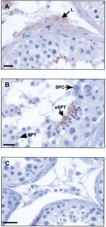
tu-bules (Fig. 5, A and B). In interstitial tissue, the specific
staining was clearly located in Leydig cells (Fig. 5A),
where in seminiferous tubules the signal was observed in
pachytene spermatocytes, early spermatids, and with a
particularly high intensity in elongated spermatids (Fig.
5B). No signal was detected in sections incubated with
preimmune serum instead of the anti-HBD-1 polyclonal
antibody (Fig. 5C).
In sperm smears, specific HBD-1 staining was observed
FIG. 5. Immunolocalization of the beta-defensin HBD-1 in human testis sections. An anti-HBD-1 rabbit polyclonal antiserum was used at a dilu-tion of 1:500 and immunoreacdilu-tion was revealed with diaminobenzidine. Sections were counterstained with Masson hemalun. A) In the intersti-tium, immunostaining was also visualized in Leydig cells (L). Bar 5 25 mm. B) Discrete immunolabeling was detected within the seminiferous tubule in pachytene spermatocytes (SPC), whereas early spermatids (SPT) and elongated spermatids (eSPT) displayed a strong signal. Bar5 25 mm. C) Negative control was performed using the corresponding preimmune serum. Bar5 50 mm.
FIG. 6. Immunolocalization of the beta-defensin HBD-1 in human sperm smears. An anti-HBD-1 rabbit polyclonal antiserum was used at a dilution of 1:500 and immunoreaction was revealed with AEC or diami-nobenzidine (insert). Smears were counterstained with Masson hemalun. A) Very localized immunolabeling was detected in spermatozoa (arrow), whereas the seminal plasma was strongly stained (arrowhead). Bar5 5 mm. Insert, close view of positive immunolabeling on the lower head portion of spermatozoa (arrow). Bar5 5 mm. B) Negative control was performed using the appropriate preimmune serum. Bar5 5 mm.
on the lower head portion of spermatozoa, and diffuse
spe-cific staining was also seen in seminal plasma (Fig. 6, A
vs. B).
In parallel experiments to detect HBD-2 using a specific
polyclonal antibody [35], no testicular labeling was
ob-served (data not shown).
Western Blot Analysis of HBD-1 in Human
Seminal Plasma
Western blots of cationic extracts concentrated from
hu-man seminal plasma were performed in order to ascertain
the presence of soluble HBD-1 (Fig. 7). Four isoforms of
HBD-1 were detected. The predominant band appeared to
correspond to the 44-amino acid (aa) form of HBD-1 as
assessed by comparison with a recombinant HBD-1 peptide
resolved in parallel. The upper band most probably
corre-sponds to the 47 aa form of HBD-1 by comparison with
the two HBD-1 isoforms (44 and 47 aa, respectively)
pro-duced by the control Caco-2 cell line [17].
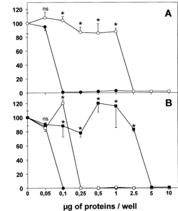
Antimicrobial Activity Assay of Human Semen
In preliminary studies, antimicrobial activity of human
seminal plasma was assessed using E. coli D22 and 363
strains, M. luteus A270 strain, and B. megaterium MA
strain (data not shown). B. megaterium was chosen for
de-tailed studies because it appeared to be the most sensitive
to antimicrobial activity in seminal plasma. Human seminal
plasma was found to be microbicidal to B. megaterium in
a concentration-dependent manner (Fig. 8A). This activity
FIG. 7. Detection of hBD-1 in the cationic fraction of human seminal plasma. Cationic peptides from seminal plasma were subjected to AU-PAGE analysis followed by immunoblotting and probing with anti-hBD-1 antibody. A recombinant 44 amino acid form of hBD-1, rhBD44-1 at 5
and 10 ng, was used as a reference. HBD-1 forms in seminal plasma are indicated by arrows on the right.
FIG. 8. An antibacterial assay of human seminal plasma and its cationic extract activity againstB. megaterium (MA strain). A) Growth inhibition was determined in triplicate after 16 h of incubation with different amounts of protein extracts from crude human seminal plasma (open cir-cles), and the cationic extract of seminal plasma (closed circles). B) Growth inhibition was also determined for the cationic extract of seminal plasma (closed circles) in the presence of 150 mM NaCl (open squares) or 20 mM DTT (closed squares). Each plot is the mean 6 SEM of 3 replicate A620nmdeterminations. ns, nonsignificant; *P, 0.001 compared
with the respective control as determined by the Studentt-test.
was at least 10 times augmented in the cationic extracts
prepared from this material compared with the activity
gen-erated by the crude fraction of seminal plasma. The
anti-microbial activity of the cationic extract was much lower
in the presence of 20 mM DTT, a reducing agent for
di-sulfide bonds (Fig. 8B), whereas it was partially diminished
in the presence of 150 mM NaCl, a classical inhibitor of
b-defensin antimicrobial peptides (Fig. 8B).
DISCUSSION
Innate immunity constitutes a first line of defense against
an initial challenge by pathogens [36]. Increasing evidence
has accumulated for the major role played by cationic
an-timicrobial peptides in preventing the onset of infection in
many organisms [37, 38]. Defensins in mammals, and
par-ticularly in humans, have been studied in great detail over
the past 10 years, and their presence and bioactivity has
now been established in several organs and fluids [10]. In
this context, it is particularly noticeable that no systematic
study has been reported to search for such peptides in the
male reproductive tract and in particular in the testis, an
organ known to have developed a potent anti-infectious
de-fense system [3].
To our knowledge, a systematic approach involving the
study of the expression and distribution of all known
de-fensins in a given species and in three different species such
as the present study has never before been undertaken,
ei-ther in the testis or epididymis, or in any oei-ther tissue or
organ. Our RT-PCR results show that in rat testis, all known
a- and b-defensins are expressed with the exception of
RNP-4, whereas rat epididymis expresses
a-defensins
RNP1–2, RNP-4, and
b-defensins RBD-1 and RBD-2.
In mice, our results are consistent with those of
Grand-jean et al. who found the cryptdin-1, -2, -3, and -6 group
in mouse testis using immunohistochemistry [39]. Our
study also reveals that CRS1C, mBD-1, and mBD-2 are
present in the testis as well as in the epididymis, whereas
the murine
b-defensins mBD-3 and mBD-4 were expressed
only in the epididymis and the
a-defensin CRS4C was
ab-sent in the testis and epididymis.
The recent identification in the rat epididymis of a new
peptide—Bin1b—that exhibits structural characteristics and
antimicrobial activity similar to that of
b-defensins [40],
together with the present data on defensin expression in rat
and mouse epididymis indicates that this secretory organ is
an important site of antimicrobial peptide production in the
male reproductive system. In addition to the defensins
iden-tified here and to Bin1b, the human cationic antimicrobial
protein-18 (hCAP-18) was identified in the epithelium of
human epididymis [41]. Finally, a recent work suggests that
the human EP2 gene codes at least nine mRNA variants
that are specifically expressed in the epididymis, among
which are
b-defensin-like variants [42]. That the
epididy-mis could be a key site for antimicrobial activity is
com-patible with its function of protecting spermatozoa during
their transit and storage.
Transcripts for all known human defensins were shown
to be expressed by the human testis, with the consistent
exception of HBD-2 and HBD-3. The absence of the latter
contrasts with the results of Bals et al. [35], who noted a
very faint expression of HBD-2 mRNA within the testis.
HBD-2 peptide is well known to be an inducible defensin
(for a review see [43]). Because the suppliers offer no
in-formation about the origin and physiological status of the
tissue samples used to generate RNA master blots, it cannot
be excluded that the testicular sample analyzed by Bals et
al. was inflamed, thus explaining the presence of low levels
of the HBD-2 transcript. The HBD-1 mRNA was recently
localized at very low levels in the human prostate and
tes-tis, together with those of
a-defensins HNP1–3 [44]. The
present data on HNP1–3 is in accordance with this latter
study. However, in contrast to our findings, the authors
failed to detect HD-5 and HD-6 defensins in the human
testis. The discrepancy may result from the high
interindi-vidual variability of testicular defensin expression that was
consistently observed in the present study for HBD-1. This
variability was observed using immunohistochemistry,
Western blot analysis, and even RT-PCR, as our results
originate from multiple experiments performed on
numer-ous independent testicular and semen samples. In addition,
our own positive RT-PCR results concerning the expression
of HNP1–3 transcripts in the whole human testis, together
with the data of Zhao et al. [44] may be reconciled with
our immunohistochemistry experiments using a commercial
anti-HNP1–3 (Bachem, Bubendorf, Switzerland) that failed
to detect the peptides in any testicular cell, whereas a strong
specific signal was observed within blood vessels irrigating
the interstitial compartment (data not shown).
Due to the lack of availability of human testes for
pre-paring isolated cells, we focused on rat testes in order to
study the cellular distribution of the testicular defensins. In
the interstitium, resident macrophages express most of the
defensins studied, whereas the testosterone-producing
Ley-dig cells express only the
b-defensin RBD-2, suggesting
the preeminence of the former in the antimicrobial defense
within this compartment of the testis. Compared with the
latter compartment, the seminiferous tubule appears to be
very well equipped to react to a bacterial attack because its
different cellular constituents express all studied defensins
with the unique exception of
a-defensin RNP-4. In fact,
peritubular cells and Sertoli cells express most of the
a-and
b-defensins studied, whereas the germ cell line
ex-presses only a limited number of defensins. Thus,
sper-matogonia express only
a-defensins, but at relatively high
levels. Pachytene spermatocytes express only
b-defensins,
whereas spermatids and their cytoplasmic lobes express
both types. The variety of defensins expressed by the
Ser-toli and peritubular cells most probably has a self-defense
function but could also play a role in the protection of the
germ cells against pathogens, which is indeed of prime
im-portance because their infection could result in the total
destruction of the germ line. We hypothesize that the
com-plementary pattern of expression of defensins with different
spectra of activities between the different somatic and germ
cells is likely to generate an efficient antimicrobial defense
network system within the seminiferous tubule.
We also demonstrate that HBD-1 is expressed in the
hu-man germ line from pachytene spermatocytes to late
sper-matids. Furthermore, the expression of HBD-1 was also
located in the human Leydig cell cytoplasm, strongly
sug-gesting that a key function such as testosterone production
occurs under direct antimicroorganism protection, an
ob-servation previously made by us with antiviral proteins [5].
Regardless of human or rat tissues, the resurgence of
ex-pression of a strong antimicrobial system in late spermatids
is probably indicative of the need for spermatozoa, which
are no longer protected by testicular somatic cells, to
con-tribute to their own defense while being transported within
the male excurrent ducts and then in the female
reproduc-tive tract. In this context it is important to note the
dem-onstration of the presence of the transcripts for defensins
HD-5, HBD-1, and HNP1–3 in human ejaculated
sperma-tozoa. Because human semen from normal donors often
contains leukocytes (for a review see [45]), we cannot
ex-clude the possibility that the HNP1–3 transcripts evidenced
here in the spermatozoa fraction resulted from the presence
of neutrophils that constitutively express HNP1–3 [11].
In-deed, neutrophils were traced by RT-PCR in the HNP1–3
positive ejaculates at our disposal using a couple of probes
specific from the leukocyte common antigen CD45 (data
not shown). In contrast, the presence of HBD-1, the only
defensin for which we could access an antibody, was
un-equivocally demonstrated in spermatozoa by
immunocyto-chemistry, more precisely at the lower head portion of the
cells. Of note is that neutrophils present in the ejaculates
also displayed a marked labeling for HBD-1 (data not
shown). A strong specific labeling for HBD-1 was also
ob-served in the seminal plasma where multiple soluble forms
of the defensin were evidenced. This most probably reflects
variable N-terminal proteolytic processing of the peptide
[17, 46].
That human seminal plasma displays antibacterial
prop-erties has been known for decades, although the active
agent or agents had not been identified [47, 48]. That
ac-tivity of the cationic extract of seminal plasma prepared
here was partially reduced by NaCl probably indicates that
HBD-1 and other possible
b-defensins present in the
cat-ionic extract contribute to their antibacterial activity, as the
latter are salt-sensitive [49, 50]. The moderate salt
concen-tration that prevails in the semen [51] is highly favorable
to defensin-type activities. However, our data also suggest
that other molecules with antibacterial properties that have
not yet been characterized are present in the enriched
cat-ionic extract of the sperm. In this respect, the present study
clearly demonstrates the presence of another defensin
tran-script (HD-5) in ejaculated sperm.
What could be the origin of the seminal HBD-1 forms?
Because HBD-1 is present in spermatozoa and less mature
germ cells, and because HBD-1 is known to be secreted
[17], the germ line could represent a significant source of
this defensin. Furthermore, a recent study demonstrates the
widespread expression of HBD-1 transcript in various
hu-man organs, mostly secretory glands (kidney, salivary
glands, trachea, placenta, prostate, testis . . . ) and epithelial
cells derived from trachea, bronchi, small airways, and the
mammary gland [44]. It is therefore quite possible that the
peptide is also produced along the male reproductive tracts
by at least other accessory glands besides the prostate,
whose secretions contribute to the constitution of seminal
plasma. Moreover, the presence of antibacterial substances
in prostatic fluid was also suggested [52, 53]. In order to
clarify this, the possible production of HBD-1 by the
epi-didymis, seminal vesicles, and prostate is currently under
investigation in our laboratory. In any case, the
antimicro-bial activity demonstrated in the seminal plasma ensures a
complementary protection to the self-protecting equipment
of spermatozoa.
Beside their antimicrobial activities, additional roles of
defensins, such as recruitment of inflammatory cells and
wound healing [36], could be significant in the physiology
and pathology of testicular function.
ACKNOWLEDGMENTS
We thank Dr. M.H. Metz-Boutigue (INSERM U.338, Strasbourg France) for the generous gift of bacteria, and E. Valore (UCLA School of Medicine, Los Angeles, CA) for her gift of recombinant HBD-1 and her valuable advice on AU-PAGE/Western blot techniques. We also acknowl-edge Dr. J.M. Wilson (Institute for Human Gene Therapy, The Wistar Institute, Philadelphia, PA) for the gift of the anti-HBD-2 antibody.
REFERENCES
1. Mikuz G, Damjanov I. Inflammation of the testis, epididymis, peri-testicular membranes and scrotum. Pathol Annu 1982; 17:101–128. 2. Purvis K, Christiansen E. Infection in the male reproductive tract.
Impact, diagnosis and treatment in relation to male infertility. Int J Androl 1993; 16:1–13.
3. Dejucq N, Je´gou B. Viruses in the mammalian male genital tract and their effects on the reproductive system. Microbiol Mol Biol Rev 2001; 65:208–231.
4. Dejucq N, Dugast I, Ruffault A, van der Meide PH, Je´gou B. Inter-feron-alpha and -gamma expression in the rat testis. Endocrinology 1995; 136:4925–4931.
5. Dejucq N, Chousterman S, Je´gou B. The testicular antiviral defense system: localization, expression, and regulation of 2959 oligoadenylate synthetase, double-stranded RNA-activated protein kinase, and Mx proteins in the rat seminiferous tubule. J Cell Biol 1997; 139:865– 873.
6. Dejucq N, Lie´nard MO, Guillaume E, Dorval I, Je´gou B. Expression of interferons alpha and gamma in testicular interstitial tissue and spermatogonia of the rat. Endocrinology 1998; 139:3081–3087. 7. Dejucq N, Lie´nard MO, Je´gou B. Interferons and interferon-induced
antiviral proteins in the testis. J Reprod Immunol 1998; 41:291–300. 8. Boman HG. Gene-encoded peptide antibiotics and the concept of in-nate immunity: an update review. Scand J Immunol 1998; 48:15–25. 9. Hancock RE, Falla T, Brown M. Cationic bactericidal peptides. Adv
Microb Physiol 1995; 37:135–175.
10. Lehrer RI, Ganz T. Antimicrobial peptides in mammalian and insect host defence. Curr Opin Immunol 1999; 11:23–27.
11. Daher KA, Selsted ME, Lehrer RI. Direct inactivation of viruses by human granulocyte defensins. J Virol 1986; 60:1068–1074.
12. Nakashima H, Yamamoto N, Masuda M, Fujii N. Defensins inhibit HIV replication in vitro [letter]. AIDS 1993; 7:1129.
13. White SH, Wimley WC, Selsted ME. Structure, function, and mem-brane integration of defensins. Curr Opin Struct Biol 1995; 5:521– 527.
14. Ganz T. Extracellular release of antimicrobial defensins by human polymorphonuclear leukocytes. Infect Immun 1987; 55:568–571. 15. Jones DE, Bevins CL. Paneth cells of the human small intestine
ex-press an antimicrobial peptide gene. J Biol Chem 1992; 267:23216– 23225.
16. Jones DE, Bevins CL. Defensin-6 mRNA in human Paneth cells: im-plications for antimicrobial peptides in host defense of the human bowel. FEBS Lett 1993; 315:187–192.
17. Valore EV, Park CH, Quayle AJ, Wiles KR, McCray PB Jr, Ganz T. Human beta-defensin-1: an antimicrobial peptide of urogenital tissues. J Clin Invest 1998; 101:1633–1642.
18. Toebosch AM, Robertson DM, Klaij IA, de Jong FH, Grootegoed JA. Effects of FSH and testosterone on highly purified rat Sertoli cells: inhibin alpha-subunit mRNA expression and inhibin secretion are en-hanced by FSH but not by testosterone. J Endocrinol 1989; 122:757– 762.
19. Galdieri M, Ziparo E, Palombi F, Russo MA, Stefanini M. Pure Sertoli cell cultures: a new model for the study of somatic-germ cell inter-actions. J Androl 1981; 5:249–259.
20. Skinner MK, Fritz IB. Testicular peritubular cells secrete a protein under androgen control that modulates Sertoli cell functions. Proc Natl Acad Sci U S A 1985; 82:114–118.
21. Klinefelter GR, Kelce WR, Hardy MP. Isolation and Culture of Leydig Cells From Adult Rats. San Diego: Academic Press; 1993.
22. Pineau C, Syed V, Bardin CW, Je´gou B, Cheng CY. Germ cell-con-ditioned medium contains multiple factors that modulate the secretion of testins, clusterin, and transferrin by Sertoli cells. J Androl 1993; 14:87–98.
23. Guillaume E, Dupaix A, Moertz E, Courtens J, Je´gou B, Pineau C. Proteome analysis of spermatogonia: identification of a first set of 53 proteins. Proteome; 2000. Available online at: DOI 10.1007/ s102160000003.
24. Shi SR, Cote RJ, Taylor CR. Antigen retrieval immunohistochemistry: past, present, and future. J Histochem Cytochem 1997; 45:327–343. 25. Lehrer RI, Rosenman M, Harwig SS, Jackson R, Eisenhauer P.
Ultra-sensitive assays for endogenous antimicrobial polypeptides. J Immu-nol Methods 1991; 137:167–173.
26. Yount NY, Wang MS, Yuan J, Banaiee N, Ouellette AJ, Selsted ME. Rat neutrophil defensins. Precursor structures and expression during neutrophilic myelopoiesis. J Immunol 1995; 155:4476–4484. 27. Condon MR, Viera A, D’Alessio M, Diamond G. Induction of a rat
enteric defensin gene by hemorrhagic shock. Infect Immun 1999; 67: 4787–4793.
28. Jia HP, Mills JN, Barahmand-Pour F, Nishimura D, Mallampali RK, Wang G, Wiles K, Tack BF, Bevins CL, McCray PB Jr. Molecular
cloning and characterization of rat genes encoding homologues of human beta-defensins. Infect Immun 1999; 67:4827–4833.
29. Huttner KM, Selsted ME, Ouellette AJ. Structure and diversity of the murine cryptdin gene family [published erratum appears in Genomics 1995; 25:762]. Genomics 1994; 19:448–453.
30. Ouellette AJ, Lualdi JC. A novel mouse gene family coding for cat-ionic, cysteine-rich peptides. Regulation in small intestine and cells of myeloid origin [published erratum appears in J Biol Chem 1994; 269:18702]. J Biol Chem 1990; 265:9831–9837.
31. Huttner KM, Kozak CA, Bevins CL. The mouse genome encodes a single homolog of the antimicrobial peptide human beta-defensin 1. FEBS Lett 1997; 413:45–49.
32. Morrison GM, Davidson DJ, Dorin JR. A novel mouse beta defensin, Defb2, which is upregulated in the airways by lipopolysaccharide. FEBS Lett 1999; 442:112–116.
33. Bals R, Wang X, Meegalla RL, Wattler S, Weiner DJ, Nehls MC, Wilson JM. Mouse beta-defensin 3 is an inducible antimicrobial pep-tide expressed in the epithelia of multiple organs. Infect Immun 1999; 67:3542–3547.
34. Jia HP, Wowk SA, Schutte BC, Lee SK, Vivado A, Tack BF, Bevins CL, McCray PB Jr. A novel murine beta-defensin expressed in tongue, esophagus, and trachea. J Biol Chem 2000; 275:33314–33320. 35. Bals R, Wang X, Wu Z, Freeman T, Bafna V, Zasloff M, Wilson JM.
Human beta-defensin 2 is a salt-sensitive peptide antibiotic expressed in human lung. J Clin Invest 1998; 102:874–880.
36. Kaiser V, Diamond G. Expression of mammalian defensin genes. J Leukoc Biol 2000; 68:779–784.
37. Hancock RE, Lehrer R. Cationic peptides: a new source of antibiotics. Trends Biotechnol 1998; 16:82–88.
38. Hancock RE, Diamond G. The role of cationic antimicrobial peptides in innate host defences. Trends Microbiol 2000; 8:402–410. 39. Grandjean V, Vincent S, Martin L, Rassoulzadegan M, Cuzin F.
An-timicrobial protection of the mouse testis: synthesis of defensins of the cryptdin family. Biol Reprod 1997; 57:1115–1122.
40. Li P, Chan HC, He B, So SC, Chung YW, Shang Q, Zhang YD, Zhang YL. An antimicrobial peptide gene found in the male reproductive system of rats. Science 2001; 291:1783–1785.
41. Malm J, Sorensen O, Persson T, Frohm-Nilsson M, Johansson B, Bjar-tell A, Lilja H, Stahle-Backdahl M, Borregaard N, Egesten A. The human cationic antimicrobial protein (hCAP-18) is expressed in the epithelium of human epididymis, is present in seminal plasma at high concentrations, and is attached to spermatozoa. Infect Immun 2000; 68:4297–4302.
42. Frohlich O, Po C, Young LG. Organization of the human gene en-coding the epididymis-specific EP2 protein variants and its relation-ship to defensin genes. Biol Reprod 2001; 64:1072–1079.
43. Schroder JM, Harder J. Human beta-defensin-2. Int J Biochem Cell Biol 1999; 31:645–651.
44. Zhao C, Wang I, Lehrer RI. Widespread expression of beta-defensin hBD-1 in human secretory glands and epithelial cells. FEBS Lett 1996; 396:319–322.
45. Barratt CL, Bolton AE, Cooke ID. Functional significance of white blood cells in the male and female reproductive tract. Hum Reprod 1990; 5:639–648.
46. Hiratsuka T, Nakazato M, Ihi T, Minematsu T, Chino N, Nakanishi T, Shimizu A, Kangawa K, Matsukura S. Structural analysis of human beta-defensin-1 and its significance in urinary tract infection. Nephron 2000; 85:34–40.
47. Rozansky R, Gurevitch J, Brzezinsky A, Eckerling B. Inhibition of the growth of Staphylococcus aureus by human semen. J Lab Clin Med 1949; 34:1526–1529.
48. Mardh PA, Colleen S. Antimicrobial activity of human seminal fluid. Scand J Urol Nephrol 1975; 9:17–23.
49. Ganz T, Lehrer RI. Defensins. Pharmacol Ther 1995; 66:191–205. 50. Singh PK, Jia HP, Wiles K, Hesselberth J, Liu L, Conway BA,
Green-berg EP, Valore EV, Welsh MJ, Ganz T, Tack BF, McCray PB Jr. Production of beta-defensins by human airway epithelia [published erratum appears in Proc Natl Acad Sci U S A 1999; 96:2569]. Proc Natl Acad Sci U S A 1998; 95:14961–14966.
51. Mann T. Les deux composants du sperme: spermatozoı¨des et plasma se´minal. In: La Biochimie du sperme. Paris: Presses Universitaires de France; 1956: 3–39.
52. Fair WR, Couch J, Wehner N. Prostatic antibacterial factor. Identity and significance. Urology 1976; 7:169–177.
53. Fair WR, Parrish RF. Antibacterial substances in prostatic fluid. Prog Clin Biol Res 1981; 75A:247–264.
54. Daher KA, Lehrer RI, Ganz T, Kronenberg M. Isolation and
terization of human defensin cDNA clones [published erratum appears in Proc Natl Acad Sci U S A 1989; 86:342]. Proc Natl Acad Sci U S A 1988; 85:7327–7331.
55. McCray PB Jr, Bentley L. Human airway epithelia express a beta-defensin. Am J Respir Cell Mol Biol 1997; 16:343–349.
56. Harder J, Bartels J, Christophers E, Schroder JM. A peptide antibiotic from human skin [letter; see comments]. Nature 1997; 387:861. 57. Harder J, Bartels J, Christophers E, Schroder JM. Isolation and
char-acterization of human beta-defensin-3, a novel human inducible pep-tide antibiotic. J Biol Chem 2001; 276:5707–5713.