HAL Id: hal-02622540
https://hal.inrae.fr/hal-02622540
Submitted on 26 May 2020
HAL is a multi-disciplinary open access
archive for the deposit and dissemination of
sci-entific research documents, whether they are
pub-lished or not. The documents may come from
teaching and research institutions in France or
abroad, or from public or private research centers.
L’archive ouverte pluridisciplinaire HAL, est
destinée au dépôt et à la diffusion de documents
scientifiques de niveau recherche, publiés ou non,
émanant des établissements d’enseignement et de
recherche français ou étrangers, des laboratoires
publics ou privés.
Distributed under a Creative Commons Attribution| 4.0 International License
Identification and characterization of core abscisic acid
(ABA) signaling components and their gene expression
profile in response to abiotic stresses in Setaria viridis
Karoline Estefani Duarte, Wagner Rodrigo de Souza, Thais Ribeiro Santiago,
Bruno Leite Sampaio, Ana Paula Ribeiro, Michelle Guitton Cotta, Barbara
Andrade Dias Brito da Cunha, Pierre Marraccini, Adilson Kenji Kobayashi,
Hugo Bruno Correa Molinari
To cite this version:
Karoline Estefani Duarte, Wagner Rodrigo de Souza, Thais Ribeiro Santiago, Bruno Leite Sampaio,
Ana Paula Ribeiro, et al.. Identification and characterization of core abscisic acid (ABA) signaling
components and their gene expression profile in response to abiotic stresses in Setaria viridis. Scientific
Reports, Nature Publishing Group, 2019, 9, �10.1038/s41598-019-40623-5�. �hal-02622540�
Identification and characterization
of core abscisic acid (ABA) signaling
components and their gene
expression profile in response to
abiotic stresses in Setaria viridis
Karoline Estefani Duarte
1,2, Wagner Rodrigo de Souza
2,3, Thaís Ribeiro Santiago
2,
Bruno Leite Sampaio
2, Ana Paula Ribeiro
2, Michelle Guitton Cotta
4,
Bárbara Andrade Dias Brito da Cunha
2, Pierre Roger René Marraccini
1,5,6,
Adilson Kenji Kobayashi
2& Hugo Bruno Correa Molinari
2Abscisic acid (ABA) is an essential phytohormone that regulates growth, development and adaptation of plants to environmental stresses. In Arabidopsis and other higher plants, ABA signal transduction involves three core components namely PYR/PYL/RCAR ABA receptors (PYLs), type 2C protein phosphatases (PP2Cs) and class III SNF-1-related protein kinase 2 (SnRK2s). In the present study, we reported the identification and characterization of the core ABA signaling components in Setaria viridis, an emerging model plant for cereals and feedstock crops presenting C4 metabolism, leading to the identification of eight PYL (SvPYL1 to 8), twelve PP2C (SvPP2C1 to 12) and eleven SnRK2 (SvSnRK2.1 through SvSnRK2.11) genes. In order to study the expression profiles of these genes, two different S.
viridis accessions (A10.1 and Ast-1) were submitted to drought, salinity and cold stresses, in addition to
application of exogenous ABA. Differential gene expression profiles were observed in each treatment and plant genotype, demonstrating variations of ABA stress responses within the same species. These differential responses to stresses were also assessed by physiological measurements such as photosynthesis, stomatal conductance and transpiration rate. This study allows a detailed analysis of gene expression of the core ABA signaling components in Setaria viridis submitted to different treatments and provides suitable targets for genetic engineering of C4 plants aiming tolerance to abiotic stresses.
Abscisic acid (ABA) is a phytohormone involved in the control of many aspects of plant growth and development including embryo maturation, cell division and elongation, seed dormancy and germination, root growth and floral induction1–5. In addition, ABA also responds to a variety of environmental stresses, including biotic and
abiotic stresses such as drought, cold and salinity6–8. Chemically, ABA is a sesquiterpene synthesized in plants
from β-carotene, via multiple enzymatic reactions that involves zeaxanthin oxidase (ZEP), 9-cis-epoxycarotenoid dioxygenase (NCED), ABA-aldehyde oxidase (AAO) and molybdenium cofactor sulfurase9 (MCSU). In abiotic
stress conditions, ABA levels increase, leading to a signaling cascade that ultimately activates plant adaptation responses to stress10. The ABA-activated signaling network was recently unraveled, and the identification of ABA
receptors is among the most important advances in stress signaling in the past decade4,11.
1Plant Biotechnology Program, Federal University of Lavras (UFLA), Lavras, MG, 37200-000, Brazil. 2Genetics and Biotechnology Laboratory, Embrapa Agroenergy (CNPAE), Brasilia, DF, 70770-901, Brazil. 3centro de ciências Naturais e Humanas, Universidade Federal do ABC (UFABC), São Bernardo do Campo, Santo André, SP, 09606-045, Brazil. 4Department of Cell Biology, University of Brasília (UnB), Brasília, DF, 70910-900, Brazil. 5ciRAD, UMR AGAP (University Montpellier, CIRAD, IRD, INRA), Montpellier, 34398, France. 6ciRAD, UMR iPMe (University Montpellier, CIRAD, IRD, Montpellier), Agricultural Genetics Institute, LMI RICE2, Hanoi, Vietnam. Correspondence and requests for materials should be addressed to H.B.c.M. (email: hugo.molinari@embrapa.br)
Received: 28 June 2018 Accepted: 19 February 2019 Published: xx xx xxxx
www.nature.com/scientificreports
www.nature.com/scientificreports/
ABA receptors were first uncovered in Arabidopsis, where three core components have been identified: the ABA receptor PYR/PYL/RCAR (PYL) protein family, the negative regulator type 2C protein phosphatase (PP2C) and the positive regulator class III SNF-1-related protein kinase 2 (SnRK2). Some of these receptors were also identified in other plant species such as sorghum, maize and rubber tree12–14. PYR proteins were identified
through genetic analyses, which found that PYR1 (Pyrabactin Resistance 1) and members of its 13 relative pro-teins (Pyrabactin Resistance 1-Like; PYL) are necessary for proper ABA signal transduction in Arabidopsis15,16.
This study also demonstrated that PYR1 binds to ABA and inhibits the group A protein phosphatases 2Cs (clade A PP2Cs), whose members include ABI1, ABI2 (ABA-insensitive 1 and 2) and HAB1 (Hypersensitive to ABA 1). The genetic evidences suggest that PP2Cs act as negative regulators of ABA-dependent pathways, and this func-tion appears to be conserved from Arabidopsis to moss17. The main targets of PP2Cs identified to date are related
to protein kinases involved as positive regulators of ABA signaling4,17. Among these kinases, the class III
SNF-1-related protein kinases 2 (SnRK2s) are the most implicated in positive regulation of ABA signaling, especially because of the strong phenotype observed in the Arabidopsis triple mutant snrk2.2/2.3/2.6, which could germinate and grow on 50 µM ABA, an abnormal phenotype demonstrated by ABA-insensitive mutants18. Some evidences
showed that SnRK2s might directly phosphorylate members of the ABF/AREB/ABI5 clade of bZIP transcription factors, promoting ABA-induced gene expression18–21. In summary, ABA binds to a PYL protein, resulting in
inhibition of PP2Cs through the ABA-PYL-PP2C complex. This complex leads to accumulation of phosphoryl-ated SnRK2s, which leads to phosphorylation of ABA-responsive element binding factors (ABFs) and subsequent ABA gene expression for appropriate cellular responses4.
Some important crops used as sources of food and feedstock belong to the Panicoideae subfamily and include cereal grains and grasses such as sugarcane (Saccharum spp.), maize (Zea mays), sorghum (Sorghum bicolor) and switchgrass22 (Panicum virgatum). Abiotic stresses such as cold, drought and salinity are among the most
delete-rious environmental stresses in these crops, responsible for great yield losses worldwide23. Despite the
advance-ments achieved in these crops towards the comprehension of the molecular and biochemical pathways associated with abiotic stresses, the complexity of the genome and the long generation times required have hindered the progress of these studies. In this context, S. viridis has emerged as a suitable C4 model species for molecular and genetic studies. It is a short, fast-growing, C4 metabolism plant, with its genome sequence available, mak-ing it a suitable model plant for genetic studies24,25. Moreover, S. viridis is highly responsive to Agrobacterium tumefaciens-mediated genetic transformation, with well-established in vitro transformation protocols24,26 and
more recently, spike-dipping transformation methods have also been proposed27,28. Genetically engineered S. viridis plants can be used in a proof-of-concept approach to evaluate phenotypes related to important
agri-cultural traits such as abiotic stress tolerance, resistance to pathogens and improved yield and biomass29–31. The
tested promising genes could be further transferred to a target crop.
In the present study, the identification and characterization of the ABA receptors PYL and the core signaling components PP2C (clade A) and SnRK2 gene families in S. viridis is reported. Since phenotypic variability in natural accessions of S. viridis has been reported28,32,33, two different S. viridis accessions (A10.1 and Ast-1) was
used to study gene expression of core ABA signaling components under drought, salinity, cold and exogenous ABA application. A total of 8 PYLs (SvPYL1 to SvPYL 8), 12 PP2Cs (SvPP2C1 to SvPP2C 12) and 11 SnRK2s (SvSnRK2.1 to SvSnRK2.11) were found in S. viridis genome. Differential gene expression was found for the dif-ferent treatments and accessions, demonstrating that even within the same species the abiotic stress responses can be variable. Gas exchange measurements also demonstrated that the two accessions studied have slightly different responses to abiotic stresses. This study provides suitable targets for genetic engineering of C4 plants aiming tolerance to abiotic stresses.
Results
Genome-wide identification and characterization of the core ABA signaling components in S. viridis.
The search for PYR/PYL, PP2C and SnRK2 genes in S. viridis genome was performed using two different strategies. For both strategies applied, similar number of genes was found. Based on amino acid sequences of S. viridis, eight, twelve and eleven putative genes of PYR/PYL, PP2C and SnRK2 were identified, respectively (Supplementary Fig. S1), and described separately below. The complete set of gene orthology for Setaria viridis (Sv) PYLs, PP2Cs and SnRK2s, in comparison with Arabidopsis thaliana (At), Oryza sativa (Os) and Sorghum bicolor (Sb) is pre-sented in Supplementary Table S1.
SvPYLs. The identified PYR/PYL genes were designated as SvPYL1 through SvPYL8. The size of their
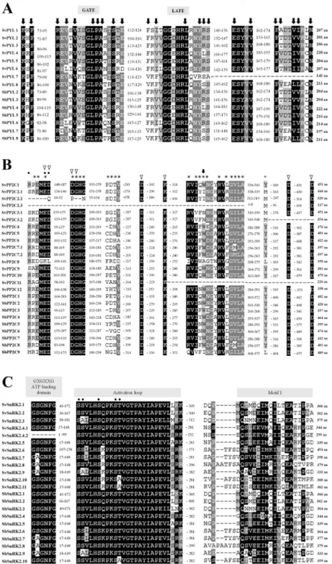
corre-sponding SvPYL proteins ranged from 141 to 220 amino acids (aa), with molecular weight (MW) from 15.11 to 23.67 KDa and pI from 5.24 to 8.88 (Table 1). They contained the polyketide cyclase 2 domain (PF10604) local-ized between the positions 43–213 aa (Fig. 1A). It was also possible to identify in SvPYL proteins a conserved motif 1 related to the well-known “GATE” and “LATCH” loop regions and motifs 2–3 involved in ABA binding (Fig. 1A).
A phylogenetic analysis was performed based on similarities to PYL proteins from Arabidopsis (AtPYLs),
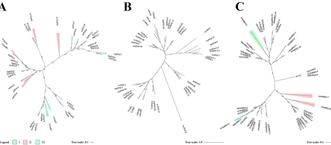
Oryza sativa (OsPYLs) and Sorghum bicolor (SbPYLs), which divided SvPYLs in subfamilies I, II and III (Fig. 2A). SvPYL1–3, SvPYL4-6 and SvPYL7-8 were classified as subclass III, II and I, respectively. With the exception of SvPYL7, all identified SvPYL proteins have orthologs in Arabidopsis (Supplementary Table S1). Intron-exon anal-ysis of SvPYL genes showed that only genes clustered into subfamily I have 2 introns while those from subfamilies II and III have none intronic regions (Fig. 3B).
SvPP2Cs. In total, 12 PP2C genes, designated as SvPP2C1 to SvPP2C12, were identified in S. viridis genome
(Fig. 2B). Three isoforms were observed for SvPP2C2 (SvPP2C2.1, SvPP2C2.2 and SvPP2C2.3) while two iso-forms were found for SvPP2C3 (SvPP2C3.1 and SvPP2C3.2) and SvPP2C7 (SvPP2C7.1 and SvPP2C7.2). The size of
SvPP2C proteins ranged from 117 (SvPP2C2.3) to 479 (SvPP2C10) amino acids with MW between 12.47 to 49.71 KDa and pI ranging from 4.35 to 8.50 (Table 1). Based on Pfam analysis, a protein domain PF00481 was identify and conserved in all putative SvPP2C proteins. Furthermore, binding residues for PYL and the cofactors Mn2+/
Mg2+ in the motifs 2, 3, 5 and 7 were also identified in SvPP2Cs (Fig. 3A). With the exception of SvPP2C2.2,
SvPP2C2.3, SvPP2C3.2 and SvPP2C11, SvPP2Cs sequences contain well-characterized functional residues and domain regulators of ABA necessary to interaction with PYLs and SnRK2s proteins (Fig. 1B). Analysis of pro-teins demonstrated that SvPP2Cs have orthologs mainly in O. sativa (OsPP2Cs) and S. bicolor (SbPP2Cs), with exception to SvPP2C8 and SvPP2C11 (Supplementary Table S1). Intro-exon analysis showed a variable numbers of introns in SvPP2C genes, with the predominance of three introns in the majority of the genes (Fig. 3B).
SvSnRK2s. Based on the presence of Pfam domain PF0069 and similarity with query sequences, 11
non-redundant SnRK2 genes (named from SvSnRK2.1 to SvSnRK2.11) were found in S. viridis genome. SnRK2.4 was the only gene presenting two isoforms, (named SvSnRK2.4.1 and SvSnRK2.4.2). The length of putative SvSnRK2 proteins ranged from 295 to 454 aa, with MW from 33.43 to 51.67 KDa and pI from 4.73 to 8.30 (Table 1). Except for SvSnRK2.4.2 protein, which lacks the ATP-binding loop domain, all these phosphatases con-tained the five important conserved motifs, including: (1) the ATP-binding domain (motif 5), (2) the activation loop (motif 2), (3) the PP2C interface residues (also called SnRK2 box), (4) the motif I (motif 6) and (5) ABA box domains, (Figs 1C and 3A). The bootstrap values deduced from the phylogenetic analysis revealed that SvSnRK2s were divided into subclasses I, II and III (Fig. 2C). The Subclass I includes SvSnRK2.6, SvSnRK2.7, SvSnRK2.8,
SvSnRK2.10 and SvSnRK2.11, with the predominance of eight introns in their corresponding genes (Fig. 3B). The
Gene Name Chr Start Final aa pI MW PFAM GeneBank ID
SvPYL1 9 34761413 34763538 207 5.24 22.13 48–192 MG766913 SvPYL2 1 2710051 2713429 201 5.97 21.75 43–180 MG766914 SvPYL3 4 35821353 35821973 206 6.71 22.13 52–198 MG766907 SvPYL4 9 46321820 46323300 220 6.62 22.93 70–213 MG766908 SvPYL5 3 15949957 15951675 204 8.88 21.58 56–197 MG766909 SvPYL6 5 39467969 39468592 207 6.75 22.70 51–194 MG766910 SvPYL7 3 5043188 5045597 141 8.74 15.11 47–131 MG766912 SvPYL8 1 1156602 1160325 211 5.92 23.67 55–198 MG766911 SvPP2C1 3 8481733 8485238 451 4.99 47.10 176–434 MG766927 SvPP2C2.1 6 387891 391296 444 5.03 47.08 113–396 MG766928 SvPP2C2.2 6 387891 391296 328 5.24 35.25 132–281 MG766929 SvPP2C2.3 6 389629 391657 117 4.35 12.47 1–104 MG766930 SvPP2C3.1 7 785451 790598 451 5.30 47.17 119–410 MG766931 SvPP2C3.2 7 786886 790598 354 6.72 36.81 116–354 MG766932 SvPP2C4 2 25703196 25705262 376 6.80 39.38 78–364 MG766933 SvPP2C5 5 40142449 40144716 401 5.87 42.51 87–390 MG766934 SvPP2C6 3 16922545 16924778 422 7.05 43.93 85–411 MG766935 SvPP2C7.1 9 47906442 47908435 397 5.69 42.35 71–330 MG766936 SvPP2C7.2 9 47906340 47908359 397 5.69 42.35 71–330 MG766937 SvPP2C8 1 1190419 1194718 358 5.79 38.52 105–345 MG766938 SvPP2C9 3 11944691 11947945 381 5.28 40.63 58–364 MG766939 SvPP2C10 5 25838987 25843846 479 4.73 49.71 176–462 MG766940 SvPP2C11 5 33933094 33934782 226 6.08 23.68 44–226 MG766941 SvPP2C12 3 9748111 9750887 398 8.50 41.23 89–376 MG766942 SvSnRK2.1 9 4714220 4719224 366 4.81 41.48 28–284 MG766915 SvSnRK2.2 9 11263105 11266229 362 4.73 40.73 23–279 MG766916 SvSnRK2.3 3 46484835 46487553 375 4.94 41.55 37–293 MG766917 SvSnRK2.4.1 9 41982274 41986678 344 5.10 39.13 4–260 MG766918 SvSnRK2.4.2 9 41982637 41986678 295 4.94 33.43 2–211 MG766919 SvSnRK2.5 2 44725651 44730688 339 5.30 38.47 4–260 MG766920 SvSnRK2.6 1 26704997 26709453 454 8.30 51.67 94–350 MG766921 SvSnRK2.7 7 19178334 19183417 358 6.00 40.99 4–260 MG766922 SvSnRK2.8 3 274209 277259 379 5.99 43.05 4–260 MG766923 SvSnRK2.9 9 35394567 35397834 333 5.40 37.90 5–261 MG766924 SvSnRK2.10 3 18332249 18337532 360 5.68 41.77 4–260 MG766925 SvSnRK2.11 5 41261960 41266866 362 6.06 42.34 4–260 MG766926
Table 1. List of SvPYL, SvPP2C and SvSnRK2 genes identified in Setaria viridis. Chr: Chromosome location; aa:
www.nature.com/scientificreports
www.nature.com/scientificreports/
Figure 1. Sequence alignment of S. viridis SvPYL, SvPP2C and SvSnRK2 proteins and SbPYL, SbPP2C and
SbSnRK2 from Sorghum bicolor. For each protein, total length is indicated in amino acids (aa). Conserved residues are shaded with black or grey. (A) Sequence alignment of eight characterized SvPYLs and x SbPYLs receptors. Conserved residues and residues involved in ligand binding in ABA receptors are marked by black arrows, and the Gate and Latch domains are indicated. (B) Sequence alignment of the 12 SvPP2Cs and the nine characterized SbPP2Cs. Residues involved in interaction with ABA, PYLs and Mg2+/Mg2+ ions are marked
by black arrows, asterisks and triangles, respectively. Phosphatase sites are marked with black points. (C) The subclass III SvSnRK2 amino acid sequences are compared with the predicted amino acid sequences of SbSnRK2s. The functional domains ATP binding site, activation loop and motif I are indicated. The phosphatase sites are marked with black points.
SvSnRK2.4, SvSnRK2.5 and SvSnRK2.9, containing mostly eight introns, were classified into subfamily II (Fig. 3B). Finally, subfamily III comprised the genes SvSnRK2.1, SvSnRK2.2 and SvSnRK2.3, with genes containing 7 to 8 introns (Fig. 3B). Orthologs of SvSnRK2s genes were found in S. bicolor and/or O. sativa (Supplementary Table S1 and Fig. 2C).
Analysis of cis-acting regulatory elements (CARE) of putative promoter regions in SvPYL, SvPP2C and
SvSnRK2 genes was performed using PlantCARE database. With exception to SvPYL1 gene, the putative promoter
regions for remaining (n = 7) SvPYL genes analyzed presented at least one of the following DNA binding domains: MYB binding site (MBS), low temperature responsive element (LTRE) and ABA-responsive element (ABRE) (Fig. 4). The ABRE DNA motif was predominant in the promoter region of SvPYL subfamily I (SvPYL7-8), with at least four of these cis-elements present in these sequences. Regarding the subfamily II, MYB, LTRE and ABRE were found in SvPYL4 and SvPYL5 promoter regions while one MBS was found in SvPYL6 promoter region.
The majority of the putative promoter regions of SvPYLs, SvPP2Cs and SvSnRK2s genes analyzed contains the ABRE element. This DNA motif was present up to 18 times in the promoter region of SvPP2C5 gene (Fig. 4). Regarding the SvSnRK2 promoter regions, at least two different CAREs were found, except in SvSnRK2.1,
Figure 2. Phylogenetic analysis of ABA core signaling protein components from Setaria viridis. Maximum
likelihood phylogeny of functionally characterized (A) SvPYLs (B) SvPP2Cs (C) SvSnRK2s proteins and their close homologs from Arabdopsis thaliana (At), Sorghum bicolor (Sb) and Oryza sativa (Os) are shown. The phylogenetic tree was constructed using FastTree 2.1.5 program. Branch color scale represents SH-like local support (red to lower values and green to higher values).
Figure 3. (A) MEME analysis of 10 conserved motifs of SvPYLs, SvPP2Cs and SvSnRK2s proteins. Different
color boxes correspond to different motifs. (B) Gene structure of ABA core signaling components. Filled boxes and single lines show exons and introns, respectively.
www.nature.com/scientificreports
www.nature.com/scientificreports/
SvSnRK2.4.1 and SvSnRK2.4.2 promoter regions. The MBS element was not detected only in SvSnRK2.4 promoter
region (Fig. 4).
Physiological responses of two natural accessions of S. viridis to abiotic stresses and
exoge-nous ABA application.
Physiological plasticity was already reported for different accessions of S. viridis that occur naturally in different locations of the world28,32,33. The physiological responses of two S. viridisacces-sions (A10.1 and Ast-1) under well-watered conditions and submitted to drought, salinity, cold and exogenous ABA application were evaluated. The accession A10.1 is originated from United States and it is frequently used for genetic transformations24,26–28, while the accession Ast-1 originates from Azerbaijan (GRIN, USDA, www.
ars-grin.gov) (Supplementary Fig. S2).
Gas exchange measurements revealed that physiological responses of A10.1 and Ast-1 to drought stress and exogenous ABA application were slightly different (Fig. 5A,B). In drought conditions, A10.1 achieved the mini-mum photosynthetic rate (A) after 36 h of water deprivation, while the Ast-1 reached the minimini-mum A after 45 h (Fig. 5A). In addition, Ast-1 plants were able to re-establish ~65% of the initial photosynthesis 4 h after rehydra-tion, while A10.1 plants could re-establish ~55% of initial A in the same condition (Fig. 5A). These results suggest that, in our experimental conditions, Ast-1 was slightly more resistant to dehydration than A10.1. Interestingly, the photosynthetic rate of Ast-1 plants was not affected after 24 h of drought, but the stomatal conductance (gs)
and the transpiration rate (E) decreased at this time point (Fig. 5A). Exogenous application of ABA had different effects on A10.1 and Ast-1 plants. As 100 µM ABA was able to inhibit A by ~40% in A10.1 plants, only ABA con-centration levels at 200 µM were able to cause a significant A inhibition in Ast-1 plants, suggesting that the acces-sion A10.1 could be more responsive (sensitive) to ABA when compared to Ast-1 (Fig. 5B). Under salinity and cold conditions, A, gs and E were similar between the accessions (Fig. 5C,D). In both accessions, 100 mM NaCl
treatment caused an inhibition of ~30% of A after 96 h, while 200 mM NaCl inhibited A by ~60% of (Fig. 5C). In our experiments, A10.1 and Ast-1 plants were grown in controlled conditions at 25 °C. The gradual decrease of temperature induced a linear inhibition of A, with the minimum A rate achieved at 5 °C in both accessions. However, during cold treatment gs and E presented a bell-shaped curve response, decreasing at 15 °C and 5 °C, but
increasing at 10 °C in both accessions (Fig. 5D).
Gene expression profile of putative core ABA signaling components in S. viridis accessions
sub-mitted to different abiotic stresses.
To investigate the expression pattern of S. viridis PYL, PP2C andFigure 4. Cis-elements in the putative promoter regions of SvPYL, SvPP2C and SvSnRK2 genes related with
SnRK2 genes, qRT-PCR experiments were performed in leaves of A10.1 and Ast-1 plants submitted to drought,
salinity, cold and exogenous ABA application. The tissues were collected during the time course of the treatments, based on the physiological responses observed previously (Fig. 5). All data represent the fold-change in expres-sion of genes when compared to non-stress conditions at the beginning of the experiment (0 h). In this study, low, moderate and strong up- or downregulation were designated as 2–3, 4–10 and >10-fold change of expression related to controls, respectively (Supplementary Fig. S3).
Drought stress.
For drought treatment, the transcripts were analyzed at 0 (control), 24, 39 and 45 h after stress application, with differential expression patterns observed between the two accessions (Fig. 6A). In the results obtained for A10.1 low levels of expression of the SvPYL4 and 7 were observed, while the expression ofSvPYL1, 2, 3 and 8 was significant in control conditions. Overall, drought stress downregulated the expression of SvPYLs, whereas the expression and SvPYL2 and 3 greatly increased after re-watering, when the transcript levels
were compared to the last time point of drought (Supplementary Fig. S4). For Ast-1 downregulation of SvPYL3 and upregulation of SvPYL1, 4, and 7 was observed. Expression of SvPYL1 increased after 39 h of drought stress, however, the levels decreased when the photosynthesis rate reached its minimum (Fig. 5A). During re-watering of Ast-1 plants, expression of SvPYL2 and 4 increased slightly when compared to dehydrated plants (Figs 6A and S4). Concerning the ABA core signaling components, all SvSnRK2s (except SvSnRK2.1) and all SvPP2Cs (except
SvPP2C2.2, 11 and 12), responded to drought in at least one time point in A10.1 plants (Figs 6A and S4). In this accession, SnRK2 genes showed differential expression patterns, some of them upregulated (like SvSnRK2.3,
4.1/4.2, 5, 6, 9, 10 and 11) and the others slightly down-regulated (i.e. SvSnRK2.2, 7 and 8), mainly in early (24 h)
or late (45 h) stages of drought. Regarding the PP2C genes of group A, expression of SvPP2C1 and SvPP2C8 decreased slightly in A10.1 plants in early stages of drought treatment, while SvPP2C2.1, 3.1/3.2, 4, 5, 6, 7.1/7.2, 9 and 10 genes were upregulated. In Ast-1, with exception to SvSnRK2.7 and 8, and excepting SvPP2C3.1/3.2, 8 and 12 genes, the expression profile of SvSnRK2 and SvPP2C genes were significantly upregulated in response to drought in at least one time point (Supplementary Fig. S4). In addition, the expression of most SvSnRK2 and
SvPP2C genes was upregulated under drought conditions and downregulated during re-watering.
Salt treatment.
For salt treatment, the transcripts were analyzed 48, 72 and 96 hours after the addition of 200 mM NaCl, which was the salt concentration that significantly affected the physiological traits in both acces-sions (Fig. 5C).Except SvPYL genes, A10.1 plants largely activated the core ABA signaling genes during salt stress (Supplementary Fig. S3). For this accession, the expression of SvSnRK2.3, 4.1/4.2, 9, 10, 11 genes and of all
SvPP2C genes was upregulated in 48 h of salt treatment, excepted for SvPP2C1 and 2.2 genes. In addition,
expres-sion of SvSnRK2.7 and 8 was downregulated under salt stress. On the other hand, expresexpres-sion of SvPYL3, 5 and 6,
SnRK2.8, 9 and 11, and SvPP2C2.1, 3.2 and 12 genes was slightly upregulated in at least one time point under salt
treatment in Ast-1 plants (Supplementary Fig. S5).
Figure 5. Physiological responses of Setaria viridis accessions submitted to abiotic stresses and exogenous
ABA treatment. Photosynthesis (A), stomatal conductance (gs) and transpiration rate (E) are shown for drought
(A), exogenous ABA application (B), salt stress (C) and cold stress (D). RW refers to plant rewatering after the drought stress period. The statistical analysis was performed and presented in the Supplementary Tables S2–S5.
www.nature.com/scientificreports
www.nature.com/scientificreports/
Cold Treatment.
For cold treatment transcript analysis, the tissues were collected 24 h after each selected temperature was reached (15, 10 and 5 °C). In A10.1 plants under cold stress, SvPYL1, 2, 3, 7 and 8 were downreg-ulated (Supplementary Fig. S6). At temperatures below 15 °C, transcripts of SvSnRK2.4.1/4.2, 5 and 9 genes wereFigure 6. Multivariate analysis of gene expression profile in different abiotic stresses in A10.1 and Ast-1
accessions of Setaria viridis. (A) Heat map of expression profile of SvPYL, SvPP2C and SvSnRK2 genes for all treatments (drought, salt, cold and exogenous ABA). (B) Principal component analysis (PCA) exhibiting the correlation between treatments (drought and salt) and SvPP2C and SvSnRK2 for the accession A10.1. (C) Principal component analysis (PCA) exhibiting the correlation between treatments (drought and salt) and
SvPP2C and SvSnRK2 for the accession Ast-1. Control-C, Stressed-S, Drought-Dro, salt-Sal, cold-Col and
slightly upregulated while SvPP2C4, 5, 7.1/7.2 and 10 genes were strongly upregulated. In Ast-1 plants, SvPYL genes showed low response to cold, with weak upregulation of SvPP2C3.2 and 8 (Supplementary Fig. S3). Overall,
SvSnRK2 genes were upregulated (~2–3 fold) during cold stress (Supplementary Fig. S3). Interestingly, the
iso-forms SvSnRK2s 4.1 and 4.2 were downregulated in Ast-1 plants at 15 °C but the decrease in temperature led to the upregulation of these genes (Supplementary Fig. S6). Moreover, 8 out of 14 SvPP2C genes in Ast-1 plants under cold stress were slightly upregulated, but SvPP2C7.1 showed the same pattern as SvSnRK4.1/4.2, being downregulated at 15 °C and upregulated at lower temperatures (Supplementary Fig. S6).
Treatment by exogenous ABA.
Expression levels of genes of ABA signaling components were analyzed in plants under exogenous ABA treatment. The tissues were collected after 2, 4 and 6 h after ABA application at 100 µM and 200 µM, according to physiological responses observed for A10.1 and Ast-1 plants, respectively (Fig. 5B). PYL receptor genes responded differently between A10.1 and Ast-1, as observed in Supplementary Fig. S3.In A10.1 accession, SvPYL8 was significantly downregulated after ABA application, while in Ast-1 sig-nificant downregulation was observed for SvPYL3 and 4. In A10.1 plants, SvPYL4 was upregulated in a time course-dependent manner under ABA treatment, while SnRK2.3 was strongly downregulated after 2 h of exoge-nously applied ABA (Supplementary Fig. S7). However, SvSnRK2.3 was weakly upregulated in Ast-1 accession in the same time, demonstrating contrasting effects of ABA application among the accessions. The PP2C genes were mostly upregulated in both accessions, especially after 2 h of ABA application (Supplementary Fig. S7).
Multivariate analysis of the gene expression profile of core ABA signalling components under
abiotic stresses.
We performed hierarchical cluster analysis (HCA) and principal component analysis (PCA) in order to obtain a more comprehensive understanding of the expression profile of genes comprising the core ABA signalling components under different treatments (Supplementary Fig. S8). After the loading of the whole gene expression data comprising all families studied, it was observed negative coefficient values for SvPYL gene family (Supplementary Fig. 8C), indicating low level of influence of this family under our experimental conditions. When the SvPYL family was included in the HCA, it was verified two major groups (Supplementary Fig. 8A), one mostly comprising control samples (represented by the red group in Supplementary Fig. 8A) and another group comprising samples submitted to treatments (represented by the yellow group in Supplementary Fig. 8A). The PCA analysis revealed that PP2Cs genes are the most responsive components during salt and drought stresses (Supplementary Fig. 8B). In the analysis containing SvPYL gene family, however, we could not observe clear differences in the gene expression profile between the accessions A10.1 and Ast-1 and the different treatments applied. Thus, we decided to perform HCA and PCA studies excluding the data referred to SvPYL gene family. Using this approach, we were able to observe three major groups in the HCA (Supplementary Fig. 8D), where one of the groups mostly represents samples of Ast-1 accession, while the other two major groups rep-resented the studies in A10.1 accession. The PCA analysis performed after exclusion of the SvPYL gene family data also showed that PP2C genes were very responsive to drought and salt stresses (Supplementary Fig. 8E). A heat-map was constructed to demonstrate the influence of the different treatments in the expression of SvPYL,SvSnRK2 and SvPP2C genes in both accessions (Fig. 6A). The heat-map clearly showed that only few members of
SnRKs and PP2Cs were the most expressed genes in response to salt and drought conditions, especially in A10.1
accession. Moreover, our data also demonstrated that gene expression of the core ABA signalling components decreases in a time-dependent manner, suggesting that rapid ABA responses are achieved in S. viridis under abiotic stresses.
These results prompted us to perform a multivariate analysis correlating only the data corresponding to the expression of SnRKs, PP2Cs and the osmotic stresses, to rationalize which genes were expressed in each condition and if there were any significant differences between the accessions. As demonstrated in Fig. 6B,C, we observed clear differences in gene expression between A10.1 and Ast-1 genotypes submitted to salt and drought stresses. The genotype A10.1 was more responsive to the stress, and three major groups could be identified in the PCA, corresponding to controls, salt and drought (Fig. 6B). The most expressed genes in A10.1 submitted to salt stress were SnRKs 2.1, 2.9, 2.3 and PP2Cs 8, 6, 5, 7.1 and 7.2, while SnRKs 2.6, 2.10, 2.11 and PP2Cs 3.1 and 3.2 were the most expressed genes in A10.1 submitted to drought. The accession Ast-1 appeared to be less responsive to osmotic stresses, as we could not observe a clear separation in the groups correlating gene expression versus con-trols and treatments (Fig. 6C).
ABA accumulation in S. viridis submitted to abiotic stresses.
An increase in ABA levels was detected in leaves of Ast-1 and A10.1 plants after all treatments, as expected. The results were expressed as the ratio of the peak areas obtained for the samples (AABA) and peak areas from internal standard (AIS), as shown in Fig. 7.During drought and cold stresses, A10.1 plants showed an increase of ~3-fold in leaf ABA levels, with the double observed for Ast-1 (Fig. 7A,D). In salt stress conditions, these results were inverted, with Ast-1 plants having a 2-fold increase in ABA levels higher than A10.1 plants (Fig. 7C). In plants treated with exogenous ABA, the levels of ABA accumulation drastically increased when compared to non-treated plants (Fig. 7B).
Discussion
The core ABA signaling components are promising targets for plant genetic engineering towards improving important agricultural traits such as plant biomass, yield and tolerance to abiotic stresses, as this phytohormone is involved in many aspects of plant growth, development and responses to environmental changes. Some monocot, C4 plants including maize, sorghum, switchgrass and sugarcane are important crops for human and animal feed-ing, in addition to their use as biofuels feedstock34. In general, genetic transformation of these plants is laborious
www.nature.com/scientificreports
www.nature.com/scientificreports/
more difficult. Thus, the use of model plants with fast growth and short life cycles, suitable transformation pro-tocols and genome fully available is pivotal to accelerate genomic studies and, if possible, translate them to target crops. In this context, S. viridis emerged as a powerful model for C4 plants, as demonstrated by a diverse array of studies35. In order to better characterize this model plant, the main objectives of the present work were to identify,
characterize and perform a detailed analysis of the gene expression pattern of its core ABA signaling system that includes the pyrabactin-like receptors (PYL), Ser/Thr phosphatases (clade A PP2Cs) and Snf1-related protein kinases (SnRK2s). In this study, two different accessions of S. viridis, (A10.1 and Ast-1) were used and submitted to drought, salt and cold stresses, in addition to exogenously applied ABA, in order to analyze possible differences in the gene expression of this core ABA signaling system within the species.
Eight putative SvPYL genes were found in the S. viridis genome. These genes encode proteins containing the polyketide cyclase 2 domain (PF10604), which is a subfamily of Bet v 1-like superfamily, characterized by presence of a hydrophobic cavity that acts as ABA binding site4. Our analysis revealed that S. viridis has the
same number of SvPYL genes found in sorghum12, but a lower number compared to maize, which contains 11
PYL genes13, and to the more distant A. thaliana, which has 14 PYL receptors genes15,16. Besides the conserved
domain PF10604, plant PYL proteins characterized up to now are known to have the ‘GATE’ and the ‘LATCH’ conserved domains, which are β sheet loops present in all PYL protein sequences. The results presented here also demonstrated that the eight putative SvPYL proteins also contain these domains. The binding of ABA lead to con-formational changes of these ‘GATE’ and ‘LATCH’ domains, which in turn facilitate ABA-mediated interaction of PYL with the protein phosphatases PP2Cs36. In Arabidopsis, PP2C genes are categorized into 13 subfamilies, from
A to L37,38, where the clade A subfamily consisting of 9 proteins, contains 6 PP2Cs that act as negative regulators
of ABA signaling4. Based on the presence of highly conserved amino acid residues involved in PYL and SnRK2
protein interaction, in the binding of the cofactors Mn2+/Mg2+ and constituting the domain PF00481, we found
12 putative PP2C-encoding genes in S. viridis genome, all of them clustering in the clade A of other plant species PP2Cs12,39,40. However, in 4 out of 12 SvPP2Cs proteins (SvPP2Cs2.2/2.3, 3.2 and 11), the amino acid residues
involved in PYL and SnRK2 interactions were not found, suggesting that these proteins are not functional. The high orthology of SvPP2Cs with other plant PP2C deduced from the phylogenetic analyses indicate their close evolutionary relationship.
The modulation of signal transduction pathways is often controlled by reversible phosphorylation of pro-teins. In this regard, the subclass III plant-specific sucrose non-fermenting 1-related subfamily 2 (SnRK2) protein kinases have been implicated in ABA signaling as important modulators4. In Setaria, eleven putative SnRK2 genes
were identified. Based on the presence of conserved domains such as PF0069, ATP-binding loop, activation loop, PP2C interface residues, SnRK2 box and ABA box12,40–42. Similar number of SnRK2 genes was also identified in
other higher plants such as Arabidopsis, maize and sorghum, where 10 SnRK2s were found in each species12,13.
Since all the genes coding for the core factors of ABA signaling system were also identified in S. viridis, this system should function as described for other plants, with binding of PYL proteins to PP2Cs (and therefore inhibit-ing these phosphatases) in the presence of ABA, which in turn allows accumulation of phosphorylated SnRK2s responsible for subsequent phosphorylation of ABA-responsive element binding factors43 (ABFs). The ABFs are
responsible for the activation of ABA-related genes, which control different aspects of plant growth, development and responses to environmental changes.
To gain insight on global gene expression of identified ABA core components in Setaria under abiotic stresses, qRT-PCR experiments were performed using RNA extracted from leaves of A10.1 and Ast-1 accessions of S.
viridis submitted to different abiotic treatments. The time points chosen for gene expression analysis were based
on physiological measurements, particularly to select plants showing decreased rates of photosynthesis after stress application. The expression profiles of most of the genes coding for the core ABA signaling components were quite variable throughout the time between the different treatments and between the two S. viridis accessions. Our data was presented as fold change in gene expression in relation to the beginning of the experiments (time 0 h), and it is worth to note that even in control conditions most of the core ABA signaling components have var-iable expression levels throughout time. This may indicate that PYLs, SnRK2s and PP2Cs regulate their expression levels to maintain plant homeostasis even when plants are not submitted to drastic environmental changes. As the application of ABA and cold treatment did not drastically changed the expression profile of S. viridis PYL, PP2C and SnRK2 genes, the discussion will be focused on drought and salt stresses. However, physiological responses
Figure 7. LC-MS analysis of leaf ABA accumulation in A10.1 and Ast-1 accessions of Setaria viridis submitted
to (A) drought, (B) exogenous ABA, (C) salt and (D) cold. The results are shown as the peak area ratio from samples and peak areas from the internal standard (AABA/AIS). Asterisks indicate statistically significant
(Fig. 5) and accumulation of endogenous ABA levels (Fig. 7) were observed in all conditions tested, and the gene expression profile obtained is probably reflecting our specific experimental conditions.
Regulation of SvPYL genes.
In all conditions tested, expression levels of the SvPYL genes may be consid-ered low or downregulated, compared to those of SnRK2 and PP2C genes. In some species such as A. thaliana andZea mays, PYR/PYL/RCAR genes are usually expressed constitutively or immediately after the perception of stress
signals to sense changes in ABA4,13,43,44. In this work, expression analysis was performed based on decreased rate
of photosynthesis after the stress application and, possibly, the stresses were perceived before any physiological penalties could be observed, partially explaining the low levels or the downregulation of SvPYL gene expression determined in our experimental conditions. The most distinctive results from PYL gene expression analysis were noticed in salinity conditions, where the majority of the genes were downregulated in A10.1 plants in contrast to Ast-1 genotype, which did not show decrease in most of the PYL transcript levels (Supplementary Fig. S4–7). These results were corroborated after HCA and PCA studies, which demonstrated low variance levels of the
SvPYLs in the loading of PCA (Supplementary Fig. 8C), indicating that this family, in our experimental
condi-tions, does not have influence in the multivariate analysis, independent of the stress applied. In fact, we observed that the exclusion of SvPYLs from HCA and PCA analysis changed the profile of the clustering (Supplementary Fig. 8), creating a new group (shown in blue in Supplementary Fig. 8D).
Regulation of SnRK2 genes.
The SnRK2 kinase genes were differentially expressed in S. viridis, depending on the accession and the stress applied (Supplementary Fig. S4–7). Similar results were obtained when expres-sion patterns of wheat SnRK2 genes were analyzed under similar conditions, with ABA application showing the weakest stress response among the treatments45. It was observed that SvSnRK2.4, SvSnRK2.9, SvSnRK2.10and SvSnRK2.11 were the genes most up-regulated under drought and salt stresses in S. viridis, especially in Ast-1 accession. The protein orthology analysis revealed that SvSnRK2.4 and SvSnRK2.11 have similarity with AtSnRK2.7/AtSnRK2.8 and AtSnRK2.9, respectively, while SvSnRK2.9 and SvSnRK2.10 showed no orthology with other Arabidopsis kinases (Supplementary Table S1). However, SvSnRK2.9 and SvSnRK2.10 have orthology with the SbSnRK2.9 and SbSnRK2.10 kinases identified in sorghum12. The HCA and PCA studies corroborated
the results described above, clearly showing that SnRKs 2.9 and 2.11 genes were highly expressed in S. viridis during drought and salt stress episodes (Figs 6A and S4-S5). The most responsive SvSnRK2 genes to drought and salt stresses do not belong to the subclass III (SvSnRK2.1, SvSnRK2.2 and SvSnRK2.3), but to the subclasses I (SvSnRK2.10 and SvSnRK2.11) and II (SvSnRK2.4 and SvSnRK2.9). SnRK2 members of the subclass III are known to be strongly activated by ABA, acting as positive regulators of ABA signaling18,42,46,47. However, in agreement
with our analysis in S. viridis, SnRK2 genes from subclasses I and II were upregulated in the phylogenetically related plant sugarcane (Saccharum officinarum) submitted to NaCl and PEG treatments48. In addition,
sub-class II SnRK2s 2.7 and 2.8 from Arabidopsis, which have high orthology with SvSnRK2.4, have been demon-strated to participate in the regulation of some drought-responsive genes involving the ABA-responsive element binding factors AREB/ABF49. More recently, protein-protein interactions studies revealed that homo- and
het-eromerization of OST1 (subclass III AtSnRK2.6) with AtSnRKs 2.2, 2.3 and 2.8 occurred during osmotic stress in Arabidopsis. In addition, several OST1-complexed proteins were identified as type 2A protein phosphatase (PP2A) subunits, suggesting that broad interaction network between SnRK2-type protein kinases and PP2A-type protein phosphatases other than the well-established interactions of SnRK2-type protein kinases with PP2Cs can occur50. The high responsiveness of subclasses I and II SvSnRK2 genes in our experimental conditions reinforces
the results described above, especially because these SvSnRK2 genes were strongly downregulated after plant rewatering, suggesting the involvement of SvSnRKs 2.4, 2.9, 2.10 and 2.11 in drought responses of S. viridis.
Regulation of SvPP2C genes.
The group-A phosphatase PP2C genes were largely activated in drought conditions in both S. viridis accessions and in A10.1 plants under salt stress (Figs 6A and S4-S5), and these results were clearly observed after PCA analysis (Fig. 6B,C). Among the most expressed PP2C genes in S. viridis under salt and drought stresses are PP2Cs 3.2, 5, 6, 7.1 and 7.2.The PP2Cs, in addition to SnRK2s proteins, can interact with transcription factors and other phosphatases and kinases, altering their activities in response to abiotic stresses. In this regard, it has been reported that AtPP2Cs interact with kinases such as SnRK3/CIPK and the mitogen-activated kinase kinase kinase δ4 (MAP3K δ4). Ohta
et al.51 demonstrated that two Arabidopsis group A PP2Cs, ABI1 and ABI2, interact with several SnRK3/CIPKs,
which are members of the SnRK3/calcineurin B-like (CBL)-interacting protein kinase (CIPK) families, respon-sible for mediating various signaling pathways through interactions with CBL proteins. Moreover, it has been shown that SnRK3.17/CIPK3 is involved in the induction of gene expression in response to ABA, cold and high salinity52, indicating that these proteins might be involved in PP2C-mediated ABA signaling. Therefore, group-A
PP2Cs might form a complex signaling network not only with SnRK2, but with a myriad of proteins such as kinases, phosphatases, transcription factors and metabolic enzymes, to fine-tune ABA signaling in plants under abiotic stresses43. This fine-tuning is extremely important to balance the positive effects of ABA on plant survival
during stress and the detrimental effects of this hormone on plant development and growth. Our results on gene expression analysis show that PP2Cs are the most responsive components of the core ABA-signaling network in
S. viridis under abiotic stresses, corroborating the importance of PP2Cs to the fine-tuning of the complex ABA
responses in monocot plants.
Importantly, our studies performed using the gene expression profile of the core ABA signalling components showed that the expression of these genes decreases in a time-dependent manner, suggesting that rapid ABA responses are achieved under abiotic stresses in S. viridis. Thus, a careful experimental approach should be taken into consideration when studying ABA signalling using this experimental model. In addition, it was observed clear differences in gene expression of ABA signalling core components between the two accessions studied.
www.nature.com/scientificreports
www.nature.com/scientificreports/
The accession A10.1 appeared to be more responsive to osmotic stresses when compared to genotype Ast-1 (Fig. 6B,C).
Based on the results described above, current studies are being performed by our group using protein-protein interaction analysis and transgenic S. viridis A10.1 to demonstrate this complex ABA signaling network involved in abiotic stress responses in monocot plants. We are using the most expressed targets during our experimental conditions to corroborate the gene expression studies.
Physiological responses of S. viridis genotypes under different abiotic stress treatments.
In addition to the differential gene expression patterns of the core ABA signaling network components observed for A10.1 and Ast-1 accessions, the results also demonstrated that different accessions have distinctive physiological responses under abiotic stresses (Fig. 5). The different physiological responses observed for A10.1 and Ast-1 accessions under drought stress were already reported in a study by Saha et al.28 and, as discussed by the authors,these differential responses are probably due to distinctive osmotic adjustments that ultimately lead to changes in the root and leaf radial water movement28,53. As radial water movement through the xylem can be determined
by anatomical and morphological structures, the different physiological responses between the accessions is not surprising, since A10.1 and Ast-1 have distinguishing morphology (Supplementary Fig. S2). Obviously, other fac-tors such as cellular and molecular components might be involved in the physiological responses during osmotic stresses in plants, for instance aquaporins membrane water channels, chaperones and osmolytes11,53. Moreover,
the differences in gene expression and physiological traits in response to abiotic stresses observed between A10.1 and Ast-1 accessions may reflect the different geographical locations from where these accessions originated, since the environmental factors by which these accessions are submitted are also different. Thus, the distinctive responses of A10.1 and Ast-1 plants reflect the diverse adaptive mechanisms that allow plants to survive under adverse environmental conditions.
In summary, the present study allowed a detailed analysis of gene regulation of the core ABA signaling com-ponents in Setaria viridis submitted to different treatments and provided suitable targets for genetic engineering of C4 plants for tolerance to abiotic stresses.
Materials and Methods
Identification of ABA receptor PYR/PYL/PP2C/SnRK2 in Setaria viridis.
Two strategies were used to identify the core ABA components in the genome of Setaria viridis. In the first strategy, a total of 2,819, 17,744 and 1,586 protein sequences corresponding to PYR/PYL, PP2C protein phosphatases and SnRK2s, respectively, available in the National Center for Biotechnology (NCBI), were downloaded. Tblastn searches were performed using an e-value cutoff set to 10−10 against genome files of S. viridis v1.1 downloaded from Phytozome v12.1data-base (phytozome.jgi.doe.gov/pz/portal.html). The redundant sequences were removed using a custom Perl. After removal of the redundant sequences, putative sequences were screened for the existence of domains by Pfam54.
The specific domain observed was the polyketide cyclase2 domain (PF10604) to PYR/PYL, PF00481 to PP2Cs and SnRK-specific Pfam domain (PF0069). Protein sequences with no catalytic domain were excluded from the dataset. Molecular weight (MW), theoretical isoelectric point (pI) and protein length (aa) were manually calcu-lated using the EXPASy server (web.expasy.org/protparam/). Position of domain was predicted in HMMSCAN (www.ebi.ac.uk/Tools/hmmer/search/hmmscan).
The second strategy searched PYR/PYL/RCAR, PP2C and SnRK2 genes from S. viridis and their orthologs in
Arabidopsis thaliana, Oryza sativa, Sorghum bicolor in the GreenPhyl plataform (www.greenphyl.org/) using the keywords PYL/PYR/RCAR, PP2C and SnRK2. We identified a total of 35, 45 and 54 sequences for PYL/PYR/
RCAR, PP2C and SnRK2, respectively. BLASTn searches were carried out using these sequences as query against
the S. viridis genome (e-value < e−10) to isolate S. viridis genes that were further translated to compare their
corresponding polypeptides with those of other species using the MAFFT program55, available on the Galaxy
instance56 of the South Green Platform (www.southgreen.fr/).
The conserved amino acids were identified using the GeneDoc program (www.nrbsc.org/old/gfx/genedoc/). Genes that did not contain specific domains were removed. To perform phylogenetic analyses, the families were extended by searching for homolog proteins in complete proteomes of interest (i.e. S. viridis and A. thaliana). Firstly, a Hidden Markov Model (HMM) profile was built using hmmbuild of the HMMER software package57
from the core polypeptides of the family, aligned with the MAFFT program55, using a maximum of 100 iterations,
and cleaned with GBlocks58. Secondly, the complete proteome was screened with this profile using hmmsearch to
extract the family sequences.
Phylogenetic, exon/intron and motif analysis.
For phylogenetic analysis, amino acid sequences of putative PYL, PP2C and SnRK2 proteins of S. viridis, S. bicolor, O. sativa and A. thaliana were analyzed. The genes of S. viridis were named based on numbering of S. bicolor and A. thaliana orthologs genes, estimated in Proteinortho59 v5.11 with an algebraic connectivity threshold of 0.1 and e-value of 10−5. Sequences were alignedwith MUSCLE program and phylogenetic trees were constructed using FastTree 2.1.5 program60. The trees were
visualized using the online program ITOL (itol.embl.de/). The sequences data of SvPYL, SvPP2C and SvSnRK2 genes were deposited in the GenBank database under the mnemonic numbers MG766907 to MG766942. The exon/intron structure of these genes was examined using Gene Structure Display Server (GSDS, gsds.cbi.pku.edu. ch). Pfam prediction and conserved evolutionary domains were analyzed using Multiple EM for Motif Elicitation (MEME Suite 4.11.1) server software61.
Promoter analysis.
From the transcription start codon 1500 bp was used as promotor sequences of ABA sig-naling core components were retrieved using a custom script in Perl language. The analysis to identify cis-actingregulatory elements (CARE) ABRE, LTRE and MBS in the upstream DNA sequence was performed using PlantCARE program62 (bioinformatics.psb.ugent.be/webtools/plantcare/html/).
Plant material.
Seeds of S. viridis accessions A10.1 and Ast-1 were germinated in pots containing soil, sub-strate (Plantmax) and vermiculite (Agrifloc, Brasil Minérios) in a mixture of 3:1:0.5 (w/w/w). Plants were main-tained in a growth chamber (Conviron PGW40) under 16 h photoperiod at 450 μmoL m−2s−1 light intensity,25 ± 2 °C and 65% relative humidity.
Abiotic stress treatments and physiological measurements.
Before treatment, plants were grown under the conditions described above until the reproduction phase63 (RP; 32 days after planting DAP). Plantswere watered to field capacity every day and fertilized once a week with a solution of 2.5 g.L−1 Plantafol N:P:K
(20:20:20). Physiological measurements were performed in the flag leaf using an open gas exchange system with a 6 cm2 clamp-on leaf cuvette (LI-6400XT, LICOR). Photosynthetic photon flux density (PPFD) was fixed at
1,500 μmoL m−2s−1 and 400 ppm CO
2 using the built in red-blue LED light of the leaf cuvette.
Drought stress treatment.
Water-deficit stress (WS) was applied in both accessions by withholding the water supply to pots containing 32 DPG plants for 45 h. The net photosynthesis rate was monitored during WS until a decay in the photosynthesis rate was observed. After the period of WS, plants were re-watered keeping the net photosynthesis rate monitored. A well-watered (WW) set of plants at the field capacity for both accessions were grown under the same conditions with similar irrigation and fertilization regimes as described before. Leaf samples were collected from water-stressed, re-watered and control plants, frozen in liquid nitrogen, and stored at −80 °C for further analysis.Salt stress treatment.
For the salt treatment, different concentrations of NaCl were used for both A10.1 and Ast-1 accessions. The NaCl concentrations used were 0, 50, 100 and 200 mM. The plants were irrigated with salt solution every day until field capacity. Photosynthesis was measured every 24 h until 96 h after the application of NaCl solution. Leaf samples were collected during the treatment in 0, 24, 48, 72 and 96 h, frozen in liquid nitro-gen and stored at −80 °C for further analysis.Exogenous ABA treatment.
In the exogenous ABA application assay, plants in the RP were sprayed on the leaves with 0 (control), 50 and 100 μM of ABA [(±) ABA, Sigma] for A10.1 accession and 0, 100 and 200 μM exogenous ABA for Ast-1 for 6 h. The net photosynthetic rate was measured at 2 h intervals until 6 hours after the application of ABA solution. Leaves were collected after 0, 2, 4 and 6 h treatment, frozen in liquid nitrogen and stored at −80 °C for further analysis.Cold stress treatment.
For imposing the cold stress, the plants were submitted to decay of 5 °C per day in temperature. The initial temperature was 25 °C until the final temperature reached 5 °C. Photosynthesis analysis was performed every 24 h in different temperatures (25, 20, 15, 10 and 5 °C). Plants of the control group were kept at 25 °C. Leaf samples were collected in all temperatures, frozen in liquid nitrogen and stored at −80 °C for further analysis.RNA isolation and cDNA synthesis.
Total RNA was isolated from 200 mg of leaf samples using TRIzol Reagent (Thermo Scientific), according to the manufacturer’s instructions. Genomic DNA was removed using RQ1 RNase-free DNase (Promega), according to the manufacturer’s instructions. Total RNA was quantified using a NanoDrop ND-1000 Spectrophotometer (Uniscience), and RNA orintegrity was verified in agarose gel electro-phoresis. Reverse transcription reaction was carried out with 1 μg of total RNA and oligo (dT) in a total volume of 20 μL using RevertAid First Strand cDNA Synthesis Kit (Thermo Scientific), following the manufacturer’s rec-ommendations. cDNA samples were diluted (1:20) prior to use in RT-qPCR assays.qRT-PCR analysis.
Primers of ABA receptors were designed using the software PrimerQuest (IDT). RT-qPCR was carried out in a 96-well optical plate with a StepOnePlus Real-Time PCR Systems (Applied Biosystems). Reactions were performed using Platinum SYBR Green PCR SuperMix-UDG with ROX (Invitrogen), 0.2 μM of each primer and 1 μL of diluted cDNA (1:20) in a final volume of 10 μL. The following thermal cycling condition was used for all amplifications: 2 min at 50 °C min, 20 sec at 95 °C, followed by 40 amplification cycles of 95 °C for 3 sec, and 60 °C for 30 sec. After 40 cycles, the melting curve and amplification curve were checked to evaluate specific amplification. The relative expression levels of these genes were analyzed by the 2−∆∆Ct described in Schmittgen & Livak64, using the cullin (SvCUL), clathrin adaptor complex (SvCAC),translation factor SUI1 (SvSUI), eukaryotic initiation factor 4- alpha (SvelF4α) and elongation Factor 1-alpha (SveF1α) genes as the internal controls63. The amplified primers and internal controls were listed in
supplemen-tary materials (Table S6).
ABA extraction.
Leaf samples from all treated and control plants in the final set point of analysis were collected and freeze-dried. Samples were ground and 150 mg of powdered materials supplemented 50 μL of 3,5-Dichloro-4-hydroxybenzoic acid 0.1 g.L−1 (Sigma) was added (internal standard; IS) and extracted at 4 °Cfor 12 h under agitation in 10 mL of 80% methanol. The solution was centrifuged at 4 °C and 10.000 g for 5 min. The supernatant was evaporated, and the aqueous residue was adjusted to 5 ml with water before passed through preconditioned C18 SPE cartridges (3 mL, 500 mg), with 3 mL deionized water, followed by 3 mL methanol. The cartridges were washed with 1 mL 20% methanol containing 0.1% (v/v) formic acid and the retained phytohor-mones were eluted with 1 mL 80% methanol. The extract was evaporated in vacuum at room temperature and adjusted for 300 μL with water.
www.nature.com/scientificreports
www.nature.com/scientificreports/
ABA detection by HPLC-(ESI)-MS.
Leaf samples from both treated and control plants were analyzed by high performance liquid chromatography coupled to mass spectrometry (LC-MS) for determination of ABA accumulation. The samples were analyzed in a Shimadzu Nexera XR, equipped with LC-20AD- XR pumps, cou-pled to a Bruker Daltonics Amazon SL mass spectrometer with an Ion Trap analyzer and the data acquisition were carried out with the Compass Data Analysis data system (Bruker Daltonics). Chromatographic analysis of the leaf extracts for determination of ABA proportion was performed on an Agilent Eclipse Plus C18 RRHD analytical column 2.1 mm internal diameter × 50 mm length, 1.8 μM particle size placed in an oven maintained at 40 °C using 0.1% formic acid as solvent A and methanol plus 0.1% formic acid as solvent B with the following gra-dient elution program: at 0 min, it started with 20% B which was increased to 50% B in 8 min, then to 100% B in 8.5 min, continued at 100% B to 12 min for washing and was equilibrated back to 20% B from 12.1 min to 15 min at a solvent flow rate of 0.3 mL/min. Electrospray ionization method was used for mass spectrometry under the following conditions: spray voltage (negative mode = 4000 V); temperature of the capillary 180 °C. The MRM (Multiple Reaction Monitoring) mode was used for determination of ABA and IS. ABA and IS were monitored at m/z transitions of 263 → 153, 219; 204.6 → 160.7, respectively. The normalized collision energies for ABA and IS were 0.30 and 0.35, respectively.Statistical analyses.
Experimental data were analyzed using randomized block design (RBD) with repli-cations for each treatment (drought, salt, cold and exogenous ABA). Differences among treatments per sample were analyzed using unpaired t-test on GraphPad Prism software (GraphPad Software Inc., La Jolla, CA, USA).Multivariate Analysis.
The data of gene expression were submitted to multivariate analysis by unsupervised methods (Principal Component Analysis – PCA; Hierarchical Clustering Analysis – HCA) to determine the rela-tionship between the different levels of expression of the analyzed genes and the different experimental conditions which the individuals of two accessions of S. viridis (A10.1 and Ast-1) were submitted.As the first step, the data related to the levels of gene expression during all four types of stress applied in the plants (salinity, drought, cold and exogenous ABA) were organized in a single spreadsheet and subsequently submitted to a pre-treatment. First, all data was log transformed by the following equation: log 10 (x + 1), where x is the level of gene expression for each gene in each experimental condition observed, and then mean centred.
The pre-treated data were used to perform HCA analysis by Ward’s65 method 1 using the software Origin
®
Pro version 9 and a PCA analysis with bootstrap (N = 100). In addition, the data was used to create a heat-map using the software PAST 3.19, to establish the relationship between gene expression profile, stress treatments and plant accession66–69.
References
1. Lopez-Molina, L., Mongrand, S. & Chua, N. H. A postgermination developmental arrest checkpoint is mediated by abscisic acid and requires the ABI5 transcription factor in Arabidopsis. Proc. Natl. Acad. Sci. USA 98, 4782–4787 (2001).
2. Finkelstein, R. R., Gampala, S. S. L. & Rock, C. D. Abscisic acid signaling in seeds and seedlings. Plant Cell 14, S15–S45 (2002). 3. Finkelstein, R., Reeves, W., Ariizumi, T. & Steber, C. Molecular aspects of seed dormancy. Ann. Rev. Plant Biol. 59, 387–415 (2008). 4. Cutler, S. R., Rodriguez, P. L., Finkelstein, R. R. & Abrams, S. R. Abscisic acid: emergence of a core signaling network. Ann. Rev. Plant
Biol. 61, 651–679 (2010).
5. Miyakawa, T., Fujita, Y., Yamaguchi-Shinozaki, K. & Tanokura, M. Structure and function of abscisic acid receptors. Trends Plant Sci.
18, 259–266 (2013).
6. Sah, S. K., Reddy, K. R. & Li, J. Abscisic acid and abiotic stress tolerance in crop plants. Front. Plant Sci. 7, 571, https://doi. org/10.3389/fpls.2016.00571 (2016).
7. Vishwakarma, K. et al. Abscisic acid signaling and abiotic stress tolerance in plants: a review on current knowledge and future prospects. Front. Plant Sci. 8, 161, https://doi.org/10.3389/fpls.2017.00161 (2017).
8. Yoshida, T., Mogami, J. & Yamaguchi-Shinozaki, K. ABA-dependent and ABA-independent signaling in response to osmotic stress in plants. Curr. Opin. Plant Biol. 21, 133–139 (2014).
9. Nambara, E. & Marion-Poll, A. Abscisic acid biosynthesis and catabolism. Annu. Rev. Plant Biol. 56, 165–185 (2005). 10. Xiong, L., Schumaker, K. S. & Zhu, J. K. Cell signaling during cold, drought, and salt stress. Plant Cell 14, S165–S183 (2002). 11. Zhu, J. K. Abiotic stress signaling and responses in plants. Cell 167, 313–324 (2016).
12. Dalal, M. & Inupakutika, M. Transcriptional regulation of ABA core signaling component genes in sorghum (Sorghum bicolor L. Moench). Mol. Breed. 34, 1517–1525 (2014).
13. Fan, W. et al. Contrasting transcriptional responses of PYR1/PYL/RCAR ABA receptors to ABA or dehydration stress between maize seedling leaves and roots. BMC Plant Biol. 16, 99, https://doi.org/10.1186/s12870-016-0764-x (2016).
14. Guo, D. et al. Identification and characterization of the abscisic acid (ABA) receptor gene family and its expression in response to hormones in the rubber tree. Sci. Rep. 7, 45157, https://doi.org/10.1038/srep45157 (2017).
15. Ma, Y. et al. Regulators of PP2C phosphatase activity function as abscisic acid sensors. Science 324, 1064–1068 (2009).
16. Park, S. Y. et al. Abscisic acid inhibits type 2C protein phosphatases via the PYR/PYL family of START proteins. Science 324, 1068–1071 (2009).
17. Komatsu, K. et al. Functional analyses of the ABI1-related protein phosphatase type 2C reveal evolutionarily conserved regulation of abscisic acid signaling between Arabidopsis and the moss Physcomitrella patens. Plant Mol. Biol. 70, 327–340 (2009).
18. Fujii, H., Verslues, P. E. & Zhu, J. K. Identification of two protein kinases required for abscisic acid regulation of seed germination, root growth, and gene expression in Arabidopsis. Plant Cell 19, 485–494 (2007).
19. Johnson, R. R., Wagner, R. L., Verhey, S. D. & Walker-Simmons, M. K. The abscisic acid-responsive kinase PKABA1 interacts with a seed-specific abscisic acid response element-binding factor, TaABF, and phosphorylates TaABF peptide sequences. Plant Physiol.
130, 837–846 (2002).
20. Kobayashi, Y. et al. Abscisic acid-activated SNRK2 protein kinases function in the gene-regulation pathway of ABA signal transduction by phosphorylating ABA response element-binding factors. Plant J. 44, 939–949 (2005).
21. Furihata, T. et al. Abscisic acid-dependent multisite phosphorylation regulates the activity of a transcription activator AREB1. Proc. Natl. Acad. Sci. USA 103, 1988–1993 (2006).
22. Sage, R. F. & Zhu, X.-G. Exploiting the engine of C4 photosynthesis. J Exp Bot. 9, 2989–3000 (2011).
23. Pereira, A. Plant abiotic stress challenges from the changing environment. Front. Plant Sci. 7, 1123, https://doi.org/10.3389/ fpls.2016.01123 (2016).