HAL Id: hal-00908877
https://hal.archives-ouvertes.fr/hal-00908877
Submitted on 26 Nov 2013HAL is a multi-disciplinary open access archive for the deposit and dissemination of sci-entific research documents, whether they are pub-lished or not. The documents may come from teaching and research institutions in France or abroad, or from public or private research centers.
L’archive ouverte pluridisciplinaire HAL, est destinée au dépôt et à la diffusion de documents scientifiques de niveau recherche, publiés ou non, émanant des établissements d’enseignement et de recherche français ou étrangers, des laboratoires publics ou privés.
Method for Narrow Vascular Structure Segmentation in
Ct Data.
Qian Wang, Vito Giovanni Ruggieri, Jean-Philippe Verhoye, Antoine Lucas,
Bruno Miguel, Huazhong Shu, Limin Luo, Pascal Haigron
To cite this version:
Qian Wang, Vito Giovanni Ruggieri, Jean-Philippe Verhoye, Antoine Lucas, Bruno Miguel, et al.. Method for Narrow Vascular Structure Segmentation in Ct Data.. RITS 2011 (Colloque National Recherche en Imagerie et Technologies pour la Santé), Apr 2011, Rennes (CHU), France. p. �hal-00908877�
A Method for Narrow Vascular Structure Segmentation in CT Data
Qian WANG
1, 2, 4, 5, Vito Giovanni RUGGIERI
1, 2, 3, Jean-Philippe VERHOYE
1, 2, 3,
Antoine LUCAS
1, 2, 3, Bruno MIGUEL
6, 7, Huazhong SHU
4, 5, Limin LUO
4, 5, Pascal HAIGRON
1, 2, 51
INSERM, U642, Rennes, F-35000, France
2 Université de Rennes 1, LTSI, F-35000, France 3
CHU de Rennes, France
4
LIST, Southeast University, Nanjing, China
5
CRIBs, Centre de Recherche en Information Biomedicale sino-francais, Rennes / Nanjing, France / China,
6
ISIT, Clermont Ferrand, France
7
CHU de Clermont Ferrand, France
Abstract
Virtual angioscopy could be used to observe and analyze vascular diseases in order to plan endovascular interventions. Nevertheless, narrow structures observed through MSCT images can be difficult to detect. This paper presents a method to segment narrow structures such as aortic valve or aortic dissection in CT data. It is based on stick filters which were first presented to detect line-like structures or tissue boundaries in ultrasonic images. It has been used in order to keep details of thin objects while decreasing speckle noise. In this work, we propose to adapt this filter to detect thin objects in CT Images by modifying the configuration and selectivity of the filter. It could be used to improve virtual angioscopy performances for the exploration of aortic diseases. Preliminary segmentation results show that this method can preserve most of the details of valve leaflets and dissection flap.
Key words:
CT image processing, Segmentation, Aortic valve, Aortic dissection, Non-linear filter, Virtual angioscopy
I. INTRODUCTION
Virtual angioscopy could be used to observe and analyze vascular diseases in order to plan endovascular interventions. At present, different imaging modalities can be used to observe cardiovascular structures such as MSCT, MRI [7, 8], US, Dual-source CT [9], etc. MSCT shows a high spatial resolution and can also be used to acquire spatio-temporal data during the cardiac cycle. In this work we are interested in using MSCT data to assess
pathological features of cardiovascular structures such as aortic valve leaflets or aortic dissection flap. Hence, the problem proposed in this paper is to segment thin cardiovascular structures from MSCT data (valve leaflets, intimal flap), in order to observe them correctly through a virtual endoscopy navigation process [1].
Due to noise, inhomogeneous distribution of contrast medium, small size or thickness of the structure of interest, image resolution compared with the size of the structures, existence of calcified plaques, pre-processing and segmentation of vascular structures [2-4], is still a difficult issue and can affect the results of the following processes such as tissue characterization, scene analysis, path planning in virtual endoscopy navigation, etc. In this context, classic filters like Median, Mean or Gauss filter [5] may prove inefficient. Pathological vascular structures are neither circular nor elliptic. This may make less efficient some filters which are often used to segment small vessels like Frangi filter [6]. Czerwinski presented a line/boundary detector [10]. Other researchers developed some filters based on it. These filters can decrease speckle noise when keeping details of lines and boundaries in ultrasonic images [11, 12]. In the following we present an adaptation of the stick filters based approach to pre-process and segment narrow / thin structures in CT data. Preliminary results of segmentation of calcified aortic valve and of aortic dissection are presented.
II. METHODS 2.1 Stick Filtering
Stick filters were proposed to decrease speckle noise in ultrasonic images. A local rectangle neighborhood is divided into a set of symmetric or asymmetric sticks in order to emphasize selectivity along the appropriate directions for smoothing.
We divided a local cube neighborhood into an asymmetric stick set (Fig 1) and set the values of resulting voxels I' as: 1 1 ' * N i i i I g I W = =
∑
and 1 n i i W g = =∑
(1) where , 1 1 L i i j j I I L ==
∑
denotes the local mean along thei
thstick. Local median or other gray level measure could also be calculated along the stick.
N
is the number of sticks andL
is the number of voxels along the stick .In 3D condition: 2 24 48 26 N= L − L+ . 2 , 1 ( ) i L i j i j L g I I = = −
∑
is the reciprocal of local variance
along the
i
th stick. Sticks without noise voxels and boundary voxels have a smaller local variance and higher output value (Fig1).Fig1: 4 voxels asymmetric stick set and the sticks which give the higher output value in 5 different configurations. In CT images, unlike speckle noise in ultrasonic images, noise appears like short strips. If we use stick filter (1) directly, these strip noises will give a big proportion to the noise voxels and cause an over-segmentation along successive iterations.
In order to cope with the problem of over-segmentation, we introduced the following process, where
K
is an adaptive value determined by the noise within the lumen of the vascular structure:- M voxels are selected inside the vessel.
- K is given by the mean of the local variances along the sticks defined by the M voxels.
2 , , 1 1 1 ( ) * M N L i j k i k i j I I K N M = = = − =
∑∑∑
- gi is calculated in the following way:
1
ig
=
when 2 , 1 ( ) L i j i j I I K L = − >∑
2 , 1 * ( ) i L i j i j L K g I I = = −∑
when 2 , 1 ( ) L i j i j I I K L = − <∑
(2)This process was used to reduce the noise inside the vessel lumen while preserving the boundaries and structures of interest.
2.2 Segmentation and Stick Region Growing
After the previous step of preprocessing, a stick region growimg process has been implemented. Some seeds were interactively selected inside the vessel lumen at different locations (typically 4 to 6 seeds). A set of N voxels inside the vascular structure was defined by considering for every seeds the voxels belonging to their neighborhood (typically 5x5x5 voxels for each seed). For each of the N voxels, We calculated:
- 2 , 1 0 ( ) max( ) L i j i j I I Var L = − =
∑
1 i
≤ ≤
N
, the maximumvariance along the sticks of the N voxels. L is the number of voxels along the stick.
- 1 , 2 0 0 _ max(( ) ) L i j j I Var m I L = =
∑
−1 i
≤ ≤
N
, the maximum square of differences between means along the sticks. -I
0 , the mean of N voxels.In order to examine each stick of seed voxels, we computed:
1 / 0
V =Var Var and V2=Var_m Var/ _m0, with
2 , 1 ( ) L i j i j I I Var L = −
=
∑
, the variance along the sticks, and1 2 0 _ ( ) L j j I Var m I L =
=
∑
− , the square of differences between means along the sticks and mean of the N voxels.If V1<C1 and V2<C2 (C1 and C2 being 2 threshold values, from 0.3 to 0.8), the voxels along the stick are classified as belonging to the region of the seed voxel. They constitute new seed voxels. This process is iterated until no new voxel can be marked as seed.
Comparing with region growing using classical cube configuration, stick region growing is sensitive with thin structure like valve leaflets and dissection flap.
III. RESULTS
3D stick filtering and segmentation have been experimented on MSCT data sets of aortic dissection and aortic valve prosthesis. Fig 2 shows the result of stick filtering used on aortic dissection data. We represented aortic dissection flap and got scenes in virtual angioscopy (Fig 3).
We also used stick region growing to segement aortic valve prosthesis (Fig 3.1). We detected stent to limit the range of region growing and locate the virtual camera to get scene of aortic valve (Fig 3.2).
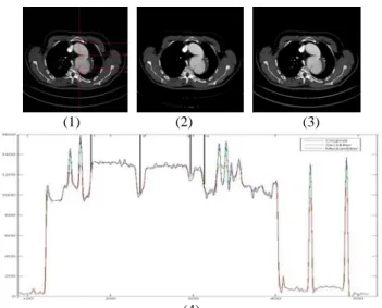
(1) (2) (3)
(4)
Fig 2: (1) Original CT image with marks and the result of: (2) 3D median filter, (3) 3D stick filter and (4) the grey levels of voxels on the marks. We can see both filters decrease noises in
aortic region (between 1 and 4) while stick filter keep more detail information of aortic dissection (3).
(1) (2)
Fig 3: Virtual angioscopy scenes of aortic dissection flap: (1) start at aortic arch and (2) connection in descending aorta.
(1) (2)
Fig 4: (1) 3D surface representation of aortic valve prosthesis extracted and segmented from patient MSCT data: stent (blue) calcified plaques (red) and leaflets (white). (2) Scene of aortic
valve leaflets in virtual angioscopy.
IV. DISCUSSION - CONCLUSION
In this work, we focused on segmentation of thin regions such as aortic valve leaflets and aortic dissection flap in MSCT data. The process of segmentation is based on a non-linear filter. It divides the local cube neighborhood into an asymmetric stick set, and emphasizes selective character along the appropriate directions for the smoothing and segmentation. We visualized 3D surface of aortic valve prosthesis and aortic dissection flap
extracted and segmented from patient MSCT data through virtual angioscopy. Further work will deal with the segmentation of time-series data sets of aortic valve, as well as its dynamic visualization through virtual angioscopy.
REFERENCES
[1]. P Haigron, ME Bellemare, O Acosta, C Göksu. Depth-map-based scene analysis for active navigation in virtual angioscopy. IEEE TRANS
MEDICAL IMAGING, VOL. 23, NO. 11,
NOVEMBER 2004
[2]. P. J.Yim, P. L. Choyke, and R. M. Summers, “Gray-scale skeletonization of small vessels in magnetic resonance angiography,” IEEE Trans. Med. Imag., vol. 19, pp. 568–576, June 2000
[3]. Li min Luo, Chafiaa Hamitouche, Jean Louis Dillenseger et al. A Moment Based Three Dimensional Edge Operator. IEEE Trans. BME, 1993, 40 (7) : 693~703.
[4]. Barrett, W.A. and Mortensen, E.N. Interactive live-wire boundary extraction. Medical Image Analysis 1(4), pp.331-341, 1997
[5]. R C Gonzalez, Digital Image Processing, the second edition
[6]. A.F. Frangi, W.J. Niessen, K.L. Vincken, and M.A. Viergever. Multiscale vessel enhancement filtering. In MICCAI, pages 130–137. Springer Verlag, 1998. [7]. J C Carr, O Simonetti, J Bundy, Cine MR
Angiography of the Heart with Segmented True Fast Imaging with Steady-State Precession. Radiology
June 2001, 219, 828-834.
[8]. T N. Bloomer, S Plein , A Radjenovic Cine MRI Using Steady State Free Precession in the Radial Long Axis Orientation is a Fast Accurate Method for Obtaining Volumetric Data of the Left Ventricle.
JOURNAL OF MAGNETIC RESONANCE IMAGING 14:685–692 (2001)
[9]. Thorsten R. C. Johnson, Konstantin Nikolaou, Bernd J. Wintersperger, Dual-source CT cardiac imaging: initial experience. Eur Radiol (2006) 16: 1409–1415DOI 10.1007/s00330-006-0298-y
[10]. R N Czerwinski., D L Jones. Line and boundary detection in speckle images. IEEE Trans. Image
Processing 7, 1700–1714 1998.
[11]. Chang-Yan Xiao , Zhang Su, Ya-zhu Chen, A diffusion stick method for speckle suppression in ultrasonic images, Pattern Recognition Letters Volume 25 , Issue 16 (December 2004) 1867 - 1877 2004
[12]. R N Czerwinski., D L Jones, 1999. Detection of lines and boundaries in speckle images-application to medical ultrasound. IEEE Trans. Med. Imag. 18, 126-136.