HAL Id: hal-01091447
https://hal-univ-rennes1.archives-ouvertes.fr/hal-01091447
Submitted on 5 Dec 2014HAL is a multi-disciplinary open access archive for the deposit and dissemination of sci-entific research documents, whether they are pub-lished or not. The documents may come from teaching and research institutions in France or abroad, or from public or private research centers.
L’archive ouverte pluridisciplinaire HAL, est destinée au dépôt et à la diffusion de documents scientifiques de niveau recherche, publiés ou non, émanant des établissements d’enseignement et de recherche français ou étrangers, des laboratoires publics ou privés.
retinitis pigmentosa in France and characterization of
biochemical and clinical features.
Gaël Manes, Tremeur Guillaumie, Werner L Vos, Aurore Devos, Isabelle
Audo, Christina Zeitz, Virginie Marquette, Xavier Zanlonghi, Sabine
Defoort-Dhellemmes, Bernard Puech, et al.
To cite this version:
Gaël Manes, Tremeur Guillaumie, Werner L Vos, Aurore Devos, Isabelle Audo, et al.. High prevalence of PRPH2 in autosomal dominant retinitis pigmentosa in France and characterization of biochemical and clinical features.. Ophthalmology: Journal of The American Academy of Ophthalmology, Elsevier, 2015, 159 (2), pp.302-314. �10.1016/j.ajo.2014.10.033�. �hal-01091447�
M
A
N
U
S
C
R
IP
T
A
C
C
E
P
T
E
D
ABSTRACTPurpose: To assess the prevalence of PRPH2 in autosomal dominant retinitis pigmentosa
(adRP), to report six novel mutations, to characterize the biochemical features of a recurrent novel mutation and to study the clinical features of adRP patients.
Design: Retrospective clinical and molecular genetic study.
Methods: Clinical investigations included visual field testing, fundus examination,
high-resolution spectral-domain optical coherence tomography (OCT), fundus autofluorescence imaging and electroretinogram (ERG) recording. PRPH2 was screened by Sanger sequencing in a cohort of 310 French families with adRP. Peripherin-2 protein was produced in yeast and analyzed by Western blot.
Results: We identified 15 mutations, including 6 novel and 9 previously reported changes in
32 families, accounting for a prevalence of 10.3% in this adRP population. We showed that a new recurrent p.Leu254Gln mutation leads to protein aggregation, suggesting abnormal folding. The clinical severity of the disease in examined patients was moderate with 78% of the eyes having 1 to 0.5 of visual acuity and 52% of the eyes retaining more than 50% of the visual field. Some patients characteristically showed vitelliform deposits or macular
involvement. In some families, pericentral RP or macular dystrophy were found in family members while widespread RP was present in other members of the same families.
Conclusions: The mutations in PRPH2 account for 10.3% of adRP in the French population,
which is higher than previously reported (0-8%) This makes PRPH2 the second most frequent adRP gene after RHO in our series. PRPH2 mutations cause highly variable phenotypes and moderate forms of adRP, including mild cases which could be underdiagnosed.
M
A
N
U
S
C
R
IP
T
A
C
C
E
P
T
E
D
High prevalence of PRPH2 in autosomal dominant retinitis
pigmentosa in France and characterization of biochemical and
clinical features
Gaël Manes,1,2,3,* Tremeur Guillaumie,2,3,4 Werner L. Vos,5 Aurore Devos,6 Isabelle Audo,7,8,9,10 Christina Zeitz,7,8,9 Virginie Marquette,1,4 Xavier Zanlonghi,11 Sabine Defoort-Dhellemmes,12 Bernard Puech,12 Saddek Mohand Said,7,8,9,10 José Alain Sahel,7,8,9,10 Sylvie Odent,13 Hélène Dollfus,14 Josseline Kaplan,15 Jean-Louis Dufier,16 Guylène Le Meur,17 Michel Weber,17 Laurence Faivre,18 Francine Behar Cohen,19 Christophe Béroud,20 Marie-Christine Picot,21 Coralie Verdier,1,2 Audrey Sénéchal,1,2,3 Corinne Baudoin,2,3,4 Béatrice Bocquet,1,2,3,4 John B. Findlay,5 Isabelle Meunier,1,2,3,4 Claire-Marie Dhaenens6,22 and Christian P. Hamel1,2,3,4
Author affiliations
1. Institut National de la Santé et de la Recherche Médicale, U1051, Institute for Neurosciences of Montpellier, Montpellier 34091, France
2. University of Montpellier 1, Montpellier 34091, France 3. University of Montpellier 2, Montpellier 34091, France
4. CHRU, Genetics of Sensory Diseases, Montpellier 34091, France
5. The Marie Curie Laboratory for Membrane Proteins, National University of Ireland, Maynooth, Co. Kildare, Ireland
6. CHRU Lille, Laboratoire de Biochimie et Biologie Moléculaire, UF Génopathies, Lille 59037, France
7. Institut National de la Santé et de la Recherche Médicale, U968, Paris 75012, France 8. Centre National de la Recherche Scientifique, UMR 7210, Paris 75012, France
9. Sorbonne Universités, Université Pierre et Marie Curie, Paris 06, UMR S968, Paris 75012, France
10. Centre Hospitalier National d’Ophtalmologie des Quinze-Vingts, INSERM, Direction de l’Hospitalisation et de l’Organisation des Soins, Centre d’Investigation Clinique 1423, Paris 75012, France
11. Clinique Ophtalmologique Sourdille, Nantes 44000, France
12. Laboratoire Neurosciences Fonctionnelles et Pathologies, CNRS FRE 2726, Hôpital Roger Salengro, Lille 59037 Cedex, France;
13. Centre Hospitalier Universitaire de Rennes 35203, France
14. Hôpitaux Universitaires de Strasbourg (HUS) , Centre de référence pour les affections rares en génétique ophtalmologique (CARGO), Strasbourg 67000, France
15. Department of Genetics, INSERM U781, Hôpital Necker Enfants Malades, Université Paris-Descartes, Paris 75015, France
16. Groupe Hospitalier Necker- Enfants Malades, Université Paris-Descartes, Paris 75015, France
17. CHU-Hotel Dieu, Service d’Ophtalmologie, Nantes 44093, France
18. Centre de génétique, hôpital d’Enfants, université de Dijon, Dijon 21000, France
19. Paris Descartes University, AP-HP Hôtel-Dieu Hospital, Department of Ophthalmology, 1 place du Parvis Notre-Dame, Paris 75006, France
20. INSERM UMR_S 910, University of Aix-Marseille, Marseille 13000, France
21. Clinical Investigation Centre, University Hospital of Montpellier, Montpellier 34295, France
M
A
N
U
S
C
R
IP
T
A
C
C
E
P
T
E
D
*Correspondence to: Gaël Manes, Inserm U1051, Genetics and therapy of retinal and optic nerve blindness, Institute for Neurosciences of Montpellier, Hôpital Saint Eloi, 80 av. Augustin Fliche, 34295 Montpellier cedex 05, France. Email: gael.manes@inserm.fr Short title:
M
A
N
U
S
C
R
IP
T
A
C
C
E
P
T
E
D
INTRODUCTIONIn the retina, the human peripherin-2 gene (PRPH2; MIM #179605), also known as RDS (retinal degeneration slow) encodes Peripherin-2, a transmembrane glycoprotein localized in the rim regions of photoreceptor outer segment discs.1–3 Peripherin-2 forms homo- and hetero-tetramers with its paralog protein ROM1 (retinal outer segment membrane protein 1; MIM #180721). These oligomers are essential for the stabilization of the disc rims and are required to pile up the discs as compact, elongated structures.4–8 Mutations in PRPH2 cause a wide range of autosomal dominant retinal dystrophies, either with involvement of the
peripheral retina such as retinitis pigmentosa,9 cone-rod dystrophy10,11 and even one case of retinitis punctata albescens,12 or with predominant involvement of the macula such as adult vitelliform macular dystrophy,13 cone dystrophy,14pattern dystrophy,15,16 and central areolar choroidal atrophy.17–20 In addition, the PRPH2 p.Leu185Pro substitution has also been associated with ROM1 mutations in a digenic form of retinitis pigmentosa.21,22
Among the variety of retinal degenerations caused by PRPH2 mutations, autosomal dominant retinitis pigmentosa (adRP) is the most frequent condition. Typical symptoms of RP include night blindness, progressive visual field constriction, eventually progressing towards total blindness after several decades.23 The prevalence of RP is approximately 1/3,500 to 1/4,000 and the mode of inheritance can be autosomal dominant (30-40%), autosomal recessive (50-60%) or X-linked (5-15%).23,24 RP is the most genetically heterogeneous clinical entity of inherited retinal disorders with 69 disease-causing genes currently known in this condition (www.sph.uth.tmc.edu/retnet) including 24 genes causing adRP. The prevalence of the known genes in adRP ranges from 26.5%25,26 to 16.6%27 for the most frequently found mutations in RHO (MIM #180380), to many genes accounting for less than 1% of the adRP families. Among those genes, the prevalence of PRPH2 mutations varies widely from 0% to 8% of the cases of adRP in cohorts of different origins but no accurate prevalence data are available for the French population.28–30 Also, as usually found in adRP, the severity of the PRPH2 genetic form is considered as moderate, but it is not known whether or not there are important
variations of severity inside the PRPH2 genetic category. Therefore, we sought for PRPH2 mutations in a large cohort of 310 adRP families originating mainly from France. We found novel mutations, characterized the biochemical features of one novel mutation and we analyzed the clinical features of the affected patients.
METHODS: Patients
Three hundred and ten index patients were included in the study. Informed and written consent was obtained for all patients participating to the study. Patients of European origins were recruited from 10 different clinical centers in France. The study (# 2008-A01238-47) received the authorization from the Sud méditerranée IV ethical board committee (# 08 10 05 from 04/11/2008), was approved by the French regulation agency for medication (AFSSAPS # B81319-70) and is registered at http://clinicaltrials.gov (# NCT01235624). The investigators followed the tenets of the Declaration of Helsinki.
Clinical investigations
Patients had standard ophthalmologic examination (refractometry, visual acuity, slit-lamp examination, applanation tonometry, and funduscopy). Kinetic visual fields were determined with a Goldmann perimeter with targets V4e, III4e and I4e. OCT measurement of the macula was performed using an OCT-3 system (Stratus model 3000, Carl Zeiss Meditec, CA) or with a spectral domain OCT (Spectralis, Heidelberg, Germany) with the software version 3.0. Autofluorescence measurements were obtained with the HRA2 Heidelberg retinal confocal
M
A
N
U
S
C
R
IP
T
A
C
C
E
P
T
E
D
angiograph (Heidelberg Engineering, Dossenheim, Germany) and fundus pictures were taken. Full-fields ERGs were recorded using a Ganzfeld apparatus (Metrovision, Pérenchies, France) with a bipolar contact lens electrode on maximally dilated pupils according to the ISCEV protocol.31
For numerical values, visual acuity was measured with Snellen charts in decimal numbers. Goldmann visual field was quantified by counting the number of subdivisions of the Goldmann grid within the areas of the V4e isopter and expressed as a percentage of the normal visual field. Correlations between visual parameters (visual acuity, visual field and ERG amplitudes) and age were investigated with the coefficient correlation of ranks of Spearman with a confidence interval at 95%, calculated by a Fisher transformation. Mutation screening
Genomic DNA was isolated from 10 ml peripheral blood leucocytes using standard salting out procedure.32 Coding exons and adjacent intronic sequences of the PRPH2 gene
(NM_000322.4; primer pairs and PCR conditions are available on request) were sequenced with an Applied Biosystems 3130xL genetic analyser (Applied Biosystems, Foster City, CA) using a BigDye Terminator cycle sequencing ready reaction kit V3.1 (Applied Biosystems, Foster City, CA) following manufacturer’s instructions. Sequence analysis and mutation identification were performed using Collection and Sequence Analysis software package (Applied Biosystems, Foster City, CA). SIFT, PolyPhen2 and Align GVGD were used to predict possible impacts of missense variants. The genomic sequence environment of putative splice-site mutations was analyzed using Human Splicing Finder and MaxEnt.
Genotyping of microsatellite markers and linkage analysis
PCR was carried out in a 25 l final volume containing 50 ng genomic DNA, 5 pmol of each primer, 0.2 mM dNTPs (MP Biochemicals, Asse-Relegen, Belgium), 2 mM MgCl2, PCR
buffer and 1 unit of DNA polymerase (AmpliTaq Gold; Applied Biosystems, Foster City, CA). Initial denaturation at 95°C for 10 minutes was followed by 35 cycles of denaturation at 94°C for 30 seconds, specific annealing temperature for 30 seconds, and extension at 72°C for 1 minute. A final extension step was performed at 72°C for 10 minutes. The PCR products were diluted and mixed with Genescan 400HD ROX size standard and subsequently analyzed on an Applied Biosystems 3130xL genetic analyzer (Applied Biosystems, Foster City, CA). Results were analyzed with GeneMapper software (version 4.0, Applied Biosystems, Foster City, CA).
Two-point LOD scores were calculated with Superlink-online
(http://bioinfo.cs.technion.ac.il/superlink-online/). The phenotype was analyzed as an autosomal dominant and fully penetrant trait with an affected allele frequency of 0.001. Peripherin-2 expression and Western blots
Wild type (WT) and p.Leu254Gln (L254Q) mutant were cloned into the pPICZ expression vector containing the c-myc epitope and the polyhistidine (His)6-tag as described before,33 the nucleotide sequence was confirmed by Eurofins MWG using automated DNA sequencing. Pichia pastoris cells (strain KM71H) were transformed with the PmeI linearised expression vector, stably transformed cells were spread on YPD plates [1% yeast extract, 2% peptone (BD), 2% glucose, 2% agar] with media containing 100 µg/ml zeocin. Cells were cultured, harvested, and stored at -80°C as described before.33 Cells were lysed upon further processing and membranes containing the WT or L254Q proteins were isolated using differential
centrifugation as described previously. The membranes were dissolved in 1% n-dodecyl-
β
-D-maltoside (DDM) using sequentially an 18G, 19G, and 25G needle. His-tagged WT or L254Q proteins were purified using Ni-NTA agarose (final buffer 10 mM NaPO4, 150 mM NaCl,M
A
N
U
S
C
R
IP
T
A
C
C
E
P
T
E
D
200 mM imidazole, and 0.1% n-dodecyl-
β
-D-maltoside). Reducing SDS-PAGE wasperformed by mixing 1:1 (v:v) with 2x loading buffer containing 1% β-mercaptoethanol and incubated for 5 minutes at room temperature prior to loading of the gel. Non-reducing SDS-PAGE was performed by mixing 1:1 (v:v) with 2x loading buffer without β-mercaptoethanol and immediate loading after mixing. Transfer to the PVDF membrane and probing – using cmyc-tagged murine monoclonal (Cell Signaling Technology, Danvers, MA) as primary and anti-mouse HRP-conjugated (Promega, Fitchburg, WI) as secondary antibody – was done as described before.
RESULTS
Identification of recurrent and novel PRPH2 mutations
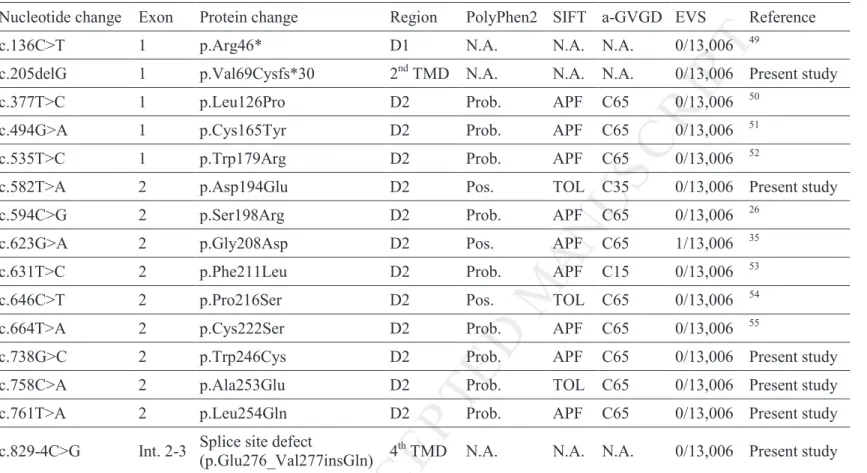
A cohort of 310 French families with autosomal dominant retinitis pigmentosa (adRP) was screened for the three exons of the PRPH2 gene (NM_000322.4). We found that 32 probands (10.3%) carried a mutation. A total of 15 different mutations were identified (Table 1). Nine of them were previously described including one nonsense (p.Arg46*) and eight missense mutations (p.Leu126Pro, p.Cys165Tyr, p.Trp179Arg, p.Ser198Arg, p.Gly208Asp,
p.Phe211Leu, p.Pro216Ser and p.Cys222Ser). Six others were novel including four missense (p.Asp194Glu, p.Trp246Cys, p.Ala253Glu and p.Leu254Gln), one frameshift
(p.Val69Cysfs*30) and one splice site (c.829-4C>G) mutations. All mutations co-segregated with the disease phenotype in available family members (Figure 1, 2). The novel mutations were not identified in 96 ethnically matched control individuals and were not present in the public human SNP databases (including dbSNP, Ensembl, HapMap, the 1000 Genomes project and Exome Variant Server).
Among the novel mutations, the truncating p.Val69Cysfs*30 mutation led to a premature termination located within the second transmembrane α-helix of peripherin-2. No affected family members were available to test the familial segregation for the p.Asp194Glu mutation (Figure 2, bottom right), but Asp194 is conserved in 16 peripherin-2 orthologs (Figure 3) and is surrounded by residues Lys193 and Arg195 which have been found mutated previously.20,34 Moreover, the substitution p.Asp194Glu was predicted to be damaging by PolyPhen2 and align-GVGD programs but not by SIFT (Table 1). For the mutations p.Trp246Cys and p.Ala253Glu, both residues at positions 246 and 253 are also evolutionary conserved (Figure 3) and Trp246 has been previously found mutated in p.Trp246Arg.35 These two mutations were predicted to be damaging by PolyPhen2, align-GVGD and SIFT but tolerated by SIFT for p.Ala253Glu (Table 1).
We identified four families (PHRC057, PHRC069, PHRC161 and PHRC162) with the novel missense mutation, c.761T>A (p.Leu254Gln), with all affected subjects heterozygous for the mutation except two homozygous brothers (II:2 and II:3) in family PHRC161. These two subjects had presumed consanguineous parents, while unaffected individuals did not carry the mutation (Figure 2, left). The evolutionary conserved Leu254 is located in the D2 loop
(Figures 3 and 8) and the substitution p.Leu254Gln is predicted to be damaging by PolyPhen2, SIFT and align-GVGD programs (Table 1). In order to investigate whether p.Leu254Gln was a founder mutation, we genotyped the microsatellite markers D6S1575, D6S1549, D6S1552, D6S282 and D6S1650 that spanned the 2.98 Mb surrounding PRPH2 in the available DNA samples in the 4 families. We found that all affected members of the four families shared an identical allele for the five markers, except patient II:2 of family PHRC161 who had a cross over between D6S1552 and D6S1549 (Figure 2, left). Since the four families originated from the same area in the south of France, this indicates a founder effect. We confirmed the linkage at this locus with microsatellite markers reaching a maximum
M
A
N
U
S
C
R
IP
T
A
C
C
E
P
T
E
D
cumulated LOD score of 4.484 for D6S1575 (Figure 2, left). Since many patients carried the p.Leu254Gln, we performed biochemical investigations of the mutated peripherin-2. The wild type (WT) and the mutated L254Q peripherin-2 proteins were expressed in yeast. We found that both purified WT and L254Q mutant showed monomers and formed dimers (Figure 4). However, aggregates, which were present in both wild type and mutated protein extracts, were much more abundant with the L254Q mutant. In addition, in the absence of the reducing agent β-mercaptoethanol in the sample buffer, the amounts of monomeric and dimeric L254Q were dramatically decreased compared to the WT. Thus, the L254Q mutant exhibited a strong tendency to form large aggregates which might suggests abnormal folding for L254Q mutant. Five independent families (PHRC011, PHRC084, PHRC197, PHRC276 and Fam716) had the c.829-4C>G mutation (Figure 2, top right). Two algorithms (Human Splicing Finder and MaxEnt) predicted that the c.829-4C>G mutation would create an acceptor splice site located three base pair upstream the natural splice site and lead to the in-frame insertion of one glutamine between amino acids 276 and 277 (p.Glu276_Val277insGln) in the fourth
transmembrane α-helix of peripherin-2 (Figure 8). In four of the five families where several family members were available, the mutation was found to co-segregate with the disease. Only individual IV:2 of the family PHRC197 harbored the mutation and was presumed to be unaffected but he was never examined. No common haplotype for five microsatellite markers (D6S1575, D6S1549, D6S1552, D6S282, and D6S1650) surrounding PRPH2 was found (data not shown) and the families were not originating from the same area suggesting that c.829-4C>G could be a mutation hot spot.
Clinical characterization of patients with PRPH2 mutations
From 27 to 67 patients were available for clinical analysis, depending on the type of
examination. On average, the age at presentation was 45.2 + 17.5 (n=44, range 13-78). The initial symptom was night blindness with an apparent age of onset at 30.8 + 13.8 (n=29, range 10-57). Almost half the patients (31/67, 46%) were emmetropic (spherical equivalent -1 to +1), 36% were myopic (SE < -1) and 18% were hypermetropic (SE > +1), showing a skew toward moderate myopia (Figure 5, top row, left).
We found that the cataract, typically present in adult patients with retinitis pigmentosa, was encountered mostly in patients older than 40 (Figure 5, top row, right). The visual acuity was variable with age (Figure 5, middle row, left), 29/81 (35.8%) eyes having a normal visual acuity (VA=1) in patients aged 32.3 + 15.2 (range 13 to 61), 34/81 (42.0%) eyes having a moderately decreased VA (0.9-0.5) in patients aged 47.9 + 15.4 (range 29 to 78), and 18/81 (22.2%) eyes having a severely decreased VA (< 0.4) in patients aged 61.2 + 6.4 (range 43 to 72). The decrease in VA was significantly correlated with age (r = -0.64; p < 0.001). The visual field also decreased progressively with age (Figure 5, middle row, right). We found that 32/62 patients (51.6%) kept more than 50% of their visual field, being aged 37.7 + 13.9 (range 16 to 59), while 30/62 (48.4%) had lost more than 50%, being aged 54.8 + 17.6 (range 16 to 78). The decrease in visual field was significantly correlated with age (r = -0.56; p < 0.001). The rod ERG (dim blue) was recordable (b wave > 10 µV) in 26/60 (43.3%) eyes from patients aged 34.2 + 16.8 (range 16 to 61) and was undetectable in 34/60 (56.7%) eyes from patients aged 54.2 + 11.5 (range 35 to 78) (Figure 5, bottom row, left). The cone ERG (30 Hz flicker) was recordable (b wave amplitude > 5 µV) in 49/54 (90.7%) eyes from patients aged 43.9 + 18.5 (range 16 to 78) and was undetectable in 5/54 (10.3%) eyes from patients aged 52.4 + 6.8 (range 45 to 58) (Figure 5, bottom row, right). Both the rod and the cone ERG decrease was correlated with age, r = -0.62 and -0.44; p < 0.001 for rod and cone function, respectively.
M
A
N
U
S
C
R
IP
T
A
C
C
E
P
T
E
D
Fundus examination revealed the presence of pigment deposits in 73% of the patients with a mean age 45 + 18. Fundus autofluorescence imaging revealed abnormalities in 62.9% (age 47 + 18) including macular autofluorescence ring, atrophic spots in periphery (Table 2). On OCT examination, the majority of patients retained their elipsoid zone at the fovea (70.4%)
whereas a minority had a cystoid macular edema (14.3%). We noticed that some patients had macular involvement with either normal, moderately reduced (Figure 6, row 1, outer left) or severely decreased (Figure 6, row 1, inner left) visual acuity. Some patients had a mild RP with a few spots of atrophy in retinal periphery and macular sparing (Figure 6, row 1, inner right). In other cases, typical pigment deposits and widespread atrophy in the mid peripheral retina was present (Figure 6, row 1, outer right). Some patients showed a pericentral
localization of the retinal lesions even if other members of the family had a widespread form (Figure 6, row 2, outer left). This was particularly evident in family PHRC281 carrying the p.Pro216Ser (Figure 6, row 2, inner left to outer right) in which a family member (III:2) had a pericentral localization of the retinal lesions sharply delimited from the unaffected peripheral retina while her sister (III:1) had a typical widespread retinitis pigmentosa. In a few
circumstances, the presence of yellow deposits was noticed, as in family PHRC305 carrying the p.Pro216Ser, in which the mother had typical retinitis pigmentosa (Figure 6, row 3, outer left) and the son a vitelliform foveal deposit but no signs of retinitis pigmentosa (Figure 6, row 3, inner left).
More clinical details were obtained for the two novel recurrent mutations c.761T>A (p.Leu254Gln) and c.829-4C>G (p.Glu276_Val277insGln) found in four and five families, respectively. In the family PHRC161 with p.Leu254Gln, the visual acuity of the two brothers homozygous for the mutation was severely decreased with 0.1 at age 63 for patient II:3 and hand motion on left eye and light perception on right eye at age 71 for patient II:2. Yet, patient II:3 still had 0.9 on the left eye at age 53, indicating that homozygosity for the
mutation did not lead to early onset severe disease. In general, clinical examination showed a progressive worsening of the visual function with age with the youngest patients being pauci-symptomatic (Figure 6, row 3, inner right) until the legal blindness stage in elder patients (Figure 6 row 3, outer right to row 4, inner left). In the family PHRC162 with p.Leu254Gln, variability in electroretinogram responses was noticed. Patient II:9 who carried the mutation still had a recordable scotopic rod ERG response and was asymptomatic while other mutation carriers of the family had undetectable scotopic ERG rod responses (Figure 7). In the five families with c.829-4C>G (p.Glu276_Val277insGln), the disease was very moderate, the fundus observation being normal (Figure 6, row 4, inner right) or with moderate lesions (Figure 6, row 4, outer right) and the ERG responses being recordable, suggesting that the insertion of an additional amino acid had a moderate pathogenic effect.
DISCUSSION
Autosomal dominant retinitis pigmentosa (adRP) is genetically heterogeneous with 24 known causatives genes so far (www.sph.uth.tmc.edu/retnet). The present study assessed the
prevalence of PRPH2, one of the major genes mutated in adRP, in a large French cohort of 310 families. We established the prevalence for PRPH2 as 10.3% in our cohort making this gene, after RHO (16.5% in French population27), the second gene most frequently found mutated in French adRP patients. Therefore, both genes account for more than a quarter (26.8%) of adRP cases in France. Prevalence studies conducted in the French population revealed that PRPF31 (MIM #606419) with 6.7%36 and RP1 (MIM #603937) with 5.3%37 are respectively the third and fourth causatives genes. Altogether, these four genes represent 38.8% of French adRP patients.
M
A
N
U
S
C
R
IP
T
A
C
C
E
P
T
E
D
Based on the literature, the prevalence of PRPH2 mutations ranges from 0% to 8% of cases of adRP in cohorts of different origins. Mutations in the PRPH2 gene appear to be rare in
Southern European adRP patients: 0% (0/48) in Italian30 and 1% (2/148) in Spanish38 patients with adRP. In comparison, the proportion of adRP due to PRPH2 mutations is higher in populations with Northern European or Asian origins: 3.5% of Northern American population with altogether dominant and recessive RP (8/227)21, in 5% of Japanese adRP cases (5/96)9, in 8% of American (17/206)29, and Swedish (3/38)28 adRP patients. With a prevalence of 10.3% in our cohort, it is higher in France than usually reported, possibly because of
underdiagnosed family members with mild disease that we describe in this study, leading to erroneously classify them as simplex cases. We found indeed that some cases presented as pericentral forms of RP, and other cases are asymptomatic or pauci-symptomatic. In general, we found that the RP associated with PRPH2 mutations is not severe, with many patients retaining useful visual acuity and visual field at middle age. There is no specific refractive error, which distinguishes this RP from the X-linked RP in which myopia is consistently found.39
This study reports 15 different mutations in PRPH2 found in 32 families from a cohort of 310 families with adRP; of these, six are novel and nine were previously reported (Table 1). The referenced mutations, represent 60% of the mutations identified in this report. Nevertheless, with 40% of novel mutations, it is still worth to screen the whole PRPH2 gene for novel changes.
Among the novel mutation, the deletion c.205delG leads to a premature stop shortening the protein to 98 residues instead of 346 amino acids (p.Val69Cysfs*30) and the truncation affects the second transmembrane α-helix of peripherin-2 (Figure 8) or, more likely, is a functional null allele. To date, 42 truncating mutations including nonsense substitutions and frameshift mutations are listed at the Human Genome Mutation Database (HGMD). Although no DNA samples from additional affected family members were available for segregation analysis, it is likely that the truncating change presented in this study is a pathogenic mutation and causes the RP phenotype in family PHRC126.
The four novel missense changes identified in this study affect evolutionary conserved amino acids (Figure 3), and are located in the large intradiscal loop domain (D2) of peripherin-2 (Figure 8), which contains most disease-causing missense mutations.40 To date, 124 PRPH2 mutations are listed at the HGMD and approximately 65% of them are located in the D2 loop of the protein emphasizing the importance of this domain. This D2 loop plays a crucial role in the dimerization of homo- or hetero-tetramers with ROM1 (retinal outer segment membrane protein 1), the homolog of peripherin-2, to form essential interactions important for disc formation and stabilization.4–8 The p.Leu254Gln substitution appears to be recurrent in the French population since it was found in four unrelated families with the same geographic origin. All the affected patients harboring the mutation share the same haplotype for five surrounding microsatellite markers (maximum cumulated LOD score of 4.484 for D6S1575) suggesting a founder effect. Two affected brothers (Figure 2, left, II:2 and II:3 in family PHRC161) were homozygous for the mutation and were expected to display a more severe phenotype, although this did not seem obvious from clinical records. It is of note that mice with a heterozygous defect in Prph2 present a loss of photoreceptor outer segments
organization while homozygous mice have no outer segments.41,42 Contrary to the null mutation presents in mice, the p.Leu254Gln mutation probably acts through a dominant-negative effect by interfering with the dimerization process. The wild type (WT) and mutant
M
A
N
U
S
C
R
IP
T
A
C
C
E
P
T
E
D
(L254Q) peripherin-2 expressed in yeast (Figure 4) migrate as monomers and dimers, but the mutant peripherin-2 shows a pronounced tendency relative to WT to form larger aggregates. This might suggest an abnormal folding for the L254Q mutant. The increased aggregation may disturb homo- and hetero-tetramers with ROM1 interactions, leading to a loss of some peripherin-2 function.
All novel mutations identified in this study were located either in the D2 loop or truncated the protein before the D2 loop. Nevertheless, one mutation (c.829-4C>G), which was predicted to lead to the in-frame insertion of one glutamine (p.Glu276_Val277insGln) was located in the fourth transmembrane α-helix of peripherin-2 (Figure 8). Three other mutations, within this last transmembrane α-helix, are mentioned in the literature; the mutation p.Gly266Asp was found in an adRP patient,43 the mutation p.Val268Ile was found in a patient with adult vitelliform macular dystrophy13 and the in-frame deletion p.Leu271del was identified in a simplex RP patient.44 The Glu276 residue is conserved in all known peripherin-2 orthologs and is substituted with a glutamine in all known ROM1 orthologs.45 The significance of this highly conserved glutamic acid at position 276 was investigated for peripherin-2 structure and function.46 The authors created a p.Glu276Gln isosteric substitution, very similar to the p.Glu276_Val277insGln predicted mutation found in our study, and they demonstrated that this conserved residue is critical for outer segment discs morphogenesis. The major
physicochemical consequence of the p.Glu276Gln substitution is a loss of ionization potential. They hypothesized that Glu276 may function as a pH sensor to regulate protein activity. Other studies suggest that Glu276 may be important for intramolecular interactions between transmembrane domains.47,48
In conclusion, we have established that the prevalence of PRPH2 is 10.3% in a French cohort of 310 adRP individuals which is higher than previously reported. We also established that PRPH2 cause highly variable phenotypes and moderate forms of adRP, including mild cases which could be underdiagnosed. Moreover, mutation analysis in a large cohort is important for the design of future clinical trials.
M
A
N
U
S
C
R
IP
T
A
C
C
E
P
T
E
D
ACKNOWLEDGMENTS / DISCLOSUREAll authors have completed and submitted the ICMJE form.
This project was supported by Union Nationale des Aveugles et Déficients Visuels (UNADEV), Programme Hospitalier de Recherche Clinique (PHRC; #12-014-0041), foundation Voir et Entendre, and foundation Fighting Blindness (FFB; CD-CL-0808-0466-CHNO (IA) and C-CMM-0907-0428-INSERM04), Fondation Dalloz (prix pour la recherche en ophtalmologie), Ville de Paris and Région Ile de France and Labex, LIFESENSES (ANR-10-LABX-65), French state funds managed by the Agence Nationale de la Recherche within the Investissements d’Avenir program (ANR-11-IDEX-0004-0)
Financial disclosures: C. Zeitz : Rétina France (financial support), Fondation pour la recherche Médicale (financial support). J.A. Sahel: Pixium Vision (consultancy, stock options), GenSight Biologics (consultancy, stock options), Sanofi-fovea (consultancy), Genesignal (consultancy) and patents (FR_10_53381, FR99/02346, EP_04_291067, EP 2005/005242, PCT/EP2005/005184, EP2006/005323, PCT/EP2008/057031,
PCT/EP2008/058672, PCT/EP2008/066878, PCT/EP2009/058447, PCT/EP2009/061764, PCT/EP2009/061464, PCT/EP2010/055053, PCT/EP2010/058622, PCT/EP2010/059059, PCT/EP2011/051378, PCT/EP2011/054071).
Contributions of Authors: design of the study (G.M., T.G., W.V., C.-M.D., C.H.); analysis and interpretation (G.M., T.G., W.V, C.-M.D., C.H.); writing the article (G.M., T.G., C.H.); critical revision of the article (G.M., T.G., I.A., C.Z., C.-M.D., C.H.); data collection (G.M., T.G., W.V, A.D., I.A., C.Z., V.M., C.B., C.V., A.S., C.B., B.B., I.M., C.-M.D., C.H.); provision of materials, patients, or resources (G.M., T.G., W.V., I.A., X.Z., S.D.D., B.P., S.M.S., J.-A.S., S.O., H.D., J.K., J.-L.D., G.L., M.W., L.F., F.B.C., M.-C.P., J.F., I.M., C.-M.D., C.H.)
We thank Jean-Louis Pasquier (Institute for Neurosciences of Montpellier, Montpellier, France) for art work.
M
A
N
U
S
C
R
IP
T
A
C
C
E
P
T
E
D
REFERENCES1. Arikawa K, Molday LL, Molday RS, Williams DS. Localization of peripherin/rds in the disk membranes of cone and rod photoreceptors: relationship to disk membrane
morphogenesis and retinal degeneration. J Cell Biol. 1992;116(3):659-667.
2. Connell G, Bascom R, Molday L, Reid D, McInnes RR, Molday RS. Photoreceptor peripherin is the normal product of the gene responsible for retinal degeneration in the rds mouse. Proc Natl Acad Sci USA. 1991;88(3):723-726.
3. Molday RS, Hicks D, Molday L. Peripherin. A rim-specific membrane protein of rod outer segment discs. Invest Ophthalmol Vis Sci. 1987;28(1):50-61.
4. Boesze-Battaglia K, Lamba OP, Napoli AA Jr, Sinha S, Guo Y. Fusion between retinal rod outer segment membranes and model membranes: a role for photoreceptor
peripherin/rds. Biochemistry. 1998;37(26):9477-9487.
5. Goldberg AF, Molday RS. Subunit composition of the peripherin/rds-rom-1 disk rim complex from rod photoreceptors: hydrodynamic evidence for a tetrameric quaternary structure. Biochemistry. 1996;35(19):6144-6149.
6. Loewen CJ, Molday RS. Disulfide-mediated oligomerization of Peripherin/Rds and Rom-1 in photoreceptor disk membranes. Implications for photoreceptor outer segment morphogenesis and degeneration. J Biol Chem. 2000;275(8):5370-5378.
7. Molday RS. Photoreceptor membrane proteins, phototransduction, and retinal degenerative diseases. The Friedenwald Lecture. Invest Ophthalmol Vis Sci. 1998;39(13):2491-2513.
8. Travis GH, Sutcliffe JG, Bok D. The retinal degeneration slow (rds) gene product is a photoreceptor disc membrane-associated glycoprotein. Neuron. 1991;6(1):61-70. 9. Kajiwara K, Hahn LB, Mukai S, Travis GH, Berson EL, Dryja TP. Mutations in the
human retinal degeneration slow gene in autosomal dominant retinitis pigmentosa. Nature. 1991;354(6353):480-483.
10. Nakazawa M, Kikawa E, Chida Y, Tamai M. Asn244His mutation of the
peripherin/RDS gene causing autosomal dominant cone-rod degeneration. Hum Mol Genet. 1994;3(7):1195-1196.
11. Nakazawa M, Kikawa E, Chida Y, Wada Y, Shiono T, Tamai M. Autosomal dominant cone-rod dystrophy associated with mutations in codon 244 (Asn244His) and codon 184 (Tyr184Ser) of the peripherin/RDS gene. Arch Ophthalmol. 1996;114(1):72-78.
12. Kajiwara K, Sandberg MA, Berson EL, Dryja TP. A null mutation in the human peripherin/RDS gene in a family with autosomal dominant retinitis punctata albescens. Nat Genet. 1993;3(3):208-212.
13. Felbor U, Schilling H, Weber BH. Adult vitelliform macular dystrophy is frequently associated with mutations in the peripherin/RDS gene. Hum Mutat. 1997;10(4):301-309.
M
A
N
U
S
C
R
IP
T
A
C
C
E
P
T
E
D
14. Fishman GA, Stone EM, Alexander KR, Gilbert LD, Derlacki DJ, Butler NS. Serine-27-phenylalanine mutation within the peripherin/RDS gene in a family with cone dystrophy. Ophthalmology. 1997;104(2):299-306.
15. Keen TJ, Inglehearn CF, Kim R, Bird AC, Bhattacharya S. Retinal pattern dystrophy associated with a 4 bp insertion at codon 140 in the RDS-peripherin gene. Hum Mol Genet. 1994;3(2):367-368.
16. Kim RY, Dollfus H, Keen TJ, et al. Autosomal dominant pattern dystrophy of the retina associated with a 4-base pair insertion at codon 140 in the peripherin/RDS gene. Arch Ophthalmol. 1995;113(4):451-455.
17. Hoyng CB, Heutink P, Testers L, Pinckers A, Deutman AF, Oostra BA. Autosomal dominant central areolar choroidal dystrophy caused by a mutation in codon 142 in the peripherin/RDS gene. Am J Ophthalmol. 1996;121(6):623-629.
18. Piguet B, Héon E, Munier FL, et al. Full characterization of the maculopathy associated with an Arg-172-Trp mutation in the RDS/peripherin gene. Ophthalmic Genet.
1996;17(4):175-186.
19. Wells J, Wroblewski J, Keen J, et al. Mutations in the human retinal degeneration slow (RDS) gene can cause either retinitis pigmentosa or macular dystrophy. Nat Genet. 1993;3(3):213-218.
20. Yanagihashi S, Nakazawa M, Kurotaki J, Sato M, Miyagawa Y, Ohguro H. Autosomal dominant central areolar choroidal dystrophy and a novel Arg195Leu mutation in the peripherin/RDS gene. Arch Ophthalmol. 2003;121(10):1458-1461.
21. Dryja TP, Hahn LB, Kajiwara K, Berson EL. Dominant and digenic mutations in the peripherin/RDS and ROM1 genes in retinitis pigmentosa. Invest Ophthalmol Vis Sci. 1997;38(10):1972-1982.
22. Kajiwara K, Berson EL, Dryja TP. Digenic retinitis pigmentosa due to mutations at the unlinked peripherin/RDS and ROM1 loci. Science. 1994;264(5165):1604-1608.
23. Hartong DT, Berson EL, Dryja TP. Retinitis pigmentosa. Lancet. 2006;368(9549):1795-1809.
24. Haim M. Epidemiology of retinitis pigmentosa in Denmark. Acta Ophthalmol Scand Suppl. 2002;(233):1-34.
25. Dryja TP, McGee TL, Reichel E, et al. A point mutation of the rhodopsin gene in one form of retinitis pigmentosa. Nature. 1990;343(6256):364-366.
26. Sullivan LS, Bowne SJ, Birch DG, et al. Prevalence of disease-causing mutations in families with autosomal dominant retinitis pigmentosa: a screen of known genes in 200 families. Invest Ophthalmol Vis Sci. 2006;47(7):3052-3064.
27. Audo I, Manes G, Mohand-Saïd S, et al. Spectrum of rhodopsin mutations in French autosomal dominant rod-cone dystrophy patients. Invest Ophthalmol Vis Sci.
M
A
N
U
S
C
R
IP
T
A
C
C
E
P
T
E
D
28. Ekström U, Ponjavic V, Andréasson S, Ehinger B, Nilsson-Ehle P, Abrahamson M. Detection of alterations in all three exons of the peripherin/RDS gene in Swedish patients with retinitis pigmentosa using an efficient DGGE system. MP, Mol Pathol. 1998;51(5):287-291.
29. Sohocki MM, Daiger SP, Bowne SJ, et al. Prevalence of mutations causing retinitis pigmentosa and other inherited retinopathies. Hum Mutat. 2001;17(1):42-51. 30. Ziviello C, Simonelli F, Testa F, et al. Molecular genetics of autosomal dominant
retinitis pigmentosa (ADRP): a comprehensive study of 43 Italian families. J Med Genet. 2005;42(7):e47.
31. Marmor MF, Fulton AB, Holder GE, et al. ISCEV Standard for full-field clinical electroretinography (2008 update). Doc Ophthalmol. 2009;118(1):69-77.
32. Miller SA, Dykes DD, Polesky HF. A simple salting out procedure for extracting DNA from human nucleated cells. Nucleic Acids Res. 1988;16(3):1215.
33. Vos WL, Vaughan S, Lall PY, McCaffrey JG, Wysocka-Kapcinska M, Findlay JBC. Expression and structural characterization of peripherin/RDS, a membrane protein implicated in photoreceptor outer segment morphology. Eur Biophys J. 2010;39(4):679-688.
34. Jacobson SG, Cideciyan AV, Kemp CM, Sheffield VC, Stone EM. Photoreceptor function in heterozygotes with insertion or deletion mutations in the RDS gene. Invest Ophthalmol Vis Sci. 1996;37(8):1662-1674.
35. Kohl S, Christ-Adler M, Apfelstedt-Sylla E, et al. RDS/peripherin gene mutations are frequent causes of central retinal dystrophies. J Med Genet. 1997;34(8):620-626. 36. Audo I, Bujakowska K, Mohand-Saïd S, et al. Prevalence and novelty of PRPF31
mutations in French autosomal dominant rod-cone dystrophy patients and a review of published reports. BMC Med Genet. 2010;11:145.
37. Audo I, Mohand-Saïd S, Dhaenens C-M, et al. RP1 and autosomal dominant rod-cone dystrophy: novel mutations, a review of published variants, and genotype-phenotype correlation. Human Mutation. 2012;33(1):73-80.
38. Millá E, Maseras M, Martínez-Gimeno M, et al. [Genetic and molecular characterization of 148 patients with autosomal dominant retinitis pigmentosa (ADRP)]. Arch Soc Esp Oftalmol. 2002;77(9):481-484.
39. Kaplan J, Pelet A, Martin C, et al. Phenotype-genotype correlations in X linked retinitis pigmentosa. J Med Genet. 1992;29(9):615-623.
40. Boon CJF, den Hollander AI, Hoyng CB, Cremers FPM, Klevering BJ, Keunen JEE. The spectrum of retinal dystrophies caused by mutations in the peripherin/RDS gene. Prog Retin Eye Res. 2008;27(2):213-235.
41. Hawkins RK, Jansen HG, Sanyal S. Development and degeneration of retina in rds mutant mice: photoreceptor abnormalities in the heterozygotes. Exp Eye Res. 1985;41(6):701-720.
M
A
N
U
S
C
R
IP
T
A
C
C
E
P
T
E
D
42. Sanyal S, Jansen HG. Absence of receptor outer segments in the retina of rds mutant mice. Neurosci Lett. 1981;21(1):23-26.
43. Kajiwara K, Berson EL, Dryja TP. Screen for mutations in the entire coding sequence of the human RDS/ peripherin gene in patients with hereditary retinal degenerations. Invest Ophthalmol Vis Sci. 1992;33(4):1149-1151.
44. Jin Z-B, Mandai M, Yokota T, et al. Identifying pathogenic genetic background of simplex or multiplex retinitis pigmentosa patients: a large scale mutation screening study. J Med Genet. 2008;45(7):465-472.
45. Goldberg AFX. Role of peripherin/rds in vertebrate photoreceptor architecture and inherited retinal degenerations. Int Rev Cytol. 2006;253:131-175.
46. Goldberg AFX, Ritter LM, Khattree N, et al. An intramembrane glutamic acid governs peripherin/rds function for photoreceptor disk morphogenesis. Invest Ophthalmol Vis Sci. 2007;48(7):2975-2986.
47. Kovalenko OV, Metcalf DG, DeGrado WF, Hemler ME. Structural organization and interactions of transmembrane domains in tetraspanin proteins. BMC Struct Biol. 2005;5:11.
48. Seigneuret M. Complete predicted three-dimensional structure of the facilitator transmembrane protein and hepatitis C virus receptor CD81: conserved and variable structural domains in the tetraspanin superfamily. Biophys J. 2006;90(1):212-227. 49. Meins M, Grüning G, Blankenagel A, et al. Heterozygous “null allele” mutation in the
human peripherin/RDS gene. Hum Mol Genet. 1993;2(12):2181-2182.
50. Renner AB, Fiebig BS, Weber BHF, et al. Phenotypic variability and long-term follow-up of patients with known and novel PRPH2/RDS gene mutations. Am J Ophthalmol. 2009;147(3):518-530.
51. Souied EH, Rozet JM, Gerber S, et al. Two novel missense mutations in the
peripherin/RDS gene in two unrelated French patients with autosomal dominant retinitis pigmentosa. Eur J Ophthalmol. 1998;8(2):98-101.
52. Bareil C, Delague V, Arnaud B, Demaille J, Hamel C, Claustres M. W179R: a novel missense mutation in the peripherin/RDS gene in a family with autosomal dominant retinitis pigmentosa. Hum Mutat. 2000;15(6):583-584.
53. Ekström U, Ponjavic V, Abrahamson M, et al. Phenotypic expression of autosomal dominant retinitis pigmentosa in a Swedish family expressing a Phe-211-Leu variant of peripherin/RDS. Ophthalmic Genet. 1998;19(1):27-37.
54. Fishman GA, Stone E, Gilbert LD, Vandenburgh K, Sheffield VC, Heckenlively JR. Clinical features of a previously undescribed codon 216 (proline to serine) mutation in the peripherin/retinal degeneration slow gene in autosomal dominant retinitis
pigmentosa. Ophthalmology. 1994;101(8):1409-1421.
55. Downs K, Zacks DN, Caruso R, et al. Molecular testing for hereditary retinal disease as part of clinical care. Arch Ophthalmol. 2007;125(2):252-258.
M
A
N
U
S
C
R
IP
T
A
C
C
E
P
T
E
D
FIGURE LEGENDSFigure 1: Pedigrees of French families with autosomal dominant retinitis pigmentosa and with recurrent mutations in PRPH2 gene identified in this study
Filled symbols indicate affected family members; squares: males; circles: females; arrows: index patients. (Top left) Family pedigrees of patients showing different recurrent PRPH2 mutations. (Top middle) Families with c.136C>T (p.Arg46*) mutation. (Top right) Families with c.494G>A (p.Cys165Tyr) mutation. (Middle left) Families with c.631T>C
(p.Phe211Leu) mutation. (Bottom left) Families with c.594C>G (p.Ser198Arg) mutation. (Bottom middle) Families with c.535T>C (p.Trp179Arg) mutation. (Bottom right) Families with c.646C>T (p.Pro216Ser) mutation.
Figure 2: Pedigrees of French families with autosomal dominant retinitis pigmentosa and with novel mutations in PRPH2 gene identified in this study
(Left) Haplotypes at the PRPH2 locus of four families showing the c.761T>A (p.Leu254Gln) mutation and surrounding microsatellite markers. The common haplotype is shown in black. (Top right) Five families with c.829-4C>G splice site mutation. (Bottom right) Family pedigrees of patients showing different novel PRPH2 mutations.
Figure 3: Conservation of amino acids affected by novel PRPH2 missense mutations identified in this study in patients with autosomal dominant retinitis pigmentosa. Multiple amino acid sequence alignment of peripherin-2 for a region surrounding the novel p.Asp194Glu, p.Trp246Cys, p.Ala253Glu and p.Leu254Gln missense mutations. The site of the mutation is indicated by an arrowhead.
Figure 4: Western blot analysis of wild type and p.Leu254Gln mutant peripherin-2 protein produced in yeast
Western blots of the wild type (WT) and the mutated p.Leu254Gln (L254Q) peripherin-2 from P. pastoris purified with Ni-NTA superflow agarose in the presence (+) or in the
absence (-) of the reducing agent β-mercaptoethanol (β-M) in the sample buffer. The blot was probed with a monoclonal anti-c-myc antibody.
Figure 5: Clinical data of the patients with autosomal dominant retinitis pigmentosa and with PRPH2 gene mutations
(Top row, left) Refractive errors were classified in 5 groups as either severe myopia (< -3[), moderate myopia ([-3;-1[), emmetropia ([-1;+1]), moderate hyperopia (]+1;+3]) or severe hyperopia (>+3[) and the percentage (y-axis) and the absolute number (above each bar) of patients are given for each group. (Top row, right) Apparent onset of cataract was classified in 4 groups depending on age and the percentage (y-axis) and the absolute number (above each bar) of patients are given for each group. Visual acuity in decimal values (Middle row, left), percentage of remaining Goldman visual field (Middle row, right), b-wave amplitude of the dim blue electroretinogram (ERG) testing rods (Bottom row, left) and peak-to-peak amplitude of the 30 Hz flicker ERG testing cone function (Bottom row, right) were plotted against age. Figure 6: Fundus imaging of patients with autosomal dominant retinitis pigmentosa and with PRPH2 gene mutations
On top of each picture, (fundus photographs, fundus autofluorescence (FAF) photographs and spectral-domain optical coherence tomography (OCT) scans), the family number (PHRC), the patient number in the family, the age, the visual acuity in decimal values are indicated. (Row 1, outer left) Left eye FAF, macular alteration, parafoveal loss of autofluorescence (arrow) with moderate decrease in visual acuity. (Row 1, inner left) Left eye color fundus photograph,
M
A
N
U
S
C
R
IP
T
A
C
C
E
P
T
E
D
pale optic disc, narrow blood vessels, pigmentary changes in the periphery and RPE changes in the macular region (arrow) correlated with severe loss of visual acuity at counting fingers (CF). (Row 1, inner right) Left eye colour fundus photographs, there are a few small spots of atrophy in retinal periphery and the macula appears normal. (Row 1, outer right, and row 2 outer left) Colour fundus photographs from two affected brothers, with, for the left eye of the patient II:3, round foveal atrophy and pigment deposits covering a large proportion of the fundus while for the right eye of the patient II:2, most of the retinal atrophy and pigment deposits are present in the macular area. (Row 2, inner left to outer right) FAF and OCT of right eyes from two sisters, with for patient III:2, a pericentral form of retinitis pigmentosa with alteration of autofluorescence within the macular area and sharp limit of the lesions (arrows) while the sister III:1 has a typical widespread retinitis pigmentosa showing small spots of retinal atrophy (stars) and a ring of autofluorescence (white arrow); OCT scans of the macula in both sisters show a conserved inner segment/outer segment line (stars) in the fovea. (Row 3, outer left and inner left) The mother IV:4 has retinitis pigmentosa with pigment deposits in retinal periphery while the son V:1 has a foveal yellow vitelliform deposit (arrow) but no retinitis pigmentosa. (Row 3, inner right to row 4, inner left) FAF imaging in 4
members of the PHRC161 family; the youngest member IV:4 has no retinal alteration, the member III:2 has many round spots of loss of autofluorescence beyond the macula (arrow) but the macula is normal, the member III:4 has a similar aspect to III:2 but there is a ring of autofluorescence around the fovea with moderate decrease of visual acuity, the oldest member II:2 has a complete loss of autofluorescence except for an open ring of remaining retina around the foveal area (arrow) and visual acuity reduced to hand motion (HM). (Row 4, inner right and outer right) Fundus photographs of the daughter II:5 show retinal lesion and slightly reduced retinal vessel diameter while fundus photographs of the mother III:3 revealed
changes typical of retinitis pigmentosa with overall moderate RPE changes and attenuated retinal vessels but no pigment deposits.
Figure 7: Variability of electroretinographic responses in family PHRC162 with autosomal dominant retinitis pigmentosa and with the PRPH2 gene mutation p.Leu254Gln
Each patient from generation II in PHRC162 was subjected to dark-adapted dim blue stimulation for rod responses and light-adapted 30 Hz flicker for cone responses. Black symbols indicate symptomatic patients, open symbols non affected or asymptomatic patients. Genotypes are shown. The 4 affected patients (II:3, II:7, II:12 and II:13) have no rod
responses and severely decreased cone responses, while the asymptomatic carrier II:9 still has rod responses, although decreased in comparison to normal responses in II:4, II:5, II:6 and II:8.
Figure 8: Summary of the novel mutations found in this study in peripherin-2 protein and in patients with autosomal dominant retinitis pigmentosa
Schematic representation of the peripherin-2 protein showing the location of the six novel mutations presented in this study. The four transmembrane α-helices and the two intradiscal loop domains D1 and D2 are schematized.
M
A
N
U
S
C
R
IP
T
A
C
C
E
P
T
E
D
Table 1. Summary of PRPH2 gene mutations identified in this study in patients with autosomal dominant retinitis pigmentosa
Nucleotide change Exon Protein change Region PolyPhen2 SIFT a-GVGD EVS Reference c.136C>T 1 p.Arg46* D1 N.A. N.A. N.A. 0/13,006 49
c.205delG 1 p.Val69Cysfs*30 2nd TMD N.A. N.A. N.A. 0/13,006 Present study c.377T>C 1 p.Leu126Pro D2 Prob. APF C65 0/13,006 50
c.494G>A 1 p.Cys165Tyr D2 Prob. APF C65 0/13,006 51 c.535T>C 1 p.Trp179Arg D2 Prob. APF C65 0/13,006 52
c.582T>A 2 p.Asp194Glu D2 Pos. TOL C35 0/13,006 Present study c.594C>G 2 p.Ser198Arg D2 Prob. APF C65 0/13,006 26
c.623G>A 2 p.Gly208Asp D2 Pos. APF C65 1/13,006 35 c.631T>C 2 p.Phe211Leu D2 Prob. APF C15 0/13,006 53 c.646C>T 2 p.Pro216Ser D2 Pos. TOL C65 0/13,006 54 c.664T>A 2 p.Cys222Ser D2 Prob. APF C65 0/13,006 55
c.738G>C 2 p.Trp246Cys D2 Prob. APF C65 0/13,006 Present study c.758C>A 2 p.Ala253Glu D2 Prob. TOL C65 0/13,006 Present study c.761T>A 2 p.Leu254Gln D2 Prob. APF C65 0/13,006 Present study c.829-4C>G Int. 2-3 Splice site defect
(p.Glu276_Val277insGln) 4 th
TMD N.A. N.A. N.A. 0/13,006 Present study
D1: D1 loop; D2: D2 loop; TMD: transmembrane helical domain; N.A.: Not applicable; Int.: intron; EVS: Exome variant server. PolyPhen2: Pos. for possibly damaging, Prob. for probably damaging. SIFT: TOL for tolerated, APF for affect protein function. a-GVGD scores amino acid substitutions on a 7-scale scoring system, from C0 (neutral) to C65 (the most likely pathogenic), and C35 is considered intermediate.
M
A
N
U
S
C
R
IP
T
A
C
C
E
P
T
E
D
Table 2: Frequency of clinical features in patients with autosomal dominant retinitis pigmentosa and PRPH2 gene mutation
Description Mean age Number of patients Percentage of positive cases
Pigment deposits 45 + 18 37 73.0%
Autofluorescence changes 47 + 18 35 62.9%
Cystoic macular edema 43 + 17 28 14.3%