HAL Id: hal-01478118
https://hal.archives-ouvertes.fr/hal-01478118
Submitted on 25 May 2018
HAL is a multi-disciplinary open access
archive for the deposit and dissemination of sci-entific research documents, whether they are pub-lished or not. The documents may come from teaching and research institutions in France or abroad, or from public or private research centers.
L’archive ouverte pluridisciplinaire HAL, est destinée au dépôt et à la diffusion de documents scientifiques de niveau recherche, publiés ou non, émanant des établissements d’enseignement et de recherche français ou étrangers, des laboratoires publics ou privés.
Clinical and Laboratory Findings in Patients with
δ-Storage Pool Disease: A Case Series.
Fabien Selle, Chloé James, Marie Tuffigo, Xavier Pillois, Jean-François
Viallard, Marie-Christine Alessi, Mathieu Fiore
To cite this version:
Fabien Selle, Chloé James, Marie Tuffigo, Xavier Pillois, Jean-François Viallard, et al.. Clinical and Laboratory Findings in Patients with δ-Storage Pool Disease: A Case Series.. Seminars in Thrombosis and Hemostasis, Thieme Publishing, 2017, 43 (01), pp.48-58. �10.1055/s-0036-1584568�. �hal-01478118�
Clinical and Laboratory Findings in Patients with δ-Storage Pool Disease: A Case Series
Fabien Selle, MD1 Chloé James, MD, PhD1,2,3,4 Marie Tuffigo, PharmD1,2,4 Xavier Pillois, PhD3,4 Jean-François Viallard, MD, PhD1,4,5 Marie-Christine Alessi, MD, PhD3,6 Mathieu Fiore, MD, PhD2,3 1 Université de Bordeaux, Bordeaux, France
2 Laboratoire d’Hématologie, CHU de Bordeaux, Pessac, France 3Centre de Référence des Pathologies Plaquettaires, France 4INSERM U1034, Biologie des Maladies Cardiovasculaires, Pessac, France
5 Service de Médecine Interne et Maladies Infectieuses, CHU de Bordeaux, Pessac, France
6 Laboratoire d’Hématologie, Aix-Marseille Université, CHU de La Timone, Marseille, France
Abstract
Platelet δ-storage pool disease (δ-SPD) is a platelet function disorder characterized by a reduction in the number or content of dense granules. Reports on δ-SPD are mostly limited to case presentations. We aimed to retrospectively describe a series of patients with δ-SPD to better characterize the disease.We studied 16 patients with congenital or
acquired δ-SPD. Lumiaggregometry, α- and δ-granules content, platelet ultrastructure, αIIbβ3 integrin, and glycoprotein Ib (GPIb) activation were assessed.Most of the patients generally demonstrate mild to moderate bleeding diathesis. Platelet aggregation studies showed moderate abnormalities with variable profiles, while all the individuals had almost complete absence of adenosine triphosphate release. Mepacrine capture, CD63 expression, and study of dense granules by electron microscopy enabled to distinguish different subtypes of δ-SPD with quantitative or qualitative defect. Surprisingly, significantly decreased GPIb expression levels after platelet activation with thrombin receptor activating peptide 50 μMwere found, suggesting that GPIb-impaired
mobilization may represent an additional feature of the disorder. In conclusion, δ-SPD represents a complex disorder with various clinical and biological aspects, requiring a great deal of expertise to be properly diagnosed.
Introduction
Platelets play a major role in hemostasis. Their activation results in shape change, adhesion, aggregation, and release of granule contents.1 Three types of granules are present in platelets: α- granules, dense (δ) granules, and lysosomes. Avast array ofmore than 300 proteins is stored in α-granules, including hemostatic and adhesive molecules, inflammatory peptides, and growth factors.2 Dense bodies contain serotonin, adenine nucleotides, calcium, and pyrophosphate, while lysosomes are composed of soluble enzymes and nonenzymatic proteins.3 Human platelets contain three to eight dense granules per platelet, each measuring 100 to 300 nm in diameter.4 δ-storage pool diseases (δ-SPD) encompass a rare and heterogeneous group of conditions characterized by defects in the number or content of platelet δ-granules.5 The causes of δ-SPD are multiple and variable and can be classified into (1) congenital diseases including Hermansky–Pudlak and Chédiak–Higashi syndromes, (2) nonsyndromic inherited platelet disorders, or (3) acquired forms, most often associated with hematologic malignancies (myeloproliferative syndrome,acute leukemia, or myelodysplastic syndromes).6
Decreased dense bodies secretion leads to a defective hemostatic response to vascular injury and patients suffer from mild to moderate hemorrhagic diathesis mainly characterizedby mucocutaneous bleedings. Thus, rapid and accurate diagnosis is crucial to initiate appropriate therapy that inturn prevents bleeding. The diagnosis must be confirmed by specialized tests,
required to demonstrate the absence or marked reduction of δ-granules (electron microscopy [EM]or mepacrine uptake) and/or their content (nucleotides measured by chemiluminescence or serotonin by high-performance liquid chromatography).7,8This platelet disorder is
considered to be a relatively rare disease, and most previous studies described clinical and laboratory findings in isolated case reports.9,10 Some case series have been also reported, but they were mainly focused on individual platelet tests.11–13Although δ-granules deficiency is the key feature, it ispresently poorly known whether other platelet structural or function
abnormalities are associated with the disorder. The aim of our work was to retrospectively describe a series of patients with δ-SPD, who were diagnosed and largely explored in the French Reference Center for Platelet Disorders of Bordeaux. These data provide novel information to better characterize the disease and enable better understanding of the contributions of structural abnormalities and defective activation pathways to the platelet phenotype.
Methods
Patients
A total of 16 patients were diagnosed in our center as having a δ-SPD of different causes. Clinical investigations included medical and family history, as well as physical examination. Signs of bleeding by sites and bleeding events requiring treatment were also assessed. Bleeding was scored according to the Bleeding Assessment Tool of the International Society on Thrombosis and Haemostasis (ISTH-BAT).14This observational study was approved by our institutional review board (Comité de Protection des Personnes Sud-Ouest et Outre Mer III, Bordeaux, France). Platelet Studies Preanalytical Procedures Venipuncture was performed after at least 5 minutes of rest. Needles with diameters between 19 and 22 gaugewere used. All tubes were inverted at least 10 times to mix the anticoagulant with the blood and prevent clotting. Platelet tests were performed within 2 hours after blood collection. Platelet Counting and Measurement of Mean Platelet Volume Blood samples were collected from patients into sodium
ethylenediaminetetraacetate (EDTA)-anticoagulated blood tubes. Platelet counts and mean platelet volume (MPV) were obtained using the Beckman Coulter LH750 cell counter (Beckman Coulter, Villepinte, France).
Platelet Aggregation Testing
Platelet aggregationwas tested in citrated platelet-rich plasma (PRP) using 1.2 mg/mL ristocetin (Stago, Asnières-sur-Seine, France), 5-μM adenosine diphosphate (ADP) (Sigma Aldrich Chimie, Lyon, France), 1-mM arachidonic acid (AA; Nu Chek Prep, Elysian, MN), 25-μM thrombin receptor activating peptide (TRAP; NeosystemSA, Strasbourg, France), 4-μMepinephrine (Helena Laboratories, Beaumont, TX), and 2 μg/μL Horm equine tendon collagen (Nycomed Pharma, Unterschleibheim, Germany) in an APACT 4004 aggregometer (Elitech, Salon de Provence, France) according to standard procedures15 and during 300 seconds of aggregation test time. In the case of normal platelet counts, test results were compared with
reference values of 30 healthy controls. Alternatively, when patients had thrombocytopenia, platelet aggregation testswere visually analyzed by hematology experts to confirm the presence of an abnormal profile.
Platelet Adenosine Triphosphate Release
Blood was anticoagulated with 0.105-M citrate and PRP was obtained after centrifugation. Platelet secretionwas recorded in real time at 37°C with stirring on a dual-channel Chrono- Log aggregometer (Chronolog Corp, Haverton, PA). The agonists used were ADP 10 μM (Chronolog Corp, Haverton, PA), collagen 2 μg/mL (Chronolog Corp, Haverton, PA), and TRAP-6 (Hart Biologicals Ltd, Queens Meadow, Hartlepool, United Kingdom). Platelet secretion was determined by measuring the release of adenosine triphosphate (ATP) using luciferin/
luciferase reagent (Kordia, Leiden, the Netherlands). Results were expressed as nmol of secreted ATP. Results of patients with platelet count <100 _ 109/L in whole blood were not
considered.
Platelet Flow Cytometry
Surface expression of human platelet glycoprotein (GP) Ib, αIIbβ3, and P-selectin were measured in PRP using a platelet calibrator kit (Biocytex, Marseille, France), according to the
manufacturer’s instructions. Platelet dense granules were also evaluated using 1.7 μM of mepacrine (Sigma Aldrich, St. Louis, MO) and/or CD63 antibody (granulophysin) (Beckman Coulter, Villepinte, France) before and after activation with TRAP. Platelets were analyzed in a Cytomics FC500 flow cytometer (Beckman Coulter, Villepinte, France) and results were expressed as mean fluorescence intensity (MFI). Histograms were generated from
measurements of 10,000 cells and data analyzed using the CXP software (Beckman Coulter, Villepinte, France).
Platelet Electron Microscopy
Standard EM for ultrastructural studies was performed for patients using venous blood taken in anticoagulant citrate dextrose solution A (ACD-A) and PRP was then prepared. After incubation at 37°C, platelets were fixed in 1.25% glutaraldehyde (Fluka, Buchs, Switzerland) in 0.1
Mphosphate buffer (pH 7.2) for 1 hour at roomtemperature. Sampleswerewashed and postfixed in 4% osmic acid containing potassium ferrocyanide (Sigma-Aldrich, St. Louis,MO) for 1 hour at roomtemperature. Following fixation, the samples were dehydrated using graded alcohols and propylene oxide and then embedded in Epon (Taab Laboratories, Reading, United Kingdom). Embedded samples were sectioned with an Ultracut E ultramicrotome (Reichert, Vienna, Austria) and stained with uranyl acetate and lead citrate (Merck, Darmstadt, Germany). Specific gridswere prepared for platelet whole-mount EM. PRP samples were harvested from
anticoagulated blood and centrifuged. The samples (5–10 μL) were spotted onto grids,
rapidly blotted, rinsed with distilled water, and air-dried. Results estimate the number of dense granules contained in a total of 100 platelets.
Statistical Analysis
To account for variability in experimental conditions among measurements, control and patient samples were handled in parallel when possible. Comparisons between patients and controls were performed with the Mann–Whitney test as appropriate and p-values of <0.05 were considered significant.
Results
Clinical Manifestations
A total of 16 patientswere included in the study (44%men and 56% women). ►Table 1 presents the clinical features of the 16 patients. In 69% (11/16) of patients, δ-SPD was considered as an acquired disorder as it was discovered during adulthood (more than 40 years old) and as an underlying primary cause was also present. In most of these cases (9/11; 82%), the
diagnosis was made in the context of amalignant hematological disorder. The type and severity of bleeding were highly variable among patients. Indeed, the ISTH bleeding score (BS) ranged from 1 to 8 with a median score of 5 (interquartile range [IQR] ¼ 4–6). The bleeding
episodeswere either spontaneous or provoked (after trauma or surgery). For two patients, the existence of the diseasewas revealedwhile theywere treated with vitamin K antagonists, which presumably elevated their bleeding risk. Patients with mild bleedings had symptoms in the form of petechiae, large ecchymosis, epistaxis, ormenorrhagia. Lastly,7/16 patients (44%) required emergency care therapy to stop hemorrhage, confirming that bleedings can on occasion be severe or life-threatening.
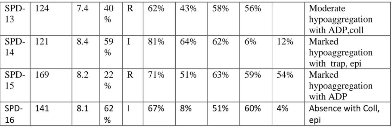
Platelet Count, Mean Platelet Volume, and Light Transmission Aggregometry Eight patients (50%) were thrombocytopenic with platelet counts ranging from 50 to 147 _ 109/L (►Table 2). Thrombocytopenia in these patientswas present in the context of an underlying hematologic disorder, while patients with congenital δ-SPD had normal platelet counts. MPV were normal in all but one case, suggesting that patientswith δ-SPD mostly produce normal-sized platelets. Results of platelet aggregation for each patient are shown in ►Table 2. In all cases, platelet aggregation tests showed moderate abnormalities in response to different agonists, with variations from one patient to another. Platelet aggregations induced by ADP, collagen, and arachidonic acid were frequently perturbed. Aggregation traces in response to ADP were also variable among patients, sometimes showing preserved primary response but lack of secondary wave. Due to the limited amount of PRP, tests were performed with epinephrine for 11 patients and, in seven of them (64%), responses were severely impaired or absent.
Platelet Adenosine Triphosphate Release
As ATP is stored in δ-granules, a decrease in its release supports a diagnosis of dense bodies’ defect. ATP release was recorded bymeasuring the luminescence from the firefly luciferin– luciferase reaction using three different agonists: 10-μM ADP, 2 μg/mL collagen, or 50-μM TRAP-6. In all tested patients, ATP release was severely decreased or absent compared with reference values, whichever the agonist used (►Fig. 1A). However, although this test can be used to screen for a δ-granules secretion defect or δ-SPD, it cannot differentiate between the two.
Mepacrine Uptake and Release
To confirmthat abnormal ATP releasewas due to δ-SPD rather than secretion defect alone, where possible, mepacrine uptake and release were studied (n ¼ 14). Mepacrine (quinacrine) is a fluorescent marker that is rapidly taken up and localized in dense granules, thus reflecting the δ-granules pool. Mepacrine uptake was reduced in patients as compared with controls (meanMFI: 3.4 _ 1.2 vs. 6.6 _ 1.6, p ¼ 0.0001). However, some patients showed only a slight decrease of mepacrine uptake, suggesting that in these cases, δ-SPD was linked to a reduction in the content of dense granules rather than to their number. By contrast, mepacrine release after platelet activation by TRAP was similar to controls (mean MFI: 1.4 _ 0.6 vs. 1.2 _ 0.6, p ¼ 0.268), showing that dense bodies were normally secreted after platelet stimulation (►Fig. 1B). CD63
Expression and Number of Dense Granules by Electron Microscopy
To confirm that results obtained using the mepacrine assay corresponded to a reduced δ-granules number, CD63 expression and dense δ-granules testing by whole mount were studied. CD63 is a marker of δ-granules that can be detected on platelet surface by
flowcytometry after agonist activation. Results of CD63 expression were available for 13 patients (►Table 3). In seven of them (54%), the expression was markedly reduced. However, results confirmed that four patients (31%) showed only a slight decrease of CD63 expression. As dense bodies are rich in calcium, they are inherently electron-opaque and can be easily counted by EM.16 The mean ratio of dense granules measured by EM (40% _ SD ¼ 29%) was also reduced when compared with controls (100 _ 12%), but percentages were very variable among patients (ranging from 0 to 106%) (►Table 3). Although CD63 expression and dense granules by EM were not completely correlated, three patients with a subnormal or normal amount of CD63 had a similar profile by EM, confirming an abnormal dense granules content in these cases rather than a reduction of their number (►Table 3).
Table 1 Patient clinical features
Abbreviations: ISTH, International Society on Thrombosis and Haemostasis; δ-SPD, δ-storage pool disease.
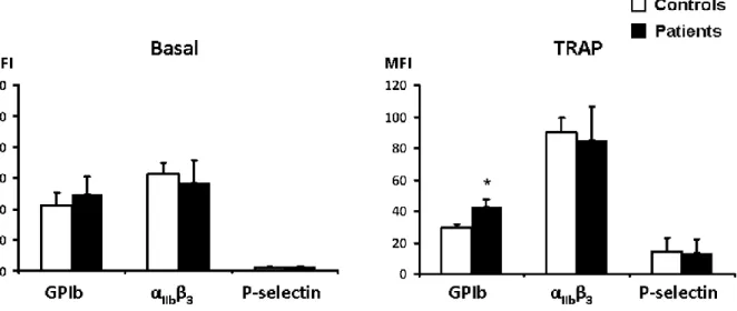
Assessment of Platelet Surface GP Expression and Activation
Measurement of platelet α-granules should be performed to rule out a deficiency that may be associated with δ-SPD. P-selectin may be measured in stimulated platelets as a marker of α-granule release. Moreover, to explore whether δ-SPD occurs in isolation or with additional defects in platelet function, expression and activation of αIIbβ3 and GPIb were
also evaluated. In the resting state, no difference was observed between patients and controls, while after platelet activation, clearance of GPIb was significantly different between patients and controls (►Fig. 2).
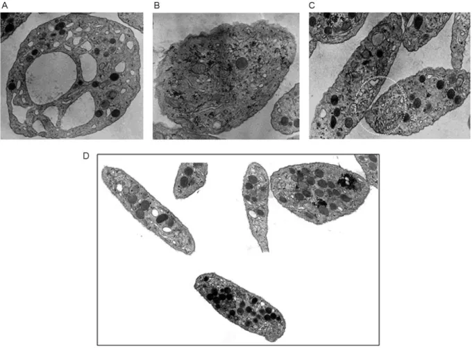
Ultrastructure of Platelets
To evaluate whether δ-SPD was associated with platelet structural abnormalities, EM was
performed, revealing no heterogeneity in platelet size and shape, but the presence of a distended open canalicular system with vacuolated appearance. α-granules content was heterogeneous, as some platelets were completely devoid of this organelle, but their mean number per platelet was comparable to controls (►Fig. 3).
Age at Causes of Treatment ISTH Comorbidities Others treatments
Patient Gender Age diagnosis δ-SPD score
SPD-1 F 5 1 Congenital None 2 Hermansky–Pudlaksyndrome
SPD-2 F 25 24 Congenital None 2 Hermansky–Pudlaksyndrome
SPD-3 F 46 5 Congenital Desmopressin 4 factor V Leiden
SPD-4 M 3 2 Congenital None 1
SPD-5 F 29 1 Congenital None 5 Eczema
SPD-6 M 48 48 Acquired None 7 CMLeukemia Hydroxycarbamide
SPD-7 M 72 71 Acquired Platelet transfusionarte rial embolization 6 CMMLeukemia type 2 progression to acute
SPD-8 F 76 74 Acquired None 4 (RAEB-1) Hydroxycarbamide
SPD-9 M 96 94 Acquired None 3 Waldenström Darbepoetin alfa
SPD-10 F 76 74 Acquired Platelet transfusion 7 Myelodysplastic syndrome
SPD-11 M 61 59 Acquired nasal cauterization 5
SPD-12 F 84 82 Acquired
red blood cell transfusion after appendicectom y Hydroxychloroquine SPD-13 M 55 54 Acquired Polytransfusion after retroperitoneal hematoma 8 SPD-14 M 69 69 Acquired none 2 SPD-15 F 46 44 Acquired Transfusion of red blood cell, platelet, 6 SPD-16 F 55 54 Acquired none 5 Septic shock Polycythemia vera Primary myelofibrosis; mechanical heart valve; congenital
toxoplasmosis
Excessive bruises in exposed areas; bleeding from minor
wounds
Chronic ITP Primary myelofibrosis;
mechanical heart valve; congenital
toxoplasmosis
Epoetin alfa and vitamin K antagonist
vitamin K antagonist Severe epistaxis ; hematoma of
the flank Primary myelofobrosis Spontaneous bruising; purpuric
eruption; oral hemorrhagic bullae; menorrhagia in the past; hemorrhage after salivary gland Retroperitoneal hematoma after radiofrequency ablation of a
kidney tumor; hematoma requiring Wound hematoma after an
inguinal lymph node biopsy Epistaxis; gum bleeding;
excessive bleeding after skin excision
Easy bruising excessive bleeding after Multiple hematoma fatal cerebral hemorrhage Hepatic subcapsular hematoma
hematuria, epistaxis
Spontaneous bruising, lower gastrointestinal bleeding palatal petechiae Epistaxis; spontaneous leg hematoma Beta-blockers; antidepressant, anticholesterol Extended hematoma of the
flank associated with a cough effort; excessive bleedings after
sinus surgery and teeth bleeding diathesis
Easy bruising
Severe epistaxis; easy bruising; menorrhagia; bleeding from
minor wounds
Excessive bleeding after an ectopic pregnancy Posttraumatic knee hematoma
Different Types of δ-Storage Pool Disease and Correlation with Bleeding Severity
In our study, we identified three different types of δ-SPD, classified as (1) severe quantitative defect (7/16; 44%), (2) partial quantitative defect (5/16; 31%), or (3) qualitative defect of dense granules (4/16; 25%) (►Fig. 4). Our aim was to investigate the relationship between bleeding severity and the type of δ-SPD. However, no correlation was found between the ISTH BS and the type of disease, perhaps due to the low number of patients in each group, given the rarity of the disorder (►Fig. 5A). Moreover, there was no difference between the BS of congenital and acquired cases (►Fig. 5B).
Table 2
Patie nt
count MP
V
ADP Risto Coll AA TRA
P Epi Interpretation (>150G/ L) (<10 fl) (>57%) (>77 %) (>73 %) (>73 %) (>74 %) (>70 %) SPD-1 322 7.3 66 % Rc 71% 42% 63% 72% Moderate hypoaggregation with all agonists SPD-2 197 8.4 82 % I 81% 31% 27% 74% 33% Marked hypoaggregation with coll, aa, epi SPD-3 170 8.5 71 % I 78% 45% 56% 73% 74% Moderate hypoaggregation with coll, aa SPD-4 348 7.1 106% 69% 3% 97% 40% Marked hypoaggregation with aa, epi SPD-5 216 8.9 48 % R 68% 57% 62% 73% 5% Moderate hypoaggregation with ADP coll aa Absence with epi
SPD-6
147 7.2 49
%
R 65% 10% 62% 58% hypoaggregation
with all agonists more specifically with coll SPD-7 50 9.5 34 % R 45% 8% 2% 25% Marked hypoaggregation with coll, aa SPD-8 72 8.1 45 % I 64% 23% 33% 56% hypoaggregation
with ADP coll, aa SPD-9 173 8.1 54 % I 61% 57% 56% 51% Moderate hypoaggregation with all agonists SPD-10 93 9.4 52 % R 56% 20% 1% 11% Marked hypoaggregation with coll, aa, epi
SPD-11
156 11.5 61
%
D 80% 68% 63% 73% 2% Absence with epi
SPD-12 66 9.8 25 % R 80% 10% 55% 80% 110% Marked hypoaggregation with ADP, coll
SPD-13 124 7.4 40 % R 62% 43% 58% 56% Moderate hypoaggregation with ADP,coll SPD-14 121 8.4 59 % I 81% 64% 62% 6% 12% Marked hypoaggregation with trap, epi SPD-15 169 8.2 22 % R 71% 51% 63% 59% 54% Marked hypoaggregation with ADP SPD-16 141 8.1 62 %
I 67% 8% 51% 60% 4% Absence with Coll,
epi
Abbreviations: ADP, adenosine diphosphate; δ-SPD, δ-storage pool disease; TRAP, thrombin receptor activating peptide.
aThreshold values derived from 30 healthy controls.
bPlatelet aggregation expressed as a percentage of maximal light transmission in response to 5-μM ADP, 4-μM epinephrine, 2 μg/mL collagen, 1-mM
arachidonic acid (AA), 25-μM TRAP, or 1.2 mg/mL ristocetin.
cR, reversible aggregation before 300 seconds; I: irreversible; D: double wave.
Table 3 Patient Type Expression of CD63a (%) Dense bodies assessed by WM (%) Type of d SPD
SPD-1 4 28 Severe quantitative defect
SPD-2 23 0 Severe quantitative defect
SPD-3 27 11 Severe quantitative defect
SPD-4 52 Partial quantitative defect
SPD-5 9 11 Severe quantitative defect
SPD-6 20 31 Severe quantitative defect
SPD-7 40 46 Severe quantitative defect
SPD-8 18 Severe quantitative defect
SPD-9 61 86 Qualitative defect
SPD-10 16 24 Severe quantitative defect
SPD-11 76 59 Qualitative defect
SPD-12 42 Partial quantitative defect
SPD-13 90 Severe quantitative defect
SPD-14 104 106 Qualitative defect
SPD-15 59 Partial quantitative defect
Figure 1
Figure 1 (A) Platelet ATP release in response to 10-μM ADP, 2 μg/mL collagen (COL), or TRAP-6 showing profound decreased secretion in
patients (n ¼ 11) compared with controls (n ¼ 30). (B) Capture of mepacrine before (not stimulated) and after platelet activation with TRAP
50 μM (n ¼ 14 for patients and n ¼ 12 for controls); _p < 0.05.
Figure 2 Flow cytometry analysis of αIIbβ3, GPIb, and P-selectin expression at basal state and after activation (n ¼ 12 for patients and n ¼ 7 for
Figure 3 : Platelet of δ-SPD patients (A, B, and C) in comparison with controls (D). Electron microscopy of thin sections of platelets with larges
Figure 5 Bleeding severity according to the type of δ-SPD (BS: bleeding score). There was no apparent
difference in BS between patients
expressing different levels of severity of δ-SPD (A), nor between acquired and congenital cases (B).
Discussion
In this monocentric study, clinical and laboratory features of 16 patients diagnosed with δ-SPD were evaluated. Our work confirms that patients suffering from dense granule deficiency generally demonstrate mild to moderate bleeding diathesis.
5 Lowe et al investigated the utility of the ISTH-BAT in patients with suspected inherited platelet function disorders, showing a median BS significantly higher (11; IQR ¼ 8–16), whatever the type of platelet defect, compared with healthy volunteers (0; IQR ¼ 0–0). In our case series, the median BS was only 5 (IQR ¼ 4–6). However, age at onset of bleeding was very high as most of the included patients had an acquired form of δ-SPD. This may have reduced the ISTHBATscore, due to fewer cumulative challenges imposed on the hemostatic system, such as surgery or pregnancy. Nevertheless, some patients also presented with severe bleeding manifestations, a finding that should be kept in mind when patients require invasive procedures. For patients affected by severe bleeding manifestations, platelet transfusions are frequently needed, while for mild to moderate bleeding, tranexamic acid or desmopressin was often sufficient.
When δ-SPD is acquired, the main clinical goal is to treat the cause of the condition and thus treatmentmay vary based on the underlying primary condition. Indeed, one patient developed
δ-SPD after a septic shock in our study, but the disease disappeared when the underlying condition was treated. Inherited δ-SPD is a very rare platelet disorder and most of the included patients were diagnosed with concomitant various hematological pathologies. Although rare,
abnormalities in platelet-dense granules have been previously reported in patients with primary myelofibrosis,17,18 chronic myeloid leukemia,17,19 myelodysplastic syndrome, 13,20–23 or immune thrombocytopenia.24 The pathogenesis of the association with these disorders remains unknown, but could result from a monoclonal alteration in the megakaryocyte cell lineage leading to decreased formation of dense granules. Patients with gray platelet syndrome (GPS), which is characterized by an isolated absence or a marked reduction of α-granules, generally displayed a moderate macrothrombocytopenia. 25,26 In contrast, δ-SPD is usually associated with a normal platelet count. In our study, most patients showed thrombocytopenia, but this was observed in the context of associated hematological disorders, thus suggesting that this feature was probably acquired. However, as in GPS, we also observed some signs of
dysmegakaryopoiesis with abnormal open canalicular system and α-granules distribution, whereas platelet size and shape were normal. Finally, these findings, observed in both hereditary and acquired forms, suggest that dense-body defects might be associated with abnormal platelet production. Surprisingly, these abnormalities were not observed in animal models with δ-SPD, as no platelet ultrastructural alterations other than those involving dense granules were detected.27 Typically, δ-SPD is associated with a lack of second wave of
aggregation in response to ADP or epinephrine, which is secondary to absent dense granule release. Although our study confirms that δ-SPD is associated with a decreased aggregation response to those agonists, platelet aggregation profiles were variable among patients. Moreover, patients affected with δ-SPD can have a normal pattern of platelet aggregation response with those agonists, a finding that we also observed in our study.28 Therefore, the definitive diagnosis of δ-SPD must be confirmed by specialized tests.
δ-SPD may be due to a deficiency of the number of dense granules or to the content of the organelle.29–32 The combination of specialized tests generally enables to distinguish between the different subtypes of δ-SPD. In our study,most of the patients (12/16) had a partial or severe quantitative defect, with a moderate decrease or a lack of dense granules, whereas 4 patients had qualitative defects of dense granules, with a normal or subnormal number of the organelle but a probable reduction of its content (►Fig. 4). Determination of the total platelet content of both ADP and ATP using lysed platelet preparations could have confirmed this hypothesis. Indeed, there are two nucleotide pools within the platelet: the metabolic pool and the dense granular one, the latter comprising approximately 60% of the total content. The ratio of ATP to ADP is of fundamental diagnostic importance as there are pronounced differences between the relative concentrations in the two pools. Any storage defects are associated with a decrease in the amount of ADP and an increased ratio of ATP to ADP.33 Nevertheless, this assay is delicate and relatively difficult to realize, and unfortunately, our laboratory does not perform nucleotide measurements. Specialized investigations must always be completed by measurement of platelet α-granules to rule out a deficiency of both granules. In our patients, the mean α-granules content per platelet was normal, although the distribution was very heterogeneous. Moreover, the release of P-selectin after platelet stimulation by TRAP 50 μM was realized in 12 patients (other patients were not tested due to insufficient sample quantity), confirming an absence of
decreased expression compared with controls. Surprisingly, a defective clearance of GPIb after platelet stimulation was observed, indicating that impaired GPIb mobilization may represent an additional feature involved in the platelet phenotype dysfunction. Nonetheless, whether down-needs to be further clarified. The principal limitation of our study is based on the fact that therewas a vast preponderance of secondary δ-SPD from mostly in the context of hematological malignancies, while congenital cases were very rare, and this could have constituted a bias analysis. The second limitation is that some of the tests might not be sufficiently specific. In addition, the controls used were healthy people and not (say) people with myeloproliferative disorders who have no bleeding symptoms, and perhaps that might have been a reasonable alternate comparison. Finally, our results confirm that δ-SPD is associated with (1) generally
mild symptoms of bleeding, but these may be sometimes severe; (2) awide variety of
pathologies; (3) a very heterogeneous platelet phenotype requiring different specialized tests; and (4) the presence of platelet ultrastructural abnormalities relating mainly to the open canalicular system and the distribution of α-granules.
Acknowledgments
The authors would like to thank Véronique Latger-Cannard for her expert opinion. Fabien Selle performed the research. Mathieu Fiore designed the study and wrote the paper. Chloé James, Jean-François Viallard, and Marie Tuffigo contributed to patients’ inclusion. Xavier Pillois analyzed the data. Marie-Christine Alessi gave her expert opinion and corrected the manuscript.
References
1 Gremmel T, Frelinger AL III, Michelson AD. Platelet physiology. Semin Thromb Hemost 2016;42(3):191–204
2 Guerrero JA, Bennett C, van der Weyden L, et al. Gray platelet syndrome: proinflammatory megakaryocytes and α-granule loss
cause myelofibrosis and confer metastasis resistance in mice. Blood 2014;124(24):3624–3635 3 von Papen M, Gambaryan S, Schütz C, Geiger J. Determination of ATP and ADP secretion from human and mouse platelets by an
HPLC assay. Transfus Med Hemother 2013;40(2):109–116 4 Ruiz FA, Lea CR, Oldfield E, Docampo R. Human platelet dense
granules contain polyphosphate and are similar to acidocalcisomes of bacteria and unicellular eukaryotes. J Biol Chem 2004;
279(43):44250–44257
5 Masliah-Planchon J, Darnige L, Bellucci S. Molecular determinants of platelet delta storage pool deficiencies: an update. Br J Haematol
2013;160(1):5–11
6 Bolton-Maggs PH, Chalmers EA, Collins PW, et al; UKHCDO. A review of inherited platelet disorders with guidelines for their
management on behalf of the UKHCDO. Br J Haematol 2006; 135(5):603–633
7 Mumford AD, Frelinger AL III, Gachet C, et al. A review of platelet secretion assays for the diagnosis of inherited platelet secretion
disorders. Thromb Haemost 2015;114(1):14–25
8 Cai H, Mullier F, Frotscher B, et al. Usefulness of flow cytometric mepacrine uptake/release combinedwith CD63 Assay in diagnosis
of patients with suspected platelet dense granule disorder. Semin Thromb Hemost 2016;42(3):282–291
9 Lowe GC, Sánchez Guiu I, Chapman O, et al; UKGAPP collaborative. Microsatellite markers as a rapid approach for autozygosity mapping
in Hermansky-Pudlak syndrome: identification of the second HPS7 mutation in a patient presenting late in life. Thromb Haemost
2013;109(4):766–768 10 Kurnik K, Bartsch I, Maul-Pavicic A, et al. Novel mutation in Hermansky-Pudlak syndrome type 2 with mild immunological phenotype. Platelets 2013;24(7):538–543
11 CattaneoM, Lecchi A, Agati B, Lombardi R, Zighetti ML. Evaluation ofplatelet function with the PFA-100 systemin patients with congenital
defects of platelet secretion. Thromb Res 1999;96(3):213–217
12 Pujol-Moix N, Hernández A, Escolar G, Español I, Martínez-Brotóns F, Mateo J. Platelet ultrastructural morphometry for diagnosis of
partial delta-storage pool disease in patients with mild platelet dysfunction and/or thrombocytopenia of unknown origin. A study
of 24 cases. Haematologica 2000;85(6):619–626
13 Gordon N, Thom J, Cole C, Baker R. Rapid detection of hereditary and acquired platelet storage pool deficiency by flow cytometry.
Br J Haematol 1995;89(1):117–123
14 Rodeghiero F, Tosetto A, Abshire T, et al; ISTH/SSC joint VWF and Perinatal/Pediatric Hemostasis Subcommittees Working Group.
ISTH/SSC bleeding assessment tool: a standardized questionnaire
and a proposal for a new bleeding score for inherited bleeding disorders. J Thromb Haemost 2010;8(9):2063–2065
15 Cattaneo M, Cerletti C, Harrison P, et al. Recommendations for the standardization of light transmission aggregometry: a consensus of the working party from the platelet physiology subcommittee of SSC/ISTH. J Thromb Haemost 2013; doi: 10.1111/jth.12231
16 White JG. Use of the electron microscope for diagnosis of platelet disorders. Semin Thromb Hemost 1998;24(2):163–168
17 Pareti FI, Gugliotta L, Mannucci L, Guarini A, Mannucci PM. Biochemical and metabolic aspects of platelet dysfunction in
chronic myeloproliferative disorders. Thromb Haemost 1982; 47(2):84–89
18 Mouly S, Youssefian T, Souni F, et al. Acquired delta-storage pool deficiency associated with idiopathic myelofibrosis. Leuk Lymphoma
2000;37(5–6):623–627
19 Mohri H. Acquired von Willebrand disease and storage pool disease in chronic myelocytic leukemia. Am J Hematol 1986;
22(4):391–401
20 Gerrard JM, McNicol A. Platelet storage pool deficiency, leukemia, and myelodysplastic syndromes. Leuk Lymphoma 1992;8(4–5):
277–281
21 Soslau G, Brodsky I. Hereditary sideroblastic anemia with associated platelet abnormalities. Am J Hematol 1989;32(4):
298–304
22 Manoharan A, Brighton T, Gemmell R, Lopez K, Moran S, Kyle P. Platelet dysfunction in myelodysplastic syndromes: a clinicopathological
study. Int J Hematol 2002;76(3):272–278
23 Malpass TW, Savage B, Hanson SR, Slichter SJ, Harker LA. Correlation between prolonged bleeding time and depletion
of platelet dense granule ADP in patients with myelodysplastic and myeloproliferative disorders. J Lab Clin Med 1984;103(6):
894–904
24 Weiss HJ, Rosove MH, Lages BA, Kaplan KL. Acquired storage pool deficiency with increased platelet-associated IgG. Report of five
cases. Am J Med 1980;69(5):711–717
25 Nurden AT, Nurden P. The gray platelet syndrome: clinical spectrum of the disease. Blood Rev 2007;21(1):21–36
26 Kahr WH, Lo RW, Li L, et al. Abnormal megakaryocyte development and platelet function in Nbeal2(-/-) mice. Blood 2013;
122(19):3349–3358
27 Ozaki K, Fujimori H, Nomura S, et al.Morphologic and hematologic characteristics of storage pool deficiency in beige rats (Chédiak-
Higashi syndrome of rats). Lab Anim Sci 1998;48(5):502–506
28 Nieuwenhuis HK, Akkerman JW, Sixma JJ. Patients with a prolonged bleeding time and normal aggregation tests may have
29 JedlitschkyG, CattaneoM, LubenowLE, et al. Role ofMRP4 (ABCC4) in platelet adenine nucleotide-storage: evidence from patients with
delta-storage pool deficiencies. Am J Pathol 2010;176(3):1097–1103
30 Lages B, Holmsen H, Weiss HJ, Dangelmaier C. Thrombin and ionophore A23187-induced dense granule secretion in storage
pool deficient platelets: evidence for impaired nucleotide storage as the primary dense granule defect. Blood 1983;61(1):154–162
31 Weiss HJ, Lages B. Studies of thromboxane B2, platelet factor 4, and fibrinopeptide A in bleeding-time blood of patients deficient in
von Willebrand factor, platelet glycoproteins Ib and IIb-IIIa, and storage granules. Blood 1993;82(2):481–490
32 Maurer-Spurej E, Pittendreigh C,Wu JK. Diagnosing platelet deltastorage pool disease in children by flow cytometry. Am J Clin
Pathol 2007;127(4):626–632
33 Harrison P, Mackie I, Mumford A, et al; British Committee for Standards in Haematology. Guidelines for the laboratory investigation