HAL Id: hal-01935771
https://hal.sorbonne-universite.fr/hal-01935771
Submitted on 27 Nov 2018
HAL is a multi-disciplinary open access archive for the deposit and dissemination of sci-entific research documents, whether they are pub-lished or not. The documents may come from teaching and research institutions in France or abroad, or from public or private research centers.
L’archive ouverte pluridisciplinaire HAL, est destinée au dépôt et à la diffusion de documents scientifiques de niveau recherche, publiés ou non, émanant des établissements d’enseignement et de recherche français ou étrangers, des laboratoires publics ou privés.
and accumulation: An fMRI study
Maxime Résibois, Jean-Yves Rotge, Pauline Delaveau, Peter Kuppens, Iven
van Mechelen, Philippe Fossati, Philippe Verduyn
To cite this version:
Maxime Résibois, Jean-Yves Rotge, Pauline Delaveau, Peter Kuppens, Iven van Mechelen, et al.. The impact of self-distancing on emotion explosiveness and accumulation: An fMRI study. PLoS ONE, Public Library of Science, 2018, 13 (11), pp.e0206889. �10.1371/journal.pone.0206889�. �hal-01935771�
The impact of self-distancing on emotion
explosiveness and accumulation: An fMRI
study
Maxime Re´siboisID1*, Jean-Yves Rotge´2,3, Pauline Delaveau2, Peter Kuppens1, Iven Van Mechelen1, Philippe Fossati2,3,4, Philippe Verduyn1,5
1 Faculty of Psychology and Educational Sciences, KU Leuven, Leuven, Belgium, 2 Inserm U 1127, CNRS UMR 7225, Sorbonne Universite´ , UPMC Univ Paris 06, UMR S 1127, Institut du Cerveau et de la Moelle, ICM-A-IHU, Social and Affective Neuroscience (SAN) Laboratory & Prisme Platform, Paris, France, 3 AP-HP, Department of Psychiatry, Pitie´-Salpêtrière Hospital, Paris, France, 4 Centre de NeuroImagerie de Recherche – CENIR, Institut du cerveau et de la moelle (ICM), Sorbonne Universite´ s, UPMC Univ Paris 06, Inserm, CNRS – Hoˆpital Pitie´-Salpêtrière, Paris, France, 5 Faculty of Psychology and Neuroscience, Maastricht University, Maastricht, Netherlands
*maxime.resibois@kuleuven.be
Abstract
Emotions unfold over time with episodes differing in explosiveness (i.e., profiles having a steep vs. a gentle start) and accumulation (i.e., profiles increasing over time vs. going back to baseline). In the present fMRI study, we wanted to replicate and extend previous findings on the psychological and neural mechanisms underlying emotion explosiveness and accu-mulation. Specifically, we aimed to: (a) replicate the finding that different neural mechanisms are associated with emotion explosiveness and accumulation, (b) replicate the finding that adopting a self-distanced (vs. self-immersed) perspective decreases emotion explosive-ness and accumulation at the level of report, and (c) examine whether adopting a self-distanced (vs. self-immersed) perspective similarly modulates activity in the brain regions associated with emotion explosiveness and accumulation. Participants in an fMRI scanner were asked to adopt a self-immersed or self-distanced perspective while reading and think-ing about negative social feedback, and to report on felt changes in negative affect durthink-ing that period using an emotion intensity profile tracking approach. We replicated previous find-ings showing that emotion explosiveness and accumulation were related to activity in regions involved in self-referential processing (such as the medial prefrontal cortex) and sustained visceral arousal (such as the posterior insula), respectively. The finding that adopting a self-distanced (vs. self-immersed) perspective lowers emotion explosiveness and accumulation was also replicated at a self-report level. However, perspective taking did not impact activity in the neural correlates of emotion explosiveness and accumulation.
a1111111111 a1111111111 a1111111111 a1111111111 a1111111111 OPEN ACCESS
Citation: Re´sibois M, Rotge´ J-Y, Delaveau P,
Kuppens P, Van Mechelen I, Fossati P, et al. (2018) The impact of self-distancing on emotion explosiveness and accumulation: An fMRI study. PLoS ONE 13(11): e0206889.https://doi.org/ 10.1371/journal.pone.0206889
Editor: Melissa A Brotman, National Institute of
Mental Health, UNITED STATES
Received: May 19, 2017 Accepted: October 22, 2018 Published: November 6, 2018
Copyright:© 2018 Re´sibois et al. This is an open access article distributed under the terms of the
Creative Commons Attribution License, which permits unrestricted use, distribution, and reproduction in any medium, provided the original author and source are credited.
Data Availability Statement: Full details of
non-thresholded statistical images have been uploaded to a public repository:https://neurovault.org/ collections/3150/. Original emotion intensity profiles and the two reconstructed intensity profiles used as BOLD user regressors, along with their score used for the ML model, have been uploaded as supplementary CSV files.
Funding: The research leading to the results
reported in this paper was supported in part by the Research Fund of KU Leuven (GOA/15/003) and by
Introduction
Emotions are dynamic processes that unfold over time. As such, studying the temporal fea-tures of emotions is a prerequisite to reach a full understanding of how emotions function [1,2]. Moreover, the fact that many forms of psychopathology (e.g., depression, post-traumatic stress disorder [3]) are characterized by disturbances in patterns of emotion unfolding only adds to the importance of research on emotion dynamics.
To study the dynamics of single emotion episodes, Frijda and colleagues [4–6] developed an intensity profile tracking approach. This approach consists of asking participants to recol-lect recent emotional episodes and to draw a curve refrecol-lecting continuous changes in emotion intensity during each episode.
In several studies, it has been shown that emotion intensity profiles collected with an inten-sity profile tracking approach can take a wide range of possible shapes reflecting the inherent complexity of emotion dynamics [1,2,7,8]. To describe this shape variability, Frijda and col-leagues used a number of dynamic features, such as the number of peaks and valleys, the inten-sity of the highest peak, and the area underneath the curve. However, these features were selected in an ad-hoc fashion. To overcome this limitation, Verduyn and colleagues [2] wanted to empirically infer dynamic features that would optimally describe variability in emotion intensity profiles. Using dimension reduction techniques, they found that the two features which explained most variability are emotion explosiveness and accumulation [2,9,10]. Emo-tion explosiveness reflects whether the profile has a steep versus a gentle start. EmoEmo-tion accu-mulation reflects whether the profile increases over time versus goes back to baseline. To better understand variability in profile shapes, one should not only examine which features optimally describe this variability, but also identify the factors influencing these feature [1].
In a recent functional magnetic resonance imaging (fMRI) study, it was found that different neural regions underlie emotion explosiveness and accumulation [11]. In particular, whereas explosiveness was found to be related to regions involved in self-referential processing such as the medial prefrontal cortex (mPFC), accumulation was related to regions underlying sus-tained visceral arousal such as the posterior insula. These findings are consistent with theoreti-cal claims in the field of emotion dynamics and emotion regulation that emotion onset and offset are partially governed by different processes [12–16]. However, as the study reported in [11] was the first attempt to uncover the neural basis of emotion explosiveness and accumula-tion, it was largely exploratory in nature and its results need to be replicated, which is also in line with recent calls for more replication studies in the field of fMRI [17].
A further issue that has been investigated is whether the perspective taken by the emotion-experiencing person may impact the emotion’s explosiveness and accumulation. Previous research has indeed found that one way that people deal with emotional events is by reflecting upon them [18], and that two types of reflection can be distinguished: adopting a self-immersed (i.e., first-person) or a self-distanced (i.e., third-person or external observer) per-spective [19–21]. In contrast to adopting a self-immersed perspective, self-distancing was found to lead individuals to experience decreased levels of emotional and physiological reactiv-ity, intrusive ideation, psychological stress and depressed affects [22–27]. However, previous research on self-distancing largely disregarded the dimension of time, with a notable exception being a study by Verduyn and colleagues [28] who found that adopting a self-distanced per-spective shortens the duration of emotional experience. However, these authors did not exam-ine the impact of perspective taking on the shape of emotion intensity unfolding. With regard to this issue, Re´sibois and colleagues asked participants in a recent study [29] to adopt either a self-immersed or a self-distanced perspective while reflecting upon negative social feedback. Adopting a self-distanced perspective was found to lead to reduced levels of both emotion
the Interuniversity Attraction Poles programme financed by the Belgian government (IAP/P7/06) to Peter Kuppens. Philippe Verduyn is supported as a postdoctoral fellow of the Research Foundation – Flanders (FWO). Philippe Fossati is supported by the French National Research Agency (Agence Nationale pour la Recherche) de la Recherche (ANR SAMENTA2012 [Sante´ Mentale et Addictions, projet SENSO]). The funders had no role in study design, data collection and analysis, decision to publish, or preparation of the manuscript.
Competing interests: The authors have declared
explosiveness and accumulation as compared to adopting a self-immersed perspective. Unfor-tunately, however, this study only relied on self-report data and did not examine the possible impact of the perspective manipulation on activity in the neural correlates of the two dynamic features underlying the variability in emotion intensity profiles.
The present study
The present study is set up to contribute to our understanding of emotion dynamics by repli-cating and extending previous findings on emotion explosiveness and accumulation with three specific aims. The first aim is to replicate seminal findings on the neural correlates of emotion explosiveness and accumulation. Consistent with Re´sibois, Verduyn, and colleagues [11], we expect explosiveness to be related to activity in the medial prefrontal cortex and accu-mulation to activity in the posterior insula. The second aim is to replicate the previously found effect of perspective taking on emotion explosiveness and accumulation at the level of self-report. Consistent with Re´sibois and colleagues [29], we expect emotional episodes to be char-acterized by lower levels of both explosiveness and accumulation when participants adopt a self-distanced versus a self-immersed perspective. The third aim is to examine the possible impact of perspective taking on the neural correlates of emotion explosiveness and accumula-tion. We expect lower activity in the medial prefrontal cortex (associated with explosiveness) and posterior insula (associated with accumulation) when participants adopt a self-distanced as compared to a self-immersed perspective.
To test these hypotheses we make use of an fMRI setup in which we induce negative emo-tions by means of negative social feedback, and ask participants to adopt a self-immersed or self-distanced perspective while reading and thinking about the feedback. Subsequently, we ask them to report on felt changes in emotion intensity using an intensity profile tracking approach. The perspective instructions, feedback form and intensity profile tracking approach were explained during a short task training. Following the procedure used in Re´sibois, Ver-duyn, and colleagues [11], non-negative matrix factorization will be used to decompose the collected intensity profiles into an explosiveness and accumulation component, which, in turn, will be used as regressors of the BOLD signal. Next, we will model the effect of the perspective taking manipulation on emotion explosiveness and accumulation at the level of self-report as well as at the level of the neural correlates of the two dynamic features under study.
Method
All variables collected in the study are mentioned and we report all experimental conditions.
Sample
A target number of 40 participants was set prior to the beginning of the study and we slightly oversampled to anticipate participants possibly not showing up at the study. Forty-two French speaking participants (22 females, mean age = 26.45, SD = 7.77, with ages ranging from 18 to 48 years old, all right handed) were thus recruited a month prior to the study through the RISC mailing list of the CNRS (France) that contains more than 10 000 people volunteering to participate in scientific experiments. These 42 participants were screened for any contraindica-tion for MRI such as claustrophobia, metallic prostheses, neurologic or psychiatric illnesses, medication or drugs intake. All participants were found to be eligible and provided written informed consent to participate in the study that took place between May and November 2015. Payment for participation was 45 Euros. A total of ten participants had to be excluded from the analyses due to (a) technical scanner issues (n = 2), (b) excessive movement (n = 1),
upset by the feedback that the experiment had to be stopped (n = 1). This resulted in a final
sample of thirty-two participants (18 females, Mean age = 26.34, SD = 7.67, with ages ranging from 18 to 48 years old). The study was approved by University Paris VI’s institutional review board.
Materials
Social feedback paradigm. Following previous studies [30–32], negative social feedback was used to induce emotions for two reasons: (a) in daily life emotions are often caused by social stimuli [33,34] and (b) social feedback elicits emotional responses that are long enough to study emotion dynamics [35]. The social feedback consisted of ratings on desirable (e.g., interesting, honest) and undesirable (e.g., stubborn, superficial) personality traits as well as on an item assessing whether the evaluator would like to have the participant as a friend (an English translation of the original feedback forms is shown inS1 Supporting Information). Negative feedback consisted of low (high) ratings on desirable (undesirable) items as well as on the evaluator’s desire to have the participant as a friend. Neutral feedback consisted of rat-ings close to the neutral scale midpoint of all items. Feedback was shown in one of two pre-specified orders, preventing the presentation of more than two consecutive trials of the same valence (negative or neutral), counterbalanced across participants.
Perspective taking instructions. Participants were asked to adopt a distanced or
self-immersed perspective when reading and thinking about the feedback. In the self-distanced perspective condition, participants were instructed to “read and think about the feedback while adopting a detached attitude with regard to this feedback, as if you were an impartial observer, a scientist who analyses the feedback objectively”. In the self-immersed perspective condition,
participants were instructed to “read and think about the feedback while concentrating on what it implies for you as a person, on what are the specific feelings you are experiencing subjectively at this feedback”. These instructions were modelled after previous studies manipulating
immersed versus distanced perspective taking [36,37].
Emotion intensity profile tracking approach. Immediately after exposure to social
feedback, participants drew with a trackball a profile reflecting continuous changes in the intensity of negative affect during the period that they read and thought about the feedback. For this purpose, a two-dimensional grid was displayed on the screen. TheX-axis represented
time and was proportionally divided into two parts corresponding to the period during which participants read (30s) and reflected upon the feedback (60s). TheY-axis represented the
intensity of negative affect and was divided into seven intervals ranging from ‘none’ to ‘very high’. The intensity labels on theY-axis were identical for self-immersed and self-distanced
trials.
Task training. To explain participants what the social feedback would look like, ensure
that they understood the perspective instructions, and familiarize them with reporting on emotion unfolding using the emotion intensity profile tracking approach, participants were walked through each screen of a practice feedback trial. First, the experimenter clarified the meaning of a self-immersed and a self-distanced perspective and answered any possible ques-tions participants had on these constructs. Next, the items constituting the social feedback were explained using a blank feedback form, and participants were reminded that they had to read the social feedback while adopting the instructed perspective. Then, participants were explained that they had to continue to think about the feedback adopting the instructed per-spective as long as a fixation cross appeared on the screen. Finally, the emotion intensity profile tracking approach was explained and participants practiced until they felt capable of drawing emotion intensity profiles.
Procedure
The experiment was divided in four phases. In phase 1 (20 min), participants wrote four brief texts on personal topics such as “Describe what is most important in your life”. Participants were made to believe that these texts would be read by five evaluators who would use the texts to assess participants’ personality. In reality, no evaluators were involved and all participants received the same feedback. To further strengthen the cover story, participants were told that the supposed evaluators would be misled themselves into thinking that each essay had been written by a different participant, supposedly allowing the experimenter to assess the stability of first impressions.
In phase 2 (20 min), participants completed several questionnaires assessing personality traits, emotion regulation dispositions, and well-being indicators. As these are not directly rel-evant for our research questions, they will be left aside in the remainder of the manuscript.
In phase 3 (50 min), after a short training, participants entered into the MRI scanner and were exposed to social feedback across two runs consisting of 10 trials each (seeFig 1for a visual representation of the structure of a trial). At the start of each run, participants were instructed to adopt a self-distanced or self-immersed perspective (manipulated within partici-pants with the order of perspectives counterbalanced across participartici-pants). Both conditions thus consisted of the same number of trials (10 each).
Finally, in phase 4 (10 min), participants went through a funnelled debriefing consisting of several questions that offered plenty of opportunities to the participants to express any suspi-cion they may have had about the veracity of the cover story (for the full list of questions, see
S1 Supporting Information). The funnelled debriefing was followed by a full debriefing reveal-ing the true purpose of the experiment.
Image acquisition
Stimuli were generated and presented with E-Prime 2.0 and projected on a Plexiglas screen mounted at the end of the scanner bore. Two functional runs were acquired on a 3T Siemens MAGNETOM PrismafitTim MR-scanner VD 13 (Siemens Medical Solutions, Erlangen, Ger-many) with Siemens standard 32-channel head coil. Participants’ head movements were restrained by foam paddings inside of the head coil. Functional images covering the whole brain were acquired using a T2�-weighted gradient echo, echo planar imaging (EPI) sequence, sensitive to blood oxygen level-dependent signal, employing the following parameters: repeti-tion time: 2040ms, echo time: 27ms, flip angle: 78˚, bandwidth: 2444Hz, matrix: 66×66, field of view: 19.8×19.8cm2, GRAPPA acceleration factor: 2. Forty sequential axial slices, with an iso-tropic voxel size of 3×3×3mm3, were acquired parallel to the anteroposterior commissure plane. Each run lasted between 1240s and 1838s (mean = 1395s, SD = 107), resulting in between 608 and 901 images (mean = 684 images, SD = 53) depending on the time participants
Fig 1. Time course of trials (in seconds). Each trial started with a screen announcing that feedback was about to be
shown and reminded participants which perspective (self-immersed or self-distanced) to adopt (Instruct).
Subsequently, while adopting the instructed perspective, participants had to read one of the negative (six trials per run) or neutral (four trials per run) feedback that was presented (Feedback), and to think about it while adopting the instructed perspective as long as a fixation cross appeared on the screen (Fixation cross). Immediately afterwards, they were asked to draw an intensity profile reflecting the changes in negative affect they experienced while reading and thinking about the feedback using the emotion intensity profile tracking approach (Drawing). To reduce carryover effects, participants were asked to relax before a new trial started (Relax). sp = self-paced.
took to draw emotion intensity profiles. Additional "dummy” volumes were acquired at the beginning of each run to allow the magnetization to stabilize to a steady state before the first real volume. High-resolution three-dimensional T1-weighted sagittal images (3D fast gradient echo inversion recovery sequence, inversion time: 900ms, repetition time: 2300ms, echo time: 2.96ms, bandwidth: 240Hz, flip angle: 9˚, matrix: 256×248, field of view: 25.6×25.6cm2, voxel size: 1×1×1mm, GRAPPA acceleration factor: 2) were acquired for anatomical localization.
Data analysis
Whereas the first author as well as two co-authors were involved in the analysis of the original dataset [11], only one of them analysed the present dataset.
Delineating emotion explosiveness and accumulation. Consistent with Re´sibois,
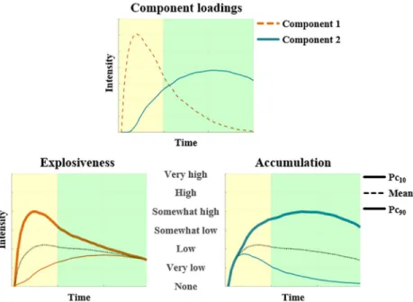
Ver-duyn, and colleagues [11], each of the obtained 384 self-reported intensity profiles following negative feedback was first transformed into a function using the linear interpolation function (interp1) implemented in MATLAB R2016b [38] and then discretised into 44 equally dis-tanced time points, corresponding to the number of images acquired during the period that participants read and thought about the feedback. These time series were subsequently decom-posed into two components using non-negative matrix factorization [39] (as implemented in MATLAB R2016b [38]). The resulting component loadings depict the dynamic features of the shape of component profiles, whereas the resulting component scores depict the extent to which each intensity profile is characterized by each of these dynamic features.
As illustrated inFig 2(top panel), the first obtained component has initial high loadings fol-lowed by a steep decrease, whereas the second obtained component has loadings that increase over time. To interpret these components [40], reconstructed profiles taking low (i.e., 10th
Fig 2. Two-component solution resulting from NNMF. Yellow (left) and green (right) backgrounds correspond to
reading and thinking about the feedback, respectively. Top: Component loadings of emotional intensity profiles over time. Bottom: Reconstructed profiles taking a high (90thpercentile), average, or low (10thpercentile) score on the
component in question and a mean score on the other component, presented according to the order of their peaks in the temporal process. This figure is based on data obtained in both self-immersed and self-distanced trials. Bottom left panel: High and low scoring profiles show an explosive and gentle start, respectively. Bottom right panel: High and low scoring profiles show emotion accumulation and recovery, respectively.
percentile), average, and high (i.e., 90thpercentile) scores on one component and mean scores on the other component were constructed (seeFig 2, bottom panel). The first component cor-responds to emotion explosiveness, with emotion intensity having either a gentle (10th percen-tile) or an explosive (90thpercentile) start. The second component corresponds to emotion accumulation, with emotion intensity either returning to baseline (10thpercentile) or accumu-lating (90thpercentile) over time. Reconstructed profiles taking low, average and high scores on one component and mean scores on the other component, separately for each perspective taking condition are available inS1 Fig.
Each intensity profile can thus be reconstructed by summing the component scores multi-plied by their corresponding loadings (i.e., adding reconstructed subprofiles). A visualization of the decomposition of intensity profiles into their reconstructed subprofiles is shown in
Fig 3.
Pre-processing of brain images. Functional scans were pre-processed with SPM8 [41], using slice-time correction, motion correction, spatial normalization to the MNI space, and spatial smoothing using a 8-mm full-width at half-maximum isotropic Gaussian kernel. Spatial normalization was performed by first co-registering the high resolution T1-weighted image to the mean functional image, normalizing the T1 to the MNI template, and applying the normal-ization parameters to the functional images.
General linear model construction. Statistical analyses were conducted using the general
linear model (GLM) framework implemented in SPM8 [41]. For each run, boxcar regressors were used to represent the first screen displaying the self-perspective instruction (self-paced). For each trial within each run, boxcar regressors were also used to represent: (a) the five-sec-ond screen notifying participants of the forthcoming feedback and reminding them of the per-spective to take, (b) the ninety-second period during which participants read and thought about the manipulated negative feedback, (c) the ninety-second period during which partici-pants read and thought about the manipulated neutral feedback, and (d) the self-paced emo-tion intensity profile drawing period, with the relaxaemo-tion period funcemo-tioning as an implicit baseline. All regressors were convolved with the canonical haemodynamic response function.
Fig 3. Original drawings (upper panel), explosiveness subprofiles (middle panel) and accumulation subprofiles (lower panel). Adding the reconstructed subprofiles closely approximates the original intensity profile. Yellow (left)
and green (right) backgrounds correspond to reading and thinking about the feedback, respectively.
Similar to Re´sibois, Verduyn, and colleagues [11], a high-pass filter of 200s was applied, and the motion realignment parameters were included as regressors of non-interest.
Neural correlates of emotion intensity profile features. To examine the neural basis of
emotion explosiveness and accumulation, we further added the reconstructed subprofiles derived from the non-negative matrix factorization (as depicted inFig 3), convolved with the haemodynamic response function, to the regression equation of the GLM presented above. This model was used to predict the BOLD signal both in a number of regions of interest and at the voxel level, and this across the two perspectives.
Two series of region of interest analysis were conducted to try to replicate earlier results on the neural correlates of emotion explosiveness and accumulation. Specifically, as replicating our previous findings was one of the key motivations for this study, in the first series of region of interest analysis we used twoglobal regions of interest that comprised all clusters that were
found to be related with emotion explosiveness (resp. emotion accumulation) in the study by Re´sibois, Verduyn and colleagues [11]. The first region of interest with all clusters found to be related with emotion explosiveness comprised the left mPFC, the left middle and superior frontal and temporal gyri, the left supramarginal gyrus, the right angular, superior temporal, lingual, and middle occipital gyri, and the right cerebellum. The second region of interest with all clusters found to be related with emotion accumulation comprised the bilateral insula (mid-posterior part) and cingulate cortex (mid-posterior part), the right claustrum and ante-rior cingulate cortex (dorsal part), the left middle frontal (dorsolateral part of the prefrontal cortex), pre/post-central, and superior temporal gyri, the left caudate body, and inferior parie-tal lobule. These were created by first saving all SPM-8’s clusters from the result table of explosiveness (resp. accumulation) as a binary image, and transforming it into a ROI using the SPM8-compatible tool MarsBar [42]. In the second series, we used the twospecific regions of
interest found by Re´sibois, Verduyn and colleagues [11] to be correlated with emotion explosiveness and accumulation, respectively (i.e., the mPFC and insula, respectively). The mPFC and insula were bilaterally defined using AAL’s [43] entire structural masks included in the SPM8-compatible tool MarsBar [42], with the insula being divided into an anterior (y > -10) and posterior (y < -10) sub-region [44]. For each region of interest, we calculated the mean value of the second-level explosiveness and accumulation BOLD regression weights by aggregating across all of their voxels, and tested for significance by means of one sample t-tests with Bonferroni correction.
In addition, voxelwise whole brain analyses were conducted to explore possible additional correlates of emotion explosiveness and accumulation. Specifically, we created statistical parametric maps for each participant and entered those into random-effects group analyses testing for significance using one samplet-tests. Similar to Re´sibois, Verduyn, and colleagues
[11], statistical parametric maps were thresholded atp<.001 (uncorrected) combined with
an extend threshold of 10 adjacent voxels, which balances Type I and Type II error rates [45,46]. To test the robustness of our findings, we also provide FWER cluster-corrected and FDR voxelwise-correctedp-values. Resulting peaks were transformed into the Talairach space
using the SPM8-compatible tool icbm2tal [47,48] and labelled using the Talairach atlas [49,50].
The effect of perspective taking on emotion explosiveness and accumulation at the self-report level. To examine the effect of the perspective taking manipulation on emotion
explosiveness and accumulation, we ran multilevel analyses using the nlme package [51] devel-oped for R [52]. In particular, the two non-negative matrix factorization scores (i.e., explosive-ness and accumulation) were predicted by a dummy predictor (0 = immersion, 1 = self-distancing) at Level 1. The intercept and slope were allowed to vary randomly across participants.
The effect of perspective taking on the neural correlates of emotion explosiveness and accumulation. To examine whether perspective taking impacts activity in the neural
corre-lates of emotion explosiveness and accumulation, two contrasts were created based on the parameters obtained from fitting the general linear model described above without the recon-structed subprofiles (i.e., only containing boxcar regressors). The contrasts compared neural activity during the period that participants adopted a self-distanced (SD) perspective to the period that participants adopted a self-immersed (SI) perspective and vice versa. Both con-trasts were examined at the level of region of interests as well as voxels across the whole brain.
Results
Neural correlates of emotion intensity profile features
The two reconstructed subprofiles (i.e., component loadings multiplied by component scores, seeFig 3) of explosiveness and accumulation (convolved with the canonical haemodynamic response function) were used as regressors of the BOLD response across the two conditions. First, we examined whether we could replicate the findings of Re´sibois, Verduyn and col-leagues [11] by conducting region of interest analyses. Next, we explored possible additional neural correlates by conducting voxelwise whole brain analyses. It is notable that the neural correlates of explosiveness and accumulation did not depend on the type of self-perspective adopted. Indeed, contrasts comparing the neural correlates of explosiveness (accumulation) while adopting a self-distanced perspective to the neural correlates of explosiveness (accumula-tion) while adopting a self-immersed perspective were not significant, regardless of whether conducting voxelwise whole-brain analysis or region of interest analyses.
In a first series of region of interest analyses we examined whether the current explosiveness and accumulation regressors were predictive of neural activity in theglobal regions of interest
that comprised all clusters identified by Re´sibois, Verduyn and colleagues [11] to be associated with emotion explosiveness and accumulation, respectively. This was found to be the case (see
Table 1).
In a second series of region of interest analyses we examined whether the explosiveness and accumulation regressors were predictive of neural activity in thespecific regions identified by
Resibois, Verduyn and colleagues [11] to be associated with emotion explosiveness (mPFC) and accumulation (posterior insula), respectively. This was found to be the case (seeTable 2).
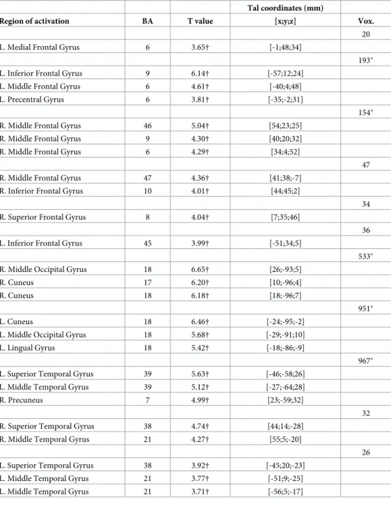
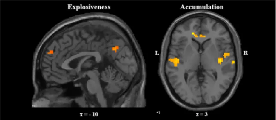
Finally, in line with previous findings [11], voxelwise whole brain analyses revealed that emotion explosiveness is related to activity in the medial prefrontal cortex, the bilateral middle frontal and superior temporal gyri, the left middle temporal gyrus, and the right middle
Table 1. Region of interest analyses predicting neural activity in the full set of clusters observed by Re´sibois, Ver-duyn & colleagues [11] to be correlated with emotion explosiveness (resp. accumulation).
Region of interest Explosiveness Accumulation
T p T p
Full set of clustersathat appeared to be correlated in [11] with explosiveness 3.54 .001 -.18 .82
Full set of clustersbthat appeared to be correlated in [11] with accumulation -4.95 1.00 3.56 .001 p-values are Bonferroni-corrected for multiple testing;
a
left mPFC, left middle and superior frontal and temporal gyri, left supramarginal gyrus, right angular, superior temporal, lingual, and middle occipital gyri, and right cerebellum;
b
bilateral insula (mid-posterior part) and cingulate cortex (mid-posterior part), right claustrum and anterior cingulate cortex (dorsal part), left middle frontal (dorsolateral part of the prefrontal cortex), pre/post-central, and superior temporal gyri, left caudate body, and inferior parietal lobule.
occipital gyrus. However, a number of regions were additionally identified with explosiveness also being related to activity in left inferior and right frontal gyri, the right middle temporal gyrus, the left precentral, middle occipital, and lingual gyri, the bilateral cuneus, and the right precuneus (SeeTable 3andFig 4).
Likewise, in line with previous findings [11], exploratory voxelwise whole brain analyses revealed that emotion accumulation is related to activity in the bilateral posterior insula, the left precentral, cingulate, middle frontal and superior temporal gyri, the left caudate body, and the right claustrum. However, a number of regions were additionally identified with accumulation also being related to activity in the left middle temporal, anterior cingulate, superior and medial frontal gyri, the left precuneus, the right paracentral lobule and caudate tail, the right middle and superior temporal gyri, and the bilateral angular gyri (SeeTable 4
andFig 4).
The effect of perspective taking on emotion explosiveness and
accumulation at the self-report level
Multilevel analysis was used to predict emotion explosiveness and accumulation scores by the perspective taking manipulation (0 = self-immersed, 1 = self-distanced). Both explosiveness (B = -174.88,β = -.34, t(351) = -3.79, p < .001, 95% confidence interval [CI] [-265.70, -84.05]) and accumulation (B = -301.46,β = -.33, t(351) = -3.35, p < .001, 95% CI [-478.65, -124.26]) were found to be lower when participants adopted a self-distanced perspective. The manipula-tion order was not related to either explosiveness (p = .28) or accumulamanipula-tion (p = .97), nor did controlling for the manipulation order alter any of the reported conclusions.
The effect of perspective taking on the neural correlates of emotion
explosiveness and accumulation
Adopting a self-distanced (vs. self-immersed) perspective did not lead (seeTable 5) to altered levels of activity in the mPFC (associated with explosiveness) or in the insula (associated with accumulation). Additional exploratory voxelwise whole-brain analyses similarly did not reveal differential neural activity depending on the self-perspective adopted. An alternative strategy to examine the neural activity associated with adopting a distanced (SD) versus a self-immersed (SI) perspective would be to use the neutral trials of the corresponding run as refer-ence categories as reflected by the following two contrasts: (1) [negative trials > neutral trials]SD> [negative trials > neutral trials]SIand (2) [negative trials > neutral trials]SD<
[neg-ative trials > neutral trials]SI. Yet, similar to the analyses reported above, this did not lead to
any significant result in region of interest or in whole brain analyses.
Table 2. Region of interest analyses predicting neural activity in thespecific regions observed by Resibois,
Ver-duyn & colleagues [11] to underlie emotion explosiveness (resp. accumulation).
ROI Explosiveness Accumulation
T p T p
mPFC 2.49 .03 1.90 .10
Insula
Anterior -.71 .99 .73 .55
Posterior -3.19 1.00 3.18 .005
mPFC = medial prefrontal cortex.p-values are Bonferroni-corrected for multiple testing.
Discussion
The overall aim of the present study was to replicate and extend previous results on the psy-chological and neural mechanisms underlying emotion explosiveness and accumulation. First of all and importantly, our findings provided an independent replication of the existence of
Table 3. Activations associated with explosiveness in whole-brain analysis.
Tal coordinates (mm)
Region of activation BA T value [x;y;z] Vox.
20
L. Medial Frontal Gyrus 6 3.65† [-1;48;34]
193�
L. Inferior Frontal Gyrus 9 6.14† [-57;12;24]
L. Middle Frontal Gyrus 6 4.61† [-40;4;48]
L. Precentral Gyrus 6 3.81† [-35;-2;31]
154�
R. Middle Frontal Gyrus 46 5.04† [54;23;25]
R. Middle Frontal Gyrus 9 4.30† [40;20;32]
R. Middle Frontal Gyrus 6 4.29† [34;4;52]
47
R. Middle Frontal Gyrus 47 4.36† [41;38;-7]
R. Inferior Frontal Gyrus 10 4.01† [44;45;2]
34
R. Superior Frontal Gyrus 8 4.04† [7;35;46]
36
L. Inferior Frontal Gyrus 45 3.99† [-51;34;5]
533�
R. Middle Occipital Gyrus 18 6.65† [26;-93;5]
R. Cuneus 17 6.20† [10;-96;4]
R. Cuneus 18 6.18† [18;-96;7]
951�
L. Cuneus 18 6.46† [-24;-95;-2]
L. Middle Occipital Gyrus 18 5.68† [-29;-91;10]
L. Lingual Gyrus 18 5.42† [-18;-86;-9]
967�
L. Superior Temporal Gyrus 39 5.63† [-46;-58;26]
L. Middle Temporal Gyrus 39 5.12† [-27;-64;28]
R. Precuneus 7 4.99† [23;-59;32]
32
R. Superior Temporal Gyrus 38 4.74† [44;14;-28]
R. Middle Temporal Gyrus 21 4.27† [55;5;-20]
26
L. Superior Temporal Gyrus 38 3.92† [-45;20;-23]
L. Middle Temporal Gyrus 21 3.77† [-51;9;-25]
L. Middle Temporal Gyrus 21 3.71† [-56;5;-17]
Allps<.001 uncorrected, number of voxels>10 per cluster. BA = Brodmann’s areas. Vox. = Voxels per cluster. L = left. R = right.
†p<.05 voxelwise FDR-corrected. �p<.05 FWE-corrected at cluster level.
distinctive correlates of emotion explosiveness and emotion accumulation with data being acquired by another experimenter and using another fMRI scanner than in the study of Re´si-bois, Verduyn, and colleagues [11]. Specifically, in the two series of region of interest analyses and in the voxelwise whole brain analyses, emotion explosiveness appeared again to be associ-ated with regions of self-referential processing (such as the medial prefrontal cortex), whereas emotion accumulation appeared again to be associated with regions of sustained monitoring of visceral arousal and the sensory component of social exclusion (such as the posterior insula). These findings corroborate that onset- and offset-bound processes have distinct neural corre-lates, which is consistent with emotion dynamic frameworks distinguishing between two key emotion unfolding phases [10–14,16]: an onset phase (associated with explosiveness) and an offset phase (associated with accumulation), which were found in the present study to be the two main constituents underlying change in emotional experience over time. It further emphasizes the need to take temporal dynamics into account when studying the neural basis of emotions, which resonates with recent calls to put time on the research agenda of affective neuroscience [53–55].
Providing further evidence for an association between emotion explosiveness and self-refer-ential processing, in the current study explosiveness was additionally found to be associated with activity in the precuneus and the temporo-parietal junction, which are two regions of the default-mode network [56–58]. Moreover, as further evidence for an association between emo-tion accumulaemo-tion and sustained monitoring of visceral arousal, emoemo-tion accumulaemo-tion addi-tionally appeared to be associated with activity in the primary sensory cortex [54,59,60]. In addition, emotion accumulation was also associated with the dorsal anterior cingulate cortex, providing suggestive evidence for the social feedback having resulted in feelings of social exclu-sion, even though it should be noted that no significant activity in the anterior insula was observed [61]. However, future studies using non-social or more basic emotion-eliciting sti-muli (e.g., emotional pictures as used in [62]) are needed to examine the degree to which the neural correlates of explosiveness and accumulation generalize across contexts. It is possible that studies using such more basic stimuli would identify primary emotion areas such as the amygdala or the anterior insula as key neural correlates of emotion explosiveness and accumu-lation [63–65]. This is especially likely given the negative association found in the literature between meaning making regions (associated with explosiveness in the present study), and the anterior insula and amygdala [62]. Yet, one could alternatively argue that activity in the ante-rior insula and amygdala is perhaps associated with emotion intensity per se regardless of the
Fig 4. Neural correlates of emotion explosiveness and accumulation. Left panel: mPFC activation associated with
emotion explosiveness. Right panel: Insula activation associated with emotion accumulation. Coordinates in the Talairach space.
Table 4. Activations associated with accumulation in whole-brain analysis.
Region of activation BA T value Tal coordinates (mm) Vox. [x;y;z] 18 R. Insula 13 4.01† [32;-10;21] R. Insula 13 3.97† [29;-22;25] 835� L. Precentral Gyrus 6 5.77† [-43;-7;20] R. Paracentral Lobule 31 5.10† [4;-29;46] L. Precentral Gyrus 4 4.72† [-27;-27;48] 42 L. Anterior Cingulate 32 4.54† [-12;33;11] L. Anterior Cingulate 24 4.29† [-1;31;5] 14 L. Caudate Body 4.15† [-9;16;20] L. Cingulate Gyrus 24 3.56† [-4;18;26] 16 L. Caudate Body 3.99† [-15;-28;29] L. Cingulate Gyrus 31 3.96† [-18;-36;26] 12 R. Caudate Tail 4.75† [21;-43;12] 49
L. Superior Frontal Gyrus 8 4.56† [-15;24;45]
L. Medial Frontal Gyrus 8 4.06† [-10;30;46]
27
L. Middle Frontal Gyrus 6 4.50† [-35;13;46]
23
L. Superior Frontal Gyrus 9 4.42† [-15;46;28]
19
L. Angular Gyrus 39 4.33† [-54;-59;34]
30
R. Angular Gyrus 39 4.13† [54;-59;33]
R. Middle Temporal Gyrus 39 4.09† [51;-67;27]
R. Superior Temporal Gyrus 39 3.88† [57;-58;20]
324�
R. Superior Temporal Gyrus 42 5.49† [63;-29;14]
R. Superior Temporal Gyrus 41 4.56† [38;-32;16]
R. Claustrum 4.38† [38;-17;4]
12
L. Superior Temporal Gyrus 22 4.20† [-57;-60;15]
L. Middle Temporal Gyrus 39 3.72† [-54;-66;20]
38
L. Precuneus 19 4.63† [-41;-76;35]
Allps<.001 uncorrected, number of voxels>10 per cluster. BA = Brodmann’s areas. Vox. = Voxels per cluster. L = left. R = right.
†p<.05 voxelwise FDR-corrected. �p<.05 FWE-corrected at cluster level.
stage of emotion unfolding (i.e., overall levels of intensity rather than specific dynamic fea-tures) as found in previous research using emotional movies to induce emotion [66,67]. How-ever, it is notable that, in the present study where we used complex social stimuli, we did not find an effect of our manipulation on any primary emotion areas despite that self-distancing was found to overall lower self-reported emotion intensity.
Second, we indeed replicated that adopting a self-distanced (vs. a self-immersed) perspec-tive decreases both emotion explosiveness and accumulation (i.e., lowering overall emotion intensity). This demonstrates that perspective taking can modulate how people initially react to emotional stimuli, at least when being instructed to use this regulation strategy beforehand. Moreover, it suggests support to psychological therapies that provide people the tools to take distance, including cognitive-behavioural, acceptance-based, and mindfulness therapies [68], such that they may be better equipped to deal with potential future stressors. However, future research including clinical populations and using stimuli eliciting higher degrees of distress are necessary to justify this conjecture.
Third, unexpectedly, adopting a self-distanced (vs. a self-immersed) perspective did not lead to altered levels of activity in the mPFC (associated with explosiveness) or in the insula (associated with accumulation). Although it is difficult to interpret null findings, one could at least speculate that this result might be due to the fact that perspective taking may recruit regions that are also correlated with emotion explosiveness and accumulation, cancelling out the possibility of finding a significant decrease of activity in these regions when adopting a self-distanced (vs. a self-immersed) perspective. This tentative explanation could be especially plausible to at least partially explain why adopting a self-distanced perspective was not found to be associated with a decreased activity in the medial prefrontal cortex (associated with explosiveness), as this region has been consistently found to underlie processes of reappraisal [69–72], including perspective taking [73,74]. This interpretation might especially hold given the use of social feedback as an emotion-eliciting stimulus. The predictive and reactive control systems (PARCS) framework [75] indeed theorizes that regions of the predictive system, including the DMN, underlie both (a) adopting an observer perspective and (b) making mean-ing of negative feedback that challenges internal models about oneself and the world. Thus, future studies might benefit from using emotional stimuli that do not require extensive self-referential processing (e.g., emotional pictures) to further explore the role of perspective taking on the neural correlates of explosiveness and accumulation.
Finally, although intensity levels reported in the present study are similar to other studies using a negative feedback procedure [10,11,31,32], future research eliciting stronger emotions are needed to get a better understanding of the neural mechanisms underlying emotion dynamics.
Table 5. Regions of interest analyses comparing activity while adopting a self-distanced vs. self-immersed per-spective when exposed to negative feedback.
Label SD > SI SI > SD T p T p mPFC 1.61 .17 -1.61 1.00 Insula Anterior .51 .67 -.51 .97 Posterior .45 .70 -.45 .96
SD = self-distancing. SI = self-immersion. mPFC = medial prefrontal cortex. p-values are Bonferroni-corrected for multiple testing.
Supporting information
S1 Supporting Information.
(DOCX)
S1 Fig. Two-component solution resulting from NNMF, separately for each perspective taking condition.
(TIF)
S1 Dataset. Emotion intensity profiles as drawn by participants in E-Prime.
(CSV)
S2 Dataset. Reconstructed explosiveness subprofiles used as BOLD regressors.
(CSV)
S3 Dataset. Reconstructed accumulation subprofiles used as BOLD regressors.
(CSV)
S1 File. Binary mask of regions of interests used in ROI analyses.
(ZIP)
Author Contributions
Conceptualization: Maxime Re´sibois, Jean-Yves Rotge´, Pauline Delaveau, Peter Kuppens,
Iven Van Mechelen, Philippe Fossati, Philippe Verduyn.
Data curation: Maxime Re´sibois. Formal analysis: Maxime Re´sibois.
Funding acquisition: Peter Kuppens, Iven Van Mechelen, Philippe Fossati, Philippe Verduyn. Investigation: Maxime Re´sibois.
Methodology: Maxime Re´sibois, Jean-Yves Rotge´, Pauline Delaveau, Peter Kuppens, Iven Van
Mechelen, Philippe Fossati, Philippe Verduyn.
Project administration: Maxime Re´sibois. Resources: Philippe Fossati.
Software: Maxime Re´sibois.
Supervision: Peter Kuppens, Iven Van Mechelen, Philippe Fossati, Philippe Verduyn. Validation: Maxime Re´sibois, Peter Kuppens, Iven Van Mechelen, Philippe Fossati, Philippe
Verduyn.
Visualization: Maxime Re´sibois.
Writing – original draft: Maxime Re´sibois.
Writing – review & editing: Maxime Re´sibois, Jean-Yves Rotge´, Pauline Delaveau, Peter
Kup-pens, Iven Van Mechelen, Philippe Fossati, Philippe Verduyn.
References
1. Frijda NH. The laws of emotion. Mahwah, NJ, US: Lawrence Erlbaum; 2007.
2. Verduyn P, Van Mechelen I, Tuerlinckx F, Meers K, Van Coillie H. Intensity profiles of emotional experi-ence over time. Cogn Emot. 2009; 23: 1427–1443.https://doi.org/10.1080/02699930902949031
3. American Psychiatric Association. Diagnostic and Statistical Manual of Mental Disorder. 5th ed. Wash-ington, DC: American Psychiatric Publishing, Inc; 2013.https://doi.org/10.1176/appi.books.
9780890425596.744053
4. Frijda NH, Mesquita B, Sonnemans J, Van Goozen S. The duration of affective phenomena or emo-tions, sentiments and passions. In: Strongman KT, editor. International review of studies on emotion. Chichester, UK: Wiley; 1991. pp. 187–225.
5. Sonnemans J, Frijda NH. The structure of subjective emotional intensity. Cogn Emot. 1994; 8: 329–350.https://doi.org/10.1080/02699939408408945
6. Sonnemans J, Frijda NH. The determinants of subjective emotional intensity. Cogn Emot. 1995; 9: 483–506.https://doi.org/10.1080/02699939508408977
7. Heylen J, Verduyn P, Van Mechelen I, Ceulemans E. Variability in anger intensity profiles: Structure and predictive basis. Cogn Emot. 2015; 29: 168–177.https://doi.org/10.1080/02699931.2014.896783
PMID:24641250
8. Heylen J, Ceulemans E, Van Mechelen I, Verduyn P. KSC-N: Clustering of hierarchical time profile data. Psychometrika. 2016; 81: 411–433.https://doi.org/10.1007/s11336-014-9433-xPMID:25491164
9. Verduyn P, Van Mechelen I, Frederix E. Determinants of the shape of emotion intensity profiles. Cogn Emot. 2012; 26: 1486–1495.https://doi.org/10.1080/02699931.2012.662152PMID:22360656
10. Re´sibois M, Kalokerinos EK, Verleysen G, Kuppens P, Van Mechelen I, Fossati P, et al. The relation between rumination and temporal features of emotion intensity. Cogn Emot. 2017; Advance online pub-lication.https://doi.org/10.1080/02699931.2017.1298993PMID:28278734
11. Re´sibois M, Verduyn P, Delaveau P, Rotge´ J-Y, Kuppens P, Van Mechelen I, et al. The neural basis of emotions varies over time: different regions go with onset- and offset-bound processes underlying emo-tion intensity. Soc Cogn Affect Neurosci. 2017; 12: 1261–1271.https://doi.org/10.1093/scan/nsx051
PMID:28402478
12. Brans K, Verduyn P. Intensity and duration of negative emotions: Comparing the role of appraisals and regulation strategies. PLoS One. 2014; 9: e92410.https://doi.org/10.1371/journal.pone.0092410PMID:
24670979
13. Davidson RJ. Affective style and affective disorders: Perspectives from affective neuroscience. Cogn Emot. 1998; 12: 307–330.https://doi.org/10.1080/026999398379628
14. Koole SL. The psychology of emotion regulation: An integrative review. Cogn Emot. 2009; 23: 4–41.
https://doi.org/10.1080/02699930802619031
15. Verduyn P, Brans K. The relationship between extraversion, neuroticism and aspects of trait affect. Pers Individ Dif. Elsevier Ltd; 2012; 52: 664–669.https://doi.org/10.1016/j.paid.2011.12.017
16. Kuppens P, Verduyn P. Looking at emotion regulation through the window of emotion dynamics. Psy-chol Inq. 2015; 26: 72–79.https://doi.org/10.1080/1047840X.2015.960505
17. Poldrack RA, Baker CI, Durnez J, Gorgolewski KJ, Matthews PM, MunafòMR, et al. Scanning the hori-zon: towards transparent and reproducible neuroimaging research. Nat Rev Neurosci. 2017; 18: 115–126.https://doi.org/10.1038/nrn.2016.167PMID:28053326
18. Papageorgiou C, Wells A. Metacognitive beliefs about rumination in recurrent major depression. Cogn Behav Pract. 2001; 8: 160–164.https://doi.org/10.1016/S1077-7229(01)80021-3
19. Nigro G, Neisser U. Point of view in personal memories. Cogn Psychol. 1983; 15: 467–482.https://doi. org/10.1016/0010-0285(83)90016-6
20. Robinson JA, Swanson KL. Field and observer modes of remembering. Memory. 1993; 1: 169–184.
https://doi.org/10.1080/09658219308258230PMID:7584265
21. Kross E, Ayduk O. Chapter Two—Self-Distancing: Theory, Research, and Current Directions. In: Olson JM, editor. Advances in Experimental Social Psychology. Academic P. Elsevier Inc.; 2017. pp. 81–136.
https://doi.org/10.1016/bs.aesp.2016.10.002
22. Ayduk O¨ , Kross E. Enhancing the pace of recovery: Self-distanced analysis of negative experiences reduces blood pressure reactivity. Psychol Sci. 2008; 19: 229–31.https://doi.org/10.1111/j.1467-9280. 2008.02073.xPMID:18315794
23. Ayduk O¨ , Kross E. From a distance: Implications of spontaneous self-distancing for adaptive self-reflec-tion. J Pers Soc Psychol. 2010; 98: 809–829.https://doi.org/10.1037/a0019205PMID:20438226
24. Denny BT, Ochsner KN. Behavioral effects of longitudinal training in cognitive reappraisal. Emotion. 2014; 14: 425–433.https://doi.org/10.1037/a0035276PMID:24364856
25. Kross E, Ayduk O¨ . Facilitating adaptive emotional analysis: Distinguishing distanced-analysis of depressive experiences from immersed-analysis and distraction. Personal Soc Psychol Bull. 2008; 34: 924–38.https://doi.org/10.1177/0146167208315938PMID:18469151
26. Kross E, Gard D, Deldin P, Clifton J, Ayduk O¨ . “Asking why” from a distance: Its cognitive and emotional consequences for people with major depressive disorder. J Abnorm Psychol. 2012; 121: 559–569.
https://doi.org/10.1037/a0028808PMID:22708885
27. Penner LA, Guevarra DA, Harper FWK, Taub J, Phipps S, Albrecht TL, et al. Self-distancing buffers high trait anxious pediatric cancer caregivers against short- and longer-term distress. Clin Psychol Sci. 2016; 4: 629–640.https://doi.org/10.1177/2167702615602864PMID:27617183
28. Verduyn P, Van Mechelen I, Kross E, Chezzi C, Van Bever F. The relationship between self-distancing and the duration of negative and positive emotional experiences in daily life. Emotion. 2012; 12: 1248–1263.https://doi.org/10.1037/a0028289PMID:22642344
29. Re´sibois M, Kuppens P, Van Mechelen I, Fossati P., Verduyn P. Depression severity moderates the relation between self-distancing and features of emotion unfolding. Pers Individ Dif. 2018; 123: 119–124.https://doi.org/10.1016/j.paid.2017.11.018
30. Bushman BJ, Baumeister RF. Threatened egotism, narcissism, self-esteem, and direct and displaced aggression: Does self-love or self-hate lead to violence? J Pers Soc Psychol. 1998; 75: 219–229.
https://doi.org/10.1037/0022-3514.75.1.219PMID:9686460
31. Eisenberger NI, Inagaki TK, Muscatell KA, Byrne Haltom KE, Leary MR. The neural sociometer: Brain mechanisms underlying state self-esteem. J Cogn Neurosci. 2011; 23: 3448–3455.https://doi.org/10. 1162/jocn_a_00027PMID:21452934
32. Harmon-Jones E, Sigelman J. State anger and prefrontal brain activity: Evidence that insult-related rela-tive left-prefrontal activation is associated with experienced anger and aggression. J Pers Soc Psychol. 2001; 80: 797–803.https://doi.org/10.1037/0022-3514.80.5.797PMID:11374750
33. Britton JC, Phan KL, Taylor SF, Welsh RC, Berridge KC, Liberzon I. Neural correlates of social and non-social emotions: An fMRI study. Neuroimage. 2006; 31: 397–409.https://doi.org/10.1016/j.neuroimage. 2005.11.027PMID:16414281
34. Parkinson B. Emotions are social. Br J Psychol. 1996; 87: 663–683. https://doi.org/10.1111/j.2044-8295.1996.tb02615.xPMID:8962482
35. Wager TD, Waugh CE, Lindquist MA, Noll DC, Fredrickson BL, Taylor SF. Brain mediators of cardio-vascular responses to social threat. Part I: Reciprocal dorsal and ventral sub-regions of the medial pre-frontal cortex and heart-rate reactivity. Neuroimage. Elsevier Inc.; 2009; 47: 821–835.https://doi.org/ 10.1016/j.neuroimage.2009.05.043PMID:19465137
36. Kross E, Ayduk O¨ , Mischel W. When asking “why” does not hurt: Distinguishing rumination from reflec-tive processing of negareflec-tive emotions. Psychol Sci. 2005; 16: 709–715. https://doi.org/10.1111/j.1467-9280.2005.01600.xPMID:16137257
37. Ray RD, Wilhelm FH, Gross JJ. All in the mind’s eye? Anger rumination and reappraisal. J Pers Soc Psychol. 2008; 94: 133–145.https://doi.org/10.1037/0022-3514.94.1.133PMID:18179323
38. The MathWorks Inc. MATLAB version 9.1.0.441655 (R2016b). Natick, MA, USA; 2016.
39. Lee DD, Seung HS. Learning the parts of objects by non-negative matrix factorization. Nature. 1999; 401: 788–791.https://doi.org/10.1038/44565PMID:10548103
40. Ramsay JO, Silverman BW. Functional Data Analysis. 2nd ed. New York, NY: Springer-Verlag; 2005. 41. Wellcome Trust Centre for Neuroimaging. SPM8 [Internet]. 2009.www.fil.ion.ucl.ac.uk/spm/software/
spm8/
42. Brett M, Anton J-L, Valabregue R, Poline J-B. Region of interest analysis using the MarsBar toolbox for SPM 99. Neuroimage. 2002; 16: S497.
43. Tzourio-Mazoyer N, Landeau B, Papathanassiou D, Crivello F, Etard O, Delcroix N, et al. Automated anatomical labeling of activations in SPM using a macroscopic anatomical parcellation of the MNI MRI single-subject brain. Neuroimage. 2002; 15: 273–89.https://doi.org/10.1006/nimg.2001.0978PMID:
11771995
44. Chang LJ, Yarkoni T, Khaw MW, Sanfey AG. Decoding the role of the insula in human cognition: Func-tional parcellation and large-scale reverse inference. Cereb Cortex. 2013; 23: 739–749.https://doi.org/ 10.1093/cercor/bhs065PMID:22437053
45. Lieberman MD, Cunningham WA. Type I and Type II error concerns in fMRI research: Re-balancing the scale. Soc Cogn Affect Neurosci. 2009; 4: 423–428.https://doi.org/10.1093/scan/nsp052PMID:
20035017
46. Woo C-W, Krishnan A, Wager TD. Cluster-extent based thresholding in fMRI analyses: pitfalls and rec-ommendations. Neuroimage. Elsevier Inc.; 2014; 91: 412–9.https://doi.org/10.1016/j.neuroimage. 2013.12.058PMID:24412399
47. Laird AR, Robinson JL, McMillan KM, Tordesillas-Gutie´rrez D, Moran ST, Gonzales SM, et al. Compari-son of the disparity between Talairach and MNI coordinates in functional neuroimaging data: Validation
of the Lancaster transform. Neuroimage. Elsevier Inc.; 2010; 51: 677–683.https://doi.org/10.1016/j. neuroimage.2010.02.048PMID:20197097
48. Lancaster JL, Tordesillas-Gutie´ rrez D, Martinez M, Salinas F, Evans AC, Zilles K, et al. Bias between MNI and talairach coordinates analyzed using the ICBM-152 brain template. Hum Brain Mapp. 2007; 28: 1194–1205.https://doi.org/10.1002/hbm.20345PMID:17266101
49. Lancaster JL, Rainey LH, Summerlin JL, Freitas CS, Fox PT, Evans AC, et al. Automated labeling of the human brain: A preliminary report on the development and evaluation of a forward-transform method. Hum Brain Mapp. 1997; 5: 238–242.https://doi.org/10.1002/(SICI)1097-0193(1997)5:4<238:: AID-HBM6>3.0.CO;2-4PMID:20408222
50. Lancaster JL, Woldorff MG, Parsons LM, Liotti M, Freitas CS, Rainey L, et al. Automated Talairach Atlas labels for functional brain mapping. Hum Brain Mapp. 2000; 10: 120–131.https://doi.org/10.1002/ 1097-0193(200007)10:3<120::AID-HBM30>3.0.CO;2-8PMID:10912591
51. Pinheiro J, Bates D, DebRoy S, Sarkar D, R Core Team. {nlme}: Linear and Nonlinear Mixed Effects Models, version 3.1–131. 2017.
52. R Core Team. R: A language and environment for statistical computing, version 3.3.3. Vienna, Austria: R Foundation for Statistical Computing; 2017.
53. Davis M, Walker DL, Miles L, Grillon C. Phasic vs sustained fear in rats and humans: role of the extended amygdala in fear vs anxiety. Neuropsychopharmacology. Nature Publishing Group; 2010; 35: 105–135.https://doi.org/10.1038/npp.2009.109PMID:19693004
54. Waugh CE, Schirillo JA. Timing: A missing key ingredient in typical fMRI studies of emotion. Behav Brain Sci. 2012; 35: 170–171.https://doi.org/10.1017/S0140525X11001646PMID:22617678
55. Waugh CE, Shing EZ, Avery BM. Temporal Dynamics of Emotional Processing in the Brain. Emot Rev. 2015; 7: 323–329.https://doi.org/10.1177/1754073915590615
56. Amft M, Bzdok D, Laird AR, Fox PT, Schilbach L, Eickhoff SB. Definition and characterization of an extended social-affective default network. Brain Struct Funct. 2014; 220: 1031–1049.https://doi.org/10. 1007/s00429-013-0698-0PMID:24399179
57. Andrews-Hanna JR, Smallwood J, Spreng RN. The default network and self-generated thought: Com-ponent processes, dynamic control, and clinical relevance. Ann N Y Acad Sci. 2014; 1316: 29–52.
https://doi.org/10.1111/nyas.12360PMID:24502540
58. Buckner RL, Andrews-Hanna JR, Schacter DL. The brain’s default network: Anatomy, function, and rel-evance to disease. Ann N Y Acad Sci. 2008; 1124: 1–38.https://doi.org/10.1196/annals.1440.011
PMID:18400922
59. Eisenberger NI. The pain of social disconnection: examining the shared neural underpinnings of physi-cal and social pain. Nat Rev Neurosci. Nature Publishing Group; 2012; 13: 421–434.https://doi.org/10. 1038/nrn3231PMID:22551663
60. Kross E, Berman MG, Mischel W, Smith EE, Wager TD. Social rejection shares somatosensory repre-sentations with physical pain. Proc Natl Acad Sci U S A. 2011; 108: 6270–6275.https://doi.org/10.1073/ pnas.1102693108PMID:21444827
61. Eisenberger NI. Social pain and the brain: Controversies, questions, and where to go from here. Annu Rev Psychol. 2015; 66: 601–629.https://doi.org/10.1146/annurev-psych-010213-115146PMID:
25251482
62. Satpute AB, Shu J, Weber J, Roy M, Ochsner KN. The Functional Neural Architecture of Self-Reports of Affective Experience. Biol Psychiatry. Elsevier; 2013; 73: 631–638.https://doi.org/10.1016/j. biopsych.2012.10.001PMID:23146356
63. Bishop SJ, Duncan J, Lawrence AD. State Anxiety Modulation of the Amygdala Response to Unat-tended Threat-Related Stimuli. J Neurosci. 2004; 24: 10364–10368.https://doi.org/10.1523/ JNEUROSCI.2550-04.2004PMID:15548650
64. Ochsner KN, Bunge SA, Gross JJ, Gabrieli JDE. Rethinking feelings: an FMRI study of the cognitive regulation of emotion. J Cogn Neurosci. 2002; 14: 1215–1229.https://doi.org/10.1162/
089892902760807212PMID:12495527
65. Ochsner KN, Ray RD, Cooper JC, Robertson ER, Chopra S, Gabrieli J, et al. For better or for worse: neural systems supporting the cognitive down- and up-regulation of negative emotion. Neuroimage. 2004; 23: 483–499.https://doi.org/10.1016/j.neuroimage.2004.06.030PMID:15488398
66. Goldin PR, Hutcherson CAC, Ochsner KN, Glover GH, Gabrieli JDE, Gross JJ. The neural bases of amusement and sadness: a comparison of block contrast and subject-specific emotion intensity regres-sion approaches. Neuroimage. 2005; 27: 26–36.https://doi.org/10.1016/j.neuroimage.2005.03.018
67. Hutcherson CAC, Goldin PR, Ochsner KN, Gabrieli JDE, Barrett LF, Gross JJ. Attention and emotion: Does rating emotion alter neural responses to amusing and sad films? Neuroimage. 2005; 27: 656–668.https://doi.org/10.1016/j.neuroimage.2005.04.028PMID:15946863
68. Bernstein A, Hadash Y, Lichtash Y, Tanay G, Shepherd K, Fresco DM. Decentering and related con-structs: A critical review and meta-Cognitive processes model. Perspect Psychol Sci. 2015; 10: 1–57.
https://doi.org/10.1177/1745691615594577PMID:26385999
69. Morawetz C, Bode S, Derntl B, Heekeren HR. The effect of strategies, goals and stimulus material on the neural mechanisms of emotion regulation: A meta-analysis of fMRI studies. Neurosci Biobehav Rev. Elsevier Ltd; 2017; 72: 111–128.https://doi.org/10.1016/j.neubiorev.2016.11.014PMID:
27894828
70. Buhle JT, Silvers JA, Wager, Lopez R, Onyemekwu C, Kober H, et al. Cognitive reappraisal of emotion: A meta-analysis of human neuroimaging studies. Cereb Cortex. 2014; 24: 2981–2990.https://doi.org/ 10.1093/cercor/bht154PMID:23765157
71. Kohn N, Eickhoff SB, Scheller M, Laird AR, Fox PT, Habel U. Neural network of cognitive emotion regu-lation—an ALE meta-analysis and MACM analysis. Neuroimage. Elsevier Inc.; 2014; 87: 345–355.
https://doi.org/10.1016/j.neuroimage.2013.11.001PMID:24220041
72. Frank DW, Dewitt M, Hudgens-Haney M, Schaeffer DJ, Ball BH, Schwartz N, et al. Emotion regulation: Quantitative meta-analysis of functional activation and deactivation. Neurosci Biobehav Rev. Elsevier Ltd; 2014; 45: 202–211.https://doi.org/10.1016/j.neubiorev.2014.06.010PMID:24984244
73. Messina I, Bianco S, Sambin M, Viviani R. Executive and semantic processes in reappraisal of negative stimuli: insights from a meta-analysis of neuroimaging studies. Front Psychol. 2015; 6: 1–13.
74. Kross E, Davidson ML, Weber J, Ochsner KN. Coping with Emotions Past: The Neural Bases of Regu-lating Affect Associated with Negative Autobiographical Memories. Biol Psychiatry. Society of Biological Psychiatry; 2009; 65: 361–366.https://doi.org/10.1016/j.biopsych.2008.10.019PMID:19058792
75. Tops M, Boksem MAS, Quirin M, IJzerman H, Koole SL. Internally directed cognition and mindfulness: an integrative perspective derived from predictive and reactive control systems theory. Front Psychol. 2014; 5: 1–21.


![Table 1. Region of interest analyses predicting neural activity in the full set of clusters observed by Re´sibois, Ver- Ver-duyn & colleagues [11] to be correlated with emotion explosiveness (resp](https://thumb-eu.123doks.com/thumbv2/123doknet/14623848.547397/10.918.297.868.849.925/analyses-predicting-activity-clusters-observed-colleagues-correlated-explosiveness.webp)