HAL Id: inserm-03227598
https://www.hal.inserm.fr/inserm-03227598v2
Submitted on 25 May 2021HAL is a multi-disciplinary open access archive for the deposit and dissemination of sci-entific research documents, whether they are pub-lished or not. The documents may come from teaching and research institutions in France or abroad, or from public or private research centers.
L’archive ouverte pluridisciplinaire HAL, est destinée au dépôt et à la diffusion de documents scientifiques de niveau recherche, publiés ou non, émanant des établissements d’enseignement et de recherche français ou étrangers, des laboratoires publics ou privés.
Positive predictive values of CMV-IgM and importance
of CMV-IgG avidity testing in detecting primary
infection in three different clinical settings. A French
retrospective cohort study
Claire Périllaud-Dubois, Elise Bouthry, Abir Jadoui, Ay-Ling Leng,
Anne-Marie Roque-Afonso, Christelle Vauloup-Fellous
To cite this version:
Claire Périllaud-Dubois, Elise Bouthry, Abir Jadoui, Ay-Ling Leng, Anne-Marie Roque-Afonso, et al.. Positive predictive values of CMV-IgM and importance of CMV-IgG avidity testing in detecting primary infection in three different clinical settings. A French retrospective cohort study. Journal of Clinical Virology, Elsevier, 2020, 132, pp.104641. �10.1016/j.jcv.2020.104641�. �inserm-03227598v2�
Elsevier Editorial System(tm) for Journal of Clinical Virology
Manuscript Draft
Manuscript Number: JCV-D-20-01378R1
Title: Positive predictive values of CMV-IgM and importance of CMV-IgG avidity testing in detecting primary infection in three different clinical settings. A French retrospective cohort study.
Article Type: Full length article
Keywords: CMV; serology; IgM; avidity; positive predictive value; pregnancy
Corresponding Author: Dr. Claire Périllaud Dubois, PharmD Corresponding Author's Institution: Paul Brousse Hospital First Author: Claire Périllaud Dubois, PharmD
Order of Authors: Claire Périllaud Dubois, PharmD; Elise Bouthry; Abir Jadoui; Ay-Ling Leng; Anne-Marie Roque-Afonso; Christelle Vauloup-Fellous Abstract: Background: Diagnosis of Cytomegalovirus (CMV) primary
infection during pregnancy or in immunocompetent patients relies on serology with detection of specific CMV-IgG and IgM. In case of positive CMV-IgM in pregnant women, CMV-IgG avidity is now widely recommended, but in general population it is not currently performed.
Objective: In this study, we aimed to determine CMV-IgM positive predictive values (PPV) in different clinical settings.
Material and methods: We conducted a retrospective study on positive CMV-IgM in our virology laboratory from 2013 to 2019, in three clinical groups: screening in non-symptomatic pregnant women (group 1), pregnant women with ultrasound (US) abnormalities (group 2) and patients (general population) with clinical signs suggestive of CMV primary infection (group 3). CMV-IgG avidity had been performed in all cases allowing to evaluate PPV of positive CMV-IgM to diagnose CMV primary-infection in each group.
Results: Between 2013 and 2019, 6,859 serum samples were found positive for CMV-IgM and had been tested for CMV-IgG avidity, with 6,560 sera for group 1, 30 for group 2 and 269 for group 3. Overall, low avidity
confirming primary infection was observed respectively in 16.4% for group 1, 36.7% for group 2, and 35.3% for group 3. CMV-IgM PPV was
significantly lower in group 1 compared to groups 2 (p=0.01) and 3 (p<0.001).
Discussion: Our observations highlight the major importance of including CMV-IgG avidity in the diagnostic algorithm, whatever the clinical
situation (for immunocompetent patients), to confirm or exclude a recent CMV primary infection in case of positive CMV-IgM.
Ms. Ref. No.: JCV-D-20-01378
Title: Positive predictive values of CMV-IgM to detect primary infection in three different clinical settings. A French retrospective cohort study.
Journal of Clinical Virology Highlights (mandatory)
Highlights consist of a short collection of bullet points that convey the core findings of the article and should be submitted in a separate file in the online submission system. Please use 'Highlights' in the file name and include 3 to 5 bullet points (maximum 85 characters, including spaces, per bullet point). See the following website for more information
https://clicktime.symantec.com/3SqpMupnfFFd243aiwUggdd6H2?u=http%3A%2F%2Fwww.elsevier.c om%2Fhighlights
In case of positive CMV-IgM, CMV-IgG avidity is essential to diagnose recent primary infection.
Positive predictive value of CMV-IgM is 16.4% if systematic screening during pregnancy. Positive predictive value of CMV-IgM is 36.7% in case of US abnormalities during pregnancy. Positive predictive value of CMV-IgM is 35.3% in case of clinical signs in general population.
Reviewer #1: The authors evaluate the predictive utility of anti-CMV IgG avidity testing retrospectively in three clinical settings. (1) non-symptomatic pregnant women, (2) pregnant women with ultrasound anomalies and (3) general population with clinical signs suggestive of CMV.
The manuscript is well written and coherent and the difficulties in serological diagnoses of recent CMV infection and the impact on, and distress it can cause, during pregnancy is already documented in the literature; this manuscript adds the utility of testing in the general population with clinical symptoms as a comparator.
Minor comments:
There is no other diagnosis of recent CMV infection or outcome of pregnancy information detailed, how the assay changed over time (if at all) is not commented upon, was a control / reference included to ensure longitudinal comparability within the laboratory?
Outcome of pregnancy was not always investigated in our population and correlation between gestational age at CMV infection and outcome has already been widely described.
All laboratories in France are subjected national accreditation (ISO 15189 - COFRAC), which implies the use of CE approved assays and annual subscription (and success) to external quality controls. This ensures optimal longitudinal confidence in their results. Moreover, in the past seven years, no major innovation occurred in routine serologic diagnosis of CMV infection and assays used have well-known performances. Assays’ references are not mentioned in the manuscript but are available.
Line 32: Is further information or review of the sonographic findings to establish consistency with CMV infection?
We detailed briefly US abnormalities in the “Material and methods section”. In the modified version, we added more details on cerebral US findings. Lines 87-93:
“head perimeter < 5th centile, ventriculomegaly, hyperechogenic ventricular wall, cerebellar and brain calcifications, enlargement of pericerebral spaces, periventricular and subependymal cysts, candlestick, porencephaly, hyperechogenic periventricular halo, candlestick, abnormal gyration, corpus callosum hypoplasia.”
Line 33: Can the clinical symptoms suggestive of CMV infection for group 3 patients be listed or described, also as there is information on the clinical symptoms is it possible to estimate the time post-infection the samples were taken?
These information were added in the text. Lines 95-97:
“Mainly fever, headache, flu-like syndrome (fever + rhinitis + myalgia), arthralgia, myalgia, fatigue and/or hepatitis. Samples were collected 5 to 30 days after onset of symptoms.”
Line 198-199: The algorithm predicted 7/30 primary CMV infections and 3/30 had high avidity but gave birth to an infected child. Is this a reflection on the testing or the progression of infection dependent upon the gestational age?
Indeed, US abnormalities may be observed late in pregnancy (explaining high avidity at that time) but result of an infection in the first trimester.
Were any of group 1 neonates infected?
For this study this information was not systematically collected and is therefore not available.
The authors comment on the age variations for the general population group 3, how does this relate to the ages in the other groups, was any age stratification done for groups 1 and 2?
Both other groups only include pregnant women between 17 and 46 years old. Age stratifications were therefore not relevant for these groups.
For Figure 2 please convert commas to decimal points. We converted commas to decimal points as required.
Reviewer #4: The authors retrospectively analyzed results of 6857 serum samples that tested positive for CMV IgM. The samples were divided into three groups: 6560 samples from routine screening of pregnant women (group 1), 30 pregnant women with abnormalities observed on ultrasound (group 2), and 297 samples from non-pregnant immunocompetent individuals showing signs of CMV infection (group 3). Using CMV IgG avidity testing to follow-up all of the CMV IgM-positive samples and basing all conclusions on the avidity testing result, recent CMV infection was confirmed in only 16.5% of group 1, 36.7% of group 2, and 35.3% of group 3. The authors strongly support the concept that all CMV IgM-positive samples must be tested by CMV IgG avidity in order to determine the true CMV infection status.
Major Comments:
1. Adding mention of CMV IgG avidity testing in the title of this paper would more effectively represent the study. Perhaps something like this "Positive predictive values of CMV-IgM and importance of CMV IgG avidity testing in detecting primary infection in three different clinical settings, A French retrospective cohort study."
We thank the reviewer for his interesting comment and changed the title as suggested. Lines 1-2:
“Positive predictive values of CMV-IgM and importance of CMV IgG avidity testing in detecting primary infection in three different clinical settings. A French retrospective cohort study.”
2. In the Materials and Methods Section:
a. Line 78: "CMV serology (IgG, IgM+/-IgG avidity)" is noted. The "IgM+/-IgG avidity" notation is confusing. Clarify when IgG avidity testing is done.
We clarified our purpose. CMV IgG avidity is performed in case of positive IgG. Actually, IgG avidity could not be performed if no CMV IgG.
Line 79:
“CMV serology (IgG, IgM and IgG avidity in case of positive IgG)”
b. Line 86: Give a few examples of the "symptoms suggestive of CMV primary infection" that were used to select patients for this group.
These information were added in the text: Lines 93-95:
“Mainly fever, headache, flu-like syndrome (fever + rhinitis + myalgia), arthralgia, myalgia, fatigue and/or hepatitis.”
c. Line 95-96: It appears that low avidity values obtained with the LXL as a screening test were then confirmed by performing the VIDAS avidity test. Based on this, the reader assumes that two avidity tests are needed. Explain this further. Why were the two avidity tests needed? Also, discuss discrepancies between the two avidity tests. This is the only place in the manuscript that the dual avidity testing is mentioned. More information is definitely needed in order to give the reader the full picture of what is involved with avidity testing in the authors' algorithm and would be of considerable importance to someone considering adding CMV-IgG avidity testing.
Our strategy is based on previous published findings (C. Vauloup-Fellous, M. Berth, F. Heskia, J.-M. Dugua, et L. Grangeot-Keros, Re-evaluation of the VIDAS® cytomegalovirus (CMV) IgG avidity assay: Determination of new cut-off values based on the study of kinetics of CMV–IgG maturation, J. Clin.
Virol. Ref 19). Two avidity tests are useful only if the first screening test gives a result below 0.4. In
that case, confirmation/exclusion of primary infection is only based on the VIDAS assay. This strategy is now detailed in the manuscript.
Lines 102-117.
d. Line 99: Indicate how far apart the two consecutive samples were to be collected. Line 110:
“Negative CMV-IgG/positive CMV-IgM on two consecutive samples 21 days apart” e. Line 104: Add the information expected for the CMV-IgG avidity test for this profile. Added in the text. Lines 116-117
2. Line 112: A total of 6,857 serum samples is shown. However, Figure 1 shows 6,859 samples. Reconcile these numbers.
We corrected in abstract and in the text: 6859 Line 37 and line 124:
3. Add to the limitations of the study: There are known to be technical issues with IgM-specific testing. This should be mentioned. It is possible that some of the CMV-IgM positive results were due simply to false positives. This has been shown to occur due to high levels of analyte specific IgG, to presence of rheumatoid factors, and to other miscellaneous technical factors involved with methods that must include some sort of process to separate CMV IgG from CMV IgM. Manufacturers' stated sensitivity and specificity for CMV IgM detection may not be truly representative. Likewise, the avidity tests are not likely 100% sensitive or 100% specific. We added this suggestion in discussion. Lines 178-182:
“Moreover, there are known to be technical issues with IgM-specific testing. Indeed, false positive CMV-IgM results were shown to occur due to high levels of analyte specific IgG, to presence of rheumatoid factors, and to other miscellaneous technical factors involved with methods that must include some sort of process to separate CMV IgG from CMV IgM.”
Minor Comments:
1. Lines 2-5 of the Abstract. These sentences are awkward and a bit confusing. Revise for clarity. Lines 26-28:
Sentences revised
2. Line 46: Replace the word "first" with "most frequent" Line 47:
We changed with “most frequent” as suggested. 3. Line 56: Change "of" to "for"
Line 57:
We corrected with “for”.
4. Line 68: Change "a" to "at" Line 69:
We corrected with “at”.
5. Lines 199-203: Overall, when positive CMV-IgM is detected at that moment (clarify: does this mean late in pregnancy?). Also, change the word "strengths" to "suggests".
We made the following changes in the text: Lines 215-216:
“Overall, when positive CMV-IgM is detected when US abnormalities are observed (possibly late in pregnancy), it suggests responsibility of CMV in 47% (14/30) cases without ruling out the possibility of CMV congenital infection following maternal non primary infection if the mother is CMV-IgG positive and CMV-IgM is negative.”
6. Line 207: Change "detection of CMV-IgM is" to "positive CMV-IgM results are" and in line 208 change "case of clinical" to "cases with clinical"
Lines 223-224:
“Remarkably, positive CMV-IgM results are most often considered as indicative of a recent CMV virus infection in case with clinical signs”
7. Lines 212-216: Divide this lengthy sentence by inserting a period in Line 214 after the references shown [20,21]. Then change "and that is case" to "Also, in cases"
We made the changes as suggested. Lines 230-231:
“Our results suggest that positive CMV-IgM is less correlated with CMV recent primary infection in young people, and are consistent with the understanding that CMV-IgM can be produced throughout life as a result of CMV non primary infection or polyclonal stimulation of the immune system [20,21]. Also, in cases of positive CMV-IgM, older people may be more likely to have a primary infection than younger people.”
8. Line 217: Change "Some limitations of our analysis is" to "One limitation of our study is" and in line 218 change "require to come to hospital" to "require the patient to come to the hospital"
We modified this sentence as suggested. Lines 233-234:
“One limitation of our study is that, although very frequent, CMV primary infection is most often a mild infection that does not require the patient to come to the hospital.”
9. Line 220: Change "severest symptomatic cases come to hospital and are therefore" to "The patients with the severest symptoms come to the hospital and are the type"
We modified as suggested. Lines 235-236:
“Consequently, diagnosis of CMV primary infection is probably mostly done by general practitioners and only the patients with the severest symptoms come to the hospital and are therefore included in our data.”
10. Figure 2 legend: (A), (B), (C), and (D) are mentioned in the legend. However, these markings were not found in the Figure. Check this out.
We corrected this in the Figure 2.
11. If possible, check the entire manuscript for errors in English grammar and punctuation. We made corrections Line 55 (peri-conceptionnal) and Line 175 (CMV primary infection).
Positive predictive values of CMV-IgM and importance of CMV-IgG avidity
testing in detecting primary infection in three different clinical settings. A
French retrospective cohort study.
Claire Périllaud-Dubois 1,2,3,4, Elise Bouthry 1,2, Abir Jadoui 1, Ay-Ling Leng 1, Anne-Marie Roque-Afonso 1,4,5, Christelle Vauloup-Fellous 1,2,4,5
1
AP–HP.Université Paris-Saclay, Hôpital Paul-Brousse, Service de Virologie, , 94804 Villejuif, France
2 Groupe de Recherche sur les Infections pendant la Grossesse (GRIG), France
3 INSERM UMR1137, IAME, 75018 Paris, France
4 Université Paris-Saclay, 94804 Villejuif, France
5 INSERM U1193, 94804 Villejuif, France
Corresponding author: Dr Claire Périllaud-Dubois
AP-HP.Université Paris-Saclay, Hôpital Paul-Brousse, Service de Virologie 12-14 avenue Paul Vaillant Couturier
94804 Villejuif, France +33145593721
claire.perillaud-dubois@inserm.fr
Keywords: CMV; serology; IgM; avidity; PPV; positive predictive value; pregnancy; ultrasound abnormalities; clinical signs
Word count: 2443
Abstract
Background: Diagnosis of Cytomegalovirus (CMV) primary infection during pregnancy or in immunocompetent patients relies on serology with detection of specific CMV-IgG and IgM. In case of positive CMV-IgM in pregnant women, CMV-IgG avidity is now widely recommended, but in general population it is not currently performed.
Objective: In this study, we aimed to determine CMV-IgM positive predictive values (PPV) in different clinical settings.
Material and methods: We conducted a retrospective study on positive CMV-IgM in our virology laboratory from 2013 to 2019, in three clinical groups: screening in non-symptomatic pregnant women (group 1), pregnant women with ultrasound (US) abnormalities (group 2) and patients (general population) with clinical signs suggestive of CMV primary infection (group 3). CMV-IgG avidity had been performed in all cases allowing to evaluate PPV of positive CMV-IgM to diagnose CMV primary-infection in each group.
Results: Between 2013 and 2019, 6,859 serum samples were found positive for CMV-IgM and had been tested for CMV-IgG avidity, with 6,560 sera for group 1, 30 for group 2 and 269 for group 3. Overall, low avidity confirming primary infection was observed respectively in 16.4% for group 1, 36.7% for group 2, and 35.3% for group 3. CMV-IgM PPV was significantly lower in group 1 compared to groups 2 (p=0.01) and 3 (p<0.001).
Discussion: Our observations highlight the major importance of including CMV-IgG avidity in the diagnostic algorithm, whatever the clinical situation (for immunocompetent patients), to confirm or exclude a recent CMV primary infection in case of positive CMV-IgM.
Word count: 247
Highlights:
In case of positive CMV-IgM, CMV-IgG avidity is essential to diagnose recent primary infection.
Positive predictive value of CMV-IgM is 16.4% if systematic screening during pregnancy. Positive predictive value of CMV-IgM is 36.7% in case of US abnormalities during pregnancy. Positive predictive value of CMV-IgM is 35.3% in case of clinical signs in general population.
1
Positive predictive values of CMV-IgM and importance of CMV-IgG avidity
1testing in detecting primary infection in three different clinical settings. A
2French retrospective cohort study.
34
Claire Périllaud-Dubois 1,2,3,4, Elise Bouthry 1,2, Abir Jadoui 1, Ay-Ling Leng 1, Anne-Marie Roque-5
Afonso 1,4,5, Christelle Vauloup-Fellous 1,2,4,5 6
7
1
AP–HP.Université Paris-Saclay, Hôpital Paul-Brousse, Service de Virologie, , 94804 Villejuif, France 8
2 Groupe de Recherche sur les Infections pendant la Grossesse (GRIG), France
9
3 INSERM UMR1137, IAME, 75018 Paris, France
10
4 Université Paris-Saclay, 94804 Villejuif, France
11
5 INSERM U1193, 94804 Villejuif, France
12 13
Corresponding author: Dr Claire Périllaud-Dubois 14
AP-HP.Université Paris-Saclay, Hôpital Paul-Brousse, Service de Virologie 15
12-14 avenue Paul Vaillant Couturier 16 94804 Villejuif, France 17 +33145593721 18 claire.perillaud-dubois@inserm.fr 19
Keywords: CMV; serology; IgM; avidity; PPV; positive predictive value; pregnancy; ultrasound 20
abnormalities; clinical signs 21 Word count: 2443 22 23 Abstract 24
*Manuscript (Clean Version)
2
Background: Diagnosis of Cytomegalovirus (CMV) primary infection during pregnancy or in 25
immunocompetent patients relies on serology with detection of specific CMV-IgG and IgM. In case of 26
positive CMV-IgM in pregnant women, CMV-IgG avidity is now widely recommended, but in general 27
population it is not currently performed. 28
Objective: In this study, we aimed to determine CMV-IgM positive predictive values (PPV) in 29
different clinical settings. 30
Material and methods: We conducted a retrospective study on positive CMV-IgM in our virology 31
laboratory from 2013 to 2019, in three clinical groups: screening in non-symptomatic pregnant women 32
(group 1), pregnant women with ultrasound (US) abnormalities (group 2) and patients (general 33
population) with clinical signs suggestive of CMV primary infection (group 3). CMV-IgG avidity had 34
been performed in all cases allowing to evaluate PPV of positive CMV-IgM to diagnose CMV 35
primary-infection in each group. 36
Results: Between 2013 and 2019, 6,859 serum samples were found positive for CMV-IgM and had 37
been tested for CMV-IgG avidity, with 6,560 sera for group 1, 30 for group 2 and 269 for group 3. 38
Overall, low avidity confirming primary infection was observed respectively in 16.4% for group 1, 39
36.7% for group 2, and 35.3% for group 3. CMV-IgM PPV was significantly lower in group 1 40
compared to groups 2 (p=0.01) and 3 (p<0.001). 41
Discussion: Our observations highlight the major importance of including CMV-IgG avidity in the 42
diagnostic algorithm, whatever the clinical situation (for immunocompetent patients), to confirm or 43
exclude a recent CMV primary infection in case of positive CMV-IgM. 44
45
Introduction 46
Cytomegalovirus (CMV) is the most frequent worldwide cause of congenital viral infection with a 47
prevalence estimated between 0.5 and 1% of all live births. Congenital CMV is a major cause of 48
3
sensorineural hearing loss and mental retardation [1-3]. CMV transmission to the fetus can occur after 49
primary or secondary maternal CMV infection, with approximately the same proportion of symptoms 50
and sequelae in both situations [4-6]. At birth, 13% of congenitally infected neonates are symptomatic 51
with CMV-specific symptoms including growth restriction, microcephaly, ventriculomegaly, 52
chorioretinitis, sensorineural hearing loss, hepatitis, thrombocytopenia and a purpuric skin eruption [2-53
7]. Risk of long term sequelae is higher if CMV transmission occurs in the first or second trimester of 54
pregnancy or during peri-conceptional period [8-10]. In immunocompetent patients, CMV primary 55
infection is often asymptomatic. When symptomatic (8 to 10% of cases), primary infection is usually 56
responsible for a mild disease. Signs most frequently reported are: isolated fever, asthenia, 57
mononucleosis syndrome with cervical lymphadenopathy and/or cytolytic hepatitis [11]. 58
Diagnosis of CMV primary infection during pregnancy mainly relies on serology: detection of specific 59
CMV-IgG and IgM, associated with CMV-IgG avidity in case of positive CMV-IgM [12]. However, 60
in immunocompetent patients not pregnant, CMV-IgG avidity is usually not performed in case of 61
positive CMV-IgM. Reported clinical performances of commercial immunoassays for CMV IgM are 62
sensitivity >90% and specificity >96% and for CMV IgG avidity, specificity and sensitivity are 63
comprised between 90 and 100% depending on the assay [13-16]. CMV-IgM can possibly indicate an 64
acute or a recent infection but can also be due to other causes: long-term persisting IgM, cross-65
reaction, secondary CMV infection or nonspecific stimulation of the immune system. Consequently, 66
diagnosis of primary infection cannot rely only on a positive IgM test result. CMV-IgG avidity 67
measurement is an essential tool to confirm or exclude CMV primary-infection. CMV-IgG are initially 68
of low avidity, but will mature to high avidity at 2-4 months after primary infection [12,17,18]. 69
The main issue with CMV serology is that CMV-IgG avidity is not available in all laboratories and 70
that clinicians still too often rely on positive CMV-IgM result to diagnose CMV primary-infection. In 71
our retrospective cohort study, we aim to determine and compare CMV-IgM positive predictive value 72
(PPV) to diagnose CMV primary infection depending on the clinical situation: systematic screening 73
4
during pregnancy, presence of ultrasound abnormalities (US) during pregnancy and clinical signs 74
suggestive of CMV primary infection in general population (immunocompetent patients). 75
76
Material and methods 77
Sample collection: 78
In our hospital virology laboratory, CMV serology (IgG, IgM and IgG avidity in case of positive IgG) 79
is performed either: 80
- in non-symptomatic pregnant women during first trimester of pregnancy (systematic 81
screening) and followed in one of the two maternities in Paris South Hospitals, or in pregnant 82
women referred to our laboratory because of positive CMV-IgM detected in one of the 83
laboratories part of our network (systematic screening in other centers) (group 1); 84
- in non-symptomatic pregnant women referred to our pluridisciplinary prenatal center for US 85
abnormalities (not initially screened at beginning of pregnancy) (group 2). Sonographic 86
findings were mostly intrauterine growth retardation (IUGR) and cerebral abnormalities (head 87
perimeter < 5th centile, ventriculomegaly, hyperechogenic ventricular wall, cerebellar and 88
brain calcifications, enlargement of pericerebral spaces, periventricular and subependymal 89
cysts, candlestick, porencephaly, hyperechogenic periventricular halo, candlestick, abnormal 90
gyration, and/or corpus callosum hypoplasia); 91
- in immunocompetent patients (general population: adults and children) in case of clinical 92
symptoms suggestive of CMV primary infection (group 3), mainly fever, headache, flu-like 93
syndrome (fever + rhinitis + myalgia), arthralgia, myalgia, fatigue and/or hepatitis. Samples 94
were collected 5 to 30 days after onset of symptoms. 95
All CMV serologic results performed in one of these contexts in our laboratory between January 2013 96
and December 2019 were retrospectively analysed. In case a patient had several samples, only the 97
5
most informative (usually the first one) was kept for analysis. Serologies performed in the context of 98
transplantation or in immunocompromised patients were not included in this study. 99
100
Serology assays: 101
CMV-IgG and CMV-IgM were measured with LIAISON XL (LXL, DiaSorin®, Saluggia, Italy). In 102
case of positive CMV-IgM, our strategy is based on previous published findings [19]. In a few words, 103
we first-line perform LXL IgG avidity as a screening test. An index > 0.40 allows to exclude a recent 104
CMV primary infection. Below LXL 0.40 index threshold, a second assay is used: VIDAS 105
(bioMérieux®, Craponne, France) CMV-IgG avidity. In this case, confirmation/exclusion of primary 106
infection (more/less than 3 months before sample collection) is only based on the VIDAS assay result 107
(cutoffs used are those recommended by manufacturer: 0.4-0.65). Results allowed us to classify 108
serological profiles as follows: 109
- Negative CMV-IgG/positive CMV-IgM on two consecutive samples 21 days apart: non-110
specific IgM; 111
- Positive CMV-IgG/positive CMV-IgM/high CMV-IgG avidity (LXL > 0.4, or LXL < 0.4 and 112
VIDAS > 0.65) : recent CMV primary infection excluded; 113
- Positive CMV-IgG/positive CMV-IgM/low CMV-IgG avidity (LXL < 0.4 and VIDAS < 114
0.40): recent CMV primary infection confirmed; 115
- Positive CMV-IgG/positive CMV-IgM/moderate CMV-IgG avidity (LXL < 0.4 and VIDAS > 116
0.40 but < 0.65): recent CMV primary infection not excluded. 117
118
Statistical analysis: 119
6
For the three groups, CMV-IgM PPV to diagnose recent CMV primary infection were calculated with 120
95% confidence intervals. We calculated p-values with Chi-2 Pearson tests. 121
122
Results 123
Between 2013 and 2019, 6,859 serum samples were tested positive for CMV-IgM in our laboratory: 124
- 6,560 /6,859 (95.64%) were collected in pregnant women during systematic screening (group 125
1) 126
- 30/6,859 (0.44%) were collected in pregnant women with US abnormalities (median 127
gestational age 25 weeks of gestation (WG); range: 12-36 WG) (group 2). 128
- 269/6,859 (3.92%) were collected from immunocompetent patients with clinical symptoms of 129
CMV primary infection (group 3) 130
All 6,859 serum samples CMV-IgM positive had been tested for CMV-IgG avidity at time of 131
diagnosis. 132
Systematic screening during pregnancy (group 1) 133
CMV-IgG avidity was high in 5,486/6,560 (83.6%) cases allowing to exclude recent CMV primary 134
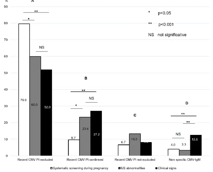
infection (< 3 months). A total of 1,074/6,560 samples collected from pregnant women for systematic 135
screening had low or moderate CMV-IgG avidity index and were considered as confirmed or not 136
excluded recent CMV primary infections (Figure 1 and Figure 2). Therefore, PPV of CMV-IgM for 137
systematic screening during pregnancy to predict a recent primary infection was 16.4% (95% CI = 138
15.5 – 17.3%). 139
Ultrasound abnormalities during pregnancy (group 2) 140
CMV-IgG avidity was high in 18/30 (60.0%) cases allowing to exclude recent CMV primary 141
infection. A total of 11/30 samples collected from pregnant women addressed for US had low or 142
7
intermediate CMV-IgG avidity index and were considered as confirmed or not excluded recent CMV 143
primary infections (Figure 1 and Figure 2). Therefore, PPV of CMV-IgM in case of US 144
abnormalities during pregnancy to predict a recent primary infection was 36.7% (95% CI = 19.5 – 145
53.9%). 146
Immunocompetent patients with clinical symptoms of CMV primary infection (group 3) 147
CMV-IgG avidity was high in 174/269 (64.7%) cases allowing to exclude recent CMV primary 148
infection (< 3 months). A total of 95/269 samples collected from patients with symptoms of acute 149
CMV primary infection had low or intermediate CMV-IgG avidity index and were considered as 150
confirmed or not excluded recent CMV primary infections (Figure 1 and Figure 2). Therefore, PPV 151
of CMV-IgM for immunocompetent patients with clinical signs to predict a recent primary infection 152
was 35.3% (95% CI = 29.6 – 41.0%). 153
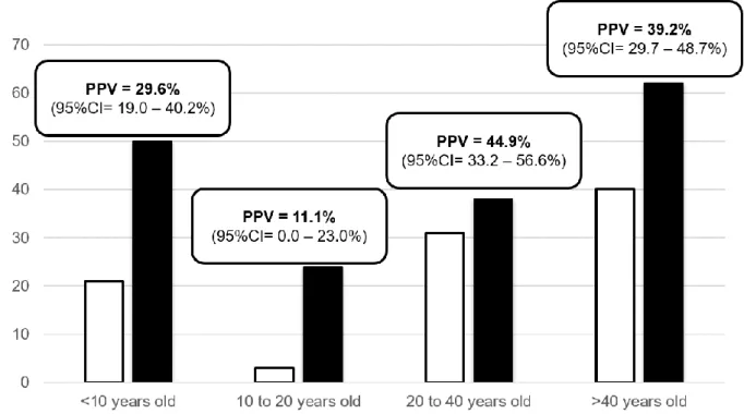
We determined CMV-IgM PPV for these 269 patients according to their age (Figure 3): 154
- less than 10 years old (n=71): CMV-IgG avidity was low or moderate in 21/71 patients 155
resulting in a PPV of 29.6% (95%CI= 19.0 – 40.5%) (p=0.50) 156
- 10 to 20 years old (n=27): CMV-IgG avidity was low or moderate in 3/27 patients resulting in 157
a PPV of 11.1% (95%CI= 0.0 – 23.0%) (p=0.01) 158
- 20 to 40 years old (n=69): CMV-IgG avidity was low or moderate in 31/69 patients resulting 159
in a PPV of 44.9% (95%CI= 33.2 – 56.6%) (p=0.20) 160
- more than 40 years old (n=102): CMV-IgG avidity was low or moderate in 40/102 patients 161
resulting in a PPV of 39.2% (95%CI= 29.7 – 48.7%) (p=0.90) 162
163
Overall, when positive CMV-IgM are observed, recent CMV primary infection is only confirmed in 164
respectively 16.4% cases for systematic screening during pregnancy (group 1), 36.7% in case of 165
ultrasound abnormalities (group 2) and 35.3% in case of clinical signs in general population (group 3). 166
8
CMV-IgM PPV was significantly lower in group 1 comparing to groups 2 (p=0.01) and 3 (p<0.001). 167
CMV-IgM PPV was not statistically different between groups 2 and 3 (p>0.90). 168
169
Discussion 170
Even if formal diagnosis of CMV primary infection is achieved with CMV-IgG 171
seroconversion, as the “first” seronegative serum is usually not available, documentation of this 172
seroconversion is rare. Usually, whatever the clinical situation, screening or clinical symptoms, only 173
one serum specimen is available. The transient CMV-IgM positivity has long been used as a 174
diagnostic marker for CMV primary infection, but it is well known that is not invariably indicative of 175
primary infection. Indeed, specific or non-specific CMV-IgM can also be present in many other 176
situations including infections with other pathogens associated with random polyclonal B cell 177
stimulation or CMV non primary infection [20,21]. Moreover, there are known to be technical issues 178
with IgM-specific testing. Indeed, false positive CMV-IgM results were shown to occur due to high 179
levels of analyte specific IgG, to presence of rheumatoid factors, and to other miscellaneous technical 180
factors involved with methods that must include some sort of process to separate CMV IgG from 181
CMV IgM. In these cases, CMV-IgG avidity is essential and in contrast to IgM, low-avidity IgG is 182
present only with primary infection, increasing over 3 to 4 months to high avidity [22]. CMV-IgG 183
avidity has thus gained diagnostic importance in identifying primary CMV infection, and several 184
commercial CMV-IgG avidity tests are currently available. Their performances to confirm a CMV 185
primary infection were reported to range from 83 to 100%, and to exclude a CMV primary infection 186
from 71 to 100% [12,16,19,23-28]. Even if not perfect, several groups have reported substantial 187
improvements in identification of at-risk pregnancies using diagnostic algorithms including CMV-IgG 188
avidity [7,15,18,22,29-31]. Indeed, it truly improves accuracy of CMV primary infection diagnosis as 189
in the specific situation of systematic screening, PPV of positive CMV-IgM is quite poor [18,31]. 190
Indeed, in our analysis, if we calculated PPV considering confirmed recent primary infection, PPV is 191
only of 9.7% (95%CI= 9.0 – 10.4%). However, from a practical point of view, we calculated PPV 192
9
considering both confirmed and not excluded recent CMV primary infection as these will probably be 193
managed like confirmed recent primary infection regarding regular ultrasound follow up. It allows 194
PPV to reach 16.4%. 195
Usefulness of serologic testing for CMV during pregnancy has been questioned since 196
congenital malformations can occur even following maternal secondary infections [6]. Although 197
screening is not recommended by any public health system because of its cost/benefit ratio, it is 198
widely adopted by many general practitioners and obstetricians in our area. Such screening provides 199
an opportunity to identify seronegative women who can be counselled about using appropriate 200
hygienic measures to prevent primary infection, especially in relation to their behavior with children, 201
who are a major source of infection. Furthermore, screening aims to diagnose CMV primary infection 202
early in pregnancy allowing women to be referred to Reference Centers for appropriate management 203
(close ultrasonography, amniocentesis and/or neonatal diagnosis…) [32]. Some authors consider that 204
screening is not justified because of its economic cost, the imperfect nature of congenital infection 205
prognostic criteria, the risk of spontaneous abortions induced by invasive tests such as amniocentesis, 206
and the few data concerning effective treatments during pregnancy. However, it is obvious that if US 207
are observed, and prenatal diagnosis of CMV congenital infection discussed, maternal CMV serology 208
should always be performed. It is particularly important especially in regions where seroprevalence is 209
around 50%, as in France, or lower because its first aim is to confirm maternal infection to CMV 210
(whatever the date of this infection) and allows to definitively exclude congenital CMV if the pregnant 211
woman is seronegative. If positive, this CMV serology usually performed late in pregnancy, might be 212
challenging to interpret. Our results show that positive CMV-IgM at time of US indicate a recent 213
infection in 23% cases (7/30) and that additionally, 3/30 women had high avidity but gave birth to an 214
infected child. Overall, when positive CMV-IgM is detected when US abnormalities are observed 215
(possibly late in pregnancy), it suggests responsibility of CMV in 47% (14/30) cases without ruling 216
out the possibility of CMV congenital infection following maternal non primary infection if the 217
mother is CMV-IgG positive and CMV-IgM is negative. However, final diagnosis will of course be 218
obtained with CMV PCR in amniotic fluid and/or in urine in the neonate at birth. 219
10 220
If CMV-IgG avidity is widely used in pregnant women, far less literature is available on the 221
usefulness of CMV-IgG avidity in general population (immunocompetent patients), and in our area it 222
is clearly not usually performed if the patient is not pregnant. Remarkably, positive CMV-IgM results 223
are most often considered as indicative of a recent CMV virus infection in case with clinical signs, but 224
they are in fact truly related to a CMV primary infection in only 27.1% cases (73/269) (low CMV IgG 225
avidity) and possibly related in 35.3% cases (low/moderate CMV IgG avidity). Interestingly, PPV was 226
significantly lower for patients between 10 to 20 years old (11.1% vs 29.6-44.9%) (p=0.01). Our 227
results suggest that positive CMV-IgM is less correlated with CMV recent primary infection in young 228
people, and are consistent with the understanding that CMV-IgM can be produced throughout life as a 229
result of CMV non primary infection or polyclonal stimulation of the immune system [20,21]. Also, in 230
cases of positive CMV-IgM, older people may be more likely to have a primary infection than 231
younger people. These also suggest that whatever the age of the patient, CMV-IgG avidity is essential 232
for accurate diagnosis. One limitation of our study is that, although very frequent, CMV primary 233
infection is most often a mild infection that does not require the patient to come to the hospital. 234
Consequently, general practitioners probably mostly do diagnosis of CMV primary infection and only 235
the patients with the severest symptoms come to the hospital and are therefore included in our data. 236
Moreover, it is possible that older people are more likely to have a severe infection compared to young 237
patients. Nevertheless, for current practice, all clinicians should be aware that, whatever the age of the 238
patient, and given the non-specific symptoms of CMV primary infection, even in case of positive 239
CMV-IgM, confirmation by avidity avoids misdiagnosis in more than 60% cases. 240
Our observations are consistent with the preexisting ones [33-37] and highlight the major 241
importance of including CMV-IgG avidity in the diagnostic algorithm, whatever the clinical situation 242
(for immunocompetent patients), to confirm or exclude a CMV primary infection in case of positive 243
CMV-IgM. 244
11 Sample CRediT author statement:
246
Claire Périllaud-Dubois: Conceptualization, Methodology, Formal analysis, Writing – original draft. 247
Elise Bouthry: Writing- Reviewing and Editing. Abir Jadoui and Ay Ling Leng: Investigation. 248
Anne-Marie Roque-Afonso: Writing- Reviewing and Editing. Christelle Vauloup-Fellous: 249
Conceptualization, Writing- Reviewing and Editing, Supervision. 250
251
Declarations of Interest: none. 252
253
Funding sources: none. 254
255 256
12 References
257 258
[1] A. Kenneson et M. J. Cannon, Review and meta-analysis of the epidemiology of congenital 259
cytomegalovirus (CMV) infection, Rev. Med. Virol., p. 253‑276, 2007, doi: 10.1002/rmv.535. 260
[2] S. C. Dollard, S. D. Grosse, et D. S. Ross, New estimates of the prevalence of neurological and 261
sensory sequelae and mortality associated with congenital cytomegalovirus infection, Rev. 262
Med. Virol., p. 355‑363, 2007, doi: 10.1002/rmv.544. 263
[3] S. Stagno et al., Primary cytomegalovirus infection in pregnancy. Incidence, transmission to 264
fetus, and clinical outcome, JAMA, p. 1904‑1908, 1986. 265
[4] S. A. Ross et al., Hearing loss in children with congenital cytomegalovirus infection born to 266
mothers with preexisting immunity, J. Pediatr., p. 332‑336, 2006, doi: 267
10.1016/j.jpeds.2005.09.030. 268
[5] M. Leruez-Ville et al., Risk Factors for Congenital Cytomegalovirus Infection Following Primary 269
and Nonprimary Maternal Infection: A Prospective Neonatal Screening Study Using Polymerase 270
Chain Reaction in Saliva, Clin. Infect. Dis. Off. Publ. Infect. Dis. Soc. Am., p. 398‑404, 2017, doi: 271
10.1093/cid/cix337. 272
[6] J. J. C. de Vries, E. W. van Zwet, F. W. Dekker, A. C. M. Kroes, P. H. Verkerk, et A. C. T. M. 273
Vossen, The apparent paradox of maternal seropositivity as a risk factor for congenital 274
cytomegalovirus infection: a population-based prediction model: Maternal seropositivity as a 275
risk factor for cCMV, Rev. Med. Virol., p. 241‑249, 2013, doi: 10.1002/rmv.1744. 276
[7] M. G. Revello et G. Gerna, Diagnosis and Management of Human Cytomegalovirus Infection in 277
the Mother, Fetus, and Newborn Infant, Clin. Microbiol. Rev., p. 680‑715, 2002, doi: 278
10.1128/CMR.15.4.680-715.2002. 279
[8] O. Picone et al., A series of 238 cytomegalovirus primary infections during pregnancy: 280
description and outcome: Outcome of maternal CMV infection during pregnancy, Prenat. 281
Diagn., p. 751‑758, 2013, doi: 10.1002/pd.4118. 282
[9] G. Enders, A. Daiminger, U. Bäder, S. Exler, et M. Enders, Intrauterine transmission and clinical 283
outcome of 248 pregnancies with primary cytomegalovirus infection in relation to gestational 284
age, J. Clin. Virol., p. 244‑246, 2011, doi: 10.1016/j.jcv.2011.07.005. 285
[10] V. Faure-Bardon et al., Sequelae of congenital cytomegalovirus (cCMV) following maternal 286
primary infection are limited to those acquired in the first trimester of pregnancy, Clin. Infect. 287
Dis. Off. Publ. Infect. Dis. Soc. Am., 2018, doi: 10.1093/cid/ciy1128. 288
[11] T. G. Wreghitt, E. L. Teare, O. Sule, R. Devi, et P. Rice, Cytomegalovirus Infection in 289
Immunocompetent Patients, Clin. Infect. Dis., p. 1603‑1606, 2003, doi: 10.1086/379711. 290
[12] C. Vauloup-Fellous, T. Lazzarotto, M. G. Revello, et L. Grangeot-Keros, Clinical evaluation of the 291
Roche Elecsys CMV IgG Avidity assay, Eur. J. Clin. Microbiol. Infect. Dis. Off. Publ. Eur. Soc. Clin. 292
Microbiol., p. 1365‑1369, 2014, doi: 10.1007/s10096-014-2080-4. 293
[13] C. Busse, A. Strubel, et P. Schnitzler, Combination of native and recombinant cytomegalovirus 294
antigens in a new ELISA for detection of CMV-specific antibodies, J. Clin. Virol., p. 137‑141, 295
2008, doi: 10.1016/j.jcv.2008.05.011. 296
[14] M. G. Revello, C. Fornara, A. Arossa, P. Zelini, et D. Lilleri, Role of human cytomegalovirus 297
(HCMV)-specific antibody in HCMV-infected pregnant women, Early Hum. Dev., p. S32‑S34, 298
2014, doi: 10.1016/S0378-3782(14)70011-8. 299
[15] M. Macé, L. Sissoeff, A. Rudent, et L. Grangeot-Keros, A serological testing algorithm for the 300
diagnosis of primary CMV infection in pregnant women: DIAGNOSIS OF CMV PRIMARY 301
INFECTION IN PREGNANT WOMEN, Prenat. Diagn., p. 861‑863, 2004, doi: 10.1002/pd.1001. 302
[16] M. L. Delforge, L. Desomberg, et I. Montesinos, Evaluation of the new LIAISON ® CMV IgG, IgM 303
and IgG Avidity II assays, J. Clin. Virol., p. 42‑45, 2015, doi: 10.1016/j.jcv.2015.09.002. 304
13
[17] S. C. Dollard, S. A. S. Staras, M. M. Amin, D. S. Schmid, et M. J. Cannon, National Prevalence 305
Estimates for Cytomegalovirus IgM and IgG Avidity and Association between High IgM Antibody 306
Titer and Low IgG Avidity, Clin. Vaccine Immunol., p. 1895‑1899, 2011, doi: 10.1128/CVI.05228-307
11. 308
[18] M. Leruez-Ville, Y. Sellier, L. J. Salomon, J. J. Stirnemann, F. Jacquemard, et Y. Ville, Prediction of 309
Fetal Infection in Cases With Cytomegalovirus Immunoglobulin M in the First Trimester of 310
Pregnancy: A Retrospective Cohort, Clin. Infect. Dis., p. 1428‑1435, 2013, doi: 311
10.1093/cid/cit059. 312
[19] C. Vauloup-Fellous, M. Berth, F. Heskia, J.-M. Dugua, et L. Grangeot-Keros, Re-evaluation of the 313
VIDAS® cytomegalovirus (CMV) IgG avidity assay: Determination of new cut-off values based on 314
the study of kinetics of CMV–IgG maturation, J. Clin. Virol., p. 118‑123, 2013, doi: 315
10.1016/j.jcv.2012.10.017. 316
[20] S. L. Nielsen, I. Sørensen, et H. K. Andersen, Kinetics of specific immunoglobulins M, E, A, and G 317
in congenital, primary, and secondary cytomegalovirus infection studied by antibody-capture 318
enzyme-linked immunosorbent assay, J. Clin. Microbiol., p. 654‑661, 1988. 319
[21] R. F. Pass, S. Stagno, W. J. Britt, et C. A. Alford, Specific cell-mediated immunity and the natural 320
history of congenital infection with cytomegalovirus, J. Infect. Dis., p. 953‑961, 1983. 321
[22] T. Lazzarotto, B. Guerra, M. Lanari, L. Gabrielli, et M. P. Landini, New advances in the diagnosis 322
of congenital cytomegalovirus infection, J. Clin. Virol. Off. Publ. Pan Am. Soc. Clin. Virol., p. 323
192‑197, 2008, doi: 10.1016/j.jcv.2007.10.015. 324
[23] T. Lazzarotto, S. Brojanac, G. T. Maine, et M. P. Landini, Search for cytomegalovirus-specific 325
immunoglobulin M: comparison between a new western blot, conventional western blot, and 326
nine commercially available assays., Clin. Diagn. Lab. Immunol., p. 483‑486, 1997, doi: 327
10.1128/CDLI.4.4.483-486.1997. 328
[24] T. Lazzarotto et al., Anticytomegalovirus (anti-CMV) immunoglobulin G avidity in identification 329
of pregnant women at risk of transmitting congenital CMV infection, Clin. Diagn. Lab. Immunol., 330
p. 127‑129, 1999. 331
[25] M. Bodéus, D. Beulné, et P. Goubau, Ability of Three IgG-Avidity Assays to Exclude Recent 332
Cytomegalovirus Infection, Eur. J. Clin. Microbiol. Infect. Dis., p. 0248‑0252, 2001, doi: 333
10.1007/s100960100484. 334
[26] M. G. Revello, G. Gorini, et G. Gerna, Clinical Evaluation of a Chemiluminescence Immunoassay 335
for Determination of Immunoglobulin G Avidity to Human Cytomegalovirus, Clin. Diagn. Lab. 336
Immunol., p. 801‑805, 2004, doi: 10.1128/CDLI.11.4.801-805.2004. 337
[27] K. Lagrou, M. Bodeus, M. Van Ranst, et P. Goubau, Evaluation of the New Architect 338
Cytomegalovirus Immunoglobulin M (IgM), IgG, and IgG Avidity Assays, J. Clin. Microbiol., p. 339
1695‑1699, 2009, doi: 10.1128/JCM.02172-08. 340
[28] M. E. Guisasola, Belé. Ramos, J. C. Sanz, I. García-Bermejo, et F. De Ory Manchón, Comparison 341
of IgG avidity assays in the confirmation of the diagnosis of cytomegalovirus primary infection: 342
IGG AVIDITY ASSAYS FOR DIAGNOSIS OF CMV INFECTION, APMIS, p. 991‑993, 2010, doi: 343
10.1111/j.1600-0463.2010.02682.x. 344
[29] T. Lazzarotto et al., Maternal IgG avidity and IgM detected by blot as diagnostic tools to identify 345
pregnant women at risk of transmitting cytomegalovirus, Viral Immunol., p. 137‑141, 2000, doi: 346
10.1089/vim.2000.13.137. 347
[30] S. C. Munro et al., Diagnosis of and screening for cytomegalovirus infection in pregnant women, 348
J. Clin. Microbiol., p. 4713‑4718, 2005, doi: 10.1128/JCM.43.9.4713-4718.2005. 349
[31] C. Vauloup-Fellous et al., Does hygiene counseling have an impact on the rate of CMV primary 350
infection during pregnancy? Results of a 3-year prospective study in a French hospital, J. Clin. 351
Virol. Off. Publ. Pan Am. Soc. Clin. Virol., p. S49-53, 2009, doi: 10.1016/j.jcv.2009.09.003. 352
14
[32] W. D. Rawlinson et al., Congenital cytomegalovirus infection in pregnancy and the neonate: 353
consensus recommendations for prevention, diagnosis, and therapy, Lancet Infect. Dis., p. 354
e177‑e188, 2017, doi: 10.1016/S1473-3099(17)30143-3. 355
[33] M. De Paschale, C. Agrappi, M. T. Manco, et P. Clerici, Positive predictive value of anti-HCMV 356
IgM as an index of primary infection, J. Virol. Methods, p. 121‑125, 2010, doi: 357
10.1016/j.jviromet.2010.05.001. 358
[34] M. Yoshida et al., Can measurement of maternal anti-cytomegalovirus immunoglobulin-M 359
antibody levels be used to screen for cytomegalovirus infection in embryos and fetuses?, J. 360
Obstet. Gynaecol. Res., p. 166‑169, 2013, doi: 10.1111/j.1447-0756.2012.01900.x. 361
[35] A. Goncé et al., Maternal IgM antibody status in confirmed fetal cytomegalovirus infection 362
detected by sonographic signs, Prenat. Diagn., p. 817‑821, 2012, doi: 10.1002/pd.3907. 363
[36] Y. Torii et al., Serological screening of immunoglobulin M and immunoglobulin G during 364
pregnancy for predicting congenital cytomegalovirus infection, BMC Pregnancy Childbirth, p. 365
205, 2019, doi: 10.1186/s12884-019-2360-1. 366
[37] Toriyabe K. et al., Anticytomegalovirus IgM titer for congenital infection in first-trimester 367
pregnancy with primary infection, J. Obstet. Gynaecol. Res., p. 1555‑1556, 2018, doi: 368
10.1111//jog.13762. 369
1
Positive predictive values of CMV-IgM
and importance of CMV-IgG avidity
1testing in detecting
primary infection in three different clinical settings. A
2French retrospective cohort study.
34
Claire Périllaud-Dubois 1,2,3,4, Elise Bouthry 1,2, Abir Jadoui 1, Ay-Ling Leng 1, Anne-Marie Roque-5
Afonso 1,4,5, Christelle Vauloup-Fellous 1,2,4,5 6
7
1
AP–HP.Université Paris-Saclay, Hôpital Paul-Brousse, Service de Virologie, , 94804 Villejuif, France 8
2 Groupe de Recherche sur les Infections pendant la Grossesse (GRIG), France
9
3 INSERM UMR1137, IAME, 75018 Paris, France
10
4 Université Paris-Saclay, 94804 Villejuif, France
11
5 INSERM U1193, 94804 Villejuif, France
12 13
Corresponding author: Dr Claire Périllaud-Dubois 14
AP-HP.Université Paris-Saclay, Hôpital Paul-Brousse, Service de Virologie 15
12-14 avenue Paul Vaillant Couturier 16 94804 Villejuif, France 17 +33145593721 18 claire.perillaud-dubois@inserm.fr 19
Keywords: CMV; serology; IgM; avidity; PPV; positive predictive value; pregnancy; ultrasound 20
abnormalities; clinical signs 21 Word count: 2443 22 23 Abstract 24
*Manuscript (Tracked Changes)
2
Background: Diagnosis of Cytomegalovirus (CMV) primary infection during pregnancy or in 25
immunocompetent patients relies on serology with detection of specific CMV-IgG and IgM. In case of 26
positive CMV-IgM in pregnant women, CMV-IgG avidity is now widely recommended, but in general 27
population it is not currently performed. 28
Objective: In this study, we aimed to determine CMV-IgM positive predictive values (PPV) in 29
different clinical settings. 30
Material and methods: We conducted a retrospective study on positive CMV-IgM in our virology 31
laboratory from 2013 to 2019, in three clinical groups: screening in non-symptomatic pregnant women 32
(group 1), pregnant women with ultrasound (US) abnormalities (group 2) and patients (general 33
population) with clinical signs suggestive of CMV primary infection (group 3). CMV-IgG avidity had 34
been performed in all cases allowing to evaluate PPV of positive CMV-IgM to diagnose CMV 35
primary-infection in each group. 36
Results: Between 2013 and 2019, 6,859 serum samples were found positive for CMV-IgM and had 37
been tested for CMV-IgG avidity, with 6,560 sera for group 1, 30 for group 2 and 269 for group 3. 38
Overall, low avidity confirming primary infection was observed respectively in 16.4% for group 1, 39
36.7% for group 2, and 35.3% for group 3. CMV-IgM PPV was significantly lower in group 1 40
compared to groups 2 (p=0.01) and 3 (p<0.001). 41
Discussion: Our observations highlight the major importance of including CMV-IgG avidity in the 42
diagnostic algorithm, whatever the clinical situation (for immunocompetent patients), to confirm or 43
exclude a recent CMV primary infection in case of positive CMV-IgM. 44
45
Introduction 46
Cytomegalovirus (CMV) is the most frequent worldwide cause of congenital viral infection with a 47
prevalence estimated between 0.5 and 1% of all live births. Congenital CMV is a major cause of 48
3
sensorineural hearing loss and mental retardation [1-3]. CMV transmission to the fetus can occur after 49
primary or secondary maternal CMV infection, with approximately the same proportion of symptoms 50
and sequelae in both situations [4-6]. At birth, 13% of congenitally infected neonates are symptomatic 51
with CMV-specific symptoms including growth restriction, microcephaly, ventriculomegaly, 52
chorioretinitis, sensorineural hearing loss, hepatitis, thrombocytopenia and a purpuric skin eruption [2-53
7]. Risk of long term sequelae is higher if CMV transmission occurs in the first or second trimester of 54
pregnancy or during peri-conceptional period [8-10]. In immunocompetent patients, CMV primary 55
infection is often asymptomatic. When symptomatic (8 to 10% of cases), primary infection is usually 56
responsible for a mild disease. Signs most frequently reported are: isolated fever, asthenia, 57
mononucleosis syndrome with cervical lymphadenopathy and/or cytolytic hepatitis [11]. 58
Diagnosis of CMV primary infection during pregnancy mainly relies on serology: detection of specific 59
CMV-IgG and IgM, associated with CMV-IgG avidity in case of positive CMV-IgM [12]. However, 60
in immunocompetent patients not pregnant, CMV-IgG avidity is usually not performed in case of 61
positive CMV-IgM. Reported clinical performances of commercial immunoassays for CMV IgM are 62
sensitivity >90% and specificity >96% and for CMV IgG avidity, specificity and sensitivity are 63
comprised between 90 and 100% depending on the assay [13-16]. CMV-IgM can possibly indicate an 64
acute or a recent infection but can also be due to other causes: long-term persisting IgM, cross-65
reaction, secondary CMV infection or nonspecific stimulation of the immune system. Consequently, 66
diagnosis of primary infection cannot rely only on a positive IgM test result. CMV-IgG avidity 67
measurement is an essential tool to confirm or exclude CMV primary-infection. CMV-IgG are initially 68
of low avidity, but will mature to high avidity at 2-4 months after primary infection [12,17,18]. 69
The main issue with CMV serology is that CMV-IgG avidity is not available in all laboratories and 70
that clinicians still too often rely on positive CMV-IgM result to diagnose CMV primary-infection. In 71
our retrospective cohort study, we aim to determine and compare CMV-IgM positive predictive value 72
(PPV) to diagnose CMV primary infection depending on the clinical situation: systematic screening 73
4
during pregnancy, presence of ultrasound abnormalities (US) during pregnancy and clinical signs 74
suggestive of CMV primary infection in general population (immunocompetent patients). 75
76
Material and methods 77
Sample collection: 78
In our hospital virology laboratory, CMV serology (IgG, IgM and IgG avidity in case of positive IgG) 79
is performed either: 80
- in non-symptomatic pregnant women during first trimester of pregnancy (systematic 81
screening) and followed in one of the two maternities in Paris South Hospitals, or in pregnant 82
women referred to our laboratory because of positive CMV-IgM detected in one of the 83
laboratories part of our network (systematic screening in other centers) (group 1); 84
- in non-symptomatic pregnant women referred to our pluridisciplinary prenatal center for US 85
abnormalities (not initially screened at beginning of pregnancy) (group 2). Sonographic 86
findings were mostly intrauterine growth retardation (IUGR) and cerebral abnormalities (head 87
perimeter < 5th centile, ventriculomegaly, hyperechogenic ventricular wall, cerebellar and 88
brain calcifications, enlargement of pericerebral spaces, periventricular and subependymal 89
cysts, candlestick, porencephaly, hyperechogenic periventricular halo, candlestick, abnormal 90
gyration, and/or corpus callosum hypoplasia); 91
- in immunocompetent patients (general population: adults and children) in case of clinical 92
symptoms suggestive of CMV primary infection (group 3), mainly fever, headache, flu-like 93
syndrome (fever + rhinitis + myalgia), arthralgia, myalgia, fatigue and/or hepatitis. Samples 94
were collected 5 to 30 days after onset of symptoms. 95
All CMV serologic results performed in one of these contexts in our laboratory between January 2013 96
and December 2019 were retrospectively analysed. In case a patient had several samples, only the 97
5
most informative (usually the first one) was kept for analysis. Serologies performed in the context of 98
transplantation or in immunocompromised patients were not included in this study. 99
100
Serology assays: 101
CMV-IgG and CMV-IgM were measured with LIAISON XL (LXL, DiaSorin®, Saluggia, Italy). In 102
case of positive CMV-IgM, our strategy is based on previous published findings [19]. In a few words, 103
we first-line perform LXL IgG avidity as a screening test. An index > 0.40 allows to exclude a recent 104
CMV primary infection. Below LXL 0.40 index threshold, a second assay is used: VIDAS 105
(bioMérieux®, Craponne, France) CMV-IgG avidity. In this case, confirmation/exclusion of primary 106
infection (more/less than 3 months before sample collection) is only based on the VIDAS assay result 107
(cutoffs used are those recommended by manufacturer: 0.4-0.65). Results allowed us to classify 108
serological profiles as follows: 109
- Negative CMV-IgG/positive CMV-IgM on two consecutive samples 21 days apart: non-110
specific IgM; 111
- Positive CMV-IgG/positive CMV-IgM/high CMV-IgG avidity (LXL > 0.4, or LXL < 0.4 and 112
VIDAS > 0.65) : recent CMV primary infection excluded; 113
- Positive CMV-IgG/positive CMV-IgM/low CMV-IgG avidity (LXL < 0.4 and VIDAS < 114
0.40): recent CMV primary infection confirmed; 115
- Positive CMV-IgG/positive CMV-IgM/moderate CMV-IgG avidity (LXL < 0.4 and VIDAS > 116
0.40 but < 0.65): recent CMV primary infection not excluded. 117
118
Statistical analysis: 119
6
For the three groups, CMV-IgM PPV to diagnose recent CMV primary infection were calculated with 120
95% confidence intervals. We calculated p-values with Chi-2 Pearson tests. 121
122
Results 123
Between 2013 and 2019, 6,859 serum samples were tested positive for CMV-IgM in our laboratory: 124
- 6,560 /6,859 (95.64%) were collected in pregnant women during systematic screening (group 125
1) 126
- 30/6,859 (0.44%) were collected in pregnant women with US abnormalities (median 127
gestational age 25 weeks of gestation (WG); range: 12-36 WG) (group 2). 128
- 269/6,859 (3.92%) were collected from immunocompetent patients with clinical symptoms of 129
CMV primary infection (group 3) 130
All 6,859 serum samples CMV-IgM positive had been tested for CMV-IgG avidity at time of 131
diagnosis. 132
Systematic screening during pregnancy (group 1) 133
CMV-IgG avidity was high in 5,486/6,560 (83.6%) cases allowing to exclude recent CMV primary 134
infection (< 3 months). A total of 1,074/6,560 samples collected from pregnant women for systematic 135
screening had low or moderate CMV-IgG avidity index and were considered as confirmed or not 136
excluded recent CMV primary infections (Figure 1 and Figure 2). Therefore, PPV of CMV-IgM for 137
systematic screening during pregnancy to predict a recent primary infection was 16.4% (95% CI = 138
15.5 – 17.3%). 139
Ultrasound abnormalities during pregnancy (group 2) 140
CMV-IgG avidity was high in 18/30 (60.0%) cases allowing to exclude recent CMV primary 141
infection. A total of 11/30 samples collected from pregnant women addressed for US had low or 142
7
intermediate CMV-IgG avidity index and were considered as confirmed or not excluded recent CMV 143
primary infections (Figure 1 and Figure 2). Therefore, PPV of CMV-IgM in case of US 144
abnormalities during pregnancy to predict a recent primary infection was 36.7% (95% CI = 19.5 – 145
53.9%). 146
Immunocompetent patients with clinical symptoms of CMV primary infection (group 3) 147
CMV-IgG avidity was high in 174/269 (64.7%) cases allowing to exclude recent CMV primary 148
infection (< 3 months). A total of 95/269 samples collected from patients with symptoms of acute 149
CMV primary infection had low or intermediate CMV-IgG avidity index and were considered as 150
confirmed or not excluded recent CMV primary infections (Figure 1 and Figure 2). Therefore, PPV 151
of CMV-IgM for immunocompetent patients with clinical signs to predict a recent primary infection 152
was 35.3% (95% CI = 29.6 – 41.0%). 153
We determined CMV-IgM PPV for these 269 patients according to their age (Figure 3): 154
- less than 10 years old (n=71): CMV-IgG avidity was low or moderate in 21/71 patients 155
resulting in a PPV of 29.6% (95%CI= 19.0 – 40.5%) (p=0.50) 156
- 10 to 20 years old (n=27): CMV-IgG avidity was low or moderate in 3/27 patients resulting in 157
a PPV of 11.1% (95%CI= 0.0 – 23.0%) (p=0.01) 158
- 20 to 40 years old (n=69): CMV-IgG avidity was low or moderate in 31/69 patients resulting 159
in a PPV of 44.9% (95%CI= 33.2 – 56.6%) (p=0.20) 160
- more than 40 years old (n=102): CMV-IgG avidity was low or moderate in 40/102 patients 161
resulting in a PPV of 39.2% (95%CI= 29.7 – 48.7%) (p=0.90) 162
163
Overall, when positive CMV-IgM are observed, recent CMV primary infection is only confirmed in 164
respectively 16.4% cases for systematic screening during pregnancy (group 1), 36.7% in case of 165
ultrasound abnormalities (group 2) and 35.3% in case of clinical signs in general population (group 3). 166