HAL Id: hal-01911331
https://hal.archives-ouvertes.fr/hal-01911331
Submitted on 26 May 2020
HAL is a multi-disciplinary open access
archive for the deposit and dissemination of
sci-entific research documents, whether they are
pub-lished or not. The documents may come from
teaching and research institutions in France or
abroad, or from public or private research centers.
L’archive ouverte pluridisciplinaire HAL, est
destinée au dépôt et à la diffusion de documents
scientifiques de niveau recherche, publiés ou non,
émanant des établissements d’enseignement et de
recherche français ou étrangers, des laboratoires
publics ou privés.
Distributed under a Creative Commons Attribution| 4.0 International License
brasiliensis, a Pathogen Responsible for Cutaneous
Nocardiosis Found in France: Phylogenetic
Relationships by Using sod and hsp65 Genes
D. Kosova-Maali, E. Bergeron, Yousef Maali, T. Durand, J. Gonzalez, D.
Mouniée, H. Sandoval Trujillo, P. Boiron, M.-C. Salinas-Carmona, V.
Rodriguez-Nava
To cite this version:
D. Kosova-Maali, E. Bergeron, Yousef Maali, T. Durand, J. Gonzalez, et al.. High Intraspecific
Genetic Diversity of Nocardia brasiliensis, a Pathogen Responsible for Cutaneous Nocardiosis Found
in France: Phylogenetic Relationships by Using sod and hsp65 Genes. BioMed Research International,
2018, 2018, pp.7314054. �10.1155/2018/7314054�. �hal-01911331�
Research Article
High Intraspecific Genetic Diversity of
Nocardia brasiliensis,
a Pathogen Responsible for Cutaneous Nocardiosis
Found in France: Phylogenetic Relationships by Using
sod and hsp65 Genes
D. Kosova-Maali,
1E. Bergeron,
1Y. Maali,
2T. Durand,
3J. Gonzalez,
4D. Mouniée,
1H. Sandoval Trujillo,
4P. Boiron,
1M.-C. Salinas-Carmona,
5and V. Rodriguez-Nava
1,31Research Group on “Bacterial Opportunistic Pathogens and Environment”, UMR Ecologie Microbienne, CNRS 5557, INRA 1418,
UCBL, Universit´e de Lyon, VetAgro Sup, Facult´e de Pharmacie, 8 avenue Rockefeller, Lyon, France
2Centre International de Recherche en Infectiologie, INSERM U1111, CNRS UMR5308, ENS de Lyon,
Team “Pathogenesis of Staphylococcal Infections”, Universit´e de Lyon 1, Lyon, France
3Laboratoire de Bact´eriologie, Institut des Agents Infectieux, Centre de Biologie et Pathologie Nord, 103 grande rue de la Croix-Rousse,
69004 Lyon, France
4Departamento de Sistemas Biol´ogicos, Universidad Aut´onoma Metropolitana-Xochimilco, Calzada del Hueso 1100,
04960 Ciudad de M´exico, Mexico
5Facultad de Medicina, Universidad Autonoma de Nuevo Leon, Monterrey, NL, Mexico
Correspondence should be addressed to V. Rodriguez-Nava; veronica.rodriguez-nava@univ-lyon1.fr Received 9 February 2018; Accepted 31 March 2018; Published 20 May 2018
Academic Editor: Charles Spencer
Copyright © 2018 D. Kosova-Maali et al. This is an open access article distributed under the Creative Commons Attribution License, which permits unrestricted use, distribution, and reproduction in any medium, provided the original work is properly cited. This study aims at genetic characterization and phylogenetic relationships of Nocardia brasiliensis focusing by using housekeeping
rrs, hsp65, and sodA genes. N. brasiliensis is the species responsible for 80% of cases of actinomycetoma, one form of cutaneous
nocardiosis which occurs mainly in tropical regions reaching immunocompetent patients in which the disease can lead to amputation. We analyze 36 indigenous cases of N. brasiliensis that happened in France. Phylogenetic analysis targeting rrs gene showed no robustness at phylogenetic nodes level. However, the use of a concatenation of hsp65 and sodA genes showed that the tested strains surprisingly ranked in 3 well-defined genotypes. Genotypes 2 and 3 were phylogenetically closer to each other and both diverged from genotype 1 sustained by a high bootstrap of 81%. This last genotype hosts all the cases of pulmonary forms (3), the sole cerebral form, and almost all the cases of immunocompromised patients (3 out of 4). Moreover, excepting one of them, all the strains belonging to this group present a susceptibility to imipenem which is not the case in the other genotypes that rarely count among them strains being susceptible to this drug. The haplotype diversity (Hd) of hsp65 (0.927) and sodA (0.885) genes was higher than that of rrs (0.824). For this gene, we obtained 16 polymorphic sites whereas, for hsp65 and sodA genes, up to 27 and 29 were identified, respectively. This study reveals that these two genes have an important genetic discriminatory power for the evaluation of the intraspecies genetic variability of N. brasiliensis and they may be useful for identification purposes at species level. This study also reveals the possible existence of a new species harbored by genotype 1.
1. Introduction
Nocardia is a genus belonging to the aerobic
actino-mycetes group of bacteria which are Gram-positive bacilli and showing branching filamentous forms [1]. They are
saprophytic ubiquitous bacteria which can be found in several environments such as fresh water and saltwater, soil, dust, decaying vegetation, and decaying fecal deposits from animals [1]. Nevertheless, these environmental bac-teria can be opportunistic pathogens and lead to human
Volume 2018, Article ID 7314054, 10 pages https://doi.org/10.1155/2018/7314054
infectious diseases called “nocardiosis” [2]. Nocardiosis can be discriminated into two groups: invasive infection, mainly caused by N. asteroides, presenting commonly as pneumonia in patients who are immunocompromised, have underlying chronic lung disease, and are with a possible dissemination to other organs [3], and cutaneous infection via a cut or abraded skin, which can be manifest clinically as (i) abscess and cellulitis, (ii) lymphangitis, (iii) skin infection secondary to dissemination, and (iv) actinomycetoma. This latter group is the most amazing infection due to their severity characterized by the presence of tumefaction, subcu-taneous nodules, destructive granulomata, fistulas, and pus [2, 4].
N. brasiliensis is the species isolated from the
major-ity (approximately 80%) of cases of cutaneous nocardio-sis, especially in actinomycetoma [2]. This species is more commonly isolated in areas with tropical or subtropical climates such as South America, Asia, and Africa. Due to false diagnosis, rural lifestyles, and poor access to care in these countries, N. brasiliensis nocardiosis constitutes a real public health problem that can lead, in the absence of treatment, to amputations and death in young populations. On the basis of epidemiological surveys conducted in France, the number of cases of nocardiosis between 2000 and 2007 according to the French Nocardiosis Observatory (OFN) was 607 with N. farcinica and N. nova being the most frequent species [5]. However, no data currently exists on the phylogenetic relationships between the indigenous N.
brasiliensis strains of tropical origin and native strains isolated
in France. Routine genus/species identification of Nocardia was based on macroscopic, microscopic, and biochemical characteristics. The methods described by Boiron et al. [6] were used to determine the decomposition of adenine, casein, hypoxanthine, tyrosine, and xanthine. In addition to the phenotype-based methods, species-level identification is mainly genetically based, nowadays. Classically, 16S rRNA
(rrs) gene sequencing is generally used for the
species-level identification [7, 8], but it fails to discriminate among some species of Nocardia because it does not have enough polymorphism to differentiate them at the species level. Multilocus sequence analysis (MLSA) using concatenated sequences of several housekeeping genes such as superoxide dismutase A (sodA) and heat shock protein 65 (hsp65) has been increasingly used to provide higher accuracy and discriminatory power in the molecular identification of
Nocardia spp. [9, 10]. Indeed, a recent study seeking to
identify new molecular targets shows that the polymorphism observed in the sodA gene sequence contains variable regions that allow the discrimination of closely related Nocardia species [9].
The aim of the present study was to perform a genetic characterization and assess the phylogenetic relationships of Nocardia brasiliensis focusing on using housekeeping
rrs, hsp65, and sodA genes, for 36 autochthonous N. brasiliensis strains isolated in France and analyzed by
the OFN between 2002 and 2012. Phenotypic charac-terization was also conducted by assessing antimicrobial resistance profiles, metabolic profiles, and culture condi-tion.
2. Materials and Methods
2.1. Bacterial Strains and Culture Media. A collection of 36
human clinical strains of N. brasiliensis was studied (Table 1). All strains were identified as such, at species level by the French Nocardiosis Observatory (OFN) by genetic approach. Moreover, six Nocardia reference strains belonging to N.
brasiliensis clade [9] were also used: N. brasiliensis ATCC
19296T (unknown), N. altamirensis DSM 44997T (karstic cave), N. boironii DSM 101696T (pus sample), N. iowensis DSM 45197T (garden soil), N. tenerifensis DSM 44704T (rhizosphere), and N. vulneris DSM 45737T (human leg wound). Prior to the assays, strains were cultured 72 hours in Bennett medium (made in the laboratory) aerobically at 37∘C.
2.2. Growth Test on Culture Media. From 0.5 McF bacterial
suspension, bacterial growth was evaluated on three culture media: (i) bromocresol purple (BCP) (Biom´erieux, Marcy l’´etoile), (ii) Bennett (made in the laboratory), and (iii) Middlebrook (Biom´erieux, Marcy l’´etoile). One hundred microliters from bacterial suspension standardized was inoc-ulated on the different plate of culture media. The plates were incubated at 37∘C and the observations were performed at 48, 72, and 96 hours.
2.3. Antimicrobial Susceptibility. The susceptibility of the
isolates to different antimicrobials was determined by disk diffusion method with a panel of 31 antibiotics (Biorad, Marnes-la-Coquette France) on Muller Hinton E medium (Biom´erieux, Marcy l’´etoile, France). Susceptibility testing was done with amikacin 30𝜇g, gentamycin 15 𝜇g, tobramycin 10𝜇g, ciprofloxacin 5 𝜇g, levofloxacin 5 𝜇g, moxifloxacin 5𝜇g, minocycline 30 𝜇g, doxycycline 30 𝜇g, tigecycline 15 𝜇g, cefotaxime 30𝜇g, ceftriaxone 30 𝜇g, cefepime 30 𝜇g, cefurox-ime 30𝜇g, amoxicillin 25 𝜇g, amoxicillin + clavulanic acid 20/10𝜇g, ampicillin 10 𝜇g, ertapenem 10 𝜇g, meropenem 10𝜇g, imipenem 10 𝜇g, vancomycin 30 𝜇g, pristinamycin 15𝜇g, erythromycin 15 𝜇g, trimethoprim + sulfamethoxazole 1.25/23.75𝜇g, rifampicin 30 𝜇g, and linezolid 30 𝜇g.
From visible colonies, bacterial suspension was done in sterile water, using a cotton swab to obtain a concentration of 0.5 McFarland according to the Clinical and Laboratory Standards Institute standard M24-A2 [11]. Seeding was done according to the swab method. In this latter, the bacterial inoculum was spread on the agar using a sterile cotton swab in three different directions. The disks were dispensed with a dispenser and the plates were incubated at 37∘C for 72 hours and read manually according to the thresholds defined in the recommendations of the SFM 2013 [12].
2.4. Substrate Degradation. The methods of Boiron et al. [6],
Goodfellow et al. [13, 14], and Goodfellow and Lechevalier [15] were used to determine the decomposition of adenine, casein, and uric acid [9]. Clinical strains of N. brasiliensis and the strains of species belonging to the N. brasiliensis clade (N. brasiliensis, N. altamirensis, N. iowensis, N. tenerifensis, N.
T a ble 1: T ab le o f cl inical st ra in s inc ludin g the typ e o f tr o p ism o bs er ve d in h ost, th eir co rr esp o n din g so dA/hs p6 5 geno ty p es, an d d ru g p heno ty p es. Sa m p le da te N at ur e o f sa m p lin g P at ien t reco rd Imm unosu p p ress ed Ge n o ty pe so dA/hs p6 5 T ro p ism P ri st ina m yc in Im ip enem Ami k acin T rimet ho p rim + su lfa m et ho xazo le 0 4 /2 0 02 In tr ao p er at ive tissue 02.5 6 N o G3 Cut an eou s R R S S 12/20 03 Pus fr o m cu ta neo u s thig h abs cess 0 4.21 Y es G1 Cut an eou s R S S S 20 0 4 Pus fr o m cu ta n eo us ab scess 0 4.101 N o G3 Cut an eou s R R S S 07/20 0 5 E xp ec to ra ti o n th en LB A 0 5.6 4 Y es G1 Lu n g R S S S 01/2 0 05 Sk in b io psy 05.1 2 N o G1 Cut an eou s R S S S 07/20 0 5 P hala nx b io p sy 05.6 3 N o G3 Cut an eou s R R S S 2005 CS F 0 5.7 7 N o G1 Br ai n R R S S 11/20 05 -32 47 N o G3 Un k n o w n R S S S 20 07 C u ta neo u s abs cess 0 7. 16 8 N o G3 Cut an eou s R R S S 10/2 0 0 8 W o u nd o f fo re he ad 0 8.17 8 N o G3 Cut an eou s R S S S 11/20 08 Su b cu ta n eo us ab scess 0 8.188 N o G3 Su b cut an eo u s R R S S 10/2 0 0 8 E lb o w ab scess 298 5 N o G3 Cut an eou s R R S S 03 /2 0 0 8 -9 0 4 4 N o G2 Un k n o w n R R S S 03/20 0 9 P us o f leg abs cess 0 9.7 1 N o G2 Cut an eou s R S S S 04 /2 009 B ro n ch ia la sp ir ati o n 09 .1 06 N o G1 Lu n g R S S S 10 /2 0 0 9 B ron ch ia la spi ra ti on 0 9. 24 4 Y es G1 Lu n g R S S S 12/20 0 9 Fin ge r skin 0 9. 28 0 N o G3 Cut an eou s R R S S 10/2 0 0 9 P us fr o m th e li p 10.16 N o G3 Cut an eou s R R S S 02/2010 Fin ger abs cess 10.3 5 N o G2 Cut an eou s R R S S 05/2010 H an d abs cess 10.8 2 N o G3 Cut an eou s R R S S 05/2010 T o e ab scess 10.9 3 Y es G3 Cut an eou s R R S S 0 9/2010 L eg w o und 10.1 4 6 N o G3 Cut an eou s R R S S 11/201 0 Pus fin ger 10.1 80 N o G3 Cut an eou s R R S S 11/201 0 L eg w o und 12 78 6 N o G2 Cut an eou s R R S S 04 /2 01 0 -14 22 9 N o G2 Un k n o w n R S S S 07/2010 Sepsis h an d 4 57 62 N o G3 Cut an eou s R R S S 2011 Pus 11.4 4 N o G2 Cut an eou s R R S S 05/2 011 C u ta n eo us abs cess 11.7 3 N o G1 Cut an eou s R S S S 05/2011 H an d w o und 11.80 N o G1 Cut an eou s R S S S 08/2011 C u ta n eo us 11.11 6 N o G2 Cut an eou s R R S S 2011 Thig h abs cess 11.1 4 0 N o G2 Cut an eou s R R S S 0 9/2011 K nee w o und 11.15 1 N o G3 Cut an eou s R R S S 2011 H and w o und 11.1 72 N o G3 Cut an eou s R R S S 12/2 011 C u ta n eo us ab scess 11.18 9 N o G3 Cut an eou s R R S S 20 12 -12.08 N o G1 Un k n o w n R S S S 02/2 012 C u ta n eo us abs cess 12.2 8 N o G3 Cut an eou s R R S S
DSM 101696T, N. brasiliensis ATCC 19296T, and N. vulneris DSM 45737T were incubated at 37∘C, and N. altamirensis DSM 44997T, N. tenerifensis DSM 44704T, and N. iowensis DSM 45197Twere incubated at 28∘C [9]. The readings were performed at 3, 7, 10, 14, 17, and 21 days.
2.5. Methods of DNA Extraction. DNA extraction from Nocardia strains was performed with achromopeptidase
according to the method reported by Rodr´ıguez-Nava et al. [10]. Colonies were picked off with a loop, and one loopful was suspended in 200𝜇L of sterile water containing a dozen glass beads and vortexed for 5 minutes. The mixture was then incubated for 15 minutes at 70∘C. The suspension supple-mented with 3.4𝜇L of achromopeptidase (Sigma, Steinheim, Germany) at 10 U/mL was incubated at 55∘C for 15 minutes. The suspensions were then centrifuged for 5 minutes at 13,000 rpm. The supernatants were stored at−20∘C until use.
2.6. Amplification and Sequencing
Gene rrs. A 606-bp fragment of the rrs gene was
ampli-fied with primers Noc1, 5 -GCTTAACACATGCAAGTCG-3, and Noc2, 5-GAATTCCAGTCTCCCCTG-3, and PCR program and reaction mixture were carried out according the recommendations of Rodr´ıguez-Nava et al. [10].
Gene hsp65. A 441-bp fragment of the hsp65 gene
encoding the 65-kDa heat shock protein was amplified with primers described by Telenti et al. (TB11: 5 -ACCAACGATGGTGTGTCCAT-3 and TB12: 5 -CTTGTCGAACCGCATACCCT-3) [16]. PCR program and reaction mixture were carried out according to the recommendations of S´anchez-Herrera et al. [17].
Gene sodA. A 440-bp fragment of the sodA gene was
amplified and sequenced with primers SodV1 (5-CAC CAY WSC AAG CAC CA-3) and SodV2 (5-CCT TAG CGT TCT GGT ACT G-3) where Y = C or T, W = A or T, and S = C or G. The amplification was also done according to the recommendations of S´anchez-Herrera et al. [17].
All resulting PCR products were sequenced and verified (Biofidal, Lyon, France).
The breakpoints for identification based in sodA and
hsp65 genes are 99% for each one [17, 18]. For the rrs gene,
a higher breakpoint of 99.6% is used, according to CLSI [19].
2.7. Phylogenetic Analysis. The rrs gene sequences which we
obtained for the 36 clinical isolates of N. brasiliensis and the reference strains were aligned manually for the comparative phylogenetic analysis using the Seaview program.
MLSA was performed using hsp65 and sodA sequences of the strains collection. The trimmed aligned sequences were concatenated in the order sodA-hsp65 to generate an 846 bp sequence using the Seaview program. The Seaview program was also used to infer the evolutionary trees according to the neighbour-joining method [20] and Kimura’s two-parameter model [21]. The robustness of the tree was performed with a bootstrap of 1000 replicates.
Taking into account the breakpoints for identification at species level of sodA and hsp65 genes individually, the breakpoint for concatenated sequence has been also fixed at 99%.
2.8. DNA Polymorphism of rrs, hsp65, and sodA Genes.
The number of haplotypes, the haplotype diversity (Hd), the number of polymorphic sites, and other variables were obtained with DnaSP software [22].
3. Results
3.1. Growth on Culture Medium. The three culture media
allowed the growth of clinical strains of N. brasiliensis. The Bennett medium showed abundant and rapid growth (48 hours). Middlebrook medium showed strong growth but also it was slightly slower (72 hours). The BCP medium presented interesting results with good rapid growth at 48 hours. N.
brasiliensis clade tested type strains showed similar patterns
to the clinical strains, except that N. boironii had a difficult growth on BCP and no growth on Middlebrook; this seems a peculiarity of this species.
3.2. Antimicrobial Susceptibilities. Eight out of 31 antibiotic
molecules tested were active on all the strains’ collection: line-zolid, tigecycline, trimethoprim + sulfamethoxazole, moxi-floxacin, amikacin, amoxicillin + clavulanic acid, tobramycin, and gentamycin. Regarding the imipenem and pristinamycin molecules, resistance was observed on the majority of clinical isolates of N. brasiliensis (Table 1).
3.3. Degradation of Substrate. The assimilation test of adenine
and uric acid proved negative for all the strains tested of the
N. brasiliensis clade including clinical and reference ones. The
casein degradation test showed that all clinical strains are able to metabolize casein except the clinical strain 12.28. In addition our result showed that some types of strains such as N. vulneris, N. tenerifensis, N. boironii, and N. iowensis are also able to degrade casein in the same way as N. brasiliensis except N. altamirensis. Casein is ultimately a marker that can be used for the phenotypic identification of the N. brasiliensis clade and not the N. brasiliensis species as it has been believed for many years.
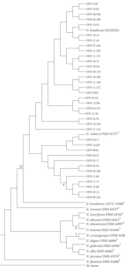
3.4. Phylogeny. Primers Noc1 and Noc2 amplified the
expected 606-bp fragment of the rrs gene for all the col-lection strains. Phylogenetic trees (Figure 1) based upon rrs showed homogeneity within clinical strains of N. brasiliensis. For this, the rrs gene is not relevant to show intraspecies diversity.
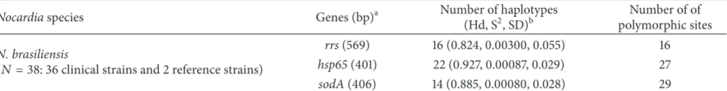
In addition, based upon the concatenation of sodA and
hsp65 housekeeping genes, the phylogenetic tree generated
(Figure 2) had several distinct genotypes: (i) genotype 1 containing clinical strains, (ii) genotype 2 harboring some clinical strains, and (iii) genotype 3 harboring some clinical strains and N. brasiliensis ATCC 19296T. For the tropical N.
brasiliensis HUJEG01 strain, it is observed that it does not
N. brasiliensis HUJEG01 M. leprae N. vulneris DSM 457374 N. iowensis DSM 451974 N. altamirensis DSM 449974 N. boironii DSM 1016964 N. globerula DSM 445964 N. tenerifensis DSM 447044 N. cyriacigeorgica DSM 444844 N. alba DSM 446844 N. elegans DSM 448904 N. abscessus DSM 444324 N. farcinica DSM 435784 N. fluminea DSM 444894 N. brasiliensis ATCC 192964 OFN 3247 OFN 10.82 OFN 08.188 OFN 09.280 OFN 10.93 OFN 10.16 OFN 11.44 OFN 07.168 OFN 11.189 OFN 11.151 OFN 10.35 OFN 45762 OFN 08.178 OFN 10.180 OFN 11.140 OFN 11.172 OFN 2985 OFN 05.63 OFN 12786 OFN 04.101 OFN 12.28 OFN 02.56 OFN 10.146 OFN 11.116 OFN 09.71 OFN 14229 OFN 9044 OFN 04.21 OFN 05.77 OFN 05.64 OFN 09.106 OFN 11.80 OFN 11.73 OFN 12.08 OFN 05.12 OFN 09.244 6 5 6 52
Figure 1: Phylogenetic distribution of rrs gene of 36 N. brasiliensis clinical strains analyzed in this study using neighbour-joining method, Kimura’s two-parameter model, and bootstrap of 1000. Only values of bootstrap significance greater than 50% (Seaview) were reported.
OFN 45762 OFN 02.56 OFN 10.180 OFN 11.172 OFN 05.63 OFN 04.101 OFN 11.151 OFN 08.188 OFN 10.146 OFN 10.93 OFN 09.280 OFN 2985 OFN 11.189 OFN 3247 OFN 10.16 OFN 12.28 OFN 10.82 OFN 08.178 OFN 07.168 OFN 9044 OFN 14229 OFN 09.71 OFN 11.44 OFN 11.140 OFN 10.35 OFN 12786 OFN 11.116 OFN 05.77 OFN 09.106 OFN 05.64 OFN 04.21 OFN 05.12 OFN 11.80 OFN 11.73 OFN 09.244 OFN 12.08 99 99 72 90 59 58 54 54 53 68 69 81 52 51 92 83 G1 G3 G2 4 4 4 4 4 4 4 4 4 4 4 4 HUJEG0145737 DSM 45197 DSMDSM44997 101696 DSM 44596 DSM 44704 DSM 44484 DSM 44684 DSM 44890 DSM 44432 DSM 43578 DSM 44489 DSM 19296 ATCC 4 N. brasiliensis M. leprae N. vulneris N. iowensis N. altamirensis N. boironii N. globerula N. tenerifensis N. cyriacigeorgica N. alba N. elegans N. abscessus N. farcinica N. fluminea N. brasiliensis
Figure 2: Phylogenetic distribution of concatenation sodA-hsp65 genes of 36 N. brasiliensis clinical strains analyzed in this study using neighbour-joining method, Kimura’s two-parameter model, and bootstrap of 1000. Only values of bootstrap significance greater than 50% were reported.
in the tree between genotypes 1 and 2. This distribution of clinical strains of N. brasiliensis in 3 different genotypes shows an intraspecies diversity rather important. To better understand the polymorphism showed by phylogenetic trees, we studied the percentages of the similarities between the sequences. The average percentages of similarities based on the rrs gene (Table 2) range from 99.39% to 99.57% between the clinical strains and the 2 reference strains of
N. brasiliensis (type and tropical strains). According to the
CLSI, the similarity percentage needed for identification at species level must be greater than or equal to a threshold of 99.6% [19]. The clinical strains that showed a similarity percentage lower than this threshold for both reference strains of N. brasiliensis were anyway considered as belonging to this species because no higher similarity percentage was obtained for any other species. In the same way, the N.
vulneris type strain was also revealed to be close to clinical
strains according to the average of percentage of similarity (98.77%). Between the 2 reference strains of N. brasiliensis the percentage of similarity is higher, up to 99.82%. The percentages of similarities based on the concatenation of the sodA-hsp65 genes (Table 2) decrease and range now from 97.99% to 99.19% between the clinical strains and the 2 reference strains of N. brasiliensis. Between the type and reference strains of N. brasiliensis the percentage of similarity does not reach 99% this time. The comparison of the 3 genotypes between them (based on the representation of each genotype by 3 clinical strains) by using sodA-hsp65 genes shows that genotypes 2 and 3 are closer to each other (98.97% of similarity). The average of the percentages of similarity between genotypes 1 and 2 were 97.97%. and 98.28% between genotypes 1 and 3. Finally this value goes up to 98.97% between genotypes 2 and 3. This means that the more distant genotypes between them are 1 and 2 and the closer ones are 2 and 3.
In parallel, an epidemiological study based on the clinical files was carried out, and the data were presented in Table 1. In order to know the link between the genetic diversity and the tropism of the clinical strains, a superposition of data was made between the phylogenetic tree obtained by the con-catenation of sodA and hsp65 and the tropism of the clinical strains (Figure 2). Thus, we can see that in genotypes 2 and 3 we have almost all the clinical strains that have a cutaneous tropism except the 08.188 strain which has a subcutaneous tropism. Regarding genotype 1 it is more heterogeneous with various tropism: (i) pulmonary, (ii) cerebral, and (iii) cutaneous. Regarding the immunocompetence of patients, we have only 4 patients who have immunodepression factors, whose strains are in genotype 1 except the 10.93 strain which is in genotype 3.
3.5. Analysis of rrs, hsp65, and sodA Genes Polymorphism.
The 36 clinical strains and 2 reference strains of N. brasiliensis studied showed (i) for rrs gene 16 polymorphic sites sharing 16 haplotypes and showing a Hd of 0.824; (ii) for hsp65 gene, 27 polymorphic sites and up to 22 different haplotypes with a Hd of 0.927; and, (iii) for sodA gene, up to 29 polymorphic sites sharing 14 haplotypes having a Hd of 0.885 (Table 3).
4. Discussion
Nocardia spp. are common soil-inhabiting bacteria that
fre-quently infect humans through traumatic injuries or inhala-tion routes and cause infecinhala-tions, such as actinomycetoma and nocardiosis, respectively. N. brasiliensis is the main aetiological agent of actinomycetoma in various countries [23]. The input data used in this study highlight the existence of indigenous cases of cutaneous and subcutaneous (such as actinomycetoma) nocardiosis caused by N. brasiliensis in France. Moreover, we can observe that N. brasiliensis is also responsible for severe cases of disseminated nocardiosis in immunocompromised patients (pulmonary and cerebral cases).
To determine whether there is an association between clinical tropism of strains and their genetic profile we per-formed genetic characterization of 36 indigenous cases of N.
brasiliensis that happened in France.
The three culture media allow the growth of clinical strains of N. brasiliensis. However, on Bennett’s medium more abundant and fast growth (48 hours) was observed. But the downside of this medium is its inaccessibility in the hospital because it is not marketable. Middlebrook medium shows strong growth but also it was slightly slower (72 hours). This medium is very expensive and not accessible to all budgets. However, it is an interesting alternative in isolating
Nocardia from a complex sample. It is a selective medium of
Mycobacteria, which promotes the growth of some Nocardia to the detriment of other external bacteria or commensal flora that may be in the biological sample analyzed. The BCP medium, used routinely in hospitals for Gram-negative bacteria, has interesting results with good fast growth (48 hours). It would therefore be advisable to use it as isolation medium for urgent cases, by the speed of growth.
Antibiograms results show resistance of most of the clin-ical strains to imipenem. This can pose therapeutic problems since it is part of molecules proposed during a phase of a general treatment for nocardiosis [24]. However, all clinical strains of N. brasiliensis were sensible to SXT and would be an effective molecule during treatments. The sensibility of
N. brasiliensis type strain to this antibiotic has already been
observed by Gilquin et al. [9].
Our study confirms that all clinical strains of N.
brasilien-sis are capable of degrading casein except 12.28 clinical strain.
As shown by Seol et al. the N. brasiliensis type strain is able to degrade casein as well [25]. However, the test on the reference strains reveals that N. vulneris, N. tenerifensis, N. boironii, and N. iowensis are also capable of degrading casein in the same way as N. brasiliensis, as also shown by Gilquin et al. [9]. This type of test is used in some countries without the necessary molecular biology tools to identify N. brasiliensis. But, now, they must be aware that with this test we target several species of clade N. brasiliensis. So, it is no longer a criterion of identification proper to N. brasiliensis.
Phylogenetic tree based on the rrs gene sequence of our collection showed a low genetic diversity resulting in low polymorphism sequence. In addition, we can note that N.
vulneris DSM 45737T, identified as a new species by Lasker et al., present a genetic sequence very close to N. brasiliensis
T a ble 2: P er cen ta ge o f simi la ri ty exp ress ed in in ter va la nd me an fo r so dA -hs p6 5 an d rr s ge ne s. N. br as il iens is HUJEG01 N. br as il iens is AT C C 19 29 6 T N. vu ln er is DS M 4 57 37 T so dA -hs p6 5 rr s sodA -hs p6 5 rr s sodA -hs p6 5 rrs N. br as il iens is AT C C 19 29 6 T Simila ri ty av era ge (%) 98.5 1 9 9.8 2 ----N. vu ln er is DS M 4 57 37 T 98.3 8 98.7 3 98.5 1 98.5 5 -N. al ta m ir ens is DS M 4 49 97 T 95.9 1 98.2 4 95.41 98.07 95.9 1 97 .47 N .b oir onii DS M 1016 9 6 T 9 6.5 3 98.07 9 6 .2 8 97. 89 9 6 .1 5 97. 29 N. io wens is DS M 4 51 97 T 94.9 1 97 .8 9 95.29 97 .7 1 94.5 4 97 .6 4 N. te n er if ens is DS M 4 47 0 4 T 9 6.2 8 9 6.67 9 6.0 3 9 6.4 9 95.9 1 9 6.0 3 Ge n o ty pe 1 Simi la ri ty ra n ge (%) (97 .7 7–98.3 8) -(97 .7 7–98.26) -(97 .5 2–98.1 4) -Simi la ri ty av era ge (%) 98.01 -97 .9 9 -97 .67 -Ge n o ty pe 2 Simi la ri ty ra n ge (%) (98.3 9–98.88) -(98.88–9 9.3 8) -(98.6 3–98.88) -Simi la ri ty av era ge (%) 98.7 3 -9 9.1 9 -98.80 -Ge n o ty pe 3 Simi la ri ty ra n ge (%) (98.1 4–98.88) -(98.26–9 9.0 0) -(98.01–98.7 6) -Simi la ri ty av era ge (%) 98.6 3 -98.7 6 -98.5 0 -All clinical st ra in s Simi la ri ty ra n ge (%) -(98.57–10 0) -(98.3 8–9 9.8 2) -(98.01–9 9.0 9) Simila ri ty av era ge (%) -9 9. 57 -9 9.3 9 -98.7 7
Table 3: DNA polymorphism of rrs, hsp65, and sodA genes from clinical N. brasiliensis strains isolated in France.
Nocardia species Genes(bp)a Number of haplotypes
(Hd, S2, SD)b
Number of of polymorphic sites
N. brasiliensis
(𝑁 = 38: 36 clinical strains and 2 reference strains)
rrs (569) 16 (0.824, 0.00300, 0.055) 16
hsp65 (401) 22 (0.927, 0.00087, 0.029) 27
sodA (406) 14 (0.885, 0.00080, 0.028) 29
aResulting fragment size without the primers sequences;bHd: haplotype (gene) diversity, S2: variance of haplotype diversity, and SD: standard deviation of
haplotype diversity.
strains [26] with percentages of similarities on average greater than 98%.
Analysis of the phylogenetic tree (Figure 2) based on the MLSA by the concatenation of sodA and hsp65 housekeeping genes showed that the isolates are surprisingly classified according to 3 genotypes. These groups were formed upon similarity percentages and existing phylogenetic distances between the sequences of the strains studied. Genotype 1 con-cerns a well-defined cluster containing 9 clinical strains only which is sustained by a bootstrap of 81%. This genotype hosts all the cases of pulmonary forms (3), the sole cerebral form, and almost all the cases of immunocompromised patients (3 out of 4). Moreover, eight out of twelve strains susceptible to imipenem can be found in this group. There is just one remaining strain in this group not presenting this kind of susceptibility. The reason may be an acquired resistance to this drug due to a previous treatment. This well-defined genotype evokes the possible existence of another species or a strong variability in this case. This may have been caused by environmental pressures in the ecosystem of these isolates which may have resulted in the selection of strains that may have acquired, by mutations or genetic transfer with other microorganisms, new virulence characters different from that of the strain type N. brasiliensis. Regarding genotypes 2 and 3, they include clinical strains and N. brasiliensis ATCC 19296T type strain. However, the discrimination between these 2 genotypes is less clear than that with genotype 1 because of being in weak bootstrap that is less than 50. The genetic differences do not allow distinguishing them properly and their phenotypic behavior remains similar. Then, it would be interesting to study on another gene capable of generating more divergences, for example, gyrB and rpoB genes, which have already successfully been used for studying the polymorphism of some other Nocardia species [27, 28].
Concerning the percentage of similarity between the type and tropical strain of N. brasiliensis, it is 99.82% according to the rrs and goes down to 98.51% with the concatenation of
sodA and hsp65. The fact of highlighting a greater
dissimilar-ity with the concatenation between the type and tropical N.
brasiliensis strain shows the advantage of the use of 2 markers
like sodA and hsp65 vis-`a-vis the rrs. The discriminatory power of these two genes may be explained by the presence of more polymorphic sites (hsp65: 27; sodA: 29) than in the case of rrs gene (16) and also by having Hd values higher than that of rrs gene (hsp65: 0.927; sodA: 0.885; rrs: 0.824).
It would be interesting to identify the genes involved in the virulence of different genotypes, including those of actinomycetoma. Interesting leads can be considered: (i) as
identification of virulence genes expressed using the RNAseq method or (ii) to identify noncoding RNAs [23]. In addition, to genomically distinguish N. brasiliensis and N. vulneris a specific PCR to N. vulneris, using a specific gene of the species, should be developed.
Conflicts of Interest
The authors declare that they have no conflicts of interest.
Acknowledgments
The authors thank Audrey Dubost for her technical support in bioinformatics analysis.
References
[1] V. Kandi, “Human Nocardia Infections: A Review of Pulmonary Nocardiosis., Human Nocardia Infections: A Review of Pul-monary Nocardiosis,” Cureus Cureus, vol. 7, no. 8, pp. e304– e304, 2015.
[2] S. Maraki, S. Chochlidakis, E. Nioti, and Y. Tselentis, “Pri-mary lymphocutaneous nocardiosis in an immunocompetent patient,” Annals of Clinical Microbiology and Antimicrobials, vol. 3, article no. 24, 2004.
[3] M. A. Saubolle and D. Sussland, “Nocardiosis: review of clinical and laboratory experience,” Journal of Clinical Microbiology, vol. 41, no. 10, pp. 4497–4501, 2003.
[4] B. A. Brown-Elliott, J. M. Brown, P. S. Conville, and R. J. Wallace Jr., “Clinical and laboratory features of the Nocardia spp. based on current molecular taxonomy,” Clinical Microbiology Reviews, vol. 19, no. 2, pp. 259–282, 2006.
[5] V. Rodriguez-Nava, D. Lebeaux, O. Lortholary, P. Boiron, and Nocardia., “Nocardia. Pr´ecis de Bact´eriologie Clinique,”
Nocardia, no. 2, 2007.
[6] P. Boiron, F. Provost, and B. Dupont, “Technical protocols,” In Methodes de laboratoire pour le diagnostic de la nocardiose, Institut Pasteur, Paris, France, 1993, 107-126.
[7] A. Betr´an, M. C. Villuendas, A. Rezusta, J. Pereira, M. J. Revillo, and V. Rodr´ıguez-Nava, “Clinical significance, antimicrobial susceptibility and molecular identification of Nocardia species isolated from children with cystic fibrosis,” Brazilian Journal of
Microbiology, vol. 47, no. 3, pp. 531–535, 2016.
[8] A. Ram´ırez-Radilla, V. Rodr´ıguez-Nava, H. V. Silva-Rojas, M. Hern´andez-Tellez, H. Sandoval, and N. Ram´ırez-Dur´an, “Phylogenetic identification of Nocardia brasiliensis strains isolated from actinomycetoma in Mexico State using species-specific primers,” Journal de Mycologie M´edicale, vol. 21, no. 2, pp. 113–117, 2011.
[9] J. M. Gilquin, B. Riviere, V. Jurado et al., “ First Case of Actinomycetoma in France Due to a Novel ,” mSphere, vol. 1, no. 6, p. e00309-16, 2016.
[10] V. Rodr´ıguez-Nava, A. Couble, G. Devulder, J.-P. Flandrois, P. Boiron, and F. Laurent, “Use of PCR-restriction enzyme pattern analysis and sequencing database for hsp65 gene-based identification of Nocardia species,” Journal of Clinical
Microbiology, vol. 44, no. 2, pp. 536–546, 2006.
[11] Susceptibility testing of Mycobacteria, Nocardiae, and other
aerobic actinomycetes, Approved Standard, vol. 56, Clinical and
Laboratory Standards Institute, Wayne, PA, USA, 2011, M24-A2.
[12] “CA-SFM, “Les recommandations du Comit´e de
l’Antibiogramme de la Soci´et´e Franc¸aise de Microbiologie,”
Paris: Soci´et´e Franc¸aise de Microbiologie, 2013.
[13] M. Goodfellow, “The genus Nocardia Trevisan,” in Topley and
Wilson’s microbiology and microbial infections, A. Balows and B.
I. Duerden, Eds., pp. 464–489, Edward Arnold, London, UK, 2 edition, 1998.
[14] M. Goodfellow, The family Nocardiaceae, The prokaryotes, Springer, New York, NY, USA, 2 edition, 1992, 1188–1213. [15] M. Goodfellow and M. P. Lechevalier, “Genus Nocardia
Tre-visan,” in Bergey’s manual of systematic bacteriology, S. T. Williams, M. E. Sharpe, and J. G. Holt, Eds., vol. 4, pp. 2350– 2361, Lippincott Williams & Wilkins, Baltimore, MD, USA, 1989.
[16] A. Telenti, F. Marchesi, M. Balz, F. Bally, E. C. B¨ottger, and T. Bodmer, “Rapid identification of mycobacteria to the species level by polymerase chain reaction and restriction enzyme analysis,” J. Clin. Microbiol, vol. 31, no. 2, pp. 175–178, 1993. [17] K. S´anchez-Herrera, H. Sandoval, D. Mouniee et al., “Molecular
identification of Nocardia species using the sodA gene: Iden-tificaci´on molecular de especies de Nocardia utilizando el gen sodA.,” New Microbes and New Infections, vol. 19, pp. 96–116, 2017.
[18] C. Y. Turenne, M. Semret, D. V. Cousins, D. M. Collins, and M. A. Behr, “Sequencing of hsp65 distinguishes among subsets of the Mycobacterium avium complex,” Journal of Clinical
Microbiology, vol. 44, no. 2, pp. 433–440, 2006.
[19] CLSI, “Interpretive Criteria for Identification of Bacteria and Fungi by DNA Target Sequencing,” in Approved Guideline. CLSI
document MM18- A. Wayne, PA, vol. CLSI document MM18- A,
Clinical and Laboratory Standards Institute, Wayne, PA, USA, 2008.
[20] J. A. Soddell, F. M. Stainsby, K. L. Eales, R. M. Kroppenstedt, R. J. Seviour, and M. Goodfellow, “Millisia gen. nov., sp. nov., an actinomycete isolated from activated sludge foam,”
Interna-tional Journal of Systematic and Evolutionary Microbiology, vol.
56, no. 4, pp. 739–744, 2006.
[21] V. A. Steingrube, R. W. Wilson, B. A. Brown et al., “Rapid identification of clinically significant species and taxa of aerobic actinomycetes, including Actinomadura, Gordona, Nocardia, Rhodococcus, Streptomyces, and Tsukamurella isolates, by DNA amplification and restriction endonuclease analysis,”
Jour-nal of Clinical Microbiology, vol. 35, no. 4, pp. 817–822, 1997.
[22] J. Rozas, A. Ferrer-Mata, J. C. S´anchez-DelBarrio et al., “DnaSP 6: DNA Sequence Polymorphism Analysis of Large Data Sets,”
Molecular Biology and Evolution, vol. 34, no. 12, pp. 3299–3302,
2017.
[23] J. S. Cruz-Rabad´an, J. Miranda-R´ıos, G. Esp´ın-Ocampo, L. J. M´endez-Tovar, H. R. Maya-Pineda, and F. Hern´andez-Hern´andez, “Non-coding RNAs are differentially expressed by
Nocardia brasiliensis in vitro and in experimental actinomyce-toma,” The Open Microbiology Journal, vol. 11, pp. 112–125, 2017. [24] S. Valdezate, N. Garrido, G. Carrasco et al., “Epidemiology and susceptibility to antimicrobial agents of the main Nocardia species in Spain,” Journal of Antimicrobial Chemotherapy, vol. 72, no. 3, pp. 754–761, 2017.
[25] C.-A. Seol, H. Sung, D.-H. Kim, M. Ji, Y.-P. Chong, and M.-N. Kim, “The first korean case of disseminated mycetoma caused by nocardia pseudobrasiliensis in a patient on long-term corticosteroid therapy for the treatment of microscopic polyangiitis,” Annals of Laboratory Medicine, vol. 33, no. 3, pp. 203–207, 2013.
[26] B. A. Lasker, M. Bell, H.-P. Klenk, C. Spr¨oer, P. Schumann, and J. M. Brown, “Nocardia vulneris sp. nov., isolated from wounds of human patients in North America,” Antonie van
Leeuwenhoek-Journal of Microbiology, vol. 106, no. 3, pp. 543–553, 2014.
[27] L. R. McTaggart, S. E. Richardson, M. Witkowska, and S. X. Zhang, “Phylogeny and identification of Nocardia species on the basis of multilocus sequence analysis,” Journal of Clinical
Microbiology, vol. 48, no. 12, pp. 4525–4533, 2010.
[28] G. Carrasco, S. Valdezate, N. Garrido, P. Villal´on, M. J. Medina-Pascual, and J. A. S´aez-Nieto, “Identification, typing, and phylogenetic relationships of the main clinical nocardia species in spain according to their gyrb and rpob genes,” Journal of
Stem Cells
International
Hindawi www.hindawi.com Volume 2018 Hindawi www.hindawi.com Volume 2018 INFLAMMATIONEndocrinology
International Journal ofHindawi www.hindawi.com Volume 2018 Hindawi www.hindawi.com Volume 2018
Disease Markers
Hindawi www.hindawi.com Volume 2018 BioMed Research InternationalOncology
Journal of Hindawi www.hindawi.com Volume 2013 Hindawi www.hindawi.com Volume 2018Oxidative Medicine and Cellular Longevity
Hindawi
www.hindawi.com Volume 2018
PPAR Research
Hindawi Publishing Corporationhttp://www.hindawi.com Volume 2013 Hindawi www.hindawi.com
The Scientific
World Journal
Volume 2018 Immunology Research Hindawi www.hindawi.com Volume 2018 Journal ofObesity
Journal of Hindawi www.hindawi.com Volume 2018 Hindawi www.hindawi.com Volume 2018 Computational and Mathematical Methods in Medicine Hindawi www.hindawi.com Volume 2018Behavioural
Neurology
Ophthalmology
Journal of Hindawi www.hindawi.com Volume 2018Diabetes Research
Journal ofHindawi
www.hindawi.com Volume 2018
Hindawi
www.hindawi.com Volume 2018
Research and Treatment
AIDS
Hindawi
www.hindawi.com Volume 2018 Gastroenterology Research and Practice
Hindawi www.hindawi.com Volume 2018