HAL Id: hal-01129777
https://hal.archives-ouvertes.fr/hal-01129777
Submitted on 29 May 2020
HAL is a multi-disciplinary open access
archive for the deposit and dissemination of
sci-entific research documents, whether they are
pub-lished or not. The documents may come from
teaching and research institutions in France or
abroad, or from public or private research centers.
L’archive ouverte pluridisciplinaire HAL, est
destinée au dépôt et à la diffusion de documents
scientifiques de niveau recherche, publiés ou non,
émanant des établissements d’enseignement et de
recherche français ou étrangers, des laboratoires
publics ou privés.
Asthenozoospermia and Alters Spermatozoa
Morphology
Pauline Tartarin, Edith Guibert, Aminata Toure, Claire Ouiste, Jocelyne
Leclerc, Nieves Sanz, Sylvain Brière, Jean-Louis Dacheux, Bernadette Delaleu,
Judith R. Mcneilly, et al.
To cite this version:
Pauline Tartarin, Edith Guibert, Aminata Toure, Claire Ouiste, Jocelyne Leclerc, et al.. Inactivation
of AMPK alpha 1 Induces Asthenozoospermia and Alters Spermatozoa Morphology. Endocrinology,
Endocrine Society, 2012, 153 (7), pp.3468 - 3481. �10.1210/en.2011-1911�. �hal-01129777�
Inactivation of AMPK
␣1 Induces Asthenozoospermia
and Alters Spermatozoa Morphology
Pauline Tartarin, Edith Guibert, Aminata Touré, Claire Ouiste, Jocelyne Leclerc,
Nieves Sanz, Sylvain Brière, Jean-Louis Dacheux, Bernadette Delaleu,
Judith R. McNeilly, Alan S. McNeilly, Jean-Pierre Brillard, Joëlle Dupont,
Marc Foretz, Benoit Viollet, and Pascal Froment
Unité Mixte de Recherche (UMR) 6175 (P.T., E.G., C.O., S.B., J.-L.D., B.D., J.P.B., J.D., P.F.), Physiologie de la Reproduction et des Comportements (Institut National dela Recherche Agronomique/Centre National dela Recherche Scientifique/Université Tours/Haras Nationaux), 37380 Nouzilly, France; Institut National de la Santé et de la Recherche Médicale Unité 1016, Institut Cochin (A.T., J.L., N.S., M.F., B.V.); and CNRS, UMR 8104 (A.T., J.L., N.S., M.F., B.V.); and Université Paris Descartes (A.T., J.L., N.S., M.F., B.V.), Sorbonne Paris Cité, 75014 Paris, France; and Medical Research Council Human Reproductive Sciences Unit (J.R.M., A.S.M.), University of Edinburgh, Queen’s Medical Research Institute, Edinburgh EH16 4TJ, Scotland, United Kingdom
AMP-activated protein kinase (AMPK), a key regulator of cellular energy homeostasis, is present in metabolic tissues (muscle and liver) and has been identified as a modulator of the female reproductive functions. However, its function in the testis has not yet been clearly defined. We have investigated the potential role of AMPK in male reproduction by using transgenic mice lacking the activity of AMPK catalytic subunit␣1 gene [␣1AMPK knockout (KO)]. In the testis, the ␣1AMPK subunit is expressed in germ cells and also in somatic cells (Sertoli and Leydig cells).␣1AMPK KO male mice show a decrease in fertility, despite no clear alteration in the testis morphology or sperm production. However, in␣1AMPK⫺/⫺mice, we demonstrate that spermatozoa have structural abnormalities and are less motile than in control mice. These spermatozoa alterations are associ-ated with a 50% decrease in mitochondrial activity, a 60% decrease in basal oxygen consumption, and morphological defects. The␣1AMPK KO male mice had high androgen levels associated with a 5-and 3-fold increase in intratesticular cholesterol 5-and testosterone concentrations, respectively. High concentrations of proteins involved in steroid production (3-hydroxysteroid dehydrogenase, cyto-chrome steroid 17 alpha-hydroxylase/17,20 lysate, and steroidogenic acute regulatory protein) were also detected in␣1AMPK⫺/⫺testes. In the pituitary, the LH and FSH concentrations tended to be lower in␣1AMPK⫺/⫺male mice, probably due to the negative feedback of the high testosterone levels. These results suggest that total␣1AMPK deficiency in male mice affects androgen production and quality of spermatozoa, leading to a decrease in fertility. (Endocrinology 153: 3468 –3481, 2012)
I
nteraction between energy balance and reproductive
sta-tus has been investigated for several decades. In humans,
anorexia and obesity can harm ovarian function and
sper-matogenesis (for review see Refs. 1 and 2). A modification
in quantity and/or quality of nutrition in female mammals
could influence ovarian cycles and the weaning-estrus
in-terval (for review, see Ref. 3).
During the last 15 yr, several messengers of the fuel
gauge have been identified to regulate fertility. Mice,
de-ficient or overexpressing hormones produced by adipose
ISSN Print 0013-7227 ISSN Online 1945-7170 Printed in U.S.A.
Copyright © 2012 by The Endocrine Society
doi: 10.1210/en.2011-1911 Received October 25, 2011. Accepted April 13, 2012. First Published Online May 11, 2012
Abbreviations: ACC, Acetyl coenzyme A carboxylase; AMPK, AMP-activated protein kinase; CREB, cAMP response element-binding protein; GATA4, transcription factor GATA-4; 3HSD, 3-hydroxysteroid dehydrogenase; JO2, oxygen consumption; KO, knockout; LKB1,
liver kinase B1; LXR, liver X receptor; MARK, microtubule affinity-regulating kinase; P450c17, cytochrome P450c17, 17 alpha-hydroxylase/17,20 lysate; PCNA, proliferation cell nuclear an-tigen; PGC-1␣, peroxisome-proliferator-activated receptor ␥ coactivator 1␣; RXR, retinoid X receptor; SER, smooth endoplasmic reticulum; SNRK, sucrose nonfermenting related kinase; TSSK, testis specific serine kinase; TUNEL, terminal deoxynucleotidyl transferase-mediated de-oxy-UTP nick end labeling; VASA, mouse homolog Drosophila Vasa; X-gal, 5-bromo-4-chloro-3-indoyl--D-galactopyranoside.
tissue such as leptin, adiponectin (4, 5), or insulin signaling
molecules (6, 7) show a decrease in fertility. At the cellular
level, several proteins have been described as key factors
involved in the relations between the energy homeostasis
and reproductive functions. These proteins are nuclear
receptors, kinases sensitive to the ATP to ADP ratio, or
transcription factors. They are involved in the control of
lipid and glucose metabolism. Several knockout (KO)
mice models have shown a relationship between
lipid/cho-lesterol metabolism and male fertility. For example, the
nuclear oxysterol receptor [liver X receptor (LXR)] forms
a heterodimer with the retinoid X receptor (RXR) and
activates gene transcription involved in cholesterol
trans-port such as the ATP-binding cassette transtrans-porters (8).
The double knockout of
␣-LXR /-LXR subunits in mice
leads to a disrupted spermatogenesis and a vacuolization
of seminiferous tubules growing with age. Spermatozoa
present structure disturbance such as hairpin flagella and
testes with high levels of intratesticular cholesterol
asso-ciated with low testosterone levels (9 –11). RXR
 is a
nu-clear receptor that interacts as a heterodimer with
differ-ent nuclear receptors such as retinoic acid receptor, LXR,
peroxisome proliferator-activated receptor, and modulate
cholesterol transport (12). KO mice lacking RXR
 have a
progressive testicular degeneration, an accumulation of
lipid droplets in Sertoli cells, and altered spermatozoa due
to malformations of acrosome and mitochondrial sheaths
(13). Similarly, the hormone-sensitive lipase disruption, a
cholesterol esterase (14), results in a marked reduction in
spermatid number and severe acrosome disturbances
lead-ing to sterility (15). Thus, control of the lipid metabolism
is closely connected to male gametogenesis. A key
regu-lator of cellular energy and cholesterol homeostasis, the
5
⬘-AMP-activated protein kinase (AMPK), has also been
de-tected in reproductive tissue (16). AMPK plays an important
role in the fatty acid and cholesterol homeostasis (17–19).
Indeed, activation of AMPK inhibits expression of
acetyl-coenzyme A carboxylase (ACC), fatty acid synthase (FAS),
and 3-hydroxy-3-methylglutaryl (HMG)-coenzyme A
re-ductase (HMGR), which are rate-limiting enzymes for
cho-lesterol and fatty acid biosynthesis. AMPK also activates
glu-cose transporter 4 (GLUT4) translocation, leading to an
increase in glucose intake (for review, see Ref. 20).
This serine threonine kinase, also named protein kinase
AMP-activated (PRKA) or sucrose nonfermenting 1
(SNF1), is ubiquitous, being present in plants (21), yeast
(22), and the animal kingdom [fish (23), birds (24), and
mammals (25)]. AMPK is a heterotrimeric complex
com-posed of a catalytic subunit
␣ and two regulatory subunits
 and ␥. The ␣-subunit is present in two isoforms, ␣1 and
␣2, and two and three isoforms for - and ␥-subunits,
respectively. AMPK is sensitive to the AMP to ATP ratio,
and its activation allows turning off of ATP-consuming
processes and stimulation of catabolic pathways.
Activa-tion of AMPK requires the phosphorylaActiva-tion of
␣-subunit
in threonine 172. The binding of AMP in regulatory
sub-units could activate AMPK directly by allosteric activation
(16, 26). Upstream kinases such as liver kinase B1 (LKB1)
and Ca
2⫹calmodulin-dependent protein kinase kinase 1
or 2 (CaMKK1 or CaMKK2) are clearly identified to
phos-phorylate AMPK (27, 28), but other upstream kinases like
TGF-
-activated kinase (TAK1) (29) or kinase suppressor
of Ras (KSR2) (30) could up-regulate AMPK activity.
Fur-thermore, AMPK is activated by metabolic hormones
lep-tin (31) and adiponeclep-tin (32), two adipokines involved in
energy homeostasis and whose receptors are also
ex-pressed in the ovary and testis.
By homology analysis of the catalytic domain of the
AMPK sequence, Manning et al. (33) identified 12
AMPK-related kinases [brain-specific kinase 1, brain-specific
ki-nase 2, nuclear related kiki-nase 1, nuclear
AMPK-related kinase 2, Qin-induced kinase, salt inducible
kinase, QSK (also named salt-inducible kinase 3,
salt-in-ducible kinase, microtubule affinity-regulating kinase
(MARK)1, MARK2, MARK3, MARK4, and maternal
embryonic leucine zipper kinase] and eight other kinases
less related to AMPK [noninducible immunity 1, sucrose
nonfermenting related kinase (SNRK), testis specific
ser-ine kinase (TSSK)1, TSSK2, TSSK3, TSSK4, small serser-ine/
threonine kinase, and hormonally upregulated neu-tumor
associated kinase]. Most of them are activated by LKB1
(33–35) and four of these AMPK-related-kinases are
found in the testis (MARK2, NIM1, SNRK, and TSSK1)
(35) (36). However, the role of these AMPK-related
ki-nases has not been clearly defined (37).
Activation of AMPK, by two synthetic drugs,
5-aminoimidazole-4-carboxamide-1-
-d-ribofuranoside
(AICA riboside) or metformin (an insulin sensitizer drug),
decreases steroid production in ovarian granulosa cells
(38). In human granulosa cells, stimulation of adiponectin
receptors (39 – 42) increases the IGF-I-induced steroid
production (43, 44). In mouse oocytes, phosphorylation
of AMPK provides a meiotic maturation signal that
im-proves meiotic resumption (45). Overall, these studies
sug-gest that AMPK is closely linked to female fertility.
AMPK may have a role in the control of male fertility.
Indeed, all AMPK subunits are found in the rat testis;
how-ever, the
␣1AMPK complexes account for most (about
75%) of the total AMPK activity in the testis (16). In
ad-dition, in vitro incubation of Sertoli cells (the nurse cells of
male germ cell lineage) with synthetic AMPK activators
leads to an increase in the production of lactate, the energy
substrate for germ cells (46). Moreover, in mice, the loss
of activity of AMPK kinase LKB1 (47) or the
AMPK-re-lated kinase MARK2 (36) induces alteration of sperm
pro-duction and morphology leading to a repro-duction in fertility
or even sterility, suggesting possible involvement of
AMPK activity in male fertility.
To test this hypothesis, we studied a transgenic mouse
model inactivated for the
␣1AMPK subunit gene and
an-alyzed sperm parameters and Sertoli and Leydig cell
functions.
Materials and Methods
Animals
The generation and characterization of the transgenic mice have been previously described by Jørgensen et al. (48). Briefly, an IRES-geo cassette (internal ribosomal entry site--galacto-sidase and neomycin phosphotransferase fusion gene) was in-serted to delete a part of the catalytic domain of␣1AMPK sub-unit from amino acids 97–157. The genotype of the offspring was first determined by PCR as described previously (48). Mice were maintained under standard conditions of light (12 h light, 12 h darkness) and temperature with ad libitum access to food and water. Animal studies described herein were reviewed and ap-proved (agreement no. 75-886) by the Directeur Départemental des Services Vétérinaires of the Préfecture de Police de Paris. The fertility assessment was estimated by counting the number of pups per litter of the following breeding: male ␣1AMPK⫹/⫺ mated with female␣1AMPK⫹/⫺, male␣1AMPK⫺/⫺mated with female␣1AMPK⫹/⫺, and male␣1AMPK⫺/⫺mated with female
␣1AMPK⫺/⫺.
Blood collection and tissue removal
Blood samples were recovered by intraorbital puncture in EDTA, centrifuged for 15 min at 3000 ⫻ g, and plasma was stored at⫺20 C. For both genotypes, animals were killed by cervical dislocation, and liver, pituitary, epididymis, testes, and spermatozoa were immediately recovered and fixed in Bouin solution for histological studies or stored at⫺80 C. Total body weight was averaged before killing, and testes were weighed im-mediately after recovery.
Morphometry of testis
The testis volume was obtained using the formula: 0.71⫻ length⫻ width2
(49). Testes, once fixed, were processed in par-affin and serially sectioned at 7m slice thickness. Round trans-verse sections of seminiferous tubules diameters (n⫽ 20 mea-surements per animal, with five ␣1AMPK⫹/⫹ and six ␣1AMPK⫺/⫺mice) were measured for each testis using an ocular
measuring device (50).
Leydig cell morphometry
The percent volume occupied by the Leydig cell nuclei was counted in 25 randomly selected areas per animal, using a 441-point square lattice at⫻400 magnification. Because the Leydig cell nucleus is spherical, the volume of the nucleus of a single Leydig cell was estimated from the equation for the volume of a sphere (4/3⫻ ⫻ r3, where r is the radius of the nucleus of the
cell). The total number of Leydig cells per testis was
deter-mined from the volume occupied by all Leydig cell nuclei in the testis volume and divided by the mean Leydig cell nucleus volume. Individual Leydig cell volume was obtained from the nucleus volume and the nucleus to cytoplasm ratio. For each animal, 45– 85 Leydig cells per testis section were measured. The mean volume of the Leydig cells was calculated as de-scribed previously (51).
Isolation of testicular cells
The preparation of spermatogonia, spermatocytes, sperma-tids, and peritubular and Leydig cells used in Fig. 1 was described by Toure et al. (52), and Sertoli cells were prepared from 20-d-old mice as described previously by Froment et al. (53). Mouse Sertoli cells were cultured at 2⫻ 105
cells per well in six-well dishes in HEPES-buffered F12/DMEM (Sigma Chemical Co., St. Louis, MO) added to 1% fetal calf serum (PAA Laboratories GmbH, Pasching, Austria) at 33 C in a humidified atmosphere of 5% CO2 in air. The medium was changed every 48 h. Cells were stimulated for 15 min with 100 ng/ml ovine FSH (Sigma) or for 48 h with 5 mMmetformin (Sigma).
Histochemical staining for
-galactosidase activity
Testes were fixed in 4% paraformaldehyde in PBS for 1 h and then embedded in Tissue-Tek O.C.T., frozen at⫺80 C, and cut into 10-m sections with a cryostat. The sections were washed with PBS, incubated in PBS containing 2 mM MgCl2, 5 mM
K3Fe(CN)6, 5 mMK4Fe(CN)6, and 1 mg/ml
5-bromo-4-chloro-3-indoyl--D-galactopyranoside (X-gal) at 30 C for 24 h. The
procedure for X-gal staining has been previously described by Papaioannou and Johnson (54). Negative controls took the form of␣1AMPK⫹/⫹mice.
Immunohistochemistry
Testes embedded in paraffin were serially sectioned at 7m slice thickness. Deparaffinized sections were hydrated, micro-waved for 5 min in an antigen unmasking solution (Vector Lab-oratories, Inc., AbCys, Paris, France), and left to cool to room temperature. The sections were then washed in a PBS bath for 5 min, and to quench endogenous peroxidase activity, they were immersed in peroxidase blocking reagent for 10 min at room temperature (Dako Cytomation, Dako, Ely, UK). After two PBS baths for 5 min, nonspecific background was prevented by in-cubation in 5% lamb serum/PBS for 30 min. Finally, sections were incubated overnight at 4 C with PBS containing primary antibody against phospho-ACC (Ser79) (1:200; Upstate Biotech-nology Inc., Lake, Placid, NY), mouse homolog Drosophila Vasa (VASA) (1:200; AbCam, Cambridge, MA), transcription factor GATA-4 (GATA4) (1:200; Santa Cruz Biotechnology, Santa Cruz, CA), proliferation cell nuclear antigen (PCNA) (1:500; Santa Cruz), and phospho-cAMP response element-binding pro-tein (CREB) (Ser 133) (1:100; Cell Signaling Technologies, Bev-erly, MA). The next day, after two PBS baths for 5 min, sections were incubated with a ready-to-use labeled polymer-horseradish peroxidase antirabbit or antimouse for 30 min at room temper-ature (Dako Cytomation Envision Plus HRP system; Dako, Ely, UK). Finally, after two baths in PBS, staining was revealed by incubation with 3,3⬘-diaminobenzidine at room temperature (Sigma Fast diaminobenzidine tablet dissolved in deionized wa-ter; Sigma). Negative controls were incubated with rabbit or mouse IgG (data not shown).
The intensity of phospho-ACC and phospho-CREB staining was estimated by using the software Scion Image version 4.02 (Scion Corp., Frederick, MD). At least 50 measurements of a 25-m2area from different sections per animal (three animals
per genotype) were made, and the mean result is expressed in arbitrary units.
Detection of apoptosis
Deparaffinized sections were hydrated, permeabilized with proteinase K, and the in situ end labeling of nuclear DNA frag-mentation was performed as described in the instruction manual (FragEL kit; Calbiochem, VWR, West Chester PA). Negative controls were terminal deoxynucleotidyl transferase free.
Sperm head counts
Testes and epididymis were thawed and cut into 2-mm pieces. To isolate spermatozoa and elongated spermatid heads from the other cells, samples were then suspended in 1 ml 0.15MNaCl,
0.05% Triton X-100 buffer. Samples were homogenized and
sonicated for 30 sec. Sperm heads were counted with a hemocytometer, and after correction for sample volume and tissue weight, the concentration was determined as previously described by Jégou et al. (55).
Sperm morphology and motility
analysis
The cauda epididymis was cut into small pieces in M2 medium (Sigma) supple-mented with 1% BSA; the sperm released into the medium were incubated 10 –15 min at 37 C in 5% CO2to allow sperm diffusion.
At least 120 spermatozoa per animal were analyzed and classified as normal or atypi-cal (microcephalic, curved spermatozoa, ir-regular head, or thinned head). The sperm suspension was also diluted for quantitative assessment of motility using a Hamilton-Thorne motility analyzer (Hamilton-Hamilton-Thorne Biosciences, Beverly, MA). For each sample, sperm curvilinear velocity and sperm straight-line velocity parameters were mea-sured as indicators of sperm movement. For each mouse (n ⫽ 6), 1000 spermatozoa were analyzed at 37 C in 100-m standard counting chambers (Leja, IMV Technolo-gies, Aigle, France).
Mitochondrial activity
Mitochondrial activity was determined by JC-1 staining (Invitrogen, Cergy-Pon-toise, France). Fresh epididymal semen was incubated with 2MJC-1-M2 medium for
15 min at 37 C, and then spermatozoa were washed and examined using standard im-munofluorescence microscopy (at least 150 spermatozoa per animal were analyzed). A green fluorescence emission was detected in the presence of low mitochondrial mem-brane potential, and an orange/red fluores-cence was emitted in mitochondria with high membrane potential.
Determination of sperm mitochondrial oxygen
consumption (JO
2)
Spermatozoa were collected in M2 medium supplemented with 5 mMsodium pyruvate according to the method previously
described by Mukai and Okuno (56) and were resuspended at a density of 2⫻ 106spermatozoa/ml. The JO
2(picomoles O2per
second per 106
cells) of the cell suspension was recorded at 37 C with an Oroboros Oxygraph-2k (Oroboros, Innsbruck, Aus-tria), and data were acquired with DatLab4 software. Basal cell respiration was determined, and then oligomycin (0.5 g/ml) and increasing concentrations of carbonyl cyanide m-chlorophe-nylhydrazone (1–2M) were subsequently added to determine
the oligomycin-sensitive JO2(basal JO2minus
oligomycin-in-sensitive JO2), which represents the cellular oxygen
consump-tion related to ATP synthesis and the maximal (uncoupled) re-spiratory rate, respectively.
FIG. 1. Expression of␣1AMPK in testis. A1 and A2, Analysis of the -galactosidase activity
and expression in the testis (gene reporter inserted in␣1AMPK gene) (48) (black arrowheads localized peritubular cells); A2, negative controls were analyzed in␣1AMPK wild-type mice (absence of the gene reporter). Scale bar, 20m. B, Western blot analysis of ␣1AMPK and ␣2AMPK subunits in the whole testis and different testicular cells in wild-type mice or ␣1AMPK⫺/⫺,␣2AMPK⫺/⫺mice (testis␣1AMPK⫺/⫺, testis␣2AMPK⫺/⫺), respectively. Liver was used as positive control (lane 1). C, Immunohistochemical staining of phospho-ACC in ␣1AMPK wild-type testis section (1 and 2) and in ␣1AMPK KO testis section (3 and 4). Scale bar,⫽20m. D, Quantification of the phospho-ACC staining intensity. Data are represented as means⫾SEM(n⫽ 3). ***, P ⬍ 0.001.
Transmission electron microscopy
Testes (n⫽ 4 mice in each genotype) and spermatozoa (n ⫽ 6 mice in each genotype) were fixed in 4% glutaraldehyde, 0.1
Msodium cacodylate buffer (Sigma) (pH 7.4) for 24 h at 4 C,
postfixed in 1% osmium tetroxide, and embedded in Eponar-aldite resin. For ultrastructure analysis, samples were serially sectioned at 70 nm slice thickness, placed on 200-mesh copper grids, and stained with uranyl acetate followed by lead citrate. Finally, sections were examined on a CM10 electron micro-scope (CM 10; Philips, Eindhoven, The Netherlands). Anal-ysis software was used for image acquisition (Soft Imaging System, Olympus, Münster, Germany), and 40 and 80 sper-matozoa in␣1AMPK⫹/⫹and␣1AMPK⫺/⫺mice, respectively, and more than 2000 mitochondria per genotype were ana-lyzed and classified as normal or exhibiting partial/total cristolysis.
Oxidative stress
The total antioxidant capacity of testicular cell lysates was evaluated by using the Cayman’s antioxidant assay kit according to the instruction manual (Cayman, Interchim, Montluçon, France). This assay is based on the ability of antioxidants (vita-mins, proteins, lipids, glutathione, uric acid, etc.) to inhibit the oxidation of 2,2⬘-azino-bis(3-ethylbenzthiazoline-6-sulfonic acid. Total antioxidant capacity was expressed as Trolox equiv-alent antioxidant capacity per milligram of testis.
Western immunoblotting
Lysates of liver, testis, epididymis, or isolated testicular cells (described above), and the protein analysis by immunoblot assay were described previously (50). Protein concentration was mea-sured by a colorimetric assay (DC assay kit; Uptima Interchim, Montluçon, France).
Primary antibodies against␣1AMPK and ␣2AMPK were a kind gift from G. Hardie (University of Dundee, Dundee, UK), 3-hydroxysteroid dehydrogenase (3HSD) and cytochrome P450c17 (P450c17, 17 alpha-hydroxylase/17,20 lysate) were purchased from Santa Cruz Biotechnology, phospho-CREB (Ser 133) from Cell Signaling Technologies, and tubulin and vinculin from Sigma. The anti-steroidogenic acute regulatory protein (StAR) and anti-p450scc (cholesterol side-chain cleavage enzyme) antibodies were generously provided by Dr. Dale Bu-chanan Hales (Department of Physiology and Biophysics, Uni-versity of Illinois at Chicago, Chicago, IL). All antibodies were used at 1/1000 dilution in Western blotting, except 3HSD and phospho-CREB antibodies diluted at 1:500. The signal was de-tected with horseradish peroxidase conjugated with specific sec-ondary antibody followed by an enhanced chemiluminescence reaction.
The band densities were estimated by using the software Scion Image version 4.0.2. The results are expressed as the signal in-tensity in arbitrary units and correspond to the average of three cell lysate signals per genotype after normalization by an internal standard (tubulin or vinculin).
Hormone assay
Testosterone and cortisol concentrations were assessed by RIA in duplicate as previously described (57, 58). For testoster-one assay, a sensitivity test was 15 pg/tube and intraassay coef-ficients of variation of 5.3%. The sensitivity of the cortisol assay was 0.25 ng/ml, and the intraassay of variation was 6.6% at 4 ng/ml. Concentrations of LH and FSH in plasma and pituitary extracts were determined by RIA using reagents supplied by the National Institute of Diabetes and Digestive and Kidney Diseases (Bethesda, MD) (rLH: rLH-RP-3; rFSH: rFSH-RP-2) as de-scribed by McNeilly and colleagues (59, 60). The sensitivity was 0.1 ng/ml (LH) and 1 ng/ml (FSH), and the intraassay coefficients of variation were less than 5%.
Lactate, cholesterol, and cAMP assay
Lactate and cholesterol concentrations were determined accord-ing to commercial spectrophotometric assays (Sigma Diagnostics procedure no. SELACT02 from Sigma, l’Isle d’Abeau Chesnes, France; cholesterol RTU, TG PAP 150 from BioMerieux, Marcy l’Etoile, France). cAMP concentration was measured by cAMP-Glo Assay (Promega, Madison, WI). Each standard and sample was assessed in duplicate.
Statistical analysis
All data are presented as the mean⫾SEM. Student’s t test was used to compare means between␣1AMPK KO samples and their corresponding control. In the case of multiple comparisons of means, statistical analysis was performed by ANOVA followed by the Newman-Keuls test. Probability valuesⱕ0.05 were con-sidered significant.
Results
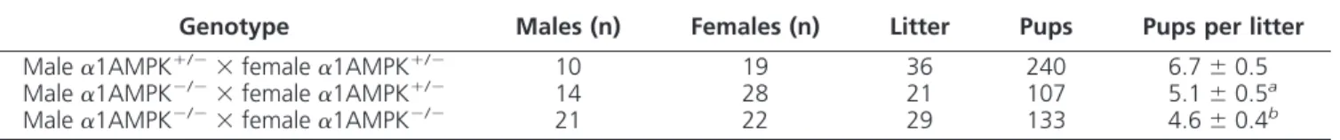
Alteration of fertility in male
␣1AMPK
ⴚ/ⴚmice
Adult males lacking the activity of
␣1AMPK have
shown a significant decrease in fertility, as attested by the
number of pups per litter when
␣1AMPK KO males were
crossed with
␣1AMPK
⫹/⫺females in comparison with
␣1AMPK
⫹/⫺males (5.1
⫾ 0.5 vs. 6.7 ⫾ 0.5 pups per litter,
respectively, Table 1).
␣1AMPK
⫺/⫺males crossed with
TABLE 1.
␣1AMPK
⫺/⫺male mice present a reduced fertility
Genotype Males (n) Females (n) Litter Pups Pups per litter
Male␣1AMPK⫹/⫺⫻ female␣1AMPK⫹/⫺ 10 19 36 240 6.7⫾ 0.5 Male␣1AMPK⫺/⫺⫻ female␣1AMPK⫹/⫺ 14 28 21 107 5.1⫾ 0.5a
Male␣1AMPK⫺/⫺⫻ female␣1AMPK⫺/⫺ 21 22 29 133 4.6⫾ 0.4b
Results are means of pups per litter of the following breeding: male␣1AMPK⫹/⫺mated with female␣1AMPK⫹/⫺(line 1), male␣1AMPK⫺/⫺mated with female␣1AMPK⫹/⫺(line 2), and male␣1AMPK⫺/⫺mated with female␣1AMPK⫺/⫺(line 3). Data are represented as mean⫾SEM.
a
P⬍ 0.05 for line 1 vs. line 2. bP⬍ 0.001 for line 1 vs. line 3.
␣1AMPK
⫺/⫺females presented a stronger decrease in
fer-tility (4.6
⫾ 0.4 pups per litter). The
␣1AMPK gene in
transgenic mice (48) was disrupted by insertion of the
re-porter gene
-galactosidase, allowing us to localize the
expression of
␣1AMPK in the testis by X-gal staining (Fig.
1A).
␣1AMPK is expressed in Leydig cells, Sertoli cells,
and in germ cells (Fig. 1).
␣1AMPK was also detected in
epithelium of the epididymal ducts (Supplemental Fig. 1,
published on The Endocrine Society’s Journals Online
web site at http://endo.endojournals.org). Absence of
␣1AMPK expression in the testis of ␣1AMPK
⫺/⫺mice
was confirmed by Western blot using
␣1 antibody (Fig.
1B); consistent with this, we observed a reduction of the
phospho-ACC-immunoreactive cells in seminiferous
tu-bules, one of the major downstream
targets of AMPK (P
⬍ 0.001) (Fig. 1, C
and D).
␣2AMPK is present in the testis
at a lower level (Fig. 1B).
As previously reported, the average
body and testis weight in
␣1AMPK KO
mice is similar to those of wild mice (61)
(Supplemental Table 1).
Morphologi-cal analysis of testis and epididymis
(Fig. 2A and Supplemental Fig. 1) has
not shown clear alteration of structure
and
polarity
between
the
two
genotypes.
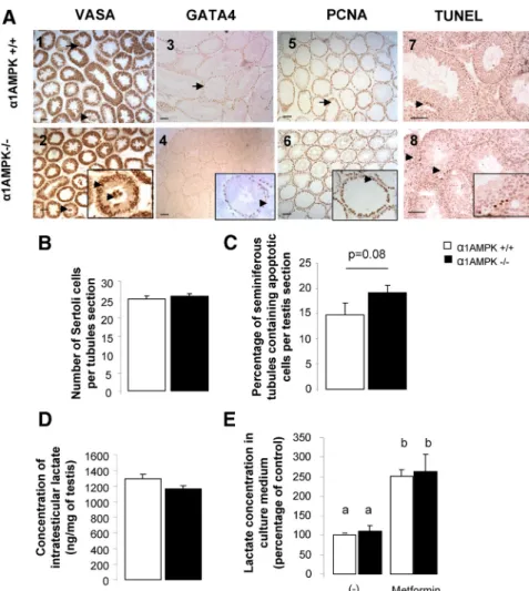
Alteration of spermatozoa in male
␣1AMPK
ⴚ/ⴚmice
To identify the cause of reduced
fer-tility, an analysis of the testis cell
compo-sition by using a specific cell marker of
germ cells (VASA-immunoreactive cells)
and their nurse cells, the Sertoli cells
(GATA4-immunoreactive cells), was
performed and did not show alteration in
␣1AMPK KO mice (Fig. 2, A1–A4 and
B). Evaluation of cell proliferation and
apoptosis was assayed by
immunostain-ing of the PCNA, and of terminal
nucleotidyl transferase-mediated
deoxy-UTP nick end labeling (TUNEL). No
clear difference in proliferation
(includ-ing mono- and multilayer of
PCNA-pos-itive cells in seminiferous tubules),
and apoptosis was noticed (Fig. 2,
A5–A8 and C). The lactate
concen-tration produced by the Sertoli cells
representing the main energetic
sub-strate for spermatozoa was also
mea-sured. No difference was observed in
testicular extract between each
geno-type (Fig. 2D) in 48 h culture medium conditioned by
␣1AMPK
⫹/⫹and
␣1AMPK
⫺/⫺Sertoli cells, even after
metformin stimulation (Fig. 2E).
Our finding of
␣1AMPK expression in spermatozoa (Fig.
3A) led us to investigate the production and quality of
␣1AMPK
⫺/⫺sperm. The numbers of spermatids and
sper-matozoa heads in testes and in cauda epididymis of wild-type
and mutant mice were compared and did not show alteration
(Fig. 3, B and C). However,
␣1AMPK KO spermatozoa had
twice as many head abnormalities as
␣1AMPK wild-type
counterparts (
␣1AMPK
⫺/⫺mice, 48% of abnormal sperm
heads;
␣1AMPK
⫹/⫹mice, 26%; P
⬍ 0.01; Fig. 3D), but
flagellum length was not altered (data not shown). Only
ab-FIG. 2. Histological analysis of␣1AMPK⫺/⫺testis. A, Testicular sections immunostained against VASA (1 and 2), GATA4 (3 and 4), PCNA (5 and 6), and apoptotic DNA fragmentation (7 and 8) from␣1AMPK⫹/⫹mice (1, 3, 5, and 7) and␣1AMPK⫺/⫺mice (2, 4, 6, and 8). Scale bar, 50m. All stages of spermatogenesis were visible, and no clear alteration was observed. Few germ cells sloughing were observed in both genotypes (arrows in A1 and A2). B, Quantification of GATA4-immunoreactive cells in seminiferous tubule (20 tubules per animal, n⫽ 4 mice). C, Percentage of seminiferous tubules with at least one TUNEL-positive nuclei (n⫽ 4 mice). D, Lactate concentration, an energy substrate for spermatozoa, in␣1AMPK⫹/⫹ and␣1AMPK⫺/⫺testis (n⫽ 5). E, Lactate secretion measured in culture medium conditioned by␣1AMPK⫹/⫹and␣1AMPK⫺/⫺Sertoli cells for 48 h in the presence or absence of 5 mM metformin (percentage of control normalized per 200,000 cells/wells). Data are represented as means⫾SEM(n⫽ 3 in triplicate). a and b, Significant differences.
normal spermatozoa with curved sheaths tended to be higher
in homozygous
␣1AMPK KO mice compared with
␣1AMPK wild-type mice (P ⫽ 0.05) (Fig. 3E). Transmission
electron microscopy analysis confirmed the neck
abnormal-ity without acrosome alteration (Fig. 3, F1–F4) and has
shown disturbances during head formation (Fig. 3, F5–F8).
These results indicate a decrease in the quality but not in the
production of spermatozoa in
␣1AMPK KO mice.
Mitochondrial defect reduces
motility of
␣1AMPK
ⴚ/ⴚspermatozoa
Sperm analysis performed with an
integrated visual optical system (IVOS)
analyzer showed a 1.7-fold increase in
static spermatozoa and a 2.4-fold
re-duction in rapid spermatozoa in
␣1AMPK KO mice compared with
wild-type mice (Fig. 4A, P
⬍ 0.05). In
␣1AMPK KO mice, spermatozoa also
exhibited a decrease in velocity (Fig.
4B) (
␣1AMPK KO mice, 186 m/sec;
␣1AMPK wild-type mice, 252 m/sec;
P
⬍ 0.05).
Because spermatozoa motility is
strongly associated with activity of
mito-chondria localized in the midpiece (62),
sperm mitochondria were evaluated by
testing mitochondrial membrane
poten-tial, measurement of the mitochondrial
respiration, and microscopic
ultrastruc-tural analysis. The percentage of active
mitochondria with a high polarization
tended to be reduced to about half in
se-men from
␣1AMPK KO mice (P ⫽ 0.07,
Fig. 4C). In addition, the measurement of
the mitochondrial respiration validated
our preliminary results because we found
a reduction of the oxygen consumption
in
␣1AMPK KO spermatozoa compared
with
␣1AMPK wild-type spermatozoa in
all the conditions tested.
␣1AMPK KO
spermatozoa
presented
significantly
lower basal JO
2rates of 60% (
␣1AMPK
wild-type spermatozoa, 2.19 pmol O
2/
sec
䡠 10
6cells;
␣1AMPK KO
spermato-zoa, 0.81 pmol O
2/sec
䡠10
6cells cells, P
⬍
0.05) (Fig. 4D). In addition,
oligomycin-sensitive and maximal JO
2were also
di-minished by 50 and 30% but without
reaching statistical significance.
Further-more,
ultrastructural
studies
of
␣1AMPK KO sperm have shown a
de-crease in the number of mitochondria as illustrated in
trans-verse sections of the midpiece region of spermatozoa (Fig. 4E,
P
⬍ 0.001) and a 3-fold increase in the percentage of
mito-chondria exhibiting dilated intermembrane space (black
ar-rows) in contrast to wild-type sperm (Fig. 4F, P
⬍ 0.05).
During spermiogenesis, abnormal arrangement of
mito-chondria along microtubules in the midpiece was also
ob-served in
␣1AMPK KO mice (Fig. 4G). In addition, the total
FIG. 3. Alteration of spermatozoa morphology in male␣1AMPK⫺/⫺mice. A, Expression of ␣1AMPK subunits protein in spermatozoa extract purified from ␣1AMPK⫹/⫹and␣1AMPK⫺/⫺ epididymis. Liver was used as positive control. B and C, Number of sperm head per milligram of testis (B) and per milligram of cauda epididymis (C) from␣1AMPK⫹/⫹mice (n⫽ 4) and ␣1AMPK⫺/⫺mice (n⫽ 4). D, Percentage of atypical sperm head in both genotypes. **, P ⬍ 0.01. E, Classification of sperm head abnormalities in␣1AMPK⫹/⫹mice (n⫽ 5) and ␣1AMPK⫺/⫺mice (n⫽ 5) in microcephalic, curved spermatozoa, irregular head, and thin head. Representative micrographs are presented under the bar graph. Data are represented as mean or percentage⫾SEM. F, Transmission electron micrographs of spermatozoa (1– 4) and spermatid head (5– 8); head and sheath morphology (1 and 2) and acrosome (3 and 4) (white arrows) from␣1AMPK⫹/⫹(1 and 3) and␣1AMPK⫺/⫺mice (2 and 4). Irregular nuclei of spermatids were observed in␣1AMPK⫺/⫺mice (6 and 8) (white arrows) compared with ␣1AMPK⫹/⫹mice (5 and 7). Scale bar, 2m.
antioxidant capacity in the whole testis
was lower in
␣1AMPK
⫺/⫺mice (Fig. 4H,
P
⬍ 0.05).
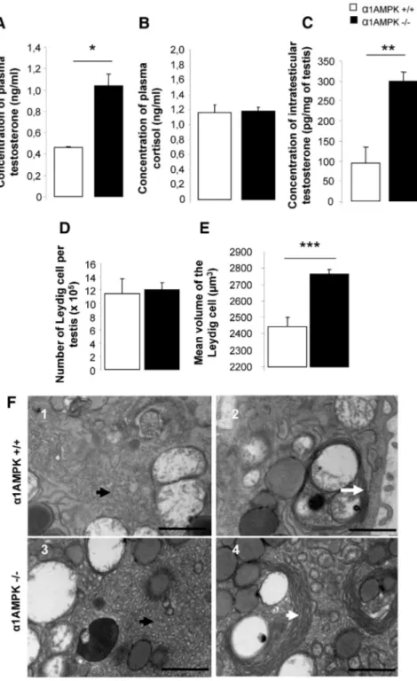
Hyperandrogenism in male
␣1AMPK
ⴚ/ⴚmice
Plasma testosterone levels were about
twice as high in
␣1AMPK KO mice
(Fig. 5A, P
⬍ 0.05) and more than
3-fold higher in testicular extract (Fig.
5C, P
⬍ 0.01) compared with
␣1AMPK
wild-type mice. Hyperandrogenism
was not associated with an alteration of
the plasma cortisol level (Fig. 5B) or an
increase in the number of Leydig cells in
the
␣1AMPK KO mice (Fig. 5D).
How-ever, the mean volume of the Leydig cell
was significantly increased (Fig. 5E). At
the ultrastructural level, Leydig cells
from
␣1AMPK KO mice exhibited an
alteration of the surface occupied by
smooth endoplasmic reticulum (SER),
involved in synthesis of lipids and
ste-roids, and the presence of larger
struc-tures of membranous whorls of SER
compared with wild-type mice (Fig.
5F). Additionally,
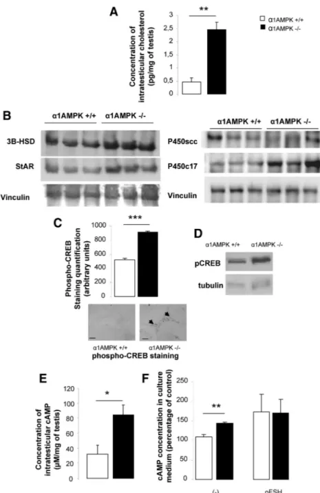
␣1AMPK KO mice
presented a 5-fold increase in
intrates-ticular cholesterol concentration, a
pre-cursor for steroid synthesis (Fig. 6A,
P
⬍ 0.01). The protein level of a
cho-lesterol carrier (StAR) and two enzymes
involved in steroid production (P450c17
and 3
HSD) was higher in ␣1AMPK
KO testicular extract (1.9-, 3.1-, and
1.8-fold increase in
␣1AMPK
⫺/⫺compared with wild-type mice,
respec-tively) (Fig. 6B). This result was
asso-ciated with an increase in
phosphory-lation of the CREB transcription factor
in Leydig cells (Fig. 6C, P
⬍ 0.001).
Phosphorylation of serine 133 is
known to activate CREB and induce
transcription of target genes such as
StAR and 3
HSD.
Hyperphosphoryla-tion of CREB was also observed on
a basal level in purified cultured
␣1AMPK KO Sertoli cells (1.5-fold
in-crease, Fig. 6D).
To test whether the higher CREB
activity is due to stimulation of the
gonadotropin receptor pathway (a G
FIG. 4. Analysis of spermatozoa mobility and characterization of mitochondria. Sperm
mobility parameters, percentage of motile spermatozoa (A) and sperm kinematics (B) of ␣1AMPK⫹/⫹mice (n⫽ 6) and␣1AMPK⫺/⫺mice (n⫽ 6), were performed by computer-assisted sperm analysis. VCL, Sperm curvilinear velocity; VSL, sperm straight-line velocity. *, P⬍ 0.05. C, Mitochondrial activity was determined by JC-1 staining and represented by percentage of high mitochondrial polarization spermatozoa (orange/red fluorescence) in ␣1AMPK⫹/⫹mice (n⫽ 5) and␣1AMPK⫺/⫺mice (n⫽ 5). Low mitochondrial membrane potential was observed in green fluorescence. D, Spermatozoa suspension (2⫻ 106
cells/ml) in M2 medium supplemented with 5 mMsodium pyruvate was transferred immediately, after isolation, into a stirrer 2-ml oxygraph vessel where JO2was recorded before and after the successive addition of 0.5g/ml oligomycin and 1–2 Mcarbonyl cyanide
m-chlorophenylhydrazone. Data are represented as means⫾SEMof n⫽ 3 different spermatozoa isolations in which measurements were performed in duplicate. *, P⬍ 0.05. E, The number of mitochondria per section of spermatozoa in the midpiece from␣1AMPK⫹/⫹ mice (n⫽ 5) and␣1AMPK⫺/⫺mice (n⫽ 5). Scale bar, 500 nm. F, Percentage of mitochondria exhibiting partial or total cristolysis was measured on transmission electron micrographs. Black arrows indicate crystolysis in mitochondria. Scale bar, 1m. G, Testis micrographs from ␣1AMPK⫺/⫺mice (3–5) exhibited abnormal arrangement of mitochondria (arrowhead) during midpiece formation in the testis compared with␣1AMPK wild-type mice (1 and 2). Scale bar, 5m. H, Intratesticular antioxidant activity measured in units called Trolox equivalent antioxidant capacity (TEAC) per milligram of testis. Data are represented as mean or percentage⫾SEM. *, P⬍ 0.05; ***, P ⬍ 0.001.
protein-coupled receptor, cAMP-dependent pathway),
the cAMP concentration was measured in the freshly
recovered testis (Fig. 6E) and in a culture medium
re-leased by purified Sertoli cells (Fig. 6F). In both cases,
basal cAMP concentrations were
in-creased in
␣1AMPK KO mice. The
high cAMP testicular concentration is
not closely linked to gonadotropin
secretion,
because
LH
plasma
(
␣1AMPK KO mice, 0.19 ⫾ 0.02 ng/
ml;
␣1AMPK wild-type mice, 0.26 ⫾
0.07 ng/ml, P
⬎ 0,05) and FSH
plasma (
␣1AMPK KO mice, 34.4 ⫾
2.1 ng/ml;
␣1AMPK wild-type mice,
38.8
⫾ 3.1 ng/ml; P ⬎ 0,05) and
pi-tuitary levels (LH:
␣1AMPK KO
mice, 476
⫾ 142 ng/mg;
␣1AMPK
wild-type mice, 775
⫾ 237 ng/mg, P ⬎
0,05; and FSH:
␣1AMPK KO mice,
1367
⫾ 193 ng/mg;
␣1AMPK
wild-type mice, 2790
⫾ 779 ng/mg, P ⫽
0,09) tended to be reduced in
␣1AMPK KO mice compared with
␣1AMPK wild-type mice.
Discussion
These data show that inactivation of
the AMPK
␣1 gene reduces male
fertil-ity. Indeed,
␣1AMPK
⫺/⫺males present
a 20% decrease in litter size per
preg-nancy associated with
hyperandro-genism and alteration in morphology
and motility of the spermatozoa.
In our study, localization of the
␣1AMPK expression has been found
in germ cells and in Leydig and Sertoli
cells in the testis. A previous study has
identified AMPK expression in the rat
testis (16), and the authors have
demonstrated by
immunoprecipita-tion that 75% of the AMPK activity is
associated with
␣1 catalytic subunit
and 25% with the
␣2 AMPK subunit in
the whole testis. Furthermore, the
ki-nase of AMPK, LKB1 (27, 47) and two
AMPK-related kinases (37), SNRK
(35) and MARK2 (36) are also
ex-pressed in the testis. Inactivation of
these genes induced alterations of
fer-tility and in some case sterility,
suggest-ing that AMPK/AMPK-related activity
is involved in testicular function.
The high testosterone level measured in
␣1AMPK
⫺/⫺mice could affect spermatogenesis. That disturbance
can-FIG. 5. A–C, Hyperandrogenism in␣1AMPK⫺/⫺male. Plasma concentrations of testosterone (A), cortisol (B), and intratesticular concentrations of testosterone (C) in ␣1AMPK⫹/⫹mice (n⫽ 5) and␣1AMPK⫺/⫺mice (n⫽ 5) were analyzed. *, P ⬍ 0.05; **, P⬍ 0.01. D and E, The number of Leydig cells (D) and their mean cell volume (E) were estimated in␣1AMPK⫹/⫹(n⫽ 3) and␣1AMPK⫺/⫺testis (n⫽ 5). ***, P ⬍ 0.001. Data are represented as mean or percentage⫾SEM. F, Transmission electron micrographs of Leydig cells; 1 and 3, altered surface occupied by SER (black arrows) in␣1AMPK⫺/⫺ Leydig cells; 2 and 4, increase in size of membranous whorls of SER (white arrows) in ␣1AMPK⫺/⫺Leydig cells. Scale bar, 1m.
not be attributed to adrenal gland disorders or to
gluco-corticoid resistance (Fig. 5B) but was associated with
hy-peractive Leydig cells as attested by the increased mean
volume, the altered endoplasmic reticulum area, the high
intratesticular cholesterol
concentra-tion, and the strong expression of
pro-teins involved in steroid production.
These results could be linked to studies
that have described the activation of
AMPK complexes by natural activators
like curcumin (63, 64) or resveratrol
(65, 66) that inhibit androgen
produc-tion by Leydig cells. In the rat ovary,
activation of AMPK reduces
progester-one secretion in granulosa cells by
in-hibition of expression of the 3
HSD
en-zyme (67) but not p450scc as observed
in our model. Furthermore, high
intra-testicular cAMP concentrations
associ-ated with high phosphorylation of
CREB suggested that gonadotropin
re-ceptors and/or a downstream signaling
pathway could be activated at a basal
level, despite recent work that
de-scribed expression of AMPK in rat
go-nadotrope pituitary cells (60). In our
model, LH and FSH pituitary
concen-trations appeared lower than in
wild-type mice (1.6- to 2-fold), suggesting
that androgen levels induced negative
feedback on gonadotropin synthesis
rather than absence of AMPK affecting
gonadotropin secretion. Indeed, Tosca
et al. (60) have shown that inhibition of
AMPK by a synthetic inhibitor,
com-pound C, did not affect LH and FSH
secretions in gonadotrope cell lines.
Additionally, different models with
high testosterone levels indicate
altera-tion of sperm quality (68, 69).
Condi-tional deletion of
␣1AMPK in Leydig
cells or administration of antiandrogen
treatment should be helpful to clarify
the role of testosterone in sperm quality
in these transgenic mice.
In various cells, AMPK has been
shown to be involved in mitochondrial
function. In their work, Nakada et al.
(70) showed that spermatogenesis is
closely linked to mitochondrial
respira-tion, and in a recent publicarespira-tion,
Pellic-cione et al. (71) associated
asthenozoo-spermia with abnormal mitochondrial ultrastructure. Thus,
we hypothesize that the motility disturbance observed in
our
␣1AMPK KO model and in the LKB1 KO model (an
AMPK kinase) showing a strong spermatozoa
abnormal-FIG. 6. Molecular alteration in steroidogenesis. A, Intratesticular concentrations of cholesterol in
␣1AMPK⫹/⫹(n⫽ 5) and␣1AMPK⫺/⫺mice (n⫽ 5). **, P ⬍ 0.01. B, Western blot analysis of StAR, 3-HSD, P450c17, and P450scc expression in ␣1AMPK⫹/⫹(lanes 1–3, three different animals) and␣1AMPK⫺/⫺testis (lanes 4 – 6, three different animals), normalized to vinculin. C, Quantification of phospho-CREB immunostaining intensity localized in Leydig cells. Bright-field photomicrographs are shown below the bar graph. Scale bar, 20m; n ⫽ 3 per genotype. ***, P⬍ 0.001. D, Western blot analysis of phospho-CREB expression in purified Sertoli cells culture of␣1AMPK⫹/⫹and␣1AMPK⫺/⫺mice. Tubulin served as a loading control. Results are representative of three independent experiments. E, Intratesticular cAMP concentration from ␣1AMPK⫹/⫹mice (n⫽ 5) and␣1AMPK⫺/⫺mice (n⫽ 6). *, P ⬍ 0.05. F, cAMP concentration in culture medium of Sertoli cells stimulated for 15 min with or without ovine FSH (oFSH) (100 ng/ml) (percentage of control normalized per 200,000 cells/wells; n⫽ 4 independent experiments). **, P⬍ 0.01. Data are represented as mean or percentage ⫾SEM.
ity (motility and head morphology) (47) was directly
linked to mitochondrial dysgenesis. Our hypothesis seems
to be confirmed, because investigation of mitochondrial
integrity in
␣1AMPK KO sperm mice has demonstrated a
50% reduction in mitochondrial activity, a decrease in
their number per sheath section, and a 3-fold rise in
ab-normal mitochondria with dilated intermembrane space
structure. Moreover, in his work, Jäger et al. (72) noted
that AMPK activation is required for
peroxisome-prolif-erator-activated receptor
␥ coactivator 1␣ (PGC-1␣)
ac-tivation, a key regulator of mitochondrial biogenesis (73),
and a study of Rodríguez-Cuenca et al. (74) showed that
testosterone reduces PGC-1
␣ mRNA levels. Therefore, in
our model, the substantial rise in testosteronemia and
ab-sence of
␣1AMPK in spermatozoa could hardly reduce
PGC-1
␣ expression and mitochondrial biogenesis. A
dys-function of mitochondria that could also induce a high
oxidative stress has been described in
red blood cells from
␣1AMPK KO mice
(75). Similarly, we have measured a
lower total antioxidant capacity in the
␣1AMPK
⫺/⫺testis. Strangely, a mouse
model inactivated for an
oxidative-stress sensor proteins like glutathione
peroxidase 4 (76, 77) is described with
structural abnormalities in
spermato-zoa closely similar to those observed in
␣1AMPK KO. Furthermore, these
mi-tochondrial malfunctions were not due
to a decline in available energy as
lac-tate substrate, produced by Sertoli cells
(78), because as shown in Fig. 2, lactate
production in vivo and in vitro (even
after metformin induction) by Sertoli
cells was similar in
␣1AMPK KO and
wild-type mice.
The Sertoli cells, closely linked to
germ cells, have an essential role in the
shaping of the spermatid head (79).
Al-though
␣1AMPK KO Sertoli cells did
not present abnormalities in structure
and nucleus polarization
(Supplemen-tal Fig. 2), observations of
abnormali-ties in sperm maturation (mitochondria
organization and head shape formation
at the spermatid stage) suggest a role of
AMPK in the cytoskeletal dynamics.
Observations from transmission
elec-tron microscopy of elongated
sperma-tids have confirmed the presence of
some disrupted Sertoli cell/germ cell
junctions in
␣1AMPK-defective testis
(Supplemental Fig. 3). In a recent study, Galardo et al. (80)
demonstrated that AMPK affects some adhesion molecule
expression and influences junction complex integrity in rat
Sertoli cells. Moreover, mice deleted for adhesion
mole-cules present in Sertoli/germ cell junctions, like nectin-2
(81) or tumor suppressor in lung cancer 1 (82) confirmed
the essential role of these molecules for sperm head
co-nfiguration, midpiece formation, and sperm motility.
Thus, we can think that AMPK has a potential role in the
cytoskeletal interaction between Sertoli and germ cells,
leading to an abnormal head shape and mitochondrial
organization around microtubules.
In conclusion, inactivation of the
␣1AMPK
predomi-nant isoform in somatic and germ cells has shown a major
malfunction of mitochondrial activity, leading mainly to a
decrease in sperm quality (morphology and mobility) and
alteration of steroid production as described in Fig. 7.
Hence, the use of an AMPK activator such as metformin,
an insulin sensitizer used in the therapeutic management
of type 2 diabetes mellitus, raises the question of its
po-tential consequences in human male fertility.
Acknowledgments
We thank Dr. Dale Buchanan Hales (Department of Physiology and Biophysics, University of Illinois at Chicago, Chicago, IL) and Dr. Grahame Hardie (University of Dundee, Dundee, UK) for generously providing the StAR, p450scc, and anti-␣1- and -␣2AMPK antibodies, respectively and Alan Scaife for the manuscript editing.
Address all correspondence and requests for reprints to: Dr. Pascal Froment Ph.D, Team Testicule Ontogenèse et Mé-tabolisme Energétique, Institut National de la Recherche Agronomique, Station de Recherche Avicole, 37380 Nouzilly, France. E-mail: pfroment@tours.inra.fr.
This work was partly supported by the national program called FERTiNERGY funded by the French National Research Agency and by the European Commission integrated project (LSHM-CT-2004-005272). P.T. was supported by a French fel-lowship from the Ministère de l’éducation et de la recherche and N.S. by a postdoctoral fellowship (Grant 2010 BLAN 1123) from the French National Research Agency.
Disclosure Summary: The authors have nothing to disclose.
References
1. ESHRE Capri Workshop Group 2006 Nutrition and reproduction in women. Hum Reprod Update 12:193–207
2. Mah PM, Wittert GA 2010 Obesity and testicular function. Mol Cell Endocrinol 316:180 –186
3. Monget P, Martin GB 1997 Involvement of insulin-like growth fac-tors in the interactions between nutrition and reproduction in female mammals. Hum Reprod 12(Suppl 1):33–52
4. Barash IA, Cheung CC, Weigle DS, Ren H, Kabigting EB, Kuijper JL,
Clifton DK, Steiner RA 1996 Leptin is a metabolic signal to the
reproductive system. Endocrinology 137:3144 –3147
5. Combs TP, Pajvani UB, Berg AH, Lin Y, Jelicks LA, Laplante M,
Nawrocki AR, Rajala MW, Parlow AF, Cheeseboro L, Ding YY, Russell RG, Lindemann D, Hartley A, Baker GR, Obici S, Deshaies Y, Ludgate M, Rossetti L, Scherer PE 2004 A transgenic mouse with
a deletion in the collagenous domain of adiponectin displays ele-vated circulating adiponectin and improved insulin sensitivity. En-docrinology 145:367–383
6. Brüning JC, Gautam D, Burks DJ, Gillette J, Schubert M, Orban PC,
Klein R, Krone W, Müller-Wieland D, Kahn CR 2000 Role of brain
insulin receptor in control of body weight and reproduction. Science 289:2122–2125
7. Burks DJ, Font de Mora J, Schubert M, Withers DJ, Myers MG,
Towery HH, Altamuro SL, Flint CL, White MF 2000 IRS-2
path-ways integrate female reproduction and energy homeostasis. Nature 407:377–382
8. Giguère V 1999 Orphan nuclear receptors: from gene to function. Endocr Rev 20:689 –725
9. Frenoux JM, Vernet P, Volle DH, Britan A, Saez F, Kocer A,
Henry-Berger J, Mangelsdorf DJ, Lobaccaro JM, Drevet JR 2004 Nuclear
oxysterol receptors, LXRs, are involved in the maintenance of mouse caput epididymidis structure and functions. J Mol Endocrinol 33: 361–375
10. Robertson KM, Schuster GU, Steffensen KR, Hovatta O, Meaney S,
Hultenby K, Johansson LC, Svechnikov K, Söder O, Gustafsson JA
2005 The liver X receptor- is essential for maintaining cholesterol homeostasis in the testis. Endocrinology 146:2519 –2530 11. Bełtowski J, Semczuk A 2010 Liver X receptor (LXR) and the
re-productive system: a potential novel target for therapeutic interven-tion. Pharmacol Rep 62:15–27
12. Repa JJ, Turley SD, Lobaccaro JA, Medina J, Li L, Lustig K, Shan
B, Heyman RA, Dietschy JM, Mangelsdorf DJ 2000 Regulation of
absorption and ABC1-mediated efflux of cholesterol by RXR het-erodimers. Science 289:1524 –1529
13. Kastner P, Mark M, Leid M, Gansmuller A, Chin W, Grondona JM,
Décimo D, Krezel W, Dierich A, Chambon P 1996 Abnormal
sper-matogenesis in RXR mutant mice. Genes Dev 10:80–92 14. Fredrikson G, Strålfors P, Nilsson NO, Belfrage P 1981
Hormone-sensitive lipase of rat adipose tissue. Purification and some proper-ties. J Biol Chem 256:6311– 6320
15. Chung S, Wang SP, Pan L, Mitchell G, Trasler J, Hermo L 2001 Infertility and testicular defects in hormone-sensitive lipase-defi-cient mice. Endocrinology 142:4272– 4281
16. Cheung PC, Salt IP, Davies SP, Hardie DG, Carling D 2000 Char-acterization of AMP-activated protein kinase gamma-subunit iso-forms and their role in AMP binding. Biochem J 346(Pt 3):659 – 669 17. Kahn BB, Alquier T, Carling D, Hardie DG 2005 AMP-activated protein kinase: ancient energy gauge provides clues to modern un-derstanding of metabolism. Cell Metab 1:15–25
18. Hardie DG, Hawley SA, Scott JW 2006 AMP-activated protein ki-nase– development of the energy sensor concept. J Physiol 574:7–15 19. Kola B, Boscaro M, Rutter GA, Grossman AB, Korbonits M 2006 Expanding role of AMPK in endocrinology. Trends Endocrinol Metab 17:205–215
20. Cantó C, Auwerx J 2010 AMP-activated protein kinase and its downstream transcriptional pathways. Cell Mol Life Sci 67:3407– 3423
21. Bouly JP, Gissot L, Lessard P, Kreis M, Thomas M 1999 Arabidopsis thaliana proteins related to the yeast SIP and SNF4 interact with AKIN␣1, an SNF1-like protein kinase. Plant J 18:541–550 22. Woods A, Munday MR, Scott J, Yang X, Carlson M, Carling D 1994
Yeast SNF1 is functionally related to mammalian AMP-activated protein kinase and regulates acetyl-CoA carboxylase in vivo. J Biol Chem 269:19509 –19515
23. Steinke D, Salzburger W, Braasch I, Meyer A 2006 A Many genes in fish have species-specific asymmetric rates of molecular evolution. BMC Genomics 7:20
24. Proszkowiec-Weglarz M, Richards MP, Ramachandran R,
McMurtry JP 2006 Characterization of the AMP-activated protein
kinase pathway in chickens. Comp Biochem Physiol B Biochem Mol Biol 143:92–106
25. Stapleton D, Mitchelhill KI, Gao G, Widmer J, Michell BJ, Teh T,
House CM, Fernandez CS, Cox T, Witters LA, Kemp BE 1996
Mammalian AMP-activated protein kinase subfamily. J Biol Chem 271:611– 614
26. Suter M, Riek U, Tuerk R, Schlattner U, Wallimann T, Neumann D 2006 Dissecting the role of 5⬘-AMP for allosteric stimulation, acti-vation, and deactivation of AMP-activated protein kinase. J Biol Chem 281:32207–32216
27. Woods A, Johnstone SR, Dickerson K, Leiper FC, Fryer LG,
Neu-mann D, Schlattner U, WalliNeu-mann T, Carlson M, Carling D 2003
LKB1 is the upstream kinase in the AMP-activated protein kinase cascade. Curr Biol 13:2004 –2008
28. Hawley SA, Pan DA, Mustard KJ, Ross L, Bain J, Edelman AM,
ki-nase kiki-nase- is an alternative upstream kinase for AMP-activated protein kinase. Cell Metab 2:9 –19
29. Xie M, Zhang D, Dyck JR, Li Y, Zhang H, Morishima M, Mann DL,
Taffet GE, Baldini A, Khoury DS, Schneider MD 2006 A pivotal role
for endogenous TGF--activated kinase-1 in the LKB1/AMP-acti-vated protein kinase energy-sensor pathway. Proc Natl Acad Sci USA 103:17378 –17383
30. Costanzo-Garvey DL, Pfluger PT, Dougherty MK, Stock JL, Boehm
M, Chaika O, Fernandez MR, Fisher K, Kortum RL, Hong EG, Jun JY, Ko HJ, Schreiner A, Volle DJ, Treece T, Swift AL, Winer M, Chen D, Wu M, Leon LR, Shaw AS, McNeish J, Kim JK, Morrison DK, Tschöp MH, Lewis RE 2009 KSR2 is an essential regulator of
AMP kinase, energy expenditure, and insulin sensitivity. Cell Metab 10:366 –378
31. Minokoshi Y, Kim YB, Peroni OD, Fryer LG, Müller C, Carling D,
Kahn BB 2002 Leptin stimulates fatty-acid oxidation by activating
AMP-activated protein kinase. Nature 415:339 –343
32. Yamauchi T, Kamon J, Minokoshi Y, Ito Y, Waki H, Uchida S,
Yamashita S, Noda M, Kita S, Ueki K, Eto K, Akanuma Y, Froguel P, Foufelle F, Ferre P, Carling D, Kimura S, Nagai R, Kahn BB, Kadowaki T 2002 Adiponectin stimulates glucose utilization and
fatty-acid oxidation by activating AMP-activated protein kinase. Nat Med 8:1288 –1295
33. Manning G, Whyte DB, Martinez R, Hunter T, Sudarsanam S 2002 The protein kinase complement of the human genome. Science 298: 1912–1934
34. Lizcano JM, Göransson O, Toth R, Deak M, Morrice NA, Boudeau
J, Hawley SA, Udd L, Mäkelä TP, Hardie DG, Alessi DR 2004 LKB1
is a master kinase that activates 13 kinases of the AMPK subfamily, including MARK/PAR-1. EMBO J 23:833– 843
35. Jaleel M, McBride A, Lizcano JM, Deak M, Toth R, Morrice NA,
Alessi DR 2005 Identification of the sucrose non-fermenting related
kinase SNRK, as a novel LKB1 substrate. FEBS Lett 579:1417–1423 36. Bessone S, Vidal F, Le Bouc Y, Epelbaum J, Bluet-Pajot MT,
Dar-mon M 1999 EMK protein kinase-null mice: dwarfism and
hypofer-tility associated with alterations in the somatotrope and prolactin pathways. Dev Biol 214:87–101
37. Bright NJ, Thornton C, Carling D 2009 The regulation and function of mammalian AMPK-related kinases. Acta Physiol (Oxf) 196: 15–26
38. Tosca L, Chabrolle C, Uzbekova S, Dupont J 2007 Effects of met-formin on bovine granulosa cells steroidogenesis: possible involve-ment of adenosine 5⬘ monophosphate-activated protein kinase (AMPK). Biol Reprod 76:368 –378
39. Karlsson C, Lindell K, Svensson E, Bergh C, Lind P, Billig H,
Carlsson LM, Carlsson B 1997 Expression of functional leptin
re-ceptors in the human ovary. J Clin Endocrinol Metab 82:4144 – 4148
40. Chabrolle C, Tosca L, Dupont J 2007 Regulation of adiponectin and its receptors in rat ovary by human chorionic gonadotrophin treat-ment and potential involvetreat-ment of adiponectin in granulosa cell steroidogenesis. Reproduction 133:719 –731
41. Tena-Sempere M, Barreiro ML 2002 Leptin in male reproduction: the testis paradigm. Mol Cell Endocrinol 188:9 –13
42. Caminos JE, Nogueiras R, Gaytán F, Pineda R, González CR,
Bar-reiro ML, Castaño JP, Malagón MM, Pinilla L, Toppari J, Diéguez C, Tena-Sempere M 2008 Novel expression and direct effects of
adiponectin in the rat testis. Endocrinology 149:3390 –3402 43. Pierre P, Froment P, Nègre D, Ramé C, Barateau V, Chabrolle C,
Lecomte P, Dupont J 2009 Role of adiponectin receptors, AdipoR1
and AdipoR2, in the steroidogenesis of the human granulosa tumor cell line, KGN. Hum Reprod 24:2890 –2901
44. Chabrolle C, Tosca L, Ramé C, Lecomte P, Royère D, Dupont J 2009 Adiponectin increases insulin-like growth factor I-induced progesterone and estradiol secretion in human granulosa cells. Fertil Steril 92:1988 –1996
45. Chen J, Hudson E, Chi MM, Chang AS, Moley KH, Hardie DG,
Downs SM 2006 AMPK regulation of mouse oocyte meiotic
re-sumption in vitro. Dev Biol 291:227–238
46. Galardo MN, Riera MF, Pellizzari EH, Cigorraga SB, Meroni SB 2007 The AMP-activated protein kinase activator, 5-aminoimida-zole-4-carboxamide-1-b-D-ribonucleoside, regulates lactate pro-duction in rat Sertoli cells. J Mol Endocrinol 39:279 –288 47. Towler MC, Fogarty S, Hawley SA, Pan DA, Martin DM, Morrice
NA, McCarthy A, Galardo MN, Meroni SB, Cigorraga SB, Ash-worth A, Sakamoto K, Hardie DG 2008 A novel short splice variant
of the tumour suppressor LKB1 is required for spermiogenesis. Biochem J 416:1–14
48. Jørgensen SB, Viollet B, Andreelli F, Frøsig C, Birk JB, Schjerling
P, Vaulont S, Richter EA, Wojtaszewski JF 2004 Knockout of the
␣2 but not ␣1 5⬘-AMP-activated protein kinase isoform abolishes 5-aminoimidazole-4-carboxamide-1--4-ribofuranosidebut not contraction-induced glucose uptake in skeletal muscle. J Biol Chem 279:1070 –1079
49. Rivkees SA, Hall DA, Boepple PA, Crawford JD 1987 Accuracy and reproducibility of clinical measures of testicular volume. J Pediatr 110:914 –917
50. Froment P, Staub C, Hembert S, Pisselet C, Magistrini M, Delaleu
B, Seurin D, Levine JE, Johnson L, Binoux M, Monget P 2004
Re-productive abnormalities in human insulin-like growth factor-bind-ing protein-1 transgenic male mice. Endocrinology 145:2080 –2091 51. Yamamoto CM, Hikim AP, Lue Y, Portugal AM, Guo TB, Hsu SY,
Salameh WA, Wang C, Hsueh AJ, Swerdloff RS 2001 Impairment
of spermatogenesis in transgenic mice with selective overexpression of Bcl-2 in the somatic cells of the testis. J Androl 22:981–991 52. Toure A, Morin L, Pineau C, Becq F, Dorseuil O, Gacon G 2001
Tat1, a novel sulfate transporter specifically expressed in human male germ cells and potentially linked to RhoGTPase signaling. J Biol Chem 276:20309 –20315
53. Froment P, Vigier M, Nègre D, Fontaine I, Beghelli J, Cosset FL,
Holzenberger M, Durand P 2007 Inactivation of the IGF-I receptor
gene in primary Sertoli cells highlights the autocrine effects of IGF-I. J Endocrinol 194:557–568
54. Papaioannou V, Johnson R 1993 Production of chimeras and ge-netically defined offspring from targeted ES cells. In: Joyner AL, ed. Gene targeting: a practical approach. Oxford, UK: IRL Press; 107– 146
55. Jégou B, Velez de la Calle JF, Bauché F 1991 Protective effect of medroxyprogesterone acetate plus testosterone against radiation-induced damage to the reproductive function of male rats and their offspring. Proc Natl Acad Sci USA 88:8710 – 8714
56. Mukai C, Okuno M 2004 Glycolysis plays a major role for aden-osine triphosphate supplementation in mouse sperm flagellar move-ment. Biol Reprod 71:540 –547
57. Hochereau-de Reviers MT, Perreau C, Pisselet C, Fontaine I,
Monet-Kuntz C 1990 Comparisons of endocrinological and testis
param-eters in 18-month-old Ile de France and Romanov rams. Domest Anim Endocrinol 7:63–73
58. Orgeur P, Naciri N, Yvore P, Bernard S, Nowak R, Schaal B, Lévy
F 1998 Artificial weaning in sheep: consequences on behavioural,
hormonal and immuno-pathological indicators of welfare. Appl Anim Behav Sci 58:87–103
59. McNeilly JR, Brown P, Mullins J, Clark AJ, McNeilly AS 1996 Characterization of the ovine LH-subunit gene: the promoter is regulated by GnRH and gonadal steroids in transgenic mice. J En-docrinol 151:481– 489
60. Tosca L, Froment P, Rame C, McNeilly JR, McNeilly AS, Maillard
V, Dupont J 2011 Metformin decreases GnRH- and activin-induced
gonadotropin secretion in rat pituitary cells: potential involvement of adenosine 5⬘ monophosphate-activated protein kinase (PRKA). Biol Reprod 84:351–362
61. Viollet B, Guigas B, Leclerc J, Hebrard S, Lantier L, Mounier R,