HAL Id: hal-01543112
https://hal.uca.fr/hal-01543112
Submitted on 27 Jun 2017
HAL is a multi-disciplinary open access archive for the deposit and dissemination of sci-entific research documents, whether they are pub-lished or not. The documents may come from teaching and research institutions in France or abroad, or from public or private research centers.
L’archive ouverte pluridisciplinaire HAL, est destinée au dépôt et à la diffusion de documents scientifiques de niveau recherche, publiés ou non, émanant des établissements d’enseignement et de recherche français ou étrangers, des laboratoires publics ou privés.
Brain mapping in stereotactic surgery: A brief overview
from the probabilistic targeting to the patient-based
anatomic mapping
Jean-Jacques Lemaire, Jerome Coste, Lemlih Ouchchane, François Caire,
Christophe Nuti, Philippe Derost, Vittorio Cristini, Jean Gabrillargues,
Simone Hemm, Franck Durif, et al.
To cite this version:
Jean-Jacques Lemaire, Jerome Coste, Lemlih Ouchchane, François Caire, Christophe Nuti, et al.. Brain mapping in stereotactic surgery: A brief overview from the probabilistic targeting to the patient-based anatomic mapping. NeuroImage, Elsevier, 2007, 37 (Suppl 1), pp.S109-S115. �10.1016/j.neuroimage.2007.05.055�. �hal-01543112�
1
Brain mapping in stereotactic surgery:
A brief overview from the probabilistic targeting to the
patient-based anatomic mapping
(Review)
Jean-Jacques Lemaire
1,9,*, Jérôme Coste
1,8, Lemlih Ouchchane
4,9, François Caire
5,9,
Christophe Nuti
6,9, Philippe Derost
2, Vittorio Cristini
7, Jean Gabrillargues
3,9,
Simone Hemm
9, Franck Durif
2and Jean Chazal
11. CHU Clermont-Ferrand, Hôpital Gabriel Montpied, Service de Neurochirurgie A, Clermont-Ferrand, F-63003, France
2. CHU Clermont-Ferrand, Hôpital Gabriel Montpied, Service de Neurologie A, Clermont-Ferrand, F-63003, France 3. CHU Clermont-Ferrand, Hôpital Gabriel Montpied, Service de Radiologie A, Clermont-Ferrand, F-63003, France 4. Univ Clermont 1, UFR Médecine, Unité de Bio statistiques, télématique et traitement d’image, Clermont-Ferrand, F-63001, France
5. CHU Limoges, Hôpital Dupuytren, Service de Neurochirurgie, Limoges, F-87042, Francef
6. CHU Saint-Etienne, Hôpital Bellevue, Service de Neurochirurgie, Saint-Etienne, F-42055, France 7. University of Texas Health Science Center, School of Health Information Sciences, Houston, TX, USA 8. Inserm, E216, Clermont-Ferrand, F-63001, France
9. Inserm, ERI 14, Clermont-Ferrand, F-63001, Franc
* Corresponding author: Fax: +33 473 752 166 jjlemaire@chu-clermontferrand.fr
In this article, we briefly review the concept of brain mapping in stereotactic surgery taking into account recent advances in stereotactic imaging. The gold standard continues to rely on probabilistic and indirect targeting, relative to a stereotactic reference, i.e., mostly the anterior (AC) and the posterior (PC) commissures. The theoretical position of a target defined on an atlas is transposed into the stereotactic space of a patient’s brain; final positioning depends on electrophysiological analysis. The method is also used to analyze final electrode or lesion position for a patient or group of patients, by projection on an atlas. Limitations are precision of definition of the AC–PC line, probabilistic location and reliability of the electrophysiological guidance. Advances in MR imaging, as from 1.5-T machines, make stereotactic references no longer mandatory and allow an anatomic mapping based on an individual patient’s brain. Direct targeting is enabled by high-quality images, an advanced anatomic knowledge and dedicated surgical software. Labeling associated with manual segmenta- tioncan help for theposition analysisalong non-conventional, interpolated planes. Analysis of final electrode or lesion position, for a patient or group of patients, could benefit from the concept of membership, the attribution of a weighted membership degree to a contact or a structure according to its level of involvement. In the future, more powerful MRI machines, diffusion tensor imaging, tractography and computational modeling will further the understanding of anatomy and deep brain stimulation effects.
2
Introduction
Brain mapping in the classical meaning of stereotactic surgery is based on intra operative electrophysiological recordings in order to locate, for a given patient, the so-called invisible targets. Since the pioneering days, the stereotactic targeting was indirect because the targets were de facto arranged in areas relative to ventricular baselines as imaging techniques failed to show the internal anatomy of the brain (Talairach et al., 1957). For most of the deep brain structures, basal ganglia, internal subdivision of thalamus and main white bundles, a reference position in relation to ventricular landmarks was proposed based on anatomic specimen studies transcribed in stereotactic atlases (Talairach et al., 1957; Schaltenbrand and Bailey, 1959). Nowadays in spite of considerable progresses in magnetic resonance imaging (MRI), few surgical teams have shifted to pure direct anatomic targeting with neither ventricular baselines and/or atlas matching nor neuronal activity recordings (Lemaire et al., 1999; Coubes et al., 2002, Caire et al., 2006; Plaha et al., 2006). This direct targeting concept depends exclusively on the visualization of the detailed internal anatomy of each patient’s brain providing a personal MRI map.
Here, we briefly review the concept of brain mapping in stereotactic surgery by indirect and direct methods of targeting, in light of recent advances in stereotactic MRI.
Background in classical stereotactic surgery with indirect probabilistic
targeting
The indirect probabilistic targeting
The gold standard in classical stereotactic surgery relies worldwide on indirect probabilistic targeting, relative to a stereotactic reference. Historically ventricle landmarks represented this reference because X-ray ventriculography (intraventricular injection of an iodized contrast agent and/or the air) was the only technique visualizing the gross internal brain morphology. With the advent of slice (computerized tomography, CT and MR) imaging, the method has shifted without redefinition of landmarks yet determined on radiographic projections. Because slice imaging can often be anatomically poorly informative in clinical routine, the ventricle landmarks continue to be used widely: the anterior (AC) and the posterior (PC) diencephalic white commissures around the third ventricle, sometimes the floor of the body of the lateral ventricle (corresponding to the
3
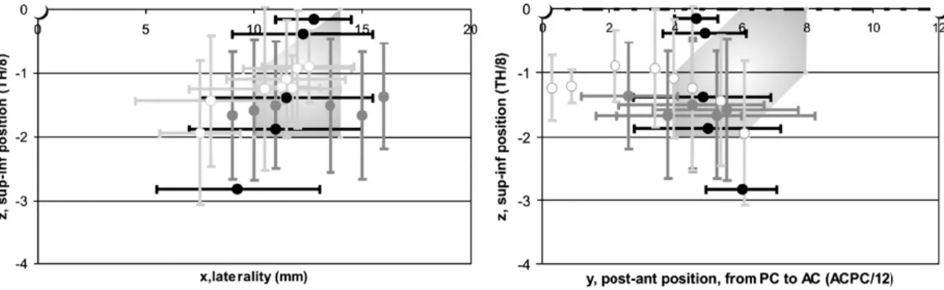
superior border of the thalamus) and the width of the third ventricle. The stereotactic coordinates of targets, in relation to the stereotactic reference, are provided by classical stereotactic atlases and stereotactic graphs (Talairach et al., 1957; Schaltenbrand and Bailey, 1959). Thus the probabilistic targeting relies on transposition of the theoretical position of a target, atlas- or graph-based, into the stereotactic space of a given patient’s brain through the ventricular stereotactic reference. The use of proportional coordinates and probabilistic location minimizes the influence of individual variability (Talairach et al., 1957; Schaltenbrand and Bailey, 1959; Velasco et al., 2001; Benabid et al., 2002) with continuous effort to optimize this approach (Nowinski et al., 2005; Yelnik et al., 2007). Although the method seems universally applicable, basic points can lead to errors of location: (1) the stereotactic locations provided by atlases and graphs can differ (e.g. Fig. 1) as an atlas relies on only one brain per plane (Schaltenbrand and Bailey, 1959) and a geometric figure represents only the area with the highest probability to find a given structure (Talairach et al.,1957; Schaltenbrand and Bailey, 1959; Benabid et al., 2002); (2) the commissural points are defined appreciably differently on their center (Schaltenbrand and Bailey, 1959; Benabid et al., 2002) or their periphery (Talairach et al., 1957) or according to different planes for the same atlas (Schaltenbrand and Bailey, 1959). Moreover the expression of stereotactic coordinates is different even if the orientation is standardized (x represents the laterality relative to the vertical midline plane going through ACPC; y represents the anterior– posterior position along the ACPC line; z represents the superior–inferior position relative to the ACPC axial plane, perpendicular to the midline plane). The units are in millimeters in case of absolute coordinates or in percentage of a standard, the ACPC line (e.g., 1/12 of ACPC) or the height of the thalamus (e.g. 1/4 of the thalamus height), in case of proportional coordinates. Coordinates are only proportional for y or z, since there is no reliable ventricle landmark to determine a standard in the frontal plane; however, a lateral proportionality can be applied (Velasco et al., 2001). Due to this probabilistic approach, it is strongly recommended to adjust the targeting by intra operative tests (electrophysiological neuronal recordings and/or clinical assessments) and multiple-tract explorations. The strength of the indirect targeting is above all the simplicity of the coordinate calculation and the important electrophysiological knowledge harvested.
4
Figure 1. Stereotactic location of the subthalamic nucleus (STN). STN projection area (pale grey surface) (Benabid
et al., 2002); projections of STN boundaries determined on axial (black dots and lines), sagittal (dark grey dots and lines) and frontal (light grey lines, white circles) slices (Schaltenbrand and Bailey, 1959). Frontal (left; white circle, projection of ACPC) and lateral (right; doted line, ACPC line; white circles, AC and PC) views (TH, thalamus height; AC, PC see text for abbreviations).
Imaging for indirect targeting
Clinical MRI, at least on the first generations of generalist machines, has non-negligible image distortion making delicate the ACPC definition and/or the location of surgical fiducials, as well as a low tissue contrast making reliable recognition of deep brain structures difficult. Therefore, matching CT with MRI was proposed (Duffner et al., 2002) in order to take the best of the two techniques, the geometric accuracy of CT plus the better ventricular anatomy on MRI. Nowadays because of the progresses of MRI machines, teams used more and more MRI to determine ACPC directly (Patel et al., 2003).
Indirect anatomic analysis of data
Even though it was originally designed to reach an invisible target for a given patient, the method of indirect location is also applied to analyze the positions of lesions (ablative surgery) or electrode contacts (deep brain stimulation) for different patients regardless of the technique of targeting (Benabid et al., 2002; Plaha et al., 2006). The aim is to determine the anatomic structures involved in the therapeutic process. The coordinates of lesions or contacts, for each patient and/or hemisphere, are displayed on an atlas or a graph. It is assumed that locations of lesions or contacts are effectively in the same structure identified during surgery; this depends mainly on surgical technical constraints and on the level of reliability of electrophysiological intraoperative guidance. The surgical constraints are linked up, among other things, with the
5
capabilities of stereotactic instrumentation. The reliability of electrophysiological guidance is not absolute (Schiff et al., 2002; Israel and Burchiel, 2004; McClelland et al., 2005) in particular because of our partial, and almost exclusively atlas-based, knowledge of “electro-anatomic” processes; a better comprehension of these latter should be allowed by a direct, patient-based, anatomic study.
The “direct” patient-based anatomic mapping in stereotactic surgery
In order to improve the surgical stereotactic method, since targets become more visible with new MRI modalities (hardware and software), a stereotactic reference is not mandatory anymore; the patient’s brain is its own reference, a direct patient-based anatomic mapping gets rational as from 1.5 T (Lemaire et al., 1999; Coubes et al., 2002; Plaha et al., 2006; Derost et al., 2007). Some teams use transitional methods, locating targets with reference to easy recognizable structures, like the red nucleus, coupled with a classic indirect atlas-based approach (Starr et al., 1999; Bejjani et al., 2000; Patel et al., 2003; Rampini et al., 2003; Andrade-Souza et al., 2005). Because of the wide range of anatomical quality of MRI sequences and the variability of surgical techniques, analysis of the literature concerning direct targeting reliability can be confusing (Andrade-Souza et al., 2005; Breit et al., 2006). However, pure direct targeting, allowed by patient-based anatomic mapping, yields benefits by reducing the number of exploration tracts and duration of intra operative tests due to optimal primary positioning (Caire et al., 2006) and by improving the analysis of relationships between a lesion or a contact and the structures implied in the clinical effect because of detailed anatomic analysis (Ulla et al., 2006). The increasing risk of hemorrhage with numerous exploration tracts (Hariz, 2002) argues for optimizing the primary positioning. A more widespread application of the method depends mostly on two factors: technical, the transfer of adequate MRI sequences; anatomic, the spread of the anatomic knowledge of nuclei and bundles related to the thalamus and basal ganglia, arranged in a complex manner and poorly known in detail. In practice, the identification and targeting of an anatomic structure rely on 3 key-points: namely, an MRI anatomic knowledge, high-quality images and dedicated surgical software. Even though anatomical knowledge is mostly based on anatomy books and stereotactic atlases, the identification of structures is facilitated by a very high field MRI anatomic reference (Lemaire et al., 2004) with contrasts similar to those used in clinical conditions and with 3D topographic analysis (Fig.2).
6
Figure 2. 4.7-T MRI reference of the human thalamus and basal ganglia. Imaging on a Biospec 4.7-T MRI system
(Bruker, GmbH, Ettlingen, Germany), anatomic specimen, 3D spin-echo sequence T1-weighted; isotropic voxel=250μm3. Isocentric images, reconstructed in the coronal (left) and axial (right) planes: Raw data (top row): labelled and highlighted structures (bottom row).
The high quality of MRI images is achieved with a small voxel size, below 1.5 mm3, and a high contrast between white and grey matters achieved with dedicated T2-weighted (Lemaire et al., 2001; Patel et al., 2003; Slavin et al., 2006) and inversion-recovery sequences (Magnotta et al., 2000; Siadoux et al., 2005). In clinical routine, there is no evidence, because of the intricate relationships between hardware and software, that 3-T magnetic field offers higher anatomic definition than 1.5-T magnetic field, at least with optimized sequences. If the anatomic images are acquired in stereotactic conditions, i.e. with the stereotactic frame locked in the head coil,
7
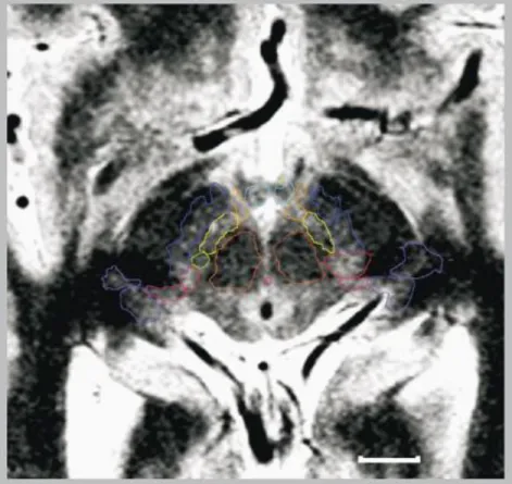
then head motion is minimized during the acquisition time of about 10 min per plane, increasing the signal/noise ratio (unpublished data). Anatomic sub structures in the area of interest, e.g. the thalamo-subthalamic region or the lenticular nucleus, can be labeled and manually highlighted (Fig.3) after identification based on the analysis of their knownrelative positions.
Figure 3. Axial 1.5-T MRI slices. Imaging (deep brain stimulation surgery, Parkinsonian) on a Sonata machine
(Siemens, GmbH, Erlangen, Germany), inversion-recovery sequence, stereotactic conditions; voxel size=0.52×0.62×2 mm3. Diencephalo-mesencephalic subthalamic structures are outlined and labeled (horizontal white bar=10 mm): substantia nigra (deep blue), red nucleus (orange), subthalamic nucleus (yellow), mammillary bodies (blue-green), nucleus of ansa lenticularis (light orange), substantia Q (green), zona incerta (red), peri peduncular nucleus (pink), lateral (light purple) and medial (deep purple) geniculate bodies.
The outlines of the structures can be helpful to interpret data on non-conventional interpolated planes reconstructed along the trajectories. Dedicated surgical software simplifies and improves the manipulation of image sets, allowing determination of the detailed anatomy, optimization of the trajectories (Fig. 4) and more detailed study of relationships between the anatomy and electrophysiological and clinical data.
8
Figure 4. 3D optimization of trajectory. Electrode implantation planned (same patient as Fig. 2) in the posterior
part of the subthalamic nucleus (yellow) near the zona incerta (red) and the Forel's fields (pale green), see Fig. 2 for the others labeled structures: 3D volume rendering of the subthalamic structures (frontal view; top left); pseudo coronal, pseudo axial and pseudo sagittal planes reconstructed along the right trajectory (clockwise from top right to bottom left).
Anatomic location of a set of lesions or contacts in a group of patients is still topical. The membership concept could incorporate the fact that a contact or a lesion can involve severalstructures. The membership degree can be weighted by a fuzzy logic method integrating the expert’s opinion concerning the level of uncertainty of the membership (Caire et al., 2006). The membership degree can also be weighted by the level of involvement of a contact into a structure and vice versa (Lemaire et al., 2005).
9
Future trends
Patient-based anatomic mapping should benefit from higher magnetic field, although beyond 3 T there are still important technical constraints preventing a routine clinical use, in particular in stereotactic conditions. But as from 1.5 T, diffusion tensor imaging (DTI) and tractography already offer new possibilities. These techniques enable the analysis of the anisotropy of brain tissue as well as fibbers constituting white bundles (Mori et al., 1999; Mori and Van Zijl, 2002; Le Bihan, 2003; Wakana et al., 2004; Hermoye et al., 2006) and several clinical applications have been published (Mukherjee, 2005; Nimsky et al., 2005; Arfanakis et al., 2006; Wilde et al., 2006). As bundles seem to participate in the DBS effects (Gabriëls et al., 2003; Hamel et al., 2003),studying their implication becomes possible. Parallel to the knowledge of the tissue anisotropy, also extracted from DTI, might be of interest because it could influence the electric current diffusion (McIntyre et al., 2004). Until now, it has been difficult to perform such imaging in stereotactic conditions, i.e. with a stereotactic frame in place, because of an important image distortion. Recently, modified surgical software yielded this approach at 1.5 T with routine stereotactic conditions. After matching of DTI with T2-weighted anatomic images and 3D tractography, it is possible to determine the main bundles of the subthalamic region potentially implied in the DBS process like the ansa lenticularis (Fig. 5).
Beyond improving stereotactic targeting, the patient-based anatomic mapping would enable new considerations for functional treatments relying on the spatial location inside specific brain areas, like radiosurgery, or the topographic diagnosis of lesions as during degenerative diseases, at least if the anatomy is not substantially modified. Furthermore, advances in predictive computational modeling (Frieboes et al., 2006) might help by reproducing in the computer the complexity and multi-dimensionality of a particular patient’s brain structure.
11
Figure 5. Fiber tracking on stereotactic 1.5-T DTI. Anisotropic diffusion images (diffusion tensor imaging [DTI];
top row, left) matched (mutual information algorithm) with anatomic images (inversion-recovery sequence, voxel size=0.52×0.62×2 mm3; top row, right) plus 3D anatomic structures (objects were created after manual outlining). DTI acquisition was performed on a 1.5-T machine (Siemens Sonata, GmbH, Erlangen, Germany): stereotactic conditions, with a Leksell G frame; 6 directions,b value=750 s/mm2, voxel size=1.8×1.8×3 mm3; image post processing with Iplan (BrainLab, Feldkirchen, Germany), color-coded fiber direction (blue for superior–inferior, red for left–right, and green for anterior–posterior) plus color map matched with 3D anatomic structures (intermediate row). Tractography (bottom row): (Top) tracking with a fractional anisotropy threshold ≥0.30 and length of fibers ≥40 mm; (Middle) volume of interest (blue box displayed on a coronal slice; insert bottom right) placed along the right trajectory on the interface between the subthalamic nucleus and the pre rubral Forel's field (same patient as Fig. 2); (Bottom) right lateral view of anatomic structures (see Figs. 2 and 3 for labels; the substantia nigra is transparent; left structures are hidden) fused with color-coded fibers according to the direction: ansa lenticularis (Al), subthalamic occipito-parietal bundle (OP b), frontal bundle going trough the anterior limb of the internal capsule (Fr b) and the dento-rubro-thalamic fascicle (Fx DRT).
References
Andrade-Souza, Y.M., Schwalb, J.M., Hamani, C., Eltahawy, H., Hoque, T., Saint-Cyr, J., Lozano, A.M., 2005. Comparison of three methods of targeting the subthalamic nucleus for chronic stimulation in Parkinson’s disease. Neurosurgery 52 (2), 360–368.
Arfanakis, K., Gui, M., Lazar, M., 2006. Optimization of white matter tractography for pre-surgical planning and image-guided surgery. Oncol. Rep. 15 (Spec no.: 1061–4).
Bejjani, B.P., Dormont, D., Pidoux, B., Yelnik, J., Damier, P., Arnulf, I., Bonnet, A.M., Marsault, C., Agid, Y., Philippon, J., Cornu, P., 2000. Bilateral subthalamic stimulation for Parkinson’s disease by using three-dimensional stereotactic magnetic resonance imaging and electrophysiological guidance. J. Neurosurg. 92, 615–625.
Benabid, A.L., Koudsie, A., Benazzouz, A., Le Bas, J.F., Pollak, P., 2002. Imaging of subthalamic nucleus and ventral intermedius of the thalamus. Mov. Disord. 17, S123–S129. Breit, S., LeBas, J.F., Koudsie, A., Schulz, J., Benazzouz, A., Pollak, P., Benabid, A.L., 2006. Pretargeting for the implantation of stimulation electrodes into the subthalamic nucleus: a comparative study of magnetic resonance imaging and ventriculography. Neurosurgery 58 (ONS Suppl. 1) (ONS-83–95).
Caire, F., Derost, P., Coste, J., Bonny, J.M., Durif, F., Frenoux, E., Villéger, A., Lemaire, J.J., 2006. Stimulation sous-thalamique dans la maladie deParkinson sévère: Étude de la localisation des contacts effectifs. Neurochirurgie 52, 15–25.
Coubes, P., Vayssiere, N., El Fertit, H., Hemm, S., Cif, L., Kienlen, J., Bonafe, A., Frerebeau, P., 2002. Deep brain stimulation for dystonia: surgical technique. Stereotact. Funct. Neurosurg. 78, 183–191.
12
Derost, P.P., Ouchchane, L., Morand, D., Ulla, M., Llorca, P.M., Barget, M., Debilly, B., Lemaire, J.J., Durif, F., 2007. Is subthalamic nucleus deep brain stimulation (DBS-STN) appropriate to manage severe Parkinson disease in an elderly population? Neurology 68, 1345–1355.
Duffner, F., Schiffbauer, H., Breit, S., Friese, S., Freudenstein, D., 2002. Relevance of image fusion for target point determination in functional neurosurgery. Acta Neurochir. 144, 445–451. Frieboes, H.B., Zheng, X., Sun, C.H., Tromberg, B., Gatenby, R., Cristini, V., 2006. An integrated computational/experimental model of tumor invasion. Cancer Res. 66, 1597–1604. Gabriëls, L., Cosyns, P., Nuttin, B., Demeulemeester, H., Gybels, J., 2003. Deep brain stimulation for treatment-refractory obsessive–compulsive disorder: psychopathological and neuropsychological outcome in three cases. Acta Psychiatr. Scand. 107, 275–282.
Hamel, W., Fietzek, U., Morsnowski, A., Schrader, B., Herzog, J., Weinert, D., Pfister, G., Muller, D., Volkmann, J., Deuschl, G., Mehdorn, H.M., 2003. Deep brain stimulation of the subthalamic nucleus in Parkinson’s disease: evaluation of active electrode contacts. J. Neurol. Neurosurg. Psychiatry 74, 1036–1046.
Hariz, M.I., 2002. Safety and risk of microelectrode recording in surgery for movement disorders. Stereotact. Funct. Neurosurg. 78, 146–157.
Hermoye, L., Saint-Martin, C., Cosnard, G., Lee, S.K., Kim, J., Nassogne, M.C., Menten, R., Clapuyt, P., Donohue, P.K., Hua, K., Wakana, S., Jiang, H., Van Zijl, P.C., Mori, S., 2006. Pediatric diffusion tensor imaging: normal database and observation of the white matter maturation in early childhood. NeuroImage 29, 493–504.
Israel, Z., Burchiel, K.J., 2004. Microelectrode Recording in Movement Disorder Surgery. Thieme, New York.
Le Bihan, D., 2003. Looking into the functional architecture of the brain with diffusion MRI. Nat. Rev., Neurosci. 4, 469–480.
Lemaire, J.J., Durif, F., Boire, J.Y., Debilly, B., Irthum, B., Chazal, J., 1999. Direct stereotactic MRI location in the globus pallidus for chronic stimulation in Parkinson’s disease. Acta Neurochir. (Wien.) 141,759–766.
Lemaire, J.J., Durif, F., Debilly, B., Blanc, O., Chazal, J., 2001. Deep brain stimulation in the subthalamic area for severe idiopathic Parkinson’s disease: location of plots in the preoperative phase and at the three month follow-up. Parkinson’s Relat. Disord. 7, S80 (Suppl.).
Lemaire, J.J., Caire, F., Bony, J.M., Kemeny, J.L., Villéger, A., Chazal, J., 2004. Contribution of 4.7-Tesla MRI in the analysis of the MRI anatomy of the human subthalamic area. Acta Neurochir. 146, 906–907.
13
Lemaire, J.J., Coste, J., Ouchchane, L., Derost, P., Ulla, M., Durif, F., Caire, F., Siadoux, S., Gabrillargues, J., Chazal, J., 2005. Stimulation électrique à haute fréquence du noyau sous thalamique dans la maladie de Parkinson sévère idiopathique: analyse du site optimale de stimulation à partir des données électrophysiologiques per opératoires et de l’IRM anatomique stéréotaxique. Neurochirurgie 519.
McIntyre, C.C., Mori, S., Sherman, D.L., Thakorc, N.V., Vitek, J.L., 2004. Electric field and stimulating influence generated by deep brain stimulation of the subthalamic nucleus. Clin. Neurophysiol. 115,589–595.
Magnotta, V.A., Gold, S., Andreasen, N.C., Ehrhardt, J.C., Yuh, T.C., 2000. Visualization of subthalamic nuclei with cortex attenuated inversion recovery MR imaging. NeuroImage 11, 341– 346.
McClelland, S., Ford, B., Senatus, P.B., Winfield, L.M., Du, Y.E., Pullman, S.L., Yu, Q., Frucht, S.J., McKhann, G.M., Goodman, R.R., 2005. Subthalamic stimulation for Parkinson disease: determination of electrode location necessary for clinical efficacy. Neurosurg. Focus 19, E12. Mori, S., Van Zijl, P.C., 2002. Fiber tracking: principles and strategies–a technical review. NMR Biomed. 15, 468–480.
Mori, S., Crain, B.J., Chacko, V.P., Van Zijl, P.C., 1999. Three-dimensional tracking of axonal projections in the brain by magnetic resonance imaging. Ann. Neurol. 45, 265–269.
Mukherjee, P., 2005. Diffusion tensor imaging and fiber tractography in acute stroke. Neuroimaging Clin. N. Am. 15, 655–665 (xii).
Nimsky, C., Ganslandt, O., Hastreiter, P., Wang, R., Benner, T., Sorensen, A.G., Fahlbusch, R., 2005. Preoperative and intraoperative diffusion tensor imaging-based fiber tracking in glioma surgery. Technique Applications. Neurosurgery 56, 130–138.
Nowinski, W.L., Belov, D., Pollak, P., Benabid, A.L., 2005. Statistical analysis of 168 bilateral subthalamic nucleus implantations by means of the probabilistic functional atlas. Oper. Neurosurg. 57, 319–330.
Patel, N.K., Plaha, P., O’Sullivan, K., McCarter, R., Heywood, P., Gill, S.S., 2003. MRI directed bilateral stimulation of the subthalamic nucleus in patients with Parkinson’s disease. J. Neurol. Neurosurg. Psychiatry 74,1631–1637.
Plaha, P., Ben-Shlomo, Y., Patel, N.K., Gill, S.S., 2006. Stimulation of the caudal zona incerta is superior to stimulation of the subthalamic nucleus in improving contralateral parkinsonism. Brain 129, 1732–1747.
Rampini, P.M., Locatelli, M., Alimehmeti, R., Tamma, F., Caputo, E., Priori, A., Pesenti, A., Rohr, M., Egidi, M., 2003. Multiple sequential image-fusion and direct MRI localisation of the subthalamic nucleus for deep brain stimulation. J. Neurosurg. Sci. 47, 33–39.
14
Schaltenbrand, G., Bailey, P., 1959. Introduction to Stereotaxis With an Atlas of the Human Brain: Volume II. Georg Thieme Verlag, Stuttgart.
Schiff, S.J., Dunagan, B.K., Worth, R.M., 2002. Failure of single-unit neuronal activity to differentiate globus pallidus internus and externus in Parkinson disease. J. Neurosurg. 97, 119– 128.
Siadoux, S., Gabrillargues, J., Coste, J., Claise, B., Chabert, E., Michel, J.L., Durif, F., Lemaire, J.J., 2005. IRM stéréotaxique de la région sous thalamique: optimisation d’une séquence de repérage pré opératoire pour la mise en place d’electrodes de stimulation profonde chronique. Neurochirurgie 51, 519.
Slavin, K.V., Thulborn, K.R., Wess, C., Nersesyan, H., 2006. Direct visualization of the human subthalamic nucleus with 3 T MR imaging. AJNR (27), 80–84.
Starr, P., Vitek, J., DeLong, M., Bakay, R., 1999. Magnetic resonance imaging-based stereotactic localization of the globus pallidus and subthalamic nucleus. Neurosurgery 44, 303–313.
Talairach, J., David, M., Tournoux, P., Corredor, H., Kvasina, T., 1957. Atlas d’anatomie stéréotaxique. Repérage radiologique indirect des noyaux gris centraux des régions mésencéphalo-sous-optiques et hypothalamiques de l’homme. Masson and Cie, Paris.
Ulla, M., Thobois, S., Lemaire, J.J., Schmitt, A., Derost, P., Broussolle, E., Llorca, P.M., Durif, F., 2006. Manic behaviour induced by deep brain stimulation in Parkinson’s disease: evidence of substantia nigra implication? J. Neurol. Neurosurg. Psychiatry 77, 1363–1366.
Velasco, F., Jiménez, F., Pérez, M.L., Carrillo-Ruiz, J.D., Velasco, A.L., Ceballos, J., Velasco, M., 2001. Electrical stimulation of the prelemniscal radiation in the treatment of Parkinson’s disease: an old target revised with new techniques. Neurosurgery 49, 293–308.
Wakana, S., Jiang, H., Nagae-Poetscher, L.M., Van Zijl, P.C., Mori, S., 2004. Fiber tract-based atlas of human white matter anatomy. Radiology 230, 77–87.
Wilde, E.A., Chu, Z., Bigler, E.D., Hunter, J.V., Fearing, M.A., Hanten, G., Newsome, M.R., Scheibel, R.S., Li, X., Levin, H.S., 2006. Diffusion tensor imaging in the corpus callosum in children after moderate to severe traumatic brain injury. J. Neurotrauma 23, 1412–1426.
Yelnik, J., Bardinet, E., Dormont, D., Malandaine, G., Ourseline, S., Tandéa, D., Karachia, C., Ayachee, N., Cornu, P., Agid, Y., 2007. A three-dimensional, histological and deformable atlas of the human basal ganglia: I. Atlas construction based on immunohistochemical and MRI data. NeuroImage 34, 618–638.