ARTICLE
Comparison of
Staphylococcus aureus isolates associated
with food intoxication with isolates from human nasal
carriers and human infections
L. Wattinger&R. Stephan&F. Layer&S. Johler
Received: 12 May 2011 / Accepted: 17 June 2011 / Published online: 15 July 2011 # Springer-Verlag 2011
Abstract Staphylococcus aureus represents an organism of striking versatility. While asymptomatic nasal colonization is widespread, it can also cause serious infections, toxinoses and life-threatening illnesses in humans and animals. Staphylo-coccal food poisoning (SFP), one of the most prevalent causes of foodborne intoxication worldwide, results from oral intake of staphylococcal enterotoxins leading to violent vomiting, diarrhea and cramps shortly upon ingestion. The aim of the present study was to compare isolates associated with SFP to isolates collected from cases of human nasal colonization and clinical infections in order to investigate the role of S. aureus colonizing and infecting humans as a possible source of SFP. Spa typing and DNA microarray profiling were used to characterize a total of 120 isolates, comprising 50 isolates collected from the anterior nares of healthy donors, 50 isolates obtained from cases of clinical infections in humans and 20 isolates related to outbreaks of staphylococcal food poisoning. Several common spa types were found among isolates of all three sources (t015, t018, t056, t084). DNA microarray results showed highly similar virulence gene profiles for isolates from all tested sources. These results suggest contamination of foodstuff with S. aureus colonizing and infecting food handlers to represent a source of SFP.
Introduction
Staphylococcus aureus is not only a commensal colonizer, but can also cause serious infections, toxinoses and life-threatening diseases, such as skin and soft tissue infections, toxic shock syndrome and septicemia. S. aureus colonizes skin and mucosa of humans and animals, with nasal carriage rates between 30% and 50% among the adult human population [1–4]. While colonization of the anterior nares is usually asymptomatic, it serves as a reservoir for the spread of the organism [1,5]. Carriers are at increased risk to develop nosocomial bacteremia which in 80% of cases is caused by the strain colonizing their nares [6,7]. The rapid emergence of antibiotic resistance among S. aureus is also known to play a crucial role in the epidemiology of staphylococcal infections. Recently, infections with methicillin resistant S. aureus (MRSA) have been estimated to constitute the leading cause of death due to one single infectious agent in the United States [8].
S. aureus also represents the cause of staphylococcal food poisoning (SFP), one of the most prevalent foodborne intoxications worldwide. SFP results from ingestion of staphylococcal enterotoxins preformed in food, typically presenting with violent emesis, nausea, diarrhea and prostra-tion. While in most cases symptoms subside spontaneously after 24 h, fatality rates range from 0.03% in the general population to 4.4% in children and the elderly [9]. As staphylococcal colonization and infection is widely spread, contamination of foodstuff by food handlers may represent a major source of SFP. As SFP isolates are difficult to obtain, to date, there is only very limited information on the original source of enterotoxigenic S. aureus strains that lead to cases of food poisoning.
Different techniques are established for typing S. aureus. The most widely used method for epidemiological
inves-L. Wattinger
:
R. Stephan:
S. Johler (*) Institute for Food Safety and Hygiene, Vetsuisse Faculty University of Zurich, Winterthurerstrasse 272,CH-8057 Zurich, Switzerland e-mail: [email protected] F. Layer
Robert Koch Institute, Wernigerode Branch, Burgstrasse 37,
tigations is spa typing, based on the determination of the polymorphic X region of the gene encoding staphylococcal protein A (spa). DNA microarray is used for rapid detection of a multitude of virulence genes (genes encoding enter-otoxins, hemolysins, leukocidins, etc.), resistance determi-nants, and typing markers. The resulting hybridization pattern can be used to assign isolates to clonal complexes [10].
In this study, spa typing and DNA microarray analysis were performed with a total of 120 S. aureus isolates, comprising S. aureus isolates obtained from nasal coloni-zation in healthy donors, isolates gained from clinical cases of infection and isolates associated with outbreaks of staphylococcal food poisoning. The objective was to compare SFP isolates to isolates obtained from S. aureus nasal colonization (SANC) and clinical cases of infection (SAI) in order to determine the role of S. aureus colonizing and infecting humans as a possible source of SFP.
Materials and methods Bacterial isolates
A total of 120 S. aureus isolates were examined, constitut-ing 50 SANC isolates, 50 SAI isolates and 20 isolates associated to outbreaks of SFP in humans. Nasal swabs of the anterior nares were collected from randomly chosen
volunteers in Switzerland between November and December 2010. Samples from both nostrils were taken using sterile cotton swabs moistened with saline. Fifty SAI isolates were obtained from the Institute of Medical Microbiol-ogy of the University of Zurich, Switzerland, between November and December 2010. The 20 SFP isolates were provided by the Bavarian Authorities for Health and Food Safety (LGL, Munich, Germany), the German National Reference Center for Staphylococci (Robert Koch Institute, Wernigerode, Germany), the Cantonal Laboratory Fribourg (Fribourg, Switzerland) and the Medical Department of the German Federal Armed Forces (Kronshagen, Germany) (Table 1). Ethical clearance was granted by the locally cognizant ethics commission (cantonal ethics commission, Zurich).
DNA extraction and species identification
Swabs were streaked directly onto rabbit plasma fibrinogen (RPF) plates (Oxoid, Basel, Switzerland), incubated at 37°C and examined for coagulase activity after 48 h. Two S. aureus typical colonies (colonies surrounded by an opaque halo) each were subcultured on RPF plates (48 h at 37°C). One colony of each plate was transferred to blood agar and incubated overnight at 37°C. DNA isolation kits were obtained from QIAGEN (Hilden, Germany) and handled according to the manufacturer's instructions. The PCR consumables were supplied by Promega (Madison, Wisconsin,
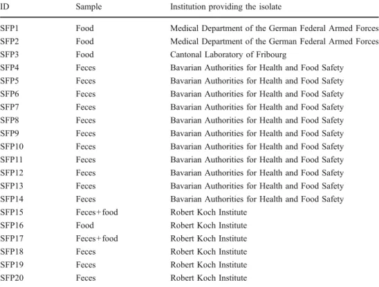
ID Sample Institution providing the isolate
SFP1 Food Medical Department of the German Federal Armed Forces
SFP2 Food Medical Department of the German Federal Armed Forces
SFP3 Food Cantonal Laboratory of Fribourg
SFP4 Feces Bavarian Authorities for Health and Food Safety
SFP5 Feces Bavarian Authorities for Health and Food Safety
SFP6 Feces Bavarian Authorities for Health and Food Safety
SFP7 Feces Bavarian Authorities for Health and Food Safety
SFP8 Feces Bavarian Authorities for Health and Food Safety
SFP9 Feces Bavarian Authorities for Health and Food Safety
SFP10 Feces Bavarian Authorities for Health and Food Safety
SFP11 Feces Bavarian Authorities for Health and Food Safety
SFP12 Feces Bavarian Authorities for Health and Food Safety
SFP13 Feces Bavarian Authorities for Health and Food Safety
SFP14 Feces Bavarian Authorities for Health and Food Safety
SFP15 Feces+food Robert Koch Institute
SFP16 Food Robert Koch Institute
SFP17 Feces+food Robert Koch Institute
SFP18 Feces Robert Koch Institute
SFP19 Feces Robert Koch Institute
SFP20 Feces Robert Koch Institute
Table 1 Staphylococcal food poisoning (SFP) isolates included in this study
USA). The DNA concentration was measured by using a Nanodrop ND-1000 UV/Vis spectrophotometer (NanoDrop Technologies, Wilmington, DE).
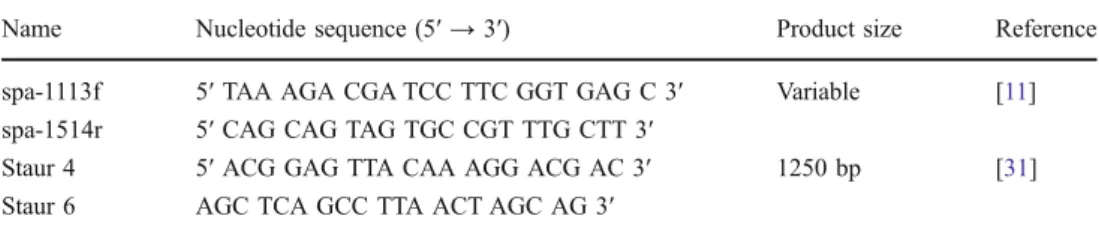
Spa typing
The sequence of the polymorphic X region of the spa gene of each S. aureus isolate was determined as described by Aires-de-Sousa et al. [11], with minor modifications. Briefly, the spa gene was amplified with spa-1113f and spa-1514r primers (Table2) using the GoTaq PCR system (Promega AG, Dübendorf, Switzerland) at the following reaction conditions: (i) 5 min at 94°C; (ii) 35x [45 s at 94°C; 45 s at 60°C; 90 s at 72°C]; (iii) 10 min at 72°C. PCR purification and sequencing was outsourced (GATC Biotech, Constance, Germany and Microsynth, Balgach, Switzerland). The sequences were assigned to spa types using the spa-server (http://www.spaserver.ridom.de/) [12]. Clonal complexes were determined using Ridom StaphType 2.0.3 software and the Based Upon Repeat Pattern (BURP) algorithm.
Microarray based genotyping
For DNA microarray profiling the StaphyType ArrayStrip platform was used according to the manufacturer's instruc-tions (Clondiag chip technologies, Jena, Germany). Similar to Coombs et al., microarray profiles were compared using SplitsTree4, a software designed to compute unrooted phylogenetic networks from molecular sequence data [13, 14]. DNA microarray gene profiles were converted to “sequence-like” strings of information, defining present genes as“A” (positive), absent genes as “T” (negative) and spots with ambiguous signal intensities as missing.
Statistical analysis
The distribution of genes among SANC, SAI, and SFP isolates was compared based on the hybridization results of the DNA microarray. SPSS Statistics 19 was used to run Pearson's chi-squared test, identifying significant associa-tions between the source the isolates were collected from and the presence of the examined genes. P-values<0.05 were considered statistically significant.
Results
Screening of nasal swabs for the presence of S. aureus showed a nasal carriage rate of 37.6% among the 133 healthy test persons.
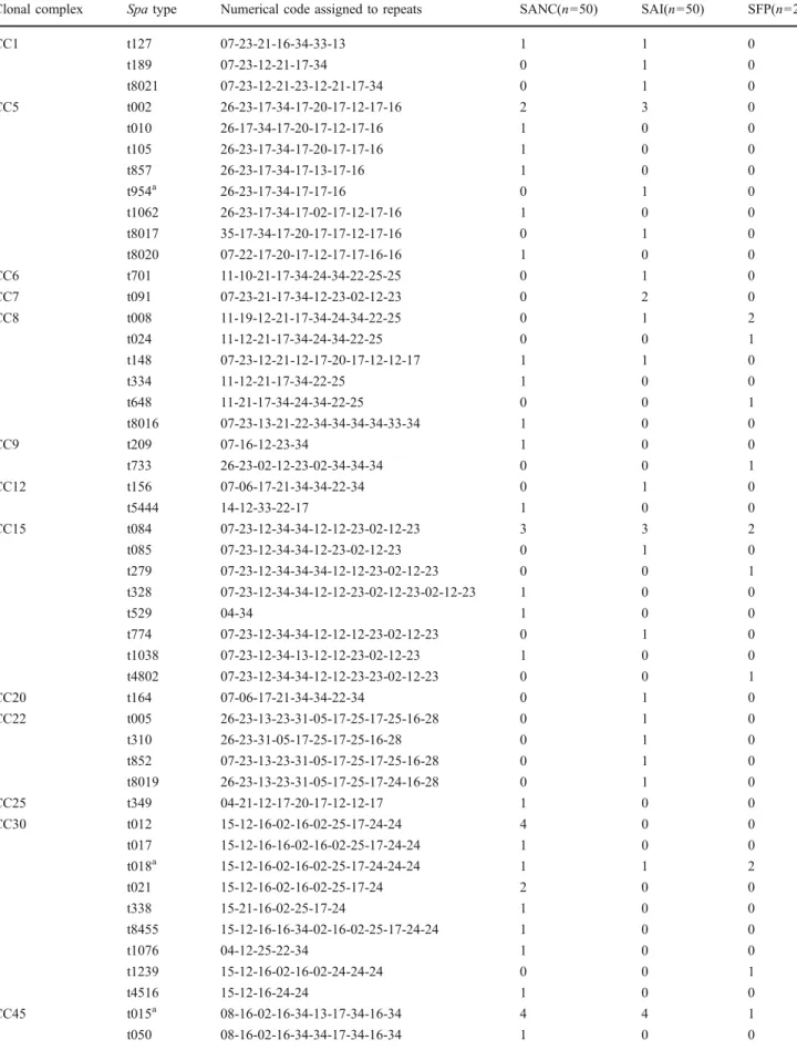
The 120 staphylococcal isolates tested, including 50 SANC, 50 SAI, as well as 20 SFP isolates, could be assigned to 20 clonal complexes comprising a total of 79 different spa types (see Table3). Among SANC, SAI, and SFP isolates, clonal complexes CC8, CC15, CC30, CC45, CC78, and CC101 could be found. Isolates from all three sources were frequently assigned to CC45 (SANC: 16%, SAI: 20%, SFP: 30%). While high prevalence of CC30 was found among SANC (24%) and SFP isolates (15%), SAI isolates were more often assigned to CC59 (14%). The 50 isolates from nasal swabs were grouped into 39 spa types with spa type t015 and t012 being found most frequently (8% each). The 50 SAI isolates grouped into 38 different spa types, with t216 representing the most common spa type (12%). The 20 isolates associated with SFP were grouped into 15 spa types. Some common spa types were found among isolates of all three sources (t015, t018, t056, and t084). Isolates obtained from nasal colonization and cases of clinical infections were overlapping in spa types t002, t127, t148, and t216. Spa type t008 was found in both clinical and food poisoning isolates of S. aureus.
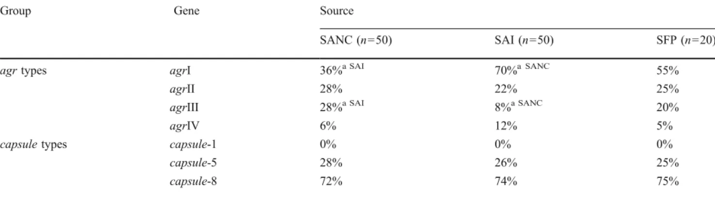
DNA microarray was used to determine gene profiles of all 120 strains. Hybridization results for agr and capsule types are depicted in Table4. While agrI was found to represent the most frequent agr type among all three sources, SANC and SAI isolates differed significantly in the number of isolates assigned to agrI (36% SANC, 70% SAI; p=0.001) and agrIII (28% SANC, 8% SAI, p=0.009).
Most isolates possessed one or several genes involved in resistance to antimicrobial agents (see Table 5). The blaZ gene conferring resistance to beta lactams was found most frequently among isolates from all three sources (SANC: 74%, SAI: 76%, SFP: 85%). Antibiotic resistance profiles were highly similar for SANC, SAI, and SFP isolates. Only fosB, which is involved in resistance to fosfomycin and bleomycin, was present in significantly higher numbers in SANC than in SAI isolates (SANC: 68%, SAI: 38%; p= 0.002). While genes involved in vancomycin resistance (vanA/B/Z) were not found, few isolates exhibited genes
Name Nucleotide sequence (5′ → 3′) Product size Reference
spa-1113f 5′ TAA AGA CGA TCC TTC GGT GAG C 3′ Variable [11]
spa-1514r 5′ CAG CAG TAG TGC CGT TTG CTT 3′
Staur 4 5′ ACG GAG TTA CAA AGG ACG AC 3′ 1250 bp [31]
Staur 6 AGC TCA GCC TTA ACT AGC AG 3′
Table 2 Primers used in this study
Table 3 Spa types and predicted clonal complexes of S. aureus nasal colonization (SANC), clinical cases of infection (SAI) and staphylococcal food poisoning (SFP) isolates investigated in this study
Clonal complex Spa type Numerical code assigned to repeats SANC(n=50) SAI(n=50) SFP(n=20)
CC1 t127 07-23-21-16-34-33-13 1 1 0 t189 07-23-12-21-17-34 0 1 0 t8021 07-23-12-21-23-12-21-17-34 0 1 0 CC5 t002 26-23-17-34-17-20-17-12-17-16 2 3 0 t010 26-17-34-17-20-17-12-17-16 1 0 0 t105 26-23-17-34-17-20-17-17-16 1 0 0 t857 26-23-17-34-17-13-17-16 1 0 0 t954a 26-23-17-34-17-17-16 0 1 0 t1062 26-23-17-34-17-02-17-12-17-16 1 0 0 t8017 35-17-34-17-20-17-17-12-17-16 0 1 0 t8020 07-22-17-20-17-12-17-17-16-16 1 0 0 CC6 t701 11-10-21-17-34-24-34-22-25-25 0 1 0 CC7 t091 07-23-21-17-34-12-23-02-12-23 0 2 0 CC8 t008 11-19-12-21-17-34-24-34-22-25 0 1 2 t024 11-12-21-17-34-24-34-22-25 0 0 1 t148 07-23-12-21-12-17-20-17-12-12-17 1 1 0 t334 11-12-21-17-34-22-25 1 0 0 t648 11-21-17-34-24-34-22-25 0 0 1 t8016 07-23-13-21-22-34-34-34-34-33-34 1 0 0 CC9 t209 07-16-12-23-34 1 0 0 t733 26-23-02-12-23-02-34-34-34 0 0 1 CC12 t156 07-06-17-21-34-34-22-34 0 1 0 t5444 14-12-33-22-17 1 0 0 CC15 t084 07-23-12-34-34-12-12-23-02-12-23 3 3 2 t085 07-23-12-34-34-12-23-02-12-23 0 1 0 t279 07-23-12-34-34-34-12-12-23-02-12-23 0 0 1 t328 07-23-12-34-34-12-12-23-02-12-23-02-12-23 1 0 0 t529 04-34 1 0 0 t774 07-23-12-34-34-12-12-12-23-02-12-23 0 1 0 t1038 07-23-12-34-13-12-12-23-02-12-23 1 0 0 t4802 07-23-12-34-34-12-12-23-23-02-12-23 0 0 1 CC20 t164 07-06-17-21-34-34-22-34 0 1 0 CC22 t005 26-23-13-23-31-05-17-25-17-25-16-28 0 1 0 t310 26-23-31-05-17-25-17-25-16-28 0 1 0 t852 07-23-13-23-31-05-17-25-17-25-16-28 0 1 0 t8019 26-23-13-23-31-05-17-25-17-24-16-28 0 1 0 CC25 t349 04-21-12-17-20-17-12-12-17 1 0 0 CC30 t012 15-12-16-02-16-02-25-17-24-24 4 0 0 t017 15-12-16-16-02-16-02-25-17-24-24 1 0 0 t018a 15-12-16-02-16-02-25-17-24-24-24 1 1 2 t021 15-12-16-02-16-02-25-17-24 2 0 0 t338 15-21-16-02-25-17-24 1 0 0 t8455 15-12-16-16-34-02-16-02-25-17-24-24 1 0 0 t1076 04-12-25-22-34 1 0 0 t1239 15-12-16-02-16-02-24-24-24 0 0 1 t4516 15-12-16-24-24 1 0 0 CC45 t015a 08-16-02-16-34-13-17-34-16-34 4 4 1 t050 08-16-02-16-34-34-17-34-16-34 1 0 0
associated with resistance to tetracycline (tetK/M) and meth-icillin (mecA). One SANC (SANC11) and four SAI isolates (SAI8, SAI9, SAI12, SAI36) possessed mecA. SANC11 was detected in a nasal swab from a female veterinarian aged 27 that could be assigned to ST398-MRSA-V (“Dutch Pig Strain”, score: 93.1%). SAI8 was isolated from a skin lesion in a 58-year-old male patient suffering from sepsis. SAI9 was detected in a pharyngeal swab from a 76-year-old male patient and was assigned to ST36/39-MRSA-II, UK-EMRSA-16 (synonym to USA 200, Irish AR7.0, Canadian MRSA-4; score: 94.3%). SAI12 was isolated from a perineal/perianal swab of an 85-year-old male and was assigned to ST45-MRSA-IV, Berlin EMRSA (synonym to USA 600-ST45-MRSA-IV, score: 91.8%). SAI 36 was isolated from a 42-year-old female
suffering from ulceration after a burn wound and could be assigned to CC78-MRSA-IV, WA MRSA-2 (score: 96.0%).
DNA microarray results for genes encoding superantigenic toxins are displayed in Table6. We tested for genes encoding staphylococcal enterotoxins (entA-entJ), enterotoxin-like proteins (entK-entR, entU), as well as exfoliative toxins (entA/B/D), toxic shock syndrome toxin (tst-1), and panton valentine leukocidin (pvl). While entA-entD were found in isolates of all three sources, entE was not detected. SFP isolates were significantly more likely to possess enterotoxin A variant entA-320 than SANC (p=0.005) and SAI isolates (p=0.002). In comparison with SFP isolates, SANC isolates exhibited enterotoxin A variant entA-N315 in significantly higher (p=0.042) and entD in significantly lower numbers.
Table 3 (continued)
Clonal complex Spa type Numerical code assigned to repeats SANC(n=50) SAI(n=50) SFP(n=20)
t073 08-16-02-16-13-17-34-16-34 0 1 0 t230 08-16-02-16-34 0 1 0 t377 04-02-12-21-17-34-22-25 0 0 1 t383 08-16-34-13-16-34 0 0 1 t445 08-16-20-16-34-13-17-34-16-34 0 1 0 t630 08-16-02-16-34-17-34-16-34 1 0 0 t950 08-16-34-17-34-16-34 0 1 0 t1270 09-34-34-34-17-34-16-34 0 0 2 t1574 08-16-02-16-34-13-13-17-34-16-34 1 0 0 t4460 08-16-02-16-17-34-16-34 0 1 0 t5599 08-16-02-16-34-13-16-34-16-34 1 0 0 t6969 08-16-13-17-34-16-34-34 0 0 1 t8454 08-16-02-16-34-16-34-13-17-34-16-34 0 1 0 CC50 t246 04-17-23-24-20-17-25 0 1 0 t8018 04-20-22-17 0 1 0 CC59 t216 04-20-17-20-17-31-16-34 2 6 0 t270 14-44-13-12-17-17-17-17-23-18-17 1 0 0 t437 04-20-17-20-17-25-34 0 1 0 CC78 t186a 07-12-21-17-13-13-34-34-33-34 0 1 0 t912 08-12-17-13-13-34-13 0 0 1 t1814 07-12-21-17-34-34-34-33-34 1 0 0 CC97 t267 07-23-12-21-17-34-34-34-33-34 0 1 0 t276 15-12-16-02-16-02-25 0 1 0 t359 07-23-12-21-17-34-34-33-34 1 0 0 CC101 t056 04-20-12-17-20-17-12-17-17 1 1 1 t2888 04-20-12-17-13-17 0 1 0 CC121 t159 14-44-13-12-17-17-23-18-17 1 0 0 t272 14-44-13-12-17-17-17-23-18-17 0 1 0 t645 14-44-13-12-17-23-18-17 1 0 0 CC398 t011b 08-16-02-25-34-24-25 1 0 0 t571 08-16-02-25-02-25-34-25 1 0 0
a spa types comprising mecA positive SAI isolates (SAI8: t954/CC5, SAI9: t018/CC30, SAI12: t015/CC45, SAI36: t186/CC78) b spa type comprising the mecA positive isolate obtained from nasal colonization SANC11
We observed an even distribution of genes belonging to the enterotoxin gene cluster (entG, entI, entM, entN, entO, entU).
SFP isolates were shown to possess entJ and entR in significantly higher numbers than isolates obtained from
Table 4 Assignment to agr and capsule types based on DNA microarray analysis. The percentages represent the fragment of S. aureus nasal colonization (SANC), clinical cases of infection (SAI) and staphylococcal
food poisoning (SFP) isolates, for which genes were determined to be present. Calculations include positive signals only, ambiguous signals were omitted
Group Gene Source
SANC (n=50) SAI (n=50) SFP (n=20)
agr types agrI 36%a SAI 70%a SANC 55%
agrII 28% 22% 25%
agrIII 28%a SAI 8%a SANC 20%
agrIV 6% 12% 5%
capsule types capsule-1 0% 0% 0%
capsule-5 28% 26% 25%
capsule-8 72% 74% 75%
a
Result for this source differs significantly from the result calculated for one (source indicated) or both other sources of isolates investigated in this study (p<0.05)
Gene Affected antibiotic Source
SANC(n=50) SAI(n=50) SFP(n=20)
mecA Methicillin 2% 8% 0%
blaZ Beta-lactam 74% 76% 85%
ermA Macrolides, lincosamides, streptogramin 8% 6% 0%
ermB Macrolides, lincosamides, streptogramin 2% 2% 5%
ermC Macrolides, lincosamides, streptogramin 2% 0% 5%
linA Lincosamide 0% 2% 0% mrsA Macrolides 0% 0% 0% mefA Macrolides 0% 0% 0% mpbBM Macrolides 0% 0% 0% vatA Streptogramin 0% 0% 0% vatB Streptogramin 0% 0% 0% vga, b Streptogramin 0% 0% 0% vgaA Streptogramin 0% 0% 0%
aacA-aphaD Aaminoglycosides (gentamicin, tobramycin) 2% 2% 0%
aadD Aminoglycosides (gentamicin, tobramycin) 0% 2% 3%
aphA Aminoglycosides (gentamicin, tobramycin) 0% 2% 0%
sat Streptothricin 0% 2% 0%
dfrA Trimethoprim 0% 4% 0%
far Fusidic acid 0% 0% 0%
mupR Mupirocin 0% 0% 0%
tetK Tetracycline 2% 8% 10%
tetM Tetracycline 2% 4% 0%
cat Chloramphenicol 0% 0% 0%
fexA Chloramphenicol 0% 2% 0%
fosB Fosfomycin, bleomycin 68%a SAI 38%a SANC 65%
vanA, Z Vancomycin 0% 0% 0%
vanB Vancomycin 0% 0% 0%
qacA, C Unspecific efflux pump 0% 4% 5%
Table 5 Genes involved in anti-biotic resistance. Percentages of S. aureus nasal colonization (SANC), clinical cases of infection (SAI) and staphylococ-cal food poisoning (SFP) isolates, for which genes were determined to be present based on DNA microarray analysis. Calculations include positive signals only, ambiguous signals were omitted
a Result for this source differs
significantly from the result cal-culated for one (source indicated) or both other sources of isolates investigated in this study (p<0.05)
nasal colonization (p=0.034 each). SAI isolates exhibited significantly higher numbers of entQ than SANC isolates (p=0.046) and significantly higher numbers of entK than both SANC (p=0.023) and SFP (p=0.040) isolates. Few isolates also possessed tst-1, pvl, and genes encoding
exfoliative toxins, with no significant differences in preva-lence among isolates of the three investigated sources.
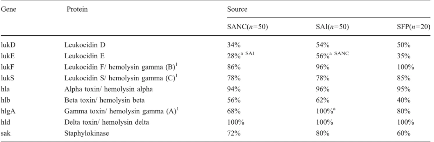
DNA microarray results for genes encoding leukocidins, hemolysins and staphylokinase are depicted in Table7. The genes were evenly distributed among isolates of all three
Group Gene SANC(n=50) SAI(n=50) SFP(n=20)
Enterotoxins entA 26% 20% 30% entA-320 6% 4% 30%a entA-N315 18%a SFP 16% 0%a SANC entB 8% 22% 5% entC 16% 26% 20% entD 2%a SFP 6% 15%a SANC entE 0% 0% 0% entG 58% 44% 50% entH 4% 4% 0% entI 64% 50% 50% entJ 2%a SFP 6% 15%a SANC
Enterotoxin-like proteins entK 4% 18%a 0%
entL 16% 26% 20%
entM 68% 50% 50%
entN 66% 50% 50%
entO 64% 50% 45%
entQ 4%a SAI 16%a SANC 0%
entR 2%a SFP 6% 15%a SANC
entU 46% 50% 50%
Exfoliative toxins etA 2% 2% 5%
etB 2% 0% 0%
etD 2% 0% 0%
Toxic shock syndrome toxin tst-1 12% 8% 15%
Panton valentine leukocidin pvl 0% 2% 6%
Table 6 Genes encoding super-antigenic toxins, such as genes coding for staphylococcal enterotoxins (entA- entJ) and enterotoxin-like proteins (entK-entR, entU), as well as exfoliative toxins (etA/B/D), toxic shock syndrome toxin (tst) and panton valentine leukocidin (pvl). Percentages of S. aureus nasal colonization (SANC), clinical cases of infection (SAI) and staphylococcal food poison-ing (SFP) isolates, for which genes were determined to be present based on DNA micro-array analysis. Calculations include positive signals only, ambiguous signals were omitted
a Result for this source differs
significantly from the result cal-culated for one (source indicated) or both other sources of isolates investigated in this study (p<0.05)
Table 7 Other virulence determinants, including genes encoding leukocidins, hemolysins, and staphylokinase. Percentages represent the fraction of S. aureus nasal colonization (SANC), clinical cases of infection (SAI) and staphylococcal food poisoning (SFP) isolates for
which the genes were determined to be present based on DNA microarray analysis. Results depicted include positive signals only, ambiguous signals were not considered for the calculation
Gene Protein Source
SANC(n=50) SAI(n=50) SFP(n=20)
lukD Leukocidin D 34% 54% 50%
lukE Leukocidin E 28%a SAI 56%a SANC 35%
lukF Leukocidin F/ hemolysin gamma (B)1 86% 96% 100%
lukS Leukocidin S/ hemolysin gamma (C)1 78% 78% 85%
hla Alpha toxin/ hemolysin alpha 94% 96% 95%
hlb Beta toxin/ hemolysin beta 56% 62% 40%
hlgA Gamma toxin/ hemolysin gamma (A)1 68% 100%a 80%
hld Delta toxin/ hemolysin delta 100% 100% 100%
sak Staphylokinase 72% 80% 60%
a
Result for this source differs significantly from the result calculated for one (source indicated) or both other sources of isolates investigated in this study (p<0.05)
sources, with the exception of lukE and hlgA. SAI isolates possessed lukE significantly more frequently than SANC isolates (p=0.005), and hlgA significantly more often than both SANC (p=0.000) and SFP isolates (p=0.017).
Comparison of microarray profiles using the SplitsTree software resulted in no source-specific clusters, but a mixed distribution of isolates of all three sources (see Fig.1).
Discussion
Screening nasal swabs for SANC isolates showed a nasal carriage rate of 38%. CC30 and CC45 represented the most common clonal complexes among nasal isolates investigated, comprising 24% and 16% of SANC isolates, respectively. These findings are consistent with a recent study conducted in Switzerland which observed a nasal carriage rate of S. aureus of 32% among healthy adults and reported CC30 and CC45 to be the most common clonal complexes among SANC
isolates, comprising 24% of nasal carriage isolates each [15]. It was reported that CC30 occurs at high frequencies and is stably maintained among human carriers worldwide [15–17]. A recent study conducted among asymptomatic carriers in Germany found CC8, CC15, CC30, and CC45 to be most common among asymptomatic carriers [18]. Among the tested SAI isolates, CC45 (20%) represented the dominant clonal complex, while a comprehensive Dutch study observed this clonal complex to be underrepresented among invasive strains [19]. A recent German study found CC8 and CC45 to be most common among S. aureus isolated from bone and joint infections [20]. The clonal complexes CC5 and CC30 that were also present among SAI isolates in our study. Isolates assigned to these clonal complexes were observed to significantly increase hematogenous complica-tions in staphylococcal infeccomplica-tions in humans [21]. Several clonal complexes found in our study among SFP isolates were also present among the investigated SANC and SAI isolates (CC8, CC15, CC30, CC45, CC78, CC101). SFP
Fig. 1 SplitsTree showing similarity between gene profiles determined by DNA microarray analysis for 120 S. aureus isolates, comprising 50 isolates obtained from nasal colonization (SANC), 50 isolates collected
from clinical cases of infection (SAI) and 20 isolates associated with staphylococcal food poisoning (SFP)
isolates were most frequently assigned to CC45 (30%), followed by CC8 (20%), CC15 (20%), and CC30 (15%). To the authors knowledge, there have been no previous studies on the distribution of clonal complexes among S. aureus isolates associated with outbreaks of SFP.
The spa types t008, t015, t018, t056, and t084 that we detected among SFP isolates were also present among SANC and SAI isolates investigated in our study. Spa types t008, t015, t056, and t084 were reported among methicillin-sensitive S. aureus causing infections in humans [22, 23] and spa type t018 was found in common MRSA clones in the United Kingdom and Denmark [24,25].
DNA microarray profiling enabled the comparison of gene profiles of isolates from nasal colonization, clinical cases of infection and SFP. Interestingly, DNA microarray profiles of isolates from all three sources were rather similar. This is consistent with a recent study that found nasal carriage isolates and clinical isolates to be closely related [26]. Interestingly, especially few significant differ-ences in prevalence rates were found when SFP and SAI isolates were compared.
Among each source of isolates investigated in this study, all agr types (agrI-IV) were found. The agrIV group was recently hypothesized to constitute a truly monophyletic group, while agrI-III might have evolved from several unrelated ancestors [10]. DNA microarray results in our study revealed a variety of isolates exhibiting differing virulence gene profiles that possessed agrIV. All isolates investigated in our study belonged to capsule type 5 or 8, which were reported to be the only capsular serotypes associated with human disease [27]. The spread of genes conferring resistance to antibiotic agents was corroborated by the antibiotic resistance determinants detected among the S. aureus investigated in our study. The most common resistance gene was blaZ, encoding penicillinase BlaZ, which enables hydrolysis of both methicillin and oxacillin, was high in isolates from all three sources (SANC: 74%, SAI: 76%, SFP: 85%). The detected prevalence rates for blaZ and mecA among SANC isolates (blaZ: 74%, mecA: 2%) are consistent with a recent German report on asymptomatic carriers, which found blaZ in 71% and mecA in 2% of staphylococcal isolates [18]. A study characteriz-ing S. aureus from bone and joint infections detected blaZ in 65% and mecA in 6% of isolates, similar to the prevalence rates of blaZ and mecA genes among SAI isolates investigated in this study (blaZ: 76%, mecA: 8%) [20].
Four out of five mecA positive isolates detected in this study were obtained from clinical cases of staphylococcal infection. The MRSA isolate obtained from a nasal swab (SANC11) of a veterinarian working in equine practice belonged to spa type t011 and clonal complex CC398, which were also found among clinical MRSA isolates
collected from a human patient and several horses in a recent Finnish study [28].
Both tested variants of entA encoding enterotoxin A, the gene responsible for most cases of SFP, were detected among SANC and SAI isolates. Interestingly, all SFP isolates possessed the entA-320 variant, which was first detected in a French field isolate in 2003 [29]. While prevalence rates of entA and entC detected among SANC isolates in this study were almost identical to those of a study conducted with nasal carriage isolates of restaurant workers in Kuwait city, we found lower prevalence rates of entB, entD, and entE [30]. While a German study reported similar rates of entB and entC among asymptomatic nasal carriers, slightly lower rates of entA, as well as higher rates of entD were found [18].
Comparison of microarray profiles using the SplitsTree software resulted in no source-specific clustering, but a mixed distribution of isolates of all three sources. In addition, in our study we found considerable overlap in spa types for SFP isolates with isolates collected from nasal colonization and clinical cases of infection. These results suggest contamination of foodstuff during preparation by food handlers that are colonized or infected by S. aureus represents a source of SFP.
Acknowledgements We thank Reinhard Zbinden and the Institute of Medical Microbiology, University of Zurich, for supplying the S. aureus isolates obtained from clinical cases of infection. We thank Barbara Schalch and the Bavarian Authorities for Health and Food Safety, Alfred Binder and the Medical Department of the German Federal Armed Forces, as well as Jean-Marie Pasquier and the Cantonal Laboratory Fribourg (Fribourg, Switzerland) for supplying isolates associated with outbreaks of staphylococcal food poisoning.
References
1. Kluytmans J, van Belkum A, Verbrugh H (1997) Nasal carriage of Staphylococcus aureus: epidemiology, underlying mechanisms, and associated risks. Clin Microbiol Rev 10(3):505–520 2. Halablab MA, Hijazi SM, Fawzi MA, Araj GF (2010) Staphylococcus
aureus nasal carriage rate and associated risk factors in individuals in the community. Epidemiol Infect 138(5):702–706
3. Munckhof WJ, Nimmo GR, Carney J, Schooneveldt JM, Huygens F, Inman-Bamber J, Tong E, Morton A, Giffard P (2008) Methicillin-susceptible, non-multiresistant methicillin-resistant and multiresistant methicillin-resistant Staphylococcus aureus infections: a clinical, epidemiological and microbiological com-parative study. Eur J Clin Microbiol Infect Dis 27(5):355–364. doi:10.1007/s10096-007-0449-3
4. Berthelot P, Grattard F, Cazorla C, Passot JP, Fayard JP, Meley R, Bejuy J, Farizon F, Pozzetto B, Lucht F (2010) Is nasal carriage of Staphylococcus aureus the main acquisition pathway for surgical-site infection in orthopaedic surgery? Eur J Clin Microbiol Infect Dis 29(4):373–382. doi:10.1007/s10096-009-0867-5
5. Kooistra-Smid M, Nieuwenhuis M, van Belkum A, Verbrugh H (2009) The role of nasal carriage in Staphylococcus aureus burn wound colonization. FEMS Immunol Med Microbiol 57(1):1–13
6. von Eiff C, Becker K, Machka K, Stammer H, Peters G, Grp S (2001) Nasal carriage as a source of Staphylococcus aureus bacteremia. New Engl J Med 344(1):11–16
7. Wertheim HFL, Vos MC, Ott A, van Belkum A, Voss A, Kluytmans JAJW, van Keulen PHJ, Vandenbroucke-Grauls CMJE, Meester MHM, Verbrugh HA (2004) Risk and outcome of nosocomial Staphylococcus aureus bacteraemia in nasal carriers versus non-carriers. Lancet 364(9435):703–705
8. Klevens RM, Morrison MA, Nadle J, Petit S, Gershman K, Ray S, Harrison LH, Lynfield R, Dumyati G, Townes JM, Craig AS, Zell ER, Fosheim GE, McDougal LK, Carey RB, Fridkin SK (2007) Invasive methicillin-resistant Staphylococcus aureus infections in the United States. JAMA 298(15):1763–1771
9. Doyle M, Beuchat L (2007) Food microbiology: fundamentals and frontiers, 3rd edn. ASM Press, Washington DC
10. Monecke S, Slickers P, Ehricht R (2008) Assignment of Staphylococcus aureus isolates to clonal complexes based on microarray analysis and pattern recognition. FEMS Immunol Med Microbiol 53(2):237–251
11. Aires-de-Sousa M, Boye K, de Lencastre H, Deplano A, Enright MC, Etienne J, Friedrich A, Harmsen D, Holmes A, Huijsdens XW, Kearns AM, Mellmann A, Meugnier H, Rasheed JK, Spalburg E, Strommenger B, Struelens MJ, Tenover FC, Thomas J, Vogel U, Westh H, Xu J, Witte W (2006) High interlaboratory reproducibility of DNA sequence-based typing of bacteria in a multicenter study. J Clin Microbiol 44(2):619–621
12. Harmsen D, Claus H, Witte W, Rothganger J, Turnwald D, Vogel U (2003) Typing of methicillin-resistant Staphylococcus aureus in a university hospital setting by using novel software for spa repeat determination and database management. J Clin Microbiol 41 (12):5442–5448
13. Huson DH, Bryant D (2006) Application of phylogenetic networks in evolutionary studies. Mol Biol Evol 23(2):254–267
14. Coombs GW, Monecke S, Ehricht R, Slickers P, Pearson JC, Tan HL, Christiansen KJ, O'Brien FG (2010) Differentiation of clonal complex 59 community-associated methicillin-resistant Staphylo-coccus aureus in Western Australia. Antimicrob Agents Chemother 54(5):1914–1921
15. Sakwinska O, Kuhn G, Balmelli C, Francioli P, Giddey M, Perreten V, Riesen A, Zysset F, Blanc DS, Moreillon P (2009) Genetic diversity and ecological success of Staphylococcus aureus strains colonizing humans. Appl Environ Microbiol 75(1):175–183 16. Ruimy R, Armand-Lefevre L, Barbier F, Ruppe E, Cocojaru R,
Mesli Y, Maiga A, Benkalfat M, Benchouk S, Hassaine H, Dufourcq JB, Nareth C, Sarthou JL, Andremont A, Feil EJ (2009) Comparisons between geographically diverse samples of carried Staphylococcus aureus. J Bacteriol 191(18):5577–5583
17. Melles DC, Tenover FC, Kuehnert MJ, Witsenboer H, Peeters JK, Verbrugh HA, van Belkum A (2008) Overlapping population structures of nasal isolates of Staphylococcus aureus from healthy Dutch and American individuals. J Clin Microbiol 46(1):235–241 18. Monecke S, Luedicke C, Slickers P, Ehricht R (2009) Molecular epidemiology of Staphylococcus aureus in asymptomatic carriers. Eur J Clin Microbiol 28(9):1159–1165. doi:10.1007/s10096-009-0752-2
19. Wertheim HF, van Leeuwen WB, Snijders S, Vos MC, Voss A, Vandenbroucke-Grauls CM, Kluytmans JA, Verbrugh HA, van
Belkum A (2005) Associations between Staphylococcus aureus genotype, infection, and in-hospital mortality: a nested case-control study. J Infect Dis 192(7):1196–1200
20. Luedicke C, Slickers P, Ehricht R, Monecke S (2010) Molecular fingerprinting of Staphylococcus aureus from bone and joint infections. Eur J Clin Microbiol 29(4):457–463. doi:10.1007/ S10096-010-0884-4
21. Fowler VG Jr, Nelson CL, McIntyre LM, Kreiswirth BN, Monk A, Archer GL, Federspiel J, Naidich S, Remortel B, Rude T, Brown P, Reller LB, Corey GR, Gill SR (2007) Potential associations between hematogenous complications and bacterial genotype in Staphylococcus aureus infection. J Infect Dis 196 (5):738–747
22. Wu D, Wang Q, Yang Y, Geng W, Yu S, Yao K, Yuan L, Shen X (2010) Epidemiology and molecular characteristics of community-associated resistant and methicillin-susceptible Staphylococcus aureus from skin/soft tissue infections in a children's hospital in Beijing, China. Diagn Microbiol Infect Dis 67(1):1–8
23. Layer F, Ghebremedhin B, König W, König B (2006) Heterogeneity of methicillin-susceptible Staphylococcus aureus strains at a German University Hospital implicates the circulating-strain pool as a potential source of emerging methicillin-resistant S. aureus clones. J Clin Microbiol 44(6):2179–2185
24. Bartels MD, Boye K, Rohde SM, Larsen AR, Torfs H, Bouchy P, Skov R, Westh H (2009) A common variant of staphylococcal cassette chromosome mec type IVa in isolates from Copenhagen, Denmark, is not detected by the BD GeneOhm methicillin-resistant Staphylococcus aureus assay. J Clin Microbiol 47 (5):1524–1527
25. Khandavilli S, Wilson P, Cookson B, Cepeda J, Bellingan G, Brown J (2009) Utility of spa typing for investigating the local epidemiology of MRSA on a UK intensive care ward. J Hosp Infect 71(1):29–35
26. Lamers RP, Stinnett JW, Muthukrishnan G, Parkinson CL, Cole AM (2011) Evolutionary analyses of Staphylococcus aureus identify genetic relationships between nasal carriage and clinical isolates. PLoS One 6(1):e16426. doi:10.1371/journal.pone.0016426
27. Melles DC, Taylor KL, Fattom AI, van Belkum A (2008) Serotyping of Dutch Staphylococcus aureus strains from carriage and infection. FEMS Immunol Med Microbiol 52(2):287–292. doi:10.1111/j.1574-695X.2008.00376.x
28. Salmenlinna S, Lyytikainen O, Vainio A, Myllyniemi AL, Raulo S, Kanerva M, Rantala M, Thomson K, Seppanen J, Vuopio J (2010) Human cases of methicillin-resistant Staphylococcus aureus CC398, Finland. Emerg Infect Dis 16(10):1626–1629 29. Letertre C, Perelle S, Dilasser F, Fach P (2003) A strategy based
on 5' nuclease multiplex PCR to detect enterotoxin genes sea to sej of Staphylococcus aureus. Mol Cell Probes 17(5):227–235 30. al Bustan MA, Udo EE, Chugh TD (1996) Nasal carriage of
enterotoxin-producing Staphylococcus aureus among restaurant workers in Kuwait City. Epidemiol Infect 116(3):319–322 31. Straub JA, Hertel C, Hammes WP (1999) A 23S rDNA-targeted
polymerase chain reaction-based system for detection of Staphylo-coccus aureus in meat starter cultures and dairy products. J Food Prot 62:1150–1156