Cardiovascular Research
1
993 ;27:2229-22?7
2229
Characterisation of Na/K-ATPase, its isoforms, and
the inotropic response
to
ouabain in isolated failing
human hearts
Olga
I
Shamraj, Ingrid L Grupp, Gunther Grupp, David Melvin, Nathalie Gradoux,
Walter Kremers, Jerry
B
Lingrel, and Alain De Pover
Objective: The aim was to determine whether failing human hearts have increased sensitivity to the inotropic
and toxic effects of ouabain, and to examine alterations in NdK-ATPase that might explain the observed higher
ouabain sensitivity.
Methods: For contractility studies, a total of 57 trabeculae were isolated from two non-
failing (death from head injury) and 10 terminally failing, explanted human hearts. After the experiment, each
trabecula was inspected under the light microscope for morphological alterations consistent with heart failure.
Samples for biochemical and molecular studies were obtained from five non-failing and 13 failing hearts. Total
NdK-ATPase was measured in desoxycholate treated homogenates and expressed per unit of tissue wet or dry
weight, DNA, protein, or myosin. Interference from residual bound digoxin due to previous therapy was
excluded. The expression of the three
ciisoforms was studied at both the mRNA level using northern blots and
the protein level by analysis of dissociation kinetics of the [3H]ouabain-enzyme complex.
Results: Trabeculae
showing morphological alterations and decreased contractility were sensitive
to
lower concentrations of ouabain
(3-100 nM) than control trabeculae (100-1000 nM); the inotropic ECSo and the minimum toxic concentration
were both reduced. [3H]Ouabain binding was significantly lower (p<<O.OOl) in failing than in non-failing hearts,
at 293(SD 74)
v
507(48) p m o l g ' wet weight. No significant change was observed in maximum ATPase turnover
rate, or in sensitivities to Na+, K', vanadate, and dihydro-ouabain. All three
ciisoforms were expressed at the
mRNA level in both normal and failing hearts.
Conclusions: This study shows conclusively, for the first time,
that failing human hearts are more sensitive to ouabain. This may be at least partly due to a mean reduction
of 42% (95% confidence interval, 26 to 56%) in the concentration of NdK-ATPase (decrease in Na,K pump
reserve), but not to an alteration in its catalytic properties or in its isoform composition.
Cardiovascular Research 1993;27:2229-2237
ardiac glycosides have been widely used in the
treatment of congestive heart failure. Their basic
C
mechanism of action consists in inhibiting N d
K-ATPase, which leads
to
a rise in intracellular free Na'
concentration and, through alteration of NdCa exchange, to
the enhancement of Ca2+ transients and of cardiac con-
traction.' However, excessive inhibition induces Ca" over-
load, causing arrhythmia, a decrease in developed tension,
and contracture.2 Although it is known that patients with
advanced heart disease are more susceptible to glycoside
induced
arrhythmia^,^
the direct effect of the drugs on the
diseased myocardium is still poorly documented. While one
study has shown that papillary muscle strips isolated from
terminally failing hearts responded to the inotropic action of
ouabain at lower concentrations than strips from non-failing
hearts, the interpretation of this difference was complicated
by the fact that the inotropic action showed an all or nothing
effect in non-failing hearts instead of a typical concentration
dependent response, and also by the absence of arrhythmia
data.4 In biochemical studies, it is still uncertain for
methodological and statistical reasons whether the density of
the glycoside receptor, the NdK-ATPase, on the cadiac cell
membrane decreases, and if so to what e ~ t e n t . ~
(This point
will be discussed in detail below.) Furthermore, the catalytic
properties of NdK-ATPase have not yet been studied in
diseased human hearts, and although the expression of three
ciisoforms at the mRNA level has been reported,67 no
information is available at the protein level.
In the present study on NdK-ATPase, contractility
experiments revealed a population of failing heart trabeculae
with increased sensitivity to the inotropic and toxic effects
of ouabain. In these experiments, concentration dependent
inotropic effects were obtained with both failing and non-
failing hearts, allowing quantitative comparison. The recent
availability of relatively large samples of human heart from
patients undergoing cardiac transplantation has made it
possible to undertake a comprehensive study of
Nd
K-ATPase properties. Thus we decided to test different
hypotheses that could account for the increased ouabain
sensitivity: (1) a switch in isoform gene expression, (2) a
decreased density of ouabain binding sites, (3) reduced
Nd
K-ATPase turnover rate, and (4) reduced Na' sensitivity. For
this study to be valid, it was critical to demonstrate the
complete recovery of NdK-ATPase from tissue samples, the
independence of the results from the reference used to
express the data (for example, tissue wet weight), and the
University of Cincinnati, College of Medicine, Ohio, USA
-Department of Molecular Genetics, Biochemistry and
Microbiology: 0 I Shamraj, J B Lingrel; Department of Pharmacology and Cell Biophysics: I L Grupp, G Grupp; Department
of Surgery: D Melvin; Ciba-Geigy Ltd, Basle, Switzerland
-Department of Cardiovascular Research: N Gradoux, A De Pover;
Medical Department, General Biometry: W Kremers. Correspondence to Dr De Pover, at K- 125.9.04, Ciba-Geigy Ltd, CH-
4002 Basle, Switzerland.
discrepancies reported i n previous studies
of
NdK-ATPase
in
human heart may be explained by the fact that these
variables were not considered, as well as by the differences
in
the methods used. The present results support the
hypothesis that a decrease
in
NdK-ATPase density underlies
the greater sensitivity
of
severely failing hearts
to
cardiac
glycosides. These investigations were approved
by
the local
ethics review committees. Preliniinary results have been
reported.h
'
Methods Contrii(.tili!J \ r i d \
'irabeculat bere taken from erplanted hearts ohtained in collaboration
with the Gni\ ersity of Cincinnati cardiac transplantation programme. Failinp he;irt\ carried the general pathological diagnoses of ischaemic, h> pi'rtrophic. or dilated cardiomyopathy. Non-failing hearts (death from head inlur! ) iwild not be implanted for technical reasons. The hearts were placed i n ice cold Krebs-Henseleit solution in the operating theatre immediately alicr removal. The mural trabeculae were excised from the walls o f both right and left atria and ventricles and stored for a short time on ice hefhrc being mounted and suspended in a 70 ml assay bath at 35°C. We chose traheculae 1 mni wide and 5 nun long. No Purkinje
tihres o r \ isiblq necrotic tihres were used. Contractility measurements
were performed as pre\ ioualy described."' BrieHy. the trabeculae were placed in a muscle holder in contact with two pointed platinum electrodes used for stiniulation. connected to a force transducer (Gra\s
i+T 03C). and studied under isometric conditions: initially they were stretched t o ii length at which Force was maximal (L,,,,). The
stimulntion rate wab 1.0 Hr. 2 to 5 ms duration, with a voltage 204 above threshold. Contractile force and its first derivative dF/dt were recorded on ;I Crass P7 polygraph. After eyuilihration for 60 min, cuniulative ouabain dose-response curves were obtained by adding iiuabain in increasing concentrations from 1 to 1000 nM every 40-60
min. The duration of exposure was long enough to bring the traheculae close to their maximum response at each concentration. When clear
s i p of toxicity became apparent (increase in resting tension. decrease
in active tension. irregularities i n contractility and/or contracture) the experiment m a \ terminated.
Iic wc m~qi~i.\iiio~i f i l r northeix uiiuly\i,s tinil hinc heriiicol sriidit~s
B e d e s the heart sample\ used above for contractility studies. samples from additional hearts were procured over a later time period for RNA isolation and biochemical studies. Samples from three failing and I3
nori-Faiiinp hertrts were collected in conjunction with the University of Cincinnati cardiac transplantation programme. Samples from two normal hearts that were wed for biochemical studie\ only were supplied hy thc International Institute tor the Advancement of Medicine tEs\ington. PA, L!SA). The material included end stage failing hearts froin patient\ with diagnoses of ischaemic. idiopathic, idiopathic dilated, dilated. hypertensive. or hypertrophic cardioniyopathy. and nowfailing hearts (death twin head injury) that could not be implanted for technical reamns. All patients with heart failure had undergone left and right heart carheterisation. echocardiography. and multiple gated iiucleiir an~iocardiograpliic scanning. Mean right atrial pressure and mean pulnionary arterial pressure were recorded from fluid filled preshurc transducers zeroed at mid-axillary level in the supine position. Cardiac output IKIS determined by thermodilution. using right atrial
in-jection and ii pulmonary arterial catheter tip thermistor. Left
ventricular ejection fraction was determined from the ratio of radioisotopic acti\it) at end systole to that in end diastole in the left ventriculai- profile ;it nuclear angiocardiography. Clinical data are
wnimarised in table 11. The principal therapy prior to cardiac transplantation had been diuretics and converting enzyme inhibitors: five patients had ~ilw been receiving digoxin. On explantation, samples from each heart were imniediatelg frozen in liquid nitrogen and stored 31 -70°C.
~~'~ll~therll f i l l " I r,\r.>
Total cellular R K A was iwlated from tissue samples and northern blot anal\ise\ carried out as described.' ' I Data obtained from three non-
failing hearts in the present study have already been reportedb and are shown for comparison onl) (fig I A ) . Brietly. 10 ~g of total RNA was iaolated and fractionated o n agarose gels and transferred to nylon inembrrtnes (hilngna NT. MSI). Total RNA from both the left and right ventricles ut' 13 diwn\ed hcarts was used to make two sets of three identical blot\. Each set wa\ hyhridised with u l . u2. and a3 specific 6Omer oligonucleotide probes (described in detail in ') and end labelled to a uniform specific activity with (y"P)-ATP (3000 Ci.mniol-'. New England Nuclear). Northern blots were exposed to x ray film for autoradiograph) and to :i Pho\phor screen for quantification with a
Figure 1 Northern blot a n a l ~ ~ s i s of' NdK-ATPuse a isofr~rrri
inRNA expression iii hiitnun heart. Each lane contains 10 pg oj
total celliilur RNA isoluted from human control tissues (kidnej ( K ) ,
skeletul muscle (S). und bruin ( B ) ) wid f r o m left unrl right
wntriciilar free wall ( L and R, respectively) ,froin three non$uilin~
himuti hearts (punel A , henrts
I ,
2, and 3)6 and from 13 disea.sedIieurts (panels B mid C, lzearts 4-16). The clussiJication of the
diseased hetrrts Liccording the type of curdiomyopathy was u s
,follo~.s: jive ischueniic (hearts
5,
7. 9, 10, and I S ) , three idiopathic(helirts 8, 13, 16), one dilated (heart 41, two idiopathic diluted
(hearts 11, 12), one hypertensive (heart 14), and one hypertrophic
(heart 6). Blots were probed with a l , a2, and a3 isoform-specific
" P tubelled probes ttnd exposed to x ruy ,film ,[or j i v e dtrys, then
stripped tirid reprobed with N -j2P labelled 18s rRNA probe, which
showed that ,some surnples were niore degrudrtl than other,s.
Phosphorimager (Molecular Dynamics), then stripped and reprobed with a ."P labelled 18s rRNA 20rner. As controls, 10 pg of total RNA from human kidney (expresses a ] ) , skeletal muscle (expresses a I and a2). and brain (expresses u l . a2. and a3) were included on each blot.
Tissire liotnogenisatiori und detergent ireutment
Pieces of frozen tissue were rapidly thawed in saline. Fat, vessels, and connective tissue were removed as well as possible with scissors. Unless otherwise stated. the following procedure was performed at
NdK-ATPase
in failing human hearts
223 1
0-6°C. A sample weighing 0.5-2 g was minced and homogenised in 10 ml of chilled buffer per g wet weight using a Polytron homogeniser, 2 s at maximum setting, twice (buffer composition: 2 mM EDTA and 10 mM Tris, pH adjusted to 7.4 with HCI). The homogenate was diluted to 30 ml with buffer and centrifuged at 100 000 g for 30 min. The pellet was homogenised in 6.5 ml buffer per g sample, with a Teflodglass Potter Elveljem homogeniser. Aliquots of these two homogenates were pooled and used for determinations of DNA, total protein, myosin, and creatine phosphokinase (CPK). The second homogenate was mixed with 3.5 ml (per g sample) of buffer supplemented with 2.86 mgm-I sodium desoxycholate (DOC). After 30 min incubation at room temperature, this mixture was frozen and kept overnight at -30°C. This treatment, used to permeabilise vesicles, revealed maximum latent N d K-ATPase activity (data not shown). After thawing, the mixture was centrifuged at 100 000 g for 2 h. The pellet was resuspended in 10 ml of buffer per g sample (buffer composition: 10 mM Tris, pH adjusted to 7.4 with HCl) and kept at -70°C until use. NdK-ATPase activity was stable for more than a year under these conditions. Four samples were processed per experimental run. No significant activity was found in the supernatants, indicating virtually complete recovery of the Na/ K-ATPase in the final suspension. In the following procedures, homogenates were stirred with vortex immediately before pipetting. Occaionally, minute fragments of non-homogenised tissue blocked the pipette tip and had to be removed. To avoid loss of material, no further attempt was made to remove unhomogenised tissue (for example, by low speed centrifugation or filtration), since we had observed that these procedures affected NdK-ATPase recovery differently in normal and diseased hearts. For all samples, NdK-ATPase activity and ['Hlouabain binding were assayed in two to three separate experiments.
NdK-ATPase assay
NdK-ATPase activity was determined by measuring inorganic phosphate (Pi) release from the ouabain sensitive hydrolysis of ATP by DOC treated homogenates as described." Protein (10-12 pg) was incubated for 60 min at 37°C in 0.1 mI medium containing (in mmo1,litre-') ATP 5, MgC1, 5, NaN' 5, NaCl 145, KCl 20, EGTA 1, and maleic acid 10 (pH adjusted to 7.4 with Tris). ATP hydrolysis never exceeded 20%. Basal activity was determined in the presence of 1 mM ouabain and represented about one third of total activity. Na/K-ATPase activity was linear with respect to time and protein concentration for all samples. Enzyme activation by Na' and K' was studied, according to the method of Skou," at a constant ionic strength by maintaining the total [Na'
+
K'] = 150 mM while the Na' and K' concentrations were varied. Na' was varied by increasing NaCl from 0 to 145 mM, while NaN3 remained constant at 5 mM. Maximum turnover rate was calculated from the ratio of maximum activity (V,J to maximum ['Hlouabain binding (B,,,), assuming one ouabain binding site per catalytic subunit. Dihydro-ouabain inhibition curves were plotted at 5mM KC1. This glycoside was used instead of ouabain or digoxin because it binds quickly to NdK-ATPase.
[3H]Ouabuin binding assay
['HIOuabain binding to DOC treated homogenates was assayed by a filtration technique as des~ribed.'~ Briefly, 2.5-3 mg protein was incubated at 37°C in 5 ml medium containing (in mmo1,litre-I): MgCI, 3, phosphoric acid 3, EGTA 1, and maleic acid 10 (pH adjusted to 7.4 with Tris), plus 1 pM ['Hlouabain (3-6 Ci.mmol-', Amersham batch diluted with ouabain). Under these conditions, complete saturation of ouabain receptors was obtained (KD<O.O1 pM) and the specific binding represents B,,,. After 10 min incubation, 0.2 ml aliquots were filtered through Whatmann G F F glass fibre filters previously soaked in 0.3% polyethylenimine. Nonspecific binding was determined in the presence of 0.1 mM ouabain and did not exceed 10% of total binding. For ['Hlouabain dissociation kinetics, 0.1 mM ouabain was added after the
10 min incubation, and 0.2 ml aliquots were rapidly filtered at various times.
Other biochemical assuys
For DNA and creatine phosphokinase (CPK) determinations, sample aliquots were diluted in appropriate buffers and centrifuged for 2 min in an Eppendorf centrifuge. DNA was determined as describedI5 and CPK assayed using Boehringer Mannheim kits at 22-23°C. For total protein and myosin determinations, proteins were extracted with 2% SDS. Total protein was estimated by the bicinchoninic acid methodI6 (reagents from Pearce, Rockford, Illjnois, US 4) and myosin levels were determined by densitometry after SDS electrophoresis according to Laemmli." Tissue dry weight was measured after freeze drying 0.2 ml of homogenate.
Statistical analysis
Clinical data were disclosed at the end of the study. The significance of observed differences between failing and non-failing hearts (unpaired) and between left and right ventricles (paired) was verified using appropriate t tests. F tests were used to check for differences linked with the aetiology of the disease (multiple groups). Differences
were accepted as significant at the p<O.OS level. Unless otherwise indicated, data represent means(SD). In figs 3 and 4, SEM is shown instead of SD to make the graphs easier to read.
Curve jtting analysis
Lindenmayer and Schwartz have shown that NdK-ATPase activation by Na' and K' followed the relationship y = V,,,/(a,b.c), where the term a described the activation for three equivalent Na' sites and the terms b and c the activation for two non-equivalent K' sites.Ix Since the resolution of this equation requires a large number of experiments, to evaluate the two cations parsimoniously we replaced a and b.c by the logistic relationship, which is equivalent to the equation of Hill,"
leading to
where y is the predicted effect, A the basal ATPase activity, BVa and BK the Hill coefficients, CNa and CK the concentrations for half maximum activity, D the maximum activity (V,,,,,), x the variable Na' concentration, and 150-x the variable K+ concentration. V,,,,, was 20% higher than the optimal activity measured at 130 mM Na' and 20 mM
K'. The data fitted to this empirical function over a wide range of cation concentrations (fig 3). Note that although the above equation applies to the case where Na' and K' hind simultaneously, it does not rule out a sequential mechanism.'*
NdK-ATPase inhibition curves were fitted to the logistic function"
where y is the predicted effect, A the basal ATPase activity, B the Hill coefficient, C the concentration for half maximum inhibition, D the control specific activity, and x the variable concentration.
['HIOuabain dissociation kinetics were fitted to the one site function" y = A
+
D,e-klx or to the function for two independent sites'" y = A+
D,e-kix+
D2ck+ where y is the predicted effect, A the non- specific binding, k, and kz the first order dissociation rate constants, D the binding at 0 time, and x the variable time.For statistical analysis, curves were fitted by using individual sample
or individual heart data whenever possible, and mean values and SD were calculated for groups of hearts. For ['Hlouabain kinetics, two site fit could not be performed with some samples because the binding was too low. In these cases, curves were fitted by using mean values of groups of hearts.
Results
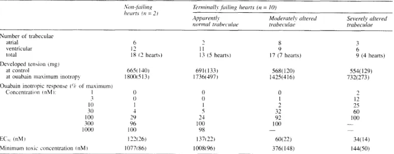
Contractility
In table I the inotropic responses to ouabain in trabeculae
from non-failing and failing hearts are listed. The data from
atrial and ventricular trabeculae were pooled, since there was
no statistical difference between the chambers. The
distinction between moderately and severely altered trabe-
culae was made after the experiment, using
a light
microscope
to evaluate the state of the myofibrils in the
contraction bands, endocardia1 thickening, vacuolisation,
scar formation, interstitial oedema, and mitochondria1 and
nuclear damage. Seventeen trabeculae from seven explanted
hearts were rated as moderately altered, and nine trabeculae
from four hearts as severely altered. It
is clear that minimal
inotropic effects of ouabain occurred at 3 and 30 nM in the
severely and moderately altered fibres compared to
100 nM
in the normal ones. The
ECsO and the minimal ouabain
toxicity showed a similar relationship. In addition, the
severely altered fibres responded with
a
significantly reduced
maximum effect to both calcium (not shown) and ouabain.
Trabeculae from two non-failing hearts served for
comparison.
NdK-ATPase
Qisoform mRNA expression
We have previously shown that
all
three
aisofonns of
N d
K-ATPase mRNAs are expressed in normal human left and
right ventricle (LV and RV, respectively) at high levels.6
Northern blots of this result (fig 1A) are shown in this
total 18 ( 2 hearts) 13 ( 5 heart\)
I)e\ eloped t e n w n (nip)
at contiol 66% 1-10] 69 1 ( 133)
'it ouabain ~ n a x i i n u ~ ~ i inotrop> 1 8 0 0 ~ 5 1 3 ) 1736(497)
Ouabain inotropic re5poiise ( I ? of maximuin j
Concenrratioii cnM 1: 1 0 0 I 0 I -30 1 100 29 300 96 1 O(X) I00 0 0 I 5 24 100 98 17 (7 hearts) 568( 120) 1425(416) 0 1 2 32 92 100 - 9 (4 hearts) 554(129) 732(273) 2 12 2s 60 I00 - - EC',. (nM) 1'326) 137122) 60(22) 34( 14)
Minimum roxic on cent ration ( n M ) 10771 86) 1008(96) 376( 148) 144(50)
Data froin atrial and ventnculirr trabeculac were pooled. Trabeculae from failing hearts were classified in three groups according to the degree of morphological alteration determined hy microscopic in\pcction (see [ex[).
manuscript to allow comparison of these data to the diseased
heart expression pattern. Northern blots
of
left and right
ventricle samples from 13 transplant recipient hearts show
that mRNAs for all three
a
isofornis are expressed in
diseased human heart (fig
1B
and
C,
hearts
4-16).
The
quality of the RNA varied considerably from sample to
sample.
as
could be seen on ethidiuni bromide stained check
gels (data not shown) and from the variability
in
the 18s
rRNA signals (that is. heart
9 (LV),
heart
14 (RV).
and heart
16
ILV)).
For each heart. the contribution of each individual
a
isoform to the total a isoform mRNA pool was calculated by
dividing thc normalised (to 185 rRNA) signal for each
isofornr by the sum
of
the normalised signals for all three.
The inem and standard deviation for left and right ventricle
normal heart samples
( n
=
6)
and diseased heart samples
(n
=22,
four samples were
too
degraded for quantification)
were calculated for cornparison purposes.
For
the normal
hearts, the results for a
1 , a2,
and
a3
isoform mRNAs were
48(SD
21
)%.26(
13)%.
and
27(
10)R of the total
a
isoform
pool,
respectively. For diseased hearts, the results for
a 1, a2.
and
a3 mKNAs
were 18(6)%, 30(12)%, and
52(12)%.
respectively. From these results, it appears that
in
normal
heart the a1 mRNA represents about half the pool. whereas
in
diseased heart
a3
represents half the pool. However. thesc
data must be interpreted with caution due to the small sample
size for the normal hearts as well as the large variability
between samples. The large variability may reflect heart to
heart variation. regional variation within one heart. prefer-
ential degradation of one
or
more
of
the isoforms, or a
combination
of
these factors. Due
to
the variable levels of
degradation, absolute levels of expression between the
different hearts were not compared.
IYei/K-ATPme
qiiciritifictitioriSamples were obtained from various parts of the left and
right ventricle free wall and from the septum (total
numbers=77.
20.
and
10,
respectively; 1-6 samples per
heart). Their numbers and locations were not predetermined,
raising the question whether the data were truly repre-
sentative of the hearts. However, differences between left
and right ventricles and septum were
not
statistically
significant. (This is in agreement with animal studies.")
Furthermore, the within heart variation did not affect the
conclusions regarding the effect of disease on Na/K-ATPase.
Therefore for the sake
of
simplicity mean values were
calculated from the different samples. reducing the data to
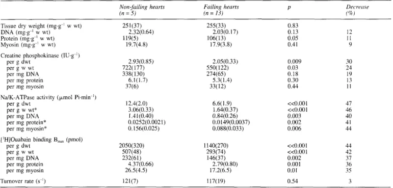
one observation per heart. Expressed per g wet weight,
[
'Hlouabain binding decreased from
507(48)
pmol in non-
failing to
293(74)
in failing hearts
(-42%,
p<<0.001). The
disparity was not related to age, which ranged from
1.5
to
60
years (table 11). Although a steep decrease in ouabain
binding during the first six months of life has been reported,
we did not test infant hearts." When hearts were grouped
according to aetiology, an analysis of variance showed that
the variability was dependent on
the
type of cardiomyopathy
(p=0.006). Table
I11
shows that the effect of disease was
also highly significant when binding data were expressed per
tissue dry weight, DNA, myosin, or total protein, instead of
tissue wet weight. Thus we could reasonably exclude the
involvement
of
tissue swelling or necrosis in this effect.
Na/
K-ATPase activity correlated closely with l'H]ouabain
Table II
IYO" Dicig Age Se.r CO LVEF RAP PAP OBG
Clinical data for the hearts used
in
biochemical study.- 3 1 15 7' 9 10 5 16" 13 8 1" I I 12" 6 14- 7
-
- NE 2 M NA NA NA N F 15 F NA NA NA N F 27 M NA N A NA N F 39 F NA N A NA NF 60 F NA NA NA IS 41 M 5 7 14 2 IS 48 M 6.0 13 12 IS 52 M 3.4 16 7 1s 54 M 4.1 17 8 IS 58 M 3.5 9 18 ID 34 M 6.8 16 3 ID 38 M 2 9 23 NA ID 47 M 3 7 20 10 D 44 M N A 20 12 DID 20 M 4.4 15 6 DID 54 F 2.8 17 10 H 32 M 3.9 21 8 HT 39 M 2.7 7 20 NA NA NA N A NA 25 42 35 27 36 26 42 35 45 31 36 3 1 50 526 511 446 572 478 301 260 184 308 290 320 330 358 289 217 222 477 246" refers to heart numbers used in fig I .
v Indicates previous digitalis therapy.
CO = cardiac output (litres.min-I); LVEF = left ventricular ejection fraction
(%): RAP = right atrial pressurc (mm Hg); PAP = pulmonary artery pressure (inm Hg): OBG = ['Hlouabain binding (pmo1.g-l wet weight); NA = n o available data: DIAG = cardiomyopathy type; N F = non-failing; IS = isch- aemic; ID = idiopathic; D =dilated: DID = dilated idiopathic; H =hyper- trophic: HT = hypertensive.
NdK-ATPase in failing human hearts
2233
Table 111 NdK-ATPase activity and other biochemical variables in human heart samples. Values are mean(SD), except those indicated
by
an
asterisk, which are median (IQR).Non-failing hearts Failing heurts P Decrrcw
( n = 5) (n = 13) f%J
Tissue dry weight (mg,g-l w wt) 25 l(37) 255(33) 0.83
DNA (mg,g-l w wt) 2.32(0.64) 2.03(0.17) 0.13 12 Protein (mg.g-' w wt) 119(5) 106(13) 0.05 1 1 Myosin (mg.g-' w wt) 19.7(4.8) 17.9(3.8) 0.41 9 Creatine phosphokinase ( I U g I ) per g dwt 2.93(0.85) per g w wt 722( 177) per mg DNA, 338(130) per mg protein 6.1 (1.7) per mg myosin 37(6) per g dwt 12.4(2.0) per g w wt* 3.06(0.33) per mg DNA I .41(0.40) per mg myosin* 0.156(0.025) per g dwt 2050(320) per g w wt 507(48) per mg DNA, 232(61) per mp protein 4.37(0.66) per mg myosin 26.5(4.5) NdK-ATPase activity ( p o l Pi.min-l)
per mg protein* 0.0252(0.0021) ['HIOuabain binding B,,, (pmol)
Turnover rate (s-') 121(7) 2.05(0.33) 550( 122) 274(65) 5.3( 1.4) 33(12) 0.009 0.03 0.18 0.30 0.44 6.6(1.9) <<O.OOl 1.64(0.37) <<0.001 0.84(0.26) 0.003 0.0149(0.0037) 0.002 O.OSS(0.033) 0.006 30 24 19 13 1 1 47 46 40 41 44 1140(270) <<O.OOl 44 293(74) <<0.001 42 146(37) 0.002 37 2.79(0.80) 0.001 36 17.2(6.5) 0.01 35 117(19) 0.54 3 NdK-ATPase activity was determined at 150 mM Na+ and 20 nM K'.
Turnover rate = NdK-ATPase activity/['H]ouabain B,,,; w wt = tissue sample
binding
(fig 2), indicating that the apparent concentration of
NdK-ATPase was altered by disease, but not its molecular
turnover rate. For comparison, creatine kinase activities are
also reported in table
111. A statistically significant decrease
of this enzyme was observed in failing hearts only when
tissue wet or dry weight was used as reference.
Potential inte ference from digoxin therapy
In hearts from patients previously treated with digoxin, the
drug may have remained bound to NdK-ATPase and
interfered with the determinations. To check for such inter-
ference, we measured
1
IJ.M [3H]ouabain binding at two
incubation times:
5
min (sufficient for complete saturation of
NdK-ATPase with [3H]ouabain) and 4 h (sufficient for
virtually complete dissociation of digoxin binding). Thus if
4r
,*D;P,
I I I
200 400 600
pmol [3H]-ouabaing' wet weight
Figure
2 Relation between NdK-ATPase activity (vertical axis)and [3H]ouabain binding in non-failing (jilled squares), ischaemic
(circles), idiopathic (triangles), idiopathic-dilated (inverted
triangles), hypertensive (diamond), hypertrophic (*), and dilated
(i) human hearts. NdK-ATPase activity was determined at 150
mM Na' and 20 mM K'. The symbols represent individual hearts.
The dotted lines represent 95% conjidence limits.
wet weight; dwt = dry weight.
digoxin was bound to NdK-ATPase, [3H]ouabain binding
should have increased after the longer incubation. We
observed no difference between the binding data after
5 min
and 4 h in any of the 18 hearts used in this study, irrespective
of whether digoxin had been prescribed before surgery (data
not shown).
We further checked whether digoxin could have been
released from NdK-ATPase during the membrane prepar-
ation as a result of homogenisation in hypotonic buffer,
ultracentrifugation, and/or treatment of the homogenate with
detergent. To estimate this effect, control homogenates were
incubated with 1 pM digoxin for
5
min in the presence
of
MgC1, and inorganic phosphate (Pi) (this is sufficient for
complete saturation of NdK-ATPase with digoxin), sub-
mitted to ultracentrifugation, and treated with DOC exactly
as described in Methods. These homogenates were then
challenged with
1 pM C3H]ouabain for
5 min. Digoxin
pretreatment inhibited
60%
of the specific ['Hlouabain
binding, indicating that DOC treatment results in the release
of 40% of the bound digoxin. After a 4 h incubation, digoxin
pretreatment had no effect on [3H]ouabain binding, indi-
cating complete release of the drug. Thus in samples from
digoxin treated patients part of the specifically bound
digoxin gets washed out during sample preparation.
Kinetics
of
NdK-ATPase activation
and
["Hlouabain
binding
No significant difference was observed in enzyme and
binding kinetics in left ventricle, right ventricle, and septum;
therefore, mean values were calculated for each heart.
Figure
3 illustrates the activation of NdK-ATPase by Na'
and
K' and its inhibition by dihydro-ouabain and vanadate.
V,,,
decreased from
3.6(0.5) pmol Pi.g-' wet weight.min-'
in non-failing hearts to 2.0(0.4) in failing hearts (p<<O.OOl),
while activation and inhibition constants and Hill
coefficients did not change. The Na' and K' sensitivities and
the molecular turnover rate
of
NdK-ATPase agree well with
data from purified preparations,''-l4
23showing that DOC
Percent 0 9 8 7 6 5 4 Di hydro-ouabain (-log M) l50 oon
characterisation.
It
is interesting to note that
a
isoforms have
different
Na-
ensitii4ies."
Figure
3
shows dissociation kinetics of the NdK-ATPase-
['W]ouabain complex. As previously reported, dissociation
kinetics can be fitted to a two site model in human heart
membranes" and to a one site niodel in kidney." This tissue
difference
cat1be accounted for by different isoenzyme
compositions. The slow complex represented
4 9 9
and
52%
of the specific sites. respectively, in non-failing
(k,
=
0.047.min ': k,
=
0.009.niin-I) and failing hearts
( k ,
=
0.052.min-':
k, =
0.01 I
.min
I).It is clear that a third high
affinity site indistinguishable from the other two niay be
present.
as
suggested by the mRNA analysis, and that makes
the identification
of
the binding sites uncertain.
Discussion
Our
initial studies showed that failing human hearts contain
trabeculae that are
more
sensitive to the inotropic and toxic
effects of ouabain. and we examined various hypotheses that
could account for the difference. An original feature of this
study
wasthat the fibres were inspected under the light
microscope after the contractility experiments. Thus.
although nccrotic fibres were discarded, the experiments
were not designed
to
look at the "best case" response
to
inotropic stimulation. We found two types
of
fibres
in
diseased hearts: apparently normal fibres and fibres showing
morphological alterations characteristic of diseased heart.
Only visibly damaged fibres showed functional changes:
( I
)reduced maximum effect of ouabain, and
( 2 )
inotropic and
toxic response\ at markedly lower concentrations of ouabain.
The fact that the inotropic responses to calcium and ouabain
were similarly reduced suggests that this reduction may be
related
to losilof
contractile material a n d o r to reduced
rnyofibrillar ATPase activity.'6 It is crucial to note that the
inotropic efkct
of
ouabain was concentration dependent
in
nowfailing tibres.
in
agreement with animal studies." The
unexplained all or nothing inotropic response of non-failing
human papillary muscle in a previous study' did not allow
a valid comparison with failing hearts. Thus the present
study
s h o wconclusively. and for the first time. an increased
sensitivity of tailing human fibres
todigitalis.
10 \ \ \ \
I
\ \ \ \ \ \ 5 t \ \ 0 60 120 180 240 Time (rnin)Figure 4 Dissociation kiiietics of the Na/K-ATPa,se-[-~H]ouabaiii
corrip1e.u in riorz-j~iiling (ti =
5.
m p t y triangles) ciridjailitig (11 = 13,,filled triatigles) hirinaii hearts. Error bars = SEM. Dotted lines
represetit the coriipirred jicncrions f o r two sites. Inset: Comparison
betnwri hrrnzr1ri heart (filled triangles) mid human kidney
(sql/(lres).
To
explain this observation, we first hypothesised that
failing hearts might express
a
higher affinity isoform of the
NdK-ATPase. Indeed, three
a
isoforms with varying
affinities for cardiac glycosides have been observed in
mammalian tissues."
For
example, the affinity
of
CY 1expressed in the kidney varies greatly among species,25
ranging from the human
a l ,
which is one
of
the most
sensitive to ouabain
(1 nM),
to the rat
a ] ,which is highly
resistant (10
pM).
In the rat,'x the affinities for
a2
and
a3
are respectively
100
and
1000
times more sensitive than the
affinity for
a 1.
In other species, the affinities
for a2
and
a3
are less precisely known, but analysis of inhibition and
binding kineti& in tissues predominantly expressing these
isoforms suggests high affinities similar to those for rat
a2
and
a3.
In human heart membranes, the dissociation kinetics
of the ['Hlouabain enzyme complex show a complex pattern
that is compatible with the presence of different isoforms, in
agreement with niRNA analysis. The slower phase (higher
affinity) is close to the monophasic dissociation observed in
human kidney. suggesting that it may represent a l . Thus,
although this
deduction
still
has
to be
verified
experimentally,' it appears that human
a1could be
5
to 10
times more sensitive to ouabain than human
a2
and
a3;
therefore an increase in a1 expression could augment the
ouabain sensitivity of failing human hearts.
In this study, we have confirmed previous reports that the
three a isoform mRNAs are expressed in both failing' and
non-failing hearts.'
'
When we quantified the mRNA levels
for
each a isoform the errors were quite large. The a
1,a.2,
and
a3mRNAs comprised
48(SD
21)%, 26(13)%,
and
27(10)%,
respectively, of the total a isoform pool in normal
hearts, and
18(6)%, 30( 12)%,
and
52( 12)%,
respectively, in
diseased hearts. Due to the large variability in the isoform
levels. we are not certain that the apparent shift towards less
a1
and more
a3
in
disease is significant.
Our
results,
including the large variability, are similar to those published
by Allen
elal,'
who report a 1:0.3:0.5 ratio for al:a2:a3
mRNA expression in both normal and diseased heart. Their
variability, when converted from SEM to SD, is similar to
ours. Although the large variability in mRNA levels of each
isoform may reflect heart to heart variation
or
regional
NdK-ATPase in failing human hearts
2235
variation within one heart, it may also be caused by selective
degradation of one or more isoforms. The degradation level
from sample to sample varied considerably, as evidenced by
a 20-fold range in
18s rRNA levels per 10
k gof total RNA.
In fact, we were surprised at the variability that Allen
et
a17
observed in the
a
1, a2,
and
a3 mRNA levels, because their
evaluation was done from slot blots instead of northern blots,
and their samples were stored in liquid nitrogen instead of
at
-70°C. Since we used northern blots, we considered our
results to be semiquantitative, especially for the
a2 mRNAs
which run at
5.7 and 6.1 kb6; the signals tend to be slightly
smeared by the
28s rRNA band and may not transfer as
efficiently as mRNAs that do not run with the rRNAs.
Moreover, in our hands, the RNA from heart samples stored
at -70°C tended to be more degraded than that from samples
stored in liquid nitrogen (data not shown). We were also
concerned about the specificity of the probes used, since they
came from rat cDNAs, which are not identical to human
sequences; the regions the probes are derived from were not
specified and the primary data used to determine probe
specificity were not shown or referenced. For our study, we
used 60mer oligonucleotides derived from published human
sequences, which we had previously shown to be isoform
specific using northem analysk6
As it is not known whether the correlation between mRNA
and the expressed protein is maintained in diseased hearts,
we have attempted to study the NdK-ATPase isoforms at the
protein level by analysing the dissociation kinetics of the
['Hlouabain enzyme complex (a technique much more
sensitive than Scatchard analysis to distinguish between
forms with similar binding site affinities7
14).The complex
pattern of dissociation kinetics is not changed in failing
hearts. Therefore there is no evidence for any change in
isoform expression at the protein level that could account for
the increased digitalis sensitivity.
We then hypothesised that failing hearts may have a lower
N d K pump reserve capacity resulting from a decrease in
Na/
K-ATPase density such that less inhibition is required to
produce pharmacological responses. Previous studies on
['Hlouabain binding site concentration in failing hearts have
led
to contradictory conclusions, for example that both
significant and non-significant decreases can
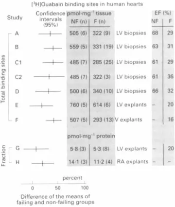
O C C U ~ . ~ 3634 36Figure
5 shows that these studies are not contradictory from
a statistical ~ t a n d p o i n t , ~ ~
since decreases between 26% and
32% fall within the confidence intervals. However, there are
important methodological differences. In one group of
studies (A to E, inclusive), small samples of tissue were
frozen, thawed, and incubated for hours in the presence of
['Hlouabain, and binding data were expressed per g wet
weight. In studies A,30 B,3'
C,32 and D33 it was concluded
there were significant decreases in
N d K pumps in failing
hearts, while study
E34 showed no significant decrease. It is
uncertain whether this discrepancy is due to the use of
explants in study
E instead
of
biopsies or of Fab fragments
to eliminate bound digoxin resulting from previous therapy,
or to insufficient statistical power (large variability and small
number of hearts). This uncertainty is compounded by the
failure of other reports, independent of the water content of
the samples, to express binding data. The potential drawback
of using small (a few mg) samples has been briefly discussed
by this group in an earlier study on necropsy samples,35 but
verification of the water content was not reported in A to E.
In another group of studies indicating that there are non-
significant decreases in binding sites (G36 and H4), large
pieces of tissue were used (several grams), but ['Hlouabain
binding was measured in a membrane fraction instead of in
Study
I1
L F
[3H10uabain binding sites in human hearts Confidence intervals (95%)
+
+
+
+
+
+
+
+
+
EF4%)
percent I 0 50 100Difference of the means of failing and non-failing groups
Figure
5
Comparison between [jH]ouabain binding data fromthis and from previous studies (for references
see
text). Conjidenceintervals of the differences between non-failing and ,failing groups
were calculated according to Gardner and Altnianz9 by using the
means, SEM, and numbers of hearts reported. Note that this
comparison is valid in so far as methods are comparable and
random sampling applied. Data have been normalised and
expressed as percent of the means of the nomfailing groups. Ejection fraction was not determined f o r nonyailing hearts in
studies E to H and is assumed to be normal (about 60%).
Diagnoses were dilated (A, C1, D, G, and
H ) ,
idiopathic dilated(B), and ischaemic (C2, E ) cardiomyopathies. Methodological
differences between studies are indicated in the jigure. Studies A to
D: biopsies were obtained from cardiac patients divided among groups according to their ejection fractions: many patients were
treated with digitalis
in
failing and nomfailing groups; the totalconcentration of specijic sites was determined in intact
endocardium and corrected for methodological errors. Study
E:
tiny pieces of myocardium were obtained from explanted hearts
( "explants "); methodology as in preceding studies except that
tissue was washed with digoxin Fob prior to binding assay. Study
F: present study. Studies
G
and H: ouabain binding was measuredto a membrane fraction isolated from lefi veniricular (LV) or right
auricular (RA) myocardium of explanted hearts; failing patients
were treated with digitalis in G, but not in H.
whole tissue extracts and expressed per mg of membrane
protein. The possibility of NdK-ATPase redistribution
between membrane fractions was not examined. In the
present study, we took care to check or minimise the
influence of such pitfalls by using large samples, ensuring
complete recovery of NdK-ATPase, using different
references to express data, and checking that interference of
bound digoxin was negligible. We further examined the
maximum turnover rate or the sodium sensitivity of
Nd
K-ATPase, which might be affected by changes in the lipid
composition of the sarcolemma, or by the action of
proteases. We did not find any change in these important
variables and, although we did not examine the ATP binding
site directly, the vanadate or phosphate binding site was not
altered. Our results suggest a mean 42% decrease of the
some other alteration in its properties.
Is
such
a
decrease sufficient to account for the increased
digitalis sensitivity? Schmidt
rt
cr17'
have estimated that the
occupation
of
digitalis receptors by disoxin is about
24% in
digitalised patients.
In
animal studies.
50-6552
inhibition
of
NdK-ATPase has been estimated to produce arrhythmias
with digitalis glycosides."
.I8I t is clear that less glycoside
may be required to produce inotropic and toxic effects if the
NdK-ATPase concentration decreases by
26-56%.
However,
because of limitations inherent
in
this study. additional proof
would
be
needed to establish a link between NdK-ATPase
concentration and inotropic responsiveness in failing human
hearts. Ouabain binding data represent apparent Na/
K-ATPace
concentrations. not actual densities. since changes
in
myocytt: number. shape, and size werc not taken into
account.
Our
data were from relatively large samples and
showed little \,ariation across the ventricles. in agreement
uith
a
previous study using necropsj samples." but in
contrast with the results
of
contractility experiments in which
trabeculae from
a
failing heart differed markedly in their
inotropic responses. Furthermore. a single (hypertrophic)
failing heart showed normal NdK-ATPase acti\,ity.
Therelore
itis unlikely that a decrease in NdK-ATPase
[ilonr
explains the increased digitalis sensitivity. Changes
distal t o Na/K-ATPase affecting. for example. sodium
permeability.
NdCa
exchange. or ATP content may also be
i n
volved
.We thanh .iohanna \ o n iler Bel-Kahn MD for her help in the microscopic e\ aluati<oii of thc human beart trabeculae. and Robert B e n i a and Gilbert Ne\\ nian for technical assistance. This \vork was
wppi)rtd i n part h) Grant POI HL22619 from thc National Institutes of Ilealth.
Ke? term\: cardiom! ctpath) : heart failure: ouabain: cardiac g1)cosides: Na/K-pump: ouahain binding: heart trabeculae: human henri
Kcceii e J I 6 .\larch 1903: accepted I 9 Augujt 1993. Time for priniar! re\ ie\v 36 tin! \ .
I Lee CO. 200 Year\ o f digitalis: the emerging central role of the sodium ion i n the control o f cardiac force. Am J Ph,~siol 2 Abete P. Vassale M. Role of intracellular Na- acti\ity in the
negative inotropy of btrophanthidin in cardiac Purkiiije fiber\. Etrr-
J Phtrrrmicd 1992;211:399409.
3 Smith TW. Antrnan EM. Friedman PL. Blatt CM. hlarsh JD. idc.;: inecbani$ms and manifestation\ of toxicit! 4 Schwinger KHG. Riihm bl. La Kosee K. Schmidt C. Schultz C.
Erdmann E. Na-channel activators increase cardiac glycoside sensitivity i n failing hunian myocardium. J Ctrrtlioi.tr.cc Phtrrriitmd 1992:19:551- h l .
5 De Poiet A. GI-upp G. Schv.rartr A. Grupp I. Coupling o f contraction through effects on Na.K-ATPase: changes in Na.K- ATPa\e i w f o r n i \ in heart disease'! Hetrr-t Foilirre
l901:6:2~1l-l I ,258.
6 Slianiwj 0 1 . Mel\in 0. I.inprt.1 JB. Exprebsion of Na.K-ATPase isoforin\ i n human heart. Biocherii Riophx\ Res Commut/
1901:179: 1134-10.
7 Allen PI). Schrnidi TA. hlarsh JD and Kjeldscn K. Na,K-ATPase expre\\icin i n normal and failing human left \.entricle. Rrrsic Rec C ~ / / d i d i992;87(
s
I KX7-91.X Gi-tipp C;. GI-upp It-. Xlclvin DB. Schuarti A. Functional evidence i n d i \ e i r d h u m a n heart tihers for multiple sensitivities of the
iiicitropii. receptor Na.K-ATPa\e (NKA). In: Membrcrrie h i q J / f , l ' t i C l
/(/: / i i o / ( i y i ~ ~ ~ / ircmport, NeLv York: Alan R Liss. 1088:215-22.
V De P o ~ e i A . Shami-:ij 01. Grtipp IL. t't trl. Characterization of total
<i\e and it\ iwforni.; in diseased human hearts. 1985:249:C367-78.
Cr/r-di/>~,ti.~c, D ~ c 1984:26:195-540.
.I
,\lo/ ( * t , l l C i d i o l 1 9 9 1 : 2 3 ( ~ ~ p p l 5):S.65.1 1 Choinczynski 1'. Sacchi N. Single-step method of RNA isolation h) acid guanidium thiocyanate-phenol-chloroform extraction. Anal Nioc/irrii 1987;162: 156-9.
2 Feige G. Leutert T. De Pover A . Na,K-ATPase isozymes in rat tissues: differential sensitivities to sodium, vanadate and dihydroouahain. In: Skou JC. Nerby JG, Maunsbach AB, Esmann M. eds. The Ncr,K-puifip. Part B: Cellular aspects. New York: Alan R Liss, 1988:377-84.
3 Skou JC. The (Na+K)-activated enzyme system and its relationship to transport of sodium and potassium. Q Rev Biopliys
1975:7:40 1-31.
4 De Pover A, Godfraind T. Interaction of ouahain with Na,K- ATPase from human heart and from guinea-pig heart. Riochern
Plinrmacol 1979;28:305 1-6.
15 Labarca C. Paigen K. A simple. rapid, and sensitive method D N A assay procedure. A m / Bioclrenz 1980;102:344-52.
I6 Siiiith PK, Krohn RI, Herinanson GT, et t i /. Measurement of protein using bicinchoninic acid. Ai7ul Biochcwi 1985;150:76-85.
17 Laeiiiinli UK. Cleavage of structural proteins during the assembly of the head of bacteriophage T4. Nrrture 1970;227:680-5. I8 Lindenmayer GE, Schwartz A. A kinetic characterization of
calcium on NaKATPase and its potential role as a link between extracellular and intracellular events: hypothesis for digitalis- induced inotropism. J M o / Cell Ctirdiol 1975;7:59 1-612.
19 Barlow R, Blake JF. Hill coefficients and the logistic equation.
T,-er7r/s Phnrinucol Sci 1989;10:440-41.
20 Boeynaems JM, Dumont JE. Outlines of' receptor theorT.
Amsterdam: ElsevierMorth Holland Biomedical Press. 1980.
2 1 Schmidt TA. Svendsen JH, Haunse S, Kjeldsen K. Quantification of the total Na,K-ATPase concentration in atria and ventricles from inaminalian species by measuring 'H-ouabain binding to intact myocardial samples. Stability to short term ischemia reperfusion. Brrsic Res Curdiol 1990;85:4 1 1-27.
21 Kjeldsen K. Gron P. Age-dependent changc in myocardial cardiac
gl! coside receptor (Na,K-pump) concentration in children. J
Ctrrdio\wsc~ Phnrrnacol 1990;15:332-7.
23 Lane LK, Copenhaver JH. Lindenniayer GE, Schwarti A. Purification and characterization of and ['H louabain binding to the transport adenosine triphosphatase from outer medulla of canine kidney. J B i d Clwm 1973;248:7 197-200.
24 Jekvell EA, Lingrel JB. Comparison of the substrate dependence
properties of the rat Na,K-ATPase e l , a2, and a3 isoforins expressed in HeLa cells. J B i d Cl7erri 1991 ;266:16925-30.
25 De Pover A. Feige G. Dissociation kinetics of [ 'H]ouabain Na.K- ATPase isoforin complexes. In: Kaplan JH, De Weer P, eds. Tl7e
s o d i i r m piirnp: i - t e t i t cledopments. New York: Rockefeller University Press 199 1 :647-5 1.
26 Pagarii ED, Alousi AA, Grant AM, Older TM. Dziuhan J, Allen PD. Changes in niyofibrillar content and Mg-ATPase activity i n ventricular tissues from patients with heart failure caused h) coronary artery disease, cardiomyopathy. or initral valve insufficiency. Circ. Res 1988;63:380-5.
27 Grupp G, Grupp 1L. Ghysel-Burton J, Godfraind T, SchwartL A. Effects of very low concentrations of ouahain on contractile force of isolated guinea-pig, rabbit and cat atria and right ventricular 1984:111-28.
I
. -
-
papillary muscles: an interinstitutional study. J Phanntrcol Exp 77wr- 1982:220: 145-5 1.
28 Sweadner KJ. Multiple digitalis receptors. A molecular perspective. Troiids Cardio\.asc Med I993;3:2-6.
29 Gardner MJ. Altman DG. Confidence intervals rather than P \ dues: estimation rather than hypothesis testing. RMJ
1986:292:746-50.
30 Norgaard A, Bagger JP, Bjerregaard P, Baandrup U, Kjeldsen K.
Thomsen PEB. Relation of left ventricular function and Na.K- pump concentration in suspected dilated cardiomyopathy. Atpi J Ctrrdiol 1988;hl: 1312-5.
3 1 Kjeldsen K. Bjerregaard P, Richter EA, Thomsen PEB, N6rgaard A. Na.K-ATPase concentration in rodent and human hcart and skeletal muscle: apparent relation to muscle perforinanor.
Car-dioimc Res 1 988:22:95- 100.
32 Norgaard A. Kjeldsen K. Human myocardial Na,K-punipa in relation to heart disease. J Appl Cwdiol l989;4:239-45.
33 Nargaard A, Bjerregaard P. Baandrup U, Kjeldsen K. Rcske- Nielsen E. Thoinsen PEB. The concentration of the Na,K-pump i n
skeletal and heart muscle in congestive heart failurc. I n / J C~rnliol
1990:26: 185-90.
31 Schmidt TA. Allen PD. Colucci WS, Marsh JD, Kjeldsen K. No
adaptation to digitalization as cvaluated by digitalis receptor (Na.K-ATPase) quantification in explanted hearts from donors without heart disease and from digitalized recipients with cnd-
NdK-ATPase in failing human hearts
2237
35 Nprrgaard A, Kjeldsen K, Hansen 0, Clausen T, Larsen CG, Larsen 37 Herzig S, Liillmann H, Mohr K, Schmitz R. Interpretation of FG. Quantification of the 3H-ouabain binding site concentration in [’Hlouabain binding in guinea-pig ventricular myocardium i n
human myocardium: a postmortem study. Curdiovasc Res relation to sodium pump activity. J Physiol ( L o r d )
19 86;20:428-35. 1988;396: 105-20.
38 Achenbach C, Daying H, Preisler R. Electrophysiological assay of glycosides in human myocardium with and without “down- glycoside-induced sodium pump inhibition in isolated sheep heart regulated” P-adrenoceptors. J Cardiovasc Phurmacol 1990; Purkinje fibers at the onset of toxicity. In: Erdmann E, Greef K,
15:692-7. Skou JC, eds: Cardiac glycosides 1785-1985. Darmstadt:
36 Schwinger RHG, Bohm M, Erdmann E. Effectiveness of cardiac

![Figure 4 Dissociation kiiietics of the Na/K-ATPa,se-[-~H]ouabaiii corrip1e.u in riorz-j~iiling (ti = 5](https://thumb-eu.123doks.com/thumbv2/123doknet/14886688.647244/6.972.102.468.154.445/figure-dissociation-kiiietics-atpa-ouabaiii-corrip-riorz-iiling.webp)