Diversity of cutinases from plant pathogenic fungi:
di¡erential and sequential expression of cutinolytic esterases
by Alternaria brassicicola
Chao-Yun Fan, Wolfram Koëller *
Department of Plant Pathology, Cornell University, New York State Agricultural Experiment Station, Geneva, NY 14456, USA Received 1 September 1997; accepted 10 October 1997
Abstract
Plant cuticles provide a protective layer that has to be penetrated by fungal pathogens. Evidence is provided for a differential and sequential induction of two classes of cutinolytic esterases by Alternaria brassicicola. Serine esterases with cutinolytic activities were expressed by conidia germinating on host surfaces. The enzymes were not induced by surface wax or cutin monomers. They were only expressed during initial (24 h) contact of conidia with cutin on host surfaces freed from wax, and with cutin in aqueous suspensions. In contrast, contact with cutin had no immediate effect on the expression of CUTAB1, a gene encoding two cutinase isozymes with crucial functions in the saprophytic utilization of cutin. Presence of a cutin monomer or prolonged exposure to cutin was required for the induction of CUTAB1 expression. The differential induction of cutinolytic esterases indicates a sequential recognition of cutin as a barrier to be penetrated and to be utilized as a carbon source in saprophytic stages.
ß 1998 Federation of European Microbiological Societies. Published by Elsevier Science B.V.
Keywords: Alternaria brassicicola; Cutinase; Cutin; Cutin monomer
1. Introduction
Many fungal pathogens encounter the plant cu-ticle, a hydroxyfatty acid polyester impregnated with waxes, as the initial barrier to be breached dur-ing penetration into their hosts. The crucial involve-ment of cutinases during this early step in host in-vasion has been suggested for numerous pathogens, with Fusarium solani (Nectria haematococca)
infect-ing pea hypocotyls as the most comprehensively studied example [1^4].
Multiple lines of evidence have indicated that cu-tinases expressed during saprophytic utilization of cutin by fungal pathogens were also crucially in-volved in the penetration of cuticles [1^4,9]. Recent results obtained with cutinase gene-disrupted mu-tants of Magnaporte grisea [5], F. solani [6^8], Alter-naria brassicicola [9] and Botrytis cinerea [10,11] have not con¢rmed this concept of a dual cutinase func-tion. Although virulence penalties reported for a gene-disrupted mutant of F. solani remain controver-sial [7,12], the disruption of cutinase genes strongly
* Corresponding author. Tel.: +1 (315) 7872375; Fax: +1 (315) 7872389; E-mail: [email protected]
expressed during saprophytic growth of the gens on cutin had no apparent e¡ect on the patho-genicity of respective mutants. Instead, cutin was no longer accepted as a saprophytic carbon source by gene-disrupted mutants of F. solani [6] and A. bras-sicicola [9].
All cutinase genes disrupted thus far and including CUTAB1 of A. brassicicola are induced by cutin monomers [1^4]. In full recognition of this induction mechanism, the original concept of cutinase involve-ment in pathogenicity implied that cutin monomers were generated by small amounts of a `sensing' cuti-nase released from conidia after their attachment to host surfaces [1^4]. In our infection of cabbage leaves with A. brassicicola, a rapid generation of cu-tin monomers during invasion of the host was not apparent; the monomer-induced cutinases Ac and Ba
encoded by CUTAB1 and strongly expressed during saprophytic utilization of cutin were not expressed during host penetration [3,9]. Instead, two di¡erent esterases with cutinolytic activities when tested in mixture were recovered from host cuticles inoculated with both the wild-type strain and CUTAB1-minus mutants [3,9]. The induction of di¡erent cutinolytic enzymes expressed either in planta or during a sap-rophytic stage was investigated in this study. 2. Materials and methods
2.1. Fungal culture and conidial germination
Wax was eluted from 80 cabbage leaves by a 10-s dip into chloroform, the chloroform solution was concentrated and spread over the surfaces of 10 glass plates (20U20 cm) equivalent to the extracted leaf area, and the chloroform was evaporated at room temperature. In order to remove chloroform traces, the plates were heated at 45³C for 4 h. Cabbage leaves freed from wax were washed in running water for 5 h. Preparation of conidia from the wild-type strain of A. brassicicola was described earlier [9,13,14]. Con-idia (2U107 ml31) were suspended in water and
misted onto glass plates, glass plates coated with cabbage leaf wax, and cabbage leaves freed from surface wax. After 24 h, the glass plates and leaves were rinsed with water as described before [9]. The rinsates were ¢ltered and freeze dried. For enzyme
expression in aqueous cultures, conidia (2U106
ml31) were suspended in water, in water amended
with 5 mg ml31 cutin, and in water containing 0.25
mg ml31of the cutin monomer
16-hydroxyhexadeca-noic acid. Conidia were removed by centrifugation after 24 h or 96 h of incubation at 22³C, and the supernatants were freeze-dried.
2.2. Detection of serine esterases and enzyme assays Freeze-dried samples were resuspended in 2 ml 10 mM HEPES pH 7.5 and centrifuged. Aliquots containing 2 nkat esterase activity were analyzed by active site-labeling with [3
H]diisopropyl£uoro-phosphate (DFP), separation of proteins by SDS-PAGE and subsequent £uorography as described elsewhere [9,13]. Esterase activity was assayed with p-nitrophenyl butyrate as substrate and cutinase ac-tivity was assayed with [3H]grapefruit cutin as
de-scribed previously [9].
2.3. Reverse transcription-polymerase chain reaction Total RNA was extracted as described previously [14] from conidia after 24 and 96 h germination in the presence of cutin, and after 24 h in the presence of 16-hydroxyhexadecanoic acid. RNA (4 Wg) was reverse-transcribed with oligo-T as primer at 42³C for 1 h.
Primers used for PCR corresponded to nucleotides 106^127 and 746^766 of CUTAB1 [14]. Ampli¢ca-tions of cDNA were performed with aliquots of the reverse-transcribed RNA fractions in 50 Wl using 2.5 mM MgCl2, 1 U Taq polymerase, 0.2 mM each of
deoxyribonucleotide triphosphates, 50 mM KCl, 10 mM Tris-HCl (pH 8.3) and 20 ng of each primer. Water and the two primers alone and as primer pair were used as negative controls. A Perkin Elmer GeneAmp PCR System 9600 was programmed as follows: 94³C for 30 s, 50³C for 30 s, 72³C for 2 min; 35 cycles. Aliquots of PCR products were electrophoresed on agarose gel (1.2%), stained with ethidium bromide and visualized with a UV trans-illuminator.
3. Results
3.1. Inductive properties of cabbage wax
Germinating conidia of A. brassicicola form simple appressoria and penetrate host cuticles within 24 h; disease symptoms are expressed 72 h after inocula-tion [3,9]. In order to re£ect this time frame of host infection, all experiments with relevance to cutinase induction during penetration were performed after 24 h of conidial germination. As before [13,14], the saprophytic stage of cutin utilization was investi-gated after 96 h. All experiments were done with the wild-type strain of A. brassicicola containing the intact cutinase gene CUTAB1 [9,13,14] and were repeated several times with very similar results. As shown in Fig. 1, serine esterases with molecular masses of 52 kDa and 26 kDa were released from conidia of A. brassicicola suspended in water. The enzymes were fully released after 4 h (data not shown) indicating a preformed nature of these ester-ases. Although the esterases were clearly di¡erent from the 31-kDa and 19-kDa cutinolytic esterases expressed on cabbage leaves [3,9] and the mono-mer-induced cutinase isozymes Ac (24 kDa) and Ba
(21 kDa) encoded by CUTAB1 [13,14], the mixture of enzymes was cutinolytically active (16 Bq h31
mg31). The same two 52-kDa and 26-kDa esterases
released by conidia suspended in water were also released from conidia germinating on glass surfaces (Fig. 1).
The expression of the 31-kDa and 19-kDa cutino-lytic esterases on host surfaces has been studied with intact leaves [3,9], and their induction by surface wax rather than cutin could not be excluded. However, the esterases released from germinating conidia ex-posed to a layer of wax removed from cabbage leaves and coated on glass plates were not di¡erent from conidia germinating in water (Fig. 1). The re-sult indicated that cabbage wax was inactive in in-ducing the cutinolytic 31-kDa and 19-kDa esterases strongly expressed on host surfaces during infection by the pathogen [3,9].
3.2. Induction of cutinolytic esterases by cutin and cutin monomers
The lack of esterase induction by surface wax mandated a study on the inductive properties of the polymer cutin. The results are summarized in Fig. 3. In the labeling experiments, equal amounts of esterases determined with p-nitrophenyl butyrate as a substrate were subjected to labeling with [3H]DFP [14], and the absence or presence of the
26-kDa esterase released under noninductive
condi-Fig. 1. SDS-PAGE and £uorography of DFP-labeled serine ester-ases under non-inductive conditions. Conidia were germinated in water (lane 1), on glass plates (lane 2) or on glass plates coated with wax isolated from cabbage leaves (lane 3). Samples were an-alyzed after 24 h.
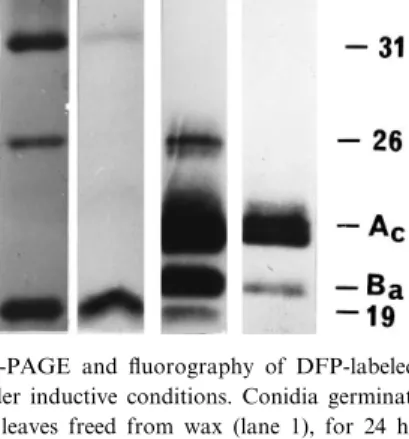
Fig. 2. SDS-PAGE and £uorography of DFP-labeled serine hy-drolases under inductive conditions. Conidia germinated for 24 h on cabbage leaves freed from wax (lane 1), for 24 h (lane 2) or 96 h (lane 3) in aqueous suspensions of apple cutin, or for 24 h in the presence of 16-hydroxyhexadeconoic acid (lane 4). Cuti-nases Ac and Ba have molecular masses of 24 and 21 kDa,
tions (Fig. 1) re£ects only the proportion of the en-zyme relative to other esterases. As shown in Fig. 2, the surface of cabbage leaves freed from wax and, thus, with cutin in immediate contact with germinat-ing conidia retained their activity of inducgerminat-ing the two esterases with molecular masses of 31 kDa and 19 kDa, which also had been recovered from wax-cov-ered leaves [3,14]. Removal of surface wax had no impact on the induction of CUTAB1 expression, as evidenced by the lack of cutinase Ac and Ba
expres-sion (Fig. 3).
The induction of the 31-kDa and 19-kDa esterases and lack of CUTAB1 expression on both intact [9] and dewaxed surfaces (Fig. 2) prompted us to inves-tigate the inductive properties of apple cutin sus-pended in aqueous medium and, thus, under the sap-rophytic conditions employed in the puri¢cation of cutinases Ac and Ba [13]. Similar to the results
ob-tained with conidia of A. brassicicola in contact with host cuticles, only the 31-kDa and 19-kDa esterases were expressed during the initial 24-h exposure phase (Fig. 2). The result con¢rmed the fungal origin of these esterases and also indicated that their induction was dependent neither on germination on cuticular
surfaces nor on the presence of host cutin. Very sim-ilar to host surfaces, cutinases Acand Baencoded by
CUTAB1 were not detected after this initial 24-h exposure of conidia to a cutin suspension. As ex-pected from our previous results [13], however, the isozymes became the predominant esterases ex-pressed after 96 h of exposure to cutin (Fig. 2).
In complete reversal, cutinases Ac and Ba were
expressed by conidia germinating for 24 h in the presence of the cutin monomer 16-hydroxypalmitic acid (Fig. 2) as a known inducer of CUTAB1 exprsion [13,14]. The cutinolytic 31-kDa and 19-kDa es-terases were not induced by the monomer (Fig. 2). 3.3. Detection of CUTAB1 mRNA under inductive
conditions
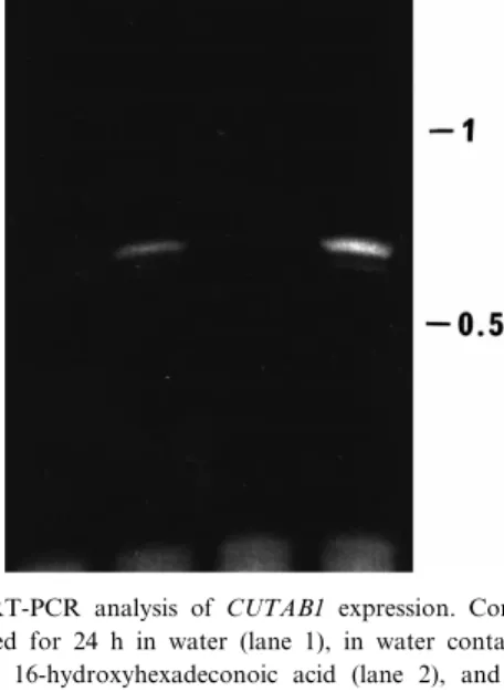
The delayed expression of CUTAB1 in the pres-ence of cutin as opposed to cutin monomers was fully con¢rmed in a RT-PCR analysis with speci¢c primers derived from the CUTAB1 sequence (Fig. 3). Conidia germinating for 24 h in water and in the presence of an aqueous cutin suspension contained no detectable CUTAB1 mRNA. As expected from the expression pattern shown in Fig. 2, gene tran-scription was evident in conidia germinating for 24 h in the presence of a cutin monomer and after 96 h of saprophytic exposure of the fungus to cutin (Fig. 3).
4. Discussion
In summary, the results described in this study suggest a regulatory sequence more complex than suggested for F. solani [1,4]. With A. brassicicola, two serine esterases with cutinolytic activity and mo-lecular masses of 52 kDa and 26 kDa were constitu-tively released shortly after conidia were suspended in water. Surface wax as the ¢rst layer encountered by conidia attaching to host surfaces was inactive in the induction of additional serine esterases. Exposure of germinating conidia to host cutin on dewaxed leaves or to nonhost cutin in aqueous suspension induced the expression of the 31-kDa and 19-kDa cutinolytic esterases described before [3,9], but cutin monomers were inactive as inducers. The situation for the well characterized cutinases Ac and Ba [13]
Fig. 3. RT-PCR analysis of CUTAB1 expression. Conidia were germinated for 24 h in water (lane 1), in water containing 0.25 mg ml31 16-hydroxyhexadeconoic acid (lane 2), and for 24 h
(lane 3) or 96 h (lane 4) in water containing 5 mg ml31apple
encoded by CUTAB1 [14] was reversed. The gene was rapidly induced by a cutin monomer, but induc-tion by cutin was delayed beyond the 24-h phase of cuticle penetration during host infection.
The sequential induction of di¡erent cutinolytic enzymes implies the existence of a regulatory `switch'. The polymer appears to be ¢rst perceived as a barrier to be penetrated in order to gain access into the host. Once the pathogen reaches a subcutic-ular location, the availability of carbohydrate sour-ces provided by the host would repress the subse-quent induction of other cutinases [1^4,13]. If such repressive conditions are not met within the normal time frame of host surface penetration, cutin mono-mers as inducers of cutinases functional in a sapro-phytic stage might slowly be generated, and cutin would then be recognized as a saprophytic carbon source.
While the activity of cutin monomers as speci¢c inducers of CUTAB1 in A. brassicicola was con-¢rmed in this study, the induction mechanism of the 31-kDa and 19-kDa cutinolytic esterases ex-pressed in the presence of cutin remains elusive. The mixture of esterases constitutively released from conidia was cutinolytically active, and these esterases might generate cutin-derived inducers dif-ferent from cutin monomers. Although highly spec-ulative, cutin oligomers generated through endo-cuti-nase action could serve this function as initial inducers. Such oligomers might slowly be hydrolyzed to cutin monomers as inducers of CUTAB1 crucial in saprophytic utilization of cutin rather than in cu-ticle penetration [9].
As summarized and discussed recently [15], it has not been demonstrated thus far that any extracellular hydrolase is crucially involved in fungal pathogene-sis. One of the explanations o¡ered was that many of these polymer-degrading enzymes are genetically re-dundant. This would imply that the disruption of one of the redundant genes is counteracted by re-lated enzymes, which in concert would overcome any recognizable virulence penalty. Our results with A. brassicicola described above might point in a dif-ferent direction. Cutinases might have evolved sepa-rately, and according to their speci¢c roles they play in either pathogenic or saprophytic niches occupied by a fungal pathogen. This alternative explanation would imply that the evolution of pathogenic and
saprophytic traits diverged more stringently than an-ticipated in the past.
In support of this hypothesis of cutinase adapta-tion to distinct ecological roles, the Fusarium cuti-nase has been described to be also active as a lipase [16,17]. Crucial involvement of the Fusarium cutinase in saprophytic degradation of the synthetic polyester polycaprolactone [18], and a close relationship be-tween the Fusarium cutinase and a lipolytic esterase gene cloned from the nonpathogenic fungus Asper-gillus oryzae [19] supports a diverse and £exible role of the enzyme in saprophytic stages of plant patho-genic fungi. The broad enzymatic activity of the Fu-sarium cutinase makes it appealing to speculate that the cutinases characterized in the past are `multi-pur-pose esterases' hydrolyzing a variety of saprophyti-cally relevant esters including cutin.
While cutinases functional in saprophytic stages of plant pathogenic fungi and induced by cutin mono-mers are exceptionally well understood [1^4,16^18], the functions and properties of cutinolytic esterases expressed by A. brassicicola during initial contact with cutin remain largely unexplored. The results described above demonstrate that such cutinase genes exist and that, in addition, the two classes of cutinases are induced by di¡erent mechanisms. The di¡erential and sequential expression of di¡erent cu-tinolytic esterases described in this study will aid in the functional characterization of the cutin-induced esterases expressed at an early stage of host invasion and in the identi¢cation of mechanisms involved in a regulatory `switch' between parasitic and saprophytic stages of a fungal pathogen.
Acknowledgments
This study was supported, in part, by a grant from USDA-NRICGP (96-35303-3815).
References
[1] Kolattukudy, P.E., Rogers, L.M., Li, D., Hwang, C.S. and Flaishman, M.A. (1995) Surface signaling in pathogenesis. Proc. Natl. Acad. Sci. USA 92, 4080^4087.
[2] Koëller, W. and Yao, C. (1996) Targets for plant protection ^ Can cutinase be counted in? In: Modern Fungicides and
Anti-fungal Compounds (Lyr, H., Russell, P.E and Sisler, H.D., Eds.), pp. 163^172. Intercept, Andover, MA.
[3] Koëller, W., Yao, C., Trail, F. and Parker, D.M. (1995) Role of cutinase in the invasion of plants. Can. J. Bot. 73, S1109^ S1118.
[4] Li, D. and Kolattukudy, P.E. (1997) Cloning of cutinase tran-scription factor 1, a transactivating protein containing Cys-6Zn-2 binuclear cluster DNA-binding motif. J. Biol. Chem. 272, 12462^12467.
[5] Sweigard J.A., Chumley, F.G. and Valent, B. (1992) Disrup-tion of a Magnaporte grisea cutinase gene. Mol. Gen. Genet. 232, 183^190.
[6] Stahl, D.J. and Schaëfer, W. (1992) Cutinase is not required for fungal pathogenicity on pea. Plant Cell 4, 621^629. [7] Stahl, D.J., Theuerkauf, A., Heitefuss, R. and Schaëfer, W.
(1994) Cutinase of Nectria haematococca (Fusarium solani f.sp. pisi) is not required for fungal virulence or organ specif-icity. Mol. Plant-Microbe Interact. 7, 713^725.
[8] Crowhurst, R.N., Binnie, S.J., Bowen, J.K., Hawthorne, B.T., Plummer, K.M., Rees, G.J., Rikkerink, E.H.A. and Temple-ton, M.D. (1997) E¡ect of disruption of a cutinase gene (cut A) on virulence and tissue speci¢city of Fusarium solani f.sp. cucurbitae race 2 toward Cucurbita maxima and C. moschata. Mol. Plant-Microbe Interact. 10, 355^368.
[9] Yao, C. and Koëller, W. (1995) Diversity of cutinases from plant pathogenic fungi: Di¡erent cutinases are expressed dur-ing saprophytic and pathogenic stages of Alternaria brassici-cola. Mol. Plant-Microbe Interact. 8, 122^130.
[10] Van der Vlugt-Bergmans, C.J.B., Wagemakers, C.A.M. and Van Kan, J.A.L. (1997) Cloning and expression of the cuti-nase A gene of Botrytis cinerea. Mol. Plant-Microbe Interact. 10, 21^29.
[11] Van Kan, J.A.L., Van't Klooster, J.W., Wagemakers, C.A.M.,
Dees, D.C.T. and Van der Vlugt-Bergmans, C.J.B. (1997) Cu-tinase A of Botrytis cinerea is expressed, but not essential, during penetration of gerbera and tomato. Mol. Plant-Mi-crobe Interact. 10, 30^38.
[12] Rogers, L.M., Flaishman, M.A. and Kolattukudy, P.E. (1994) Cutinase gene disruption in Fusarium solani f.sp. pisi decreases its virulence on pea. Plant Cell 6, 935^945.
[13] Trail, F. and Koëller, W. (1993) Diversity of cutinases from plant pathogenic fungi: Puri¢cation and characterization of two cutinases from Alternaria brassicicola. Physiol. Mol. Plant Pathol. 42, 205^220.
[14] Yao, C. and Koëller, W. (1994) Diversity of cutinases from plant pathogenic fungi: Cloning and sequence analysis of a cutinase gene from Alternaria brassicicola. Physiol. Mol. Plant Pathol. 44, 81^92
[15] Hamer, J.E. and Holden, D.W. (1997) Linking approaches in the study of fungal pathogenesis: A Commentary. Fung. Ge-net. Biol. 21, 11^16.
[16] Longhi, S., Manesse, M., Verheij, H.M., deHaas, G.H., Eg-mond, M., Knoops-Mouthuy, E. and Cambillau, C. (1997) Crystal structure of cutinase covalently inhibited by a tri-glyceride analogue. Protein Sci. 6, 275^286.
[17] Martinez, C., de Geus, P., Lauwereys, M., Matthyssens, G. and Cambillau, C. (1992) Fusarium solani cutinase is a lip-olytic enzyme with a catalytic serine accessible to solvent. Nature 356, 615^618.
[18] Murphy, C.A., Cameron, J.A., Huang, S.J. and Vinopal, R.T. (1996) Fusarium polycaprolactone depolymerase is cutinase. Appl. Environ. Microbiol. 62, 456^460.
[19] Ohnishi, D.L., Toida, J., Nakasawa, H., and Sekiguchi, J. (1995) Genome stucture and nucleotide sequence of a lipolytic enzyme gene of Aspergillus oryzae. FEMS Microbiol. Lett. 126, 145^150.