The ins(ide) and outs(ide) of dolichyl phosphate biosynthesis and recycling in the endoplasmic reticulum
10
0
0
Texte intégral
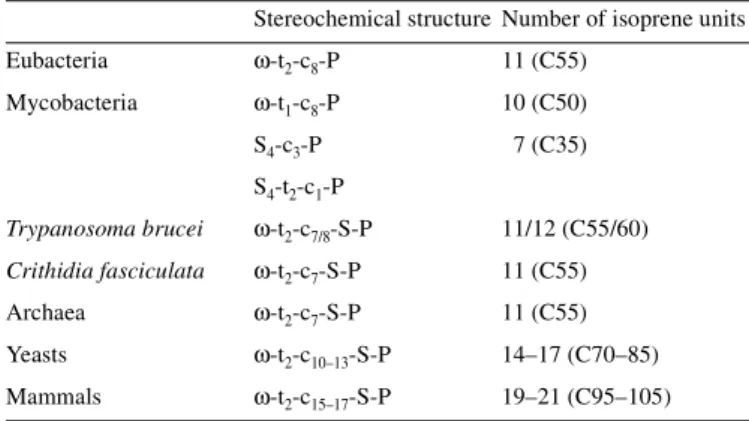
(2) B. Schenk et al.. Table I. Structure of polyprenyl and dolichyl monophosphates used as glycosyl carrier lipids in prokaryotic and eukaryotic cells. (dolichols) became saturated during evolution remains an intriguing question.. Stereochemical structure Number of isoprene units Eubacteria. ω-t2-c8-P. 11 (C55). Biosynthesis of the glycosyl carrier lipids Undec-P and Dol-P. Mycobacteria. ω-t1-c8-P. 10 (C50). Initiation and chain elongation stage catalyzed by cis-isoprenyltransferases. S4-c3-P. 7 (C35). S4-t2-c1-P Trypanosoma brucei. ω-t2-c7/8-S-P. 11/12 (C55/60). Crithidia fasciculata. ω-t2-c7-S-P. 11 (C55). Archaea. ω-t2-c7-S-P. 11 (C55). Yeasts. ω-t2-c10–13-S-P. 14–17 (C70–85). Mammals. ω-t2-c15–17-S-P. 19–21 (C95–105). Numbers in parenthesis = total numbers of carbon atoms. c = cis isoprene unit. t = trans isoprene unit. S = saturated α-isoprene unit. ω = distal terminal isoprene unit.. polyisoprenyl phosphate glycosyl carrier lipids are a family of membrane lipids varying in the number and stereoconfiguration of their linearly linked isoprene units. The chain length is species-specific with 11 isoprene residues commonly found in eubacterial and archaebacterial cells (Higashi et al., 1967; Lechner et al., 1985). Mycobacteria are unusual among eubacteria, having one containing 7 isoprene units (Wolucka and de Hoffmann, 1998), and another with 10 isoprene units (Wolucka et al., 1994). Trypanosamatids utilize dolichyl phosphates as glycosyl carrier lipids with chain lengths of 11 or 12 isoprene units (Low et al., 1991; Quesada-Allue and Parodi, 1983). Eukaryotes contain dolichols, which differ from undecaprenol and other fully unsaturated polyprenols in that the chain length is generally longer and the α-isoprene unit is saturated. The yeasts Saccharomyces cerevisiae and Schizosaccharomyces pombe contain dolichols with 14–17 isoprene units (Quellhorst et al., 1998). In mammalian cells the predominant dolichols range from 18–21 isoprene units (Rip et al., 1985). Dolichols, the longest aliphatic molecules synthesized in animal cells, are considerably longer than the fatty acyl chains of glycerophospholipids. The structures of the (C90–105) Dol-Ps found in mammalian cells are denoted as ω-t2-c15–17-S-P in Table I. Experiments conducted in many laboratories have established that the chain length and the presence (or absence) of a saturated α-isoprene unit in polyisoprenyl phosphate substrates are critical for their recognition by the enzymes that glycosylate them and utilize their glycosyl derivatives as sugar donors (Szkopinska et al., 1992; Kean et al., 1994; McLachlan and Krag, 1992, 1994; Rush et al., 1993, 1997; D’Souza-Schorey et al., 1994; Dotson et al., 1995 and other references cited in these papers). All of these reports show that, in general, each glycosyltransferase prefers the natural polyisoprenyl monophosphate as the acceptor lipid substrate. However, the functional significance of why the chain length increased and the α-isoprene units of the eukaryotic glycosyl carrier lipids 62R. Numerous laboratories have documented the presence of cisIPTase activity in crude microsomal fractions from a variety of animal tissues (Daleo et al., 1977; Grange and Adair, 1977; Wong and Lennarz, 1982; Crick et al., 1991; Ericsson et al., 1992a), yeast (Adair and Cafmeyer, 1987; Szkopinska et al., 1996), and soluble fractions from hen oviduct (Wellner and Lucas, 1979) and Ehrlich ascites tumor cells (Adair et al., 1984). In contrast to the microsomally associated cis-IPTases in mammalian tissues, bacterial cis-IPTases are apparently soluble proteins (Keenan and Allen, 1974; Baba and Allen, 1980). In all of these enzyme systems chain elongation can be initiated with F-P-P in the presence of I-P-P (Figure 1). In the bacterial systems eight cis-isoprene units are added to F-P-P, forming ω-t2-c8-P-P, which is then converted to the “active” form of the carrier lipid by a pyrophosphate phosphatase (Goldman and Strominger, 1972). A recent report indicates that the biosynthesis of the unusual heptaprenyl phosphate is derived from ω-t4-P-P, and the synthesis of decaprenyl phosphate is initiated by a reaction involving the F-P-P stereoisomer, ω-t-c-P-P, and I-P-P in mycobacteria (Crick et al., 2000). In the eukaryotic systems the chain elongation stage is catalyzed by cis-IPTases that add 12–18 isoprene units to F-P-P, forming the pyrophosphorylated intermediates, ω-t2-c12–18-P-P. Although it has been proposed that F-P-P synthase in yeast may be associated with cis-IPTase (Szkopinska et al., 1997), a direct interaction between the enzymes has not yet been demonstrated. The chain elongation process can also be initiated with the all trans-stereoisomer of geranylgeranyl pyrophosphate (ω,t,t,t,-GG-P-P) or ω,t,t,c-GG-P-P (Crick et al., 1991; Ericsson et al., 1992b) and I-P-P in vitro. Presumably, the “nascent” allylic pyrophosphate intermediate becomes firmly integrated into the cytoplasmic leaflet of the ER early in the elongation stage and continues to be extended until the cisIPTases amazingly recognize that the chain is the correct length (Figure 1). The studies reviewed herein indicate that the conversion of F-P-P to the appropriate fully unsaturated, long chain polyprenyl pyrophosphate (Poly-P-P) end product is catalyzed by a single enzyme in bacteria and yeast. Further work will be required to determine if this is true in animal cells. The biosynthesis of Dol-Ps is completed by the terminal reactions described below. Structures and subcellular localization(s) of eukaryotic cis-IPTases In early work on this class of isoprenyltransferases, cis-IPTase activities were partially purified and characterized from Micrococcus luteus (formerly called Micrococcus lysodeiktikus), Lactobacillus plantarum, and Escherichia coli (Keenan and Allen, 1974; Baba and Allen, 1980; Fujisaki et al., 1986). Cloning of the corresponding cDNAs over the past few years has now provided more definitive information on the structures of pro- and eukaryotic cis-IPTases (Table II). The first cis-IPTase.
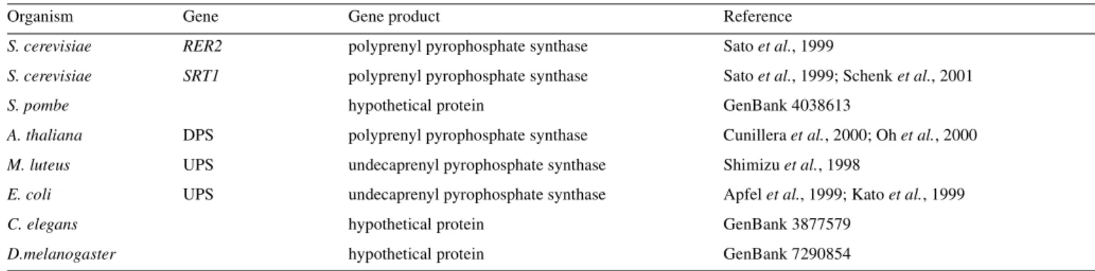
(3) Dolichyl phosphate biosynthesis. Fig. 1. Topological model for the enzymatic reactions leading to Dol-P biosynthesis de novo on the cytoplasmic face of the ER. The enzymatic reactions converting farnesyl pyrophosphate (F-P-P, structure shown), to the end product, 95-dolichyl monophosphate (95-Dol-P, structure shown) are illustrated in the upper box. cis-IPTase = cis-isoprenyltransferase; Poly-P-(P)ase = polyprenyl mono-and pyrophosphate phosphatase; PRed = polyprenol reductase; DK = dolichol kinase.RER2/SRT1 refers to yeast genes encoding separate cis-IPTases.. Table II. Gene family encoding cis-IPTase activities Organism. Gene. Gene product. Reference. S. cerevisiae. RER2. polyprenyl pyrophosphate synthase. Sato et al., 1999. S. cerevisiae. SRT1. polyprenyl pyrophosphate synthase. Sato et al., 1999; Schenk et al., 2001. hypothetical protein. GenBank 4038613. S. pombe A. thaliana. DPS. polyprenyl pyrophosphate synthase. Cunillera et al., 2000; Oh et al., 2000. M. luteus. UPS. undecaprenyl pyrophosphate synthase. Shimizu et al., 1998. E. coli. UPS. undecaprenyl pyrophosphate synthase. Apfel et al., 1999; Kato et al., 1999. C. elegans. hypothetical protein. GenBank 3877579. D.melanogaster. hypothetical protein. GenBank 7290854. (Undec-P-P synthase) to be cloned was from M. luteus (Shimizu et al., 1998). More recently, the cloning of Undec-P-P synthases from E. coli (Apfel et al., 1999; Kato et al., 1999), a cis-IPTase from Arabidopsis thaliana (Cunillera et al., 2000; Oh et al., 2000) and two cis-IPTases from S. cerevisiae (Sato et al., 1999; Schenk et al., 2001) have been reported. Homologous loci were found in many organisms, ranging from archaebacteria to C. elegans and to humans. Even though trans- and cis-IPTases both catalyze a head-to-tail condensation reaction. between I-P-P and an allylic pyrophosphate intermediate, they apparently do not share any significant sequence homology (Shimizu et al., 1998). It is noteworthy that the conserved DDXXD motif, characteristic for trans-IPTases (Chen et al., 1994) and shown to be required for catalytic activity (Wang and Ohnuma, 1999), is not present in the cis-IPTases. As recently reported in this journal (Schenk et al., 2001), the alternative cis-IPTase in S. cerevisiae extends the Poly-P-P intermediate to chain lengths similar to mammalian dolichols. 63R.
(4) B. Schenk et al.. Important clues to the domains that determine how many isoprene units are added by each cis-IPTase may be found by comparing the bacterial (C55), S. cerevisae (RER2, C75), SRT1 (C95), and Arabidopsis (C120), and ultimately the mammalian enzymes that elongate F-P-P to different chain lengths. The structure of the mammalian enzyme(s) has not yet been reported, but it will be interesting to see if those cisIPTases are structurally more closely related to SRT1 and the Arabidopsis enzyme than to RER2 and the bacterial Undec-P-P synthases. It will also be fascinating to learn how the bacterial cis-IPTases, most of which are apparently soluble enzymes, terminate the chain elongation process after adding the correct number of isoprene units with fairly high fidelity. The mammalian cis-IPTases appear to be bound firmly to microsomes, and the rat brain enzyme is highly enriched in heavy microsomes (Crick et al., 1991). Studies with rat liver suggest that peroxisomes may also be able to synthesize PolyP-P from I-P-P and F-P-P, presumably with a cis-IPTase that is distinct from the ER enzyme (Ericsson et al., 1992a). Clearly, the localization and topological arrangements of the cisIPTases involved in Dol-P biosynthesis are important subjects that warrant further investigation. The initial substrates, I-P-P and F-P-P, are soluble cytosolic intermediates, but the Poly-P-P intermediates and final products are extremely hydrophobic lipids that presumably are embedded in the ER bilayer. It is expected that the active sites of the cis-IPTases are located at the boundary of hydrophilic and hydrophobic environments in the ER. In this regard, it has been demonstrated that cis-IPTase activity has a proteasesensitive site on the cytoplasmic face of liver microsomes (Adair and Cafmeyer, 1983). Although no membranespanning helices are predicted by the structures of the yeast cis-IPTases, they are, nevertheless, membrane-bound. Further work will be required to determine the exact nature of the membrane association of eukaryotic cis-IPTases. Terminal steps in de novo pathway The terminal step in the de novo pathway for Undec-P biosynthesis in bacteria simply requires the cleavage of the pyrophosphate bond in Undec-P-P (Goldman and Strominger, 1972). In the current model for yeasts and mammalian cells, the final Poly-P-P intermediate is dephosphorylated prior to the reduction of the α-isoprene unit. Many laboratories have reported microsomal polyisoprenyl mono- and pyrophosphate phosphatase activities that could catalyze these steps (Kato et al., 1980; Wedgwood and Strominger, 1980; Appelkvist et al., 1981; Belocopitow and Boscoboinik, 1982; Scher and Waechter, 1984; Wolf et al., 1991). There is, however, no definitive proof yet for the Poly-P-P and Poly-P phosphatases, which presumably have active sites exposed on the cytoplasmic leaflet of the ER. As shown in Figure 1, the α-isoprene unit of the free polyprenol is reduced subsequently by a microsomal reductase with NADPH serving as the reductant (Sagami et al., 1993). Although Chinese hamster over (CHO) mutants that contain defects in α-reduction have been characterized (Stoll et al., 1988), there is virtually no information on the properties and structure of this enzyme. Because the recognition of the saturated α-isoprene unit is vital for the synthesis and function of the lipid intermediates as glycosyl donors, the reductase plays an indispensable role in this biosynthetic pathway. It will 64R. be extremely important to learn more about the enzymatic αreduction of the long chain polyprenol and to establish if this is the only mechanism for the formation of the saturated α-isoprene unit in dolichols. If the saturated α-isoprene unit of dolichol occurs only at the free isoprenol level, the terminal step in the de novo pathway would be catalyzed by dolichol kinase. This CTP-mediated kinase was first detected in microsomes from several mammalian tissues by Charlie Allen and his co-workers by following the conversion of [3H]dolichol to [3H]Dol-P in the presence of unlabeled CTP (Allen et al., 1978), and in brain microsomes by assaying the transfer of 32P from [γ-32P]CTP to endogenous dolichol (Burton et al., 1979). In the latter study the Dol-P formed via the kinase was shown to be formed in a membrane site, where it is available for lipid intermediate biosynthesis. The calf brain kinase is highly enriched in heavy microsomal fractions (Scher and Waechter, 1984), and the active site of the rat liver enzyme faces the cytoplasm (Adair and Cafmeyer, 1983). In S. cerevisiae, the SEC59 gene encodes an essential polypeptide component of dolichol kinase although it has not been conclusively established that it is the catalytic subunit (Heller et al., 1992). Clearly, significant progress has been made in the last two decades on these reactions, but considerably more work will be required to answer many critical questions about the enzymology, topology, and molecular biology of the terminal steps, dephosphorylation–reduction–rephosphorylation (Figure 1), in Dol-P biosynthesis. Regulation of Dol-P and lipid intermediate biosynthesis The critical role of lipid-mediated glycosylation in: (1) the proper folding and intracellular translocation of N-linked glycoproteins (Helenius and Aebi, 2001), (2) protein O- and Cmannosylation, and (3) GPI-anchor assembly emphasizes the importance of elucidating all of the mechanisms regulating DolP and lipid intermediate biosynthesis. In a number of experimental model systems (Harford et al., 1977; Lucas and Levin, 1977; Harford and Waechter, 1980; Hubbard and Robbins, 1980; Carson et al., 1981; Spiro and Spiro, 1986; Rosenwald et al., 1990) it has been shown that the level of Dol-P in the ER is one important rate-controlling factor in the biosynthesis of glycolipid intermediates and consequently protein N-glycosylation. These results emphasize the need to understand all of the factors regulating the biosynthesis of Dol-P. Changes in dolichol kinase have been reported in mammalian cells (Burton et al., 1981; Volpe et al., 1987; Eggens, 1988) and developing sea urchin embryos (Rossignol et al., 1981); shifts in the metabolic balance of the phosphorylation of dolichol and the dephosphorylation of Dol-P(P) have been implicated in controlling the level of the Dol-P pool (Scher et al., 1985; Bhat et al., 1991). However, developmental increases in cis-IPTase activity represent the best correlation with the induction of Dol-P biosynthesis and protein N-glycosylation in mammalian cells. The induction of the cis-IPTase system catalyzing the elongation stage of Dol-P biosynthesis (Figure 1) has been shown to precede large developmental increases in Dol-P and lipid intermediate biosynthesis and protein N-glycosylation in embryonic rat brain (Crick and.
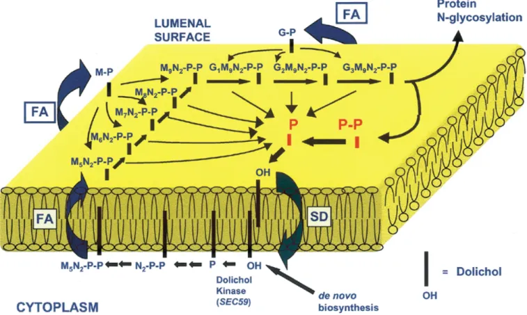
(5) Dolichyl phosphate biosynthesis. Waechter, 1994), proliferating murine B lymphocytes (Crick and Waechter, 1994) and cAMP-treated JEG-3 choriocarcinoma cells (Konrad and Merz, 1996). The recent cloning of cDNAs for several cis-IPTases from bacteria and yeast should accelerate the progress in understanding their regulation and how they are associated with the ER. The rate of formation of GlcNAc-P-P-Dol, Man-P-Dol and Glc-P-Dol in the first stage of the pathway (Figure 2) is influenced by the level of Dol-P in the ER, and there is evidence that dolichylsaccharide intermediate biosynthesis is subject to control by feedback control mechanisms. Kean and co-workers (Kean, 1985; Kean et al., 1994) have published a series of articles documenting the regulation of UDP-GlcNAc:Dol-P N-acetylglucosaminyl 1-P transferase (GPT), the enzyme responsible for GlcNAc-P-P-Dol biosynthesis, by Man-P-Dol. More recently, the same laboratory has reported evidence that ManP-Dol synthesis is enhanced by GlcNAc-P-P-Dol and that GlcNAc-P-P-Dol inhibits its own synthesis by product inhibition of GPT (Kean et al., 1999). Similarly, (GlcNAc)2-P-P-Dol inhibits the enzyme catalyzing the transfer of GlcNAc from UDP-GlcNAc to GlcNAc-P-P-Dol. At least three factors could affect the rate of conversion of Man5GlcNAc2-P-P-Dol to Glc3Man9GlcNAc2-P-P-Dol on the lumenal surface in the second stage of the pathway. In addition to the level of the lipid-mediated mannosyl- and glucosyltransferases, the lumenal reactions would be influenced by the rate at which Man5GlcNAc2-P-P-Dol, Man-P-Dol, and Glc-P-Dol. diffuse transversely from the cytoplasmic face, where they are synthesized, to the lumenal monolayer. The transbilayer movement of these three intermediates could be accelerated by a “mass action” effect as they are consumed in the lumenal mannosylation and glucosylation reactions. Similarly, it is also reasonable that the preceding lipid-mediated reactions on the lumenal leaflet could be driven by the consumption of Glc3Man9GlcNAc2-P-P-Dol, the lipid end product of the pathway, during the primary N-glycosylation reactions. The regulation of lipid intermediate biosynthesis is also likely to include other novel mechanisms. In this regard, Doerrler and Lehrman (1999) have recently reported that the unfolded protein response (UPR) stimulates the conversion of Man2–5GlcNAc2-P-P-Dol to Glc3Man9GlcNAc2-P-P-Dol. This is another mechanism that enables the UPR to maintain efficient protein folding in the ER in addition to de novo synthesis of chaperones. In view of all this information, it is clear that the regulation of Glc3Man9GlcNAc2-P-P-Dol biosynthesis is complex and multifaceted, and there is certainly much more to be learned on this topic. Topological model for the biosynthesis, utilization in the lipid intermediate pathway for protein N-glycosylation and recycling of Dol-P-P/Dol-P The current topological model for lipid intermediate biosynthesis and protein N-glycosylation was first outlined in detail by. Fig. 2. Topological model for lipid intermediate synthesis, translocation and the proposed role for Dol-P-P/Dol-P phosphatases in the recycling of Dol-P-P/Dol-P in the ER. FA = flippase assisted; SD = simple diffusion; N = N-acetylglucosamine; M = mannose; G = glucose.. 65R.
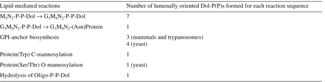
(6) B. Schenk et al.. Hirschberg and Snider (1987). The impressive work of Snider and his co-workers made a major contribution to the principal features of this early model. Snider and colleagues determined the topological orientation of the active sites of most of the biosynthetic enzymes and the various intermediates primarily by assessing protease sensitivity of the active sites and the inextractability of ConA–lipid intermediate complexes in sealed microsomal vesicles. The Hirschberg and Snider review and related articles (Lennarz, 1987; Abeijon and Hirschberg, 1992) stimulated considerable interest in this critical aspect of the dolichol pathway and provided a clear perspective on the technical complications of these topological studies and their interpretation. In Figure 2, the previous model is extended to include the entry of F-P-P into the de novo biosynthesis of Dol-P on the cytoplasmic face of the ER, and the proposed recycling of Dol-P-P/Dol-P formed on the lumenal monolayer. Newly synthesized Dol-P is available for the synthesis of Man-P-Dol, Glc-P-Dol and GlcNAc-P-P-Dol, the precursor for Man5GlcNAc2-P-P-Dol, on the cytoplasmic leaflet of the ER. In the second stage of the pathway, the synthesis of Glc3Man9GlcNAc2-P-P-Dol is completed after these three intermediates diffuse transversely to the lumenal leaflet. Recent work by Lehrman and co-workers (Anand et al., 2001) has established that the Lec35 gene product is involved in all of the lipid-mediated reactions in the second stage of the pathway. The model in Figure 2 proposes that the transbilayer movement of the polar phosphoglycosyl head groups of these three intermediates is flippase-assisted. Different experimental approaches indicate that the “flip-flopping” of this type of polyisoprenoid glycolipid in pure lipid bilayers does not occur at a biologically significant rate (Hanover and Lennarz, 1979; McCloskey and Troy, 1980). Theoretical calculations also argue that these translocation events are thermodynamically unfavorable (Lennarz, 1987), but the existence of “flippases” operating in this pathway is still hypothetical. The transport of water-soluble analogues of Man-P-Dol (Rush and Waechter, 1995) and Glc-P-Dol (Rush et al., 1998) into sealed ER vesicles has been shown to be mediated by membrane proteins that may be involved in the transbilayer movement of these intermediates. As illustrated in Figure 2, seven Dol-Ps and one Dol-P-P are discharged during each round of Glc3Man9GlcNAc2-P-P-Dol synthesis and protein N-glycosylation. It should be noted that additional Dol-P molecules are also formed on the lumenal surface as a result of reactions leading to GPI-anchor synthesis. and protein O- and C-mannosylation (Table III). In addition to these biosynthetic reactions, Dol-P/Dol-P-P can also be produced on the lumenal leaflet by the hydrolysis of oligosaccharide-P-P-Dol intermediates (see excellent review by Moore, 1999). Thus, another major question open to speculation is if these Dol-P and Dol-P-P molecules are recycled and utilized again for additional rounds of lipid intermediate biosynthesis. If so, what enzymatic and protein-mediated translocation mechanisms are required for this scheme? Here we suggest two plausible basic and testable sequences of events. First, Dol-P-P and Dol-P could be dephosphorylated on the lumenal surface, and the free polyisoprenol could diffuse transversely in an unassisted manner (Figure 2, SD). After returning to the cytoplasmic leaflet, Dol-P would be reformed by dolichol kinase. The CWH8 gene product is a phosphatase candidate that could sequentially convert Dol-P-P to dolichol in yeast on the lumenal surface (see below). Alternatively, the Dol-P-P and Dol-P discharged during the glycosylation reactions on the lumenal monolayer, could “flipflop” to the cytoplasmic leaflet. The transbilayer movement of Dol-P-P and Dol-P would presumably occur by a proteinassisted (flippase) mechanism. The necessity of a flippase is suggested by studies conducted with spin-labeled analogues that indicate that the rate of unassisted transverse diffusion of polyisoprenyl monophosphates in lipid bilayers is extremely slow (McCloskey and Troy, 1980). In this scheme, a Dol-P-P pyrophosphatase with a cytoplasmically oriented active site would be required to regenerate Dol-P to be reutilized for lipid intermediate synthesis. The presence of an ER enzyme with an active site oriented toward the cytoplasm that is capable of converting Dol-P-P to Dol-P has not yet been conclusively demonstrated. Proposed roles for Dol-P-P/Dol-P phosphatase in the recycling model The model for the recycling of Dol-P-(P) in Figure 2 clearly requires lipid phosphatases that can convert Dol-P-P to Dol-P and dephosphorylate Dol-P. These activities have been documented in microsomal fractions from yeast (Hosaka and Yamashita, 1984; Morlock et al., 1991; Carman, 1997) and many mammalian tissues (Idoyaga-Vargas et al., 1980; Wedgwood and Strominger, 1980; Appelkvist et al., 1981; Burton et al., 1981; Rip et al., 1981; Belocopitow and Boscoboinik, 1982; Scher and Waechter, 1984; Frank and Waechter, 1998), but their structures, specificity, subcellular locations, and. Table III. Lipid-mediated reactions releasing Dol-P or Dol-P-P on the lumenal surface of the ER in yeasts and mammals. 66R. Lipid-mediated reactions. Number of lumenally oriented Dol-P(P)s formed for each reaction sequence. M5N2-P-P-Dol → G3M9N2-P-P-Dol. 7. G3M9N2-P-P-Dol → G3M9N2-(Asn)Protein. 1. GPI-anchor biosynthesis. 3 (mammals and trypanosomes) 4 (yeast). Protein(Trp) C-mannosylation. 1. Protein(Ser/Thr) O-mannosylation. 1 (yeast). Hydrolysis of Oligo-P-P-Dol. 1.
(7) Dolichyl phosphate biosynthesis. topological orientation of their active sites have not been rigorously characterized. One Mg2+-independent lipid phosphatase that actively hydrolyzes Dol-P has been purified from pig brain and shown to be a 34-kDa membrane glycoprotein (Frank and Waechter, 1998). This enzyme also actively dephosphorylates phosphatidate, indicating that Dol-P may not be the primary physiological substrate in vivo. The LPP1 and DPP1 genes in S. cerevisiae encode Mg2+-independent lipid phosphatases that are capable of converting Dol-P-P to Dol-P and dephosphorylating Dol-P (Faulkner et al., 1999). Because disruption of these genes has no effect on growth, Dol-P levels or protein N-glycosylation, it is unlikely that these enzymes are responsible for the dephosphorylation of Dol-P-(P) in vivo. It was reported by van Berkel et al. (1999) that the CWH8 gene, which encodes an ER transmembrane protein, was required for normal levels of lipid intermediates and protein N-glycosylation in yeast. The presence of a phosphate-binding domain suggested that it could be involved in Dol-P-(P) metabolism. Preliminary evidence has been obtained recently that the CWH8 gene in yeast encodes a Dol-P-Pase that is a good candidate to be involved in the recycling model in Figure 2 (Rush et al., 2000). In contrast to the lipid phosphatases encoded by LPP1 and DPP1, the CWH8 phosphatase is labile in Triton X-100 but can be readily assayed in 0.4% octylglucoside. Under these conditions Cwh8p actively hydrolyzes Dol-P-P, and to a lesser extent Dol-P. It is noteworthy that the phosphatase encoded by CWH8 does not dephosphorylate phosphatidate or diacylglycerol diphosphate. Consistent with a role in the recycling model, it appears to have a chymotrypsinsensitive site that is protected in sealed ER vesicles. The recycling model described above is a variation of the model proposed by Wolf et al. (1991) in which Dol-P-P/Dol-P, which was transported to the Golgi compartment by bulk flow, would be dephosphorylated there and then rephosphorylated by dolichol kinase after being retrieved by the ER. This process could serve as a recovery mechanism for Dol-P-P/Dol-P that escapes the ER by vesicular transport.. Potential recycling of Undec-P in prokaryotic cells by similar mechanisms As noted above, the biosynthesis of numerous cell envelope components, including peptidoglycan, O-antigens, teichoic acids, and mannan in bacterial cells, involves polyisoprenoid glycosyl carrier lipids (Lennarz and Scher, 1972). In the assembly of peptidoglycan, Undec-P-P is discharged on the outer leaflet of the cytoplasmic membrane. Theoretically, the carrier lipid could be reutilized for another round of lipid intermediate biosynthesis after returning to the inner face of the cytoplasmic membrane as Undec-P-P, Undec-P, or the free polyprenol. As outlined above for the dolichol pathway, Undec-P-P could be completely dephosphorylated on the exterior face of the cytoplasmic membrane, allowing undecaprenol to diffuse transversely in an unassisted translocation event to the inner leaflet where it could be rephosphorylated by undecaprenol kinase (Sandermann and Strominger, 1971). It is also formally possible that Undec-P-P could be translocated by a flippase-assisted mechanism, and then converted to Undec-P, the “active” form of the carrier lipid on the inner face of the cytoplasmic membrane by a specific pyrophosphate. phosphatase. An Undec-P-P pyrophosphatase with a cytoplasmically oriented active site would also be necessary to complete the de novo synthesis of Undec-P from Undec-P-P in bacterial cells. Because premature cleavage of the nascent Poly-P-P intermediate would abort further chain elongation by the cis-IPTase, the recognition of the chain length of Undec-P-P by the pyrophosphate phosphatase is critical for the biosynthesis of the bacterial carrier lipid. Although all three of the required enzyme activities for these models have been documented (Sandermann and Strominger, 1971; Goldman and Strominger, 1972; Willoughby et al., 1972), the specificity and topological orientation of the active sites have not been established definitively. Similarly, there is evidence that approximately 48 Undec-Ps are formed on the exterior face of the cytoplasmic membrane in M. luteus as the membrane-associated mannan is elongated with Man-P-Undec serving as the mannosyl donor (Lennarz and Scher, 1972). In a slight variation of the mechanism for the recycling of Undec-P-P described above, Undec-P could be translocated by a flippase. Alternatively, Undec-P could be dephosphorylated on the exterior face of the cytoplasmic membrane and undecaprenol could diffuse without the assistance of a membrane protein to the inner leaflet and be rephosphorylated by undecaprenol kinase. In either case, the recycled Undec-P would be available to participate in another round of Man-P-Undec or peptidoglycan lipid intermediate synthesis. The bacterial systems still offer great potential for learning more about fundamental molecular mechanisms for the recycling of Undec-P, providing new insight into similar processes in mammalian cells. With regard to bacterial cell envelope assembly, it will be interesting to see if the Wzx gene encodes the O-antigen translocase in E. coli as proposed by Feldman et al. (1999). Important goals for future studies An attempt has been made to emphasize important questions remaining about the enzymology, regulation, and topology of Dol-P and lipid intermediate biosynthesis. Why the chain length of the polyisoprenoid glycosyl carrer lipids increased and reduction of the α-isoprene units occurred during evolution from prokaryotes to lower eukaryotes and finally to mammals remain intriguing questions. Determining how the gradual changes in chain length and appearance of saturated α-isoprene units of the polyisoprenols were required to achieve the biophysical properties required to fulfill the biochemical functions of the glycosyl carrier lipids will be challenging goals for future studies. A prospectus for work in this field should also include further investigation into the very interesting recent observation that mannosylphosphorylpolyisoprenols in mycobacterial cells are recognized by CD1c in T cells (Moody et al., 2000). It is almost shocking to realize that the dolichol pathway has been studied for over 30 years, and there is virtually nothing known about the hypothetical “flippases,” or if they actually exist, that could facilitate the transbilayer movement of Man-P-Dol, Glc-P-Dol, and Man5GlcNAc2-P-P-Dol from the cytoplasmic leaflet to the lumenal monolayer in the ER. The possible mechanisms facilitating the transbilayer movement of lipid intermediates and the potential recycling of Dol-P to be 67R.
(8) B. Schenk et al.. reutilized for additional rounds of lipid intermediate biosynthesis on the cytoplasmic leaflet of the ER certainly warrant intensive further investigation. It is very likely that answers to these difficult questions will require the application of a combination of biochemical, molecular biological and biophysical approaches. Finally, we conclude with a cautionary note. The isolation and cloning of the corresponding cDNAs of the family of alg mutants in yeast has produced an enormous amount of important information about the structure of many enzymes in the lipid intermediate pathway (Huffaker and Robbins, 1983; Herscovics and Orlean, 1993; Burda and Aebi, 1999). The yeast mutants have also provided important structural information and a valuable system for identifying mammalian homologues and related mutants in CDG patients (Korner et al., 1998; Tomita et al., 1998; Imbach et al., 1999, 2000; Takahashi et al., 2000;). However, the structure of Man-P-Dol synthase in mammalian cells (Tomita et al., 1998; Maeda et al., 1998, 2000) is more complex than in yeast, and the Lec35 gene product, which has no apparent homologue in yeast, plays a key role in the utilization of Man-P-Dol and Glc-P-Dol in mammalian cells (Anand et al., 2001). Thus, these and perhaps other more elaborate mechanisms may have evolved that will require further studies in mammalian systems to be elucidated.. Acknowledgments This article is dedicated belatedly to Dr. William Lennarz in recognition of his 65th birthday. In addition to his countless contributions to the understanding of the role of lipid intermediates in bacteria and mammalian cells, Bill’s lab has been an exceptionally stimulating training ground for many researchers (including the founding editor of this journal) in this and other important areas of biochemistry and cell biology. The authors express their appreciation to Mark Lehrman, Markus Aebi, Dean Crick, and Jeff Rush for many helpful suggestions and careful editing of this manuscript. The work done by the authors cited in this article was supported by NIH Grant GM30365 awarded to CJW, grant 31-57082.99 from the Swiss National Science Foundation awarded to Dr. Markus Aebi, and a postdoctoral fellowship awarded to FF by EMBO. Abbreviations CHO, Chinese hamster ovary; Dol-P, dolichyl monophosphate; ER, endoplasmic reticulum; F-P-P, farnesyl pyrophosphate; GPI, glycosylphosphatidylinositol; GPT, UDP-GlcNAc:Dol-P N-acetylglucosaminyl 1-P transferase; I-P-P, isopentenyl pyrophosphate; Poly-P-P, polyprenyl pyrophosphate; Undec-P, undecaprenyl monophosphate; UPR, unfolded protein response; cis-IPTase, cis-isoprenyltransferase. References Abeijon, C., and Hirschberg, C.B. (1992) Topography of glycosylation reactions in the endoplasmic reticulum. Trends Biol. Sci., 17, 32–36. Adair, W.L. Jr., and Cafmeyer, N. (1983) Topography of dolichyl phosphate synthesis in rat liver microsomes. Transbilayer arrangement of dolichol kinase and long-chain prenyltransferase. Biochim. Biophys. Acta, 751, 21–26.. 68R. Adair, W.L.J., and Cafmeyer, N. (1987) Characterization of the Saccharomyces cerevisiae cis-prenyltransferase required for dolichyl phosphate biosynthesis. Arch. Biochem. Biophys., 259, 589–596. Adair, W.L. Jr., Cafmeyer, N., and Keller, R.K. (1984) Solubilization and characterization of the long chain prenyltransferase involved in dolichyl phosphate biosynthesis. J. Biol. Chem., 259, 4441–4446. Allen, C.M. Jr., Kalin, J.R., Sack, J., and Verizzo, D. (1978) CTP-dependent dolichol phosphorylation by mammalian cell homogenates. Biochemistry, 17, 5020–5026. Anand, M., Rush, J.S., Ray, S., Doucey, M.A., Weik, J., Ware, F.E., Hofsteenge, J., Waechter, C.J., and Lehrman, M.A. (2001) Requirement of the Lec35 Gene for all known classes of monosaccharide-P-Dolichol dependent glycosyltransferase reactions in mammals. Mol. Biol. Cell (in press). Apfel, C.M., Takacs, B., Fountoulakis, M., Stieger, M., and Keck, W. (1999) Use of genomics to identify bacterial undecaprenyl pyrophosphate synthetase: cloning, expression, and characterization of the essential uppS gene. J. Bacteriol., 181, 483–492. Appelkvist, E.L., Brunk, U., and Dallner, G. (1981) Isolation of peroxisomes from rat liver using sucrose and Percoll gradients. J. Biochem. Biophys. Meth., 5, 203–217. Baba, T., and Allen, C.M. (1980) Prenyl transferases from Micrococcus luteus: characterization of undecaprenyl pyrophosphate synthetase. Arch. Biochem. Biophys., 200, 474–484. Belocopitow, E., and Boscoboinik, D. (1982) Dolichyl-phosphate phosphatase and dolichyl-diphosphate phosphatase in rat-liver microsomes. Eur. J. Biochem., 125, 167–173. Bhat, N.R., Frank, D.W., Wolf, M.J., and Waechter, C.J. (1991) Developmental changes in enzymes involved in dolichyl phosphate metabolism in cultured embryonic rat brain cells. J. Neurochem., 56, 339–344. Bugg, T.D., and Brandish, P.E. (1994) From peptidoglycan to glycoproteins: common features of lipid-linked oligosaccharide biosynthesis. FEMS Microbiol. Lett., 119, 255–262. Burda, P., and Aebi, M. (1999) The dolichol pathway of N-linked glycosylation. Biochim. Biophys. Acta, 1426, 239–257. Burton, W.A., Lucas, J.J., and Waechter, C.J. (1981) Enhanced chick oviduct dolichol kinase activity during estrogen-induced differentiation. J. Biol. Chem., 256, 632–635. Burton, W.A., Scher, M.G., and Waechter, C.J. (1979) Enzymatic phosphorylation of dolichol in central nervous tissue. J. Biol. Chem., 254, 7129–7136. Carman, G.M. (1997) Phosphatidate phosphatases and diacylglycerol pyrophosphate phosphatases in Saccharomyces cerevisiae and Escherichia coli. Biochim. Biophys. Acta, 1348, 45–55. Carson, D.D., Earles, B.J., and Lennarz, W.J. (1981) Enhancement of protein glycosylation in tissue slices by dolichylphosphate. J. Biol. Chem., 256, 11552–11557. Chen, A., Kroon, P.A., and Poulter, C.D. (1994) Isoprenyl diphosphate synthases: protein sequence comparisons, a phylogenetic tree, and predictions of secondary structure. Prot. Sci., 3, 600–607. Crick, D.C., and Waechter, C.J. (1994) Long-chain cis-isoprenyltransferase activity is induced early in the developmental program for protein N-glycosylation in embryonic rat brain cells. J. Neurochem., 62, 247–256. Crick, D.C., Rush, J.S., and Waechter, C.J. (1991) Characterization and localization of a long-chain isoprenyltransferase activity in porcine brain: proposed role in the biosynthesis of dolichyl phosphate. J. Neurochem., 57, 1354–1362. Crick, D.C., Schulbach, M.C., Zink, E.E., Macchia, M., Barontini, S., Besra, G.S., and Brennan, P.J. (2000) Polyprenyl phosphate biosynthesis in Mycobacterium tuberculosis and Mycobacterium smegmatis. J. Bacteriol., 182, 5771–578. Crick, D.C., Scocca, J.R., Rush, J.S., Frank, D.W., Krag, S.S., and Waechter, C.J. (1994) Induction of dolichyl-saccharide intermediate biosynthesis corresponds to increased long chain cis-isoprenyltransferase activity during the mitogenic response in mouse B cells. J. Biol. Chem., 269, 10559–10565. Cunillera, N., Arro, M., Fores, O., Manzano, D., and Ferrer, A. (2000) Characterization of dehydrodolichyl diphosphate synthase of Arabidopsis thaliana, a key enzyme in dolichol biosynthesis. FEBS Lett., 477, 170–174. Daleo, G.R., Hopp, H.E., Romero, P.A., and Pont Lezica, R. (1977) Biosynthesis of dolichol phosphate by subcellular fractions from liver. FEBS Lett., 81, 411–414. Doerrler, W.T., and Lehrman, M.A. (1999) Regulation of the dolichol pathway in human fibroblasts by the endoplasmic reticulum unfolded protein response. Proc. Natl Acad. Sci. USA, 96, 13050–13055..
(9) Dolichyl phosphate biosynthesis. Dotson, S.B., Rush, J.S., Ricketts, A.D., and Waechter, C.J. (1995) Mannosylphosphoryldolichol-mediated O-mannosylation of yeast glycoproteins: stereospecificity and recognition of the alpha-isoprene unit by a purified mannosyltransferase. Arch. Biochem. Biophys., 316, 773–779. D’Souza-Schorey, C., McLachlan, K.R., Krag, S.S., and Elbein, A.D. (1994) Mammalian glycosyltransferases prefer glycosyl phosphoryl dolichols rather than glycosyl phosphoryl polyprenols as substrates for oligosaccharyl synthesis. Arch. Biochem. Biophys., 308, 497–503. Eggens, I. (1988) Regulation of the level of dolichyl phosphate in human hepatomas. Acta Chem. Scand [B], 42, 247–249. Ericsson, J., Appelkvist, E.L., Thelin, A., Chojnacki, T., and Dallner, G. (1992a) Isoprenoid biosynthesis in rat liver peroxisomes. Characterization of cis-prenyltransferase and squalene synthetase. J. Biol. Chem., 267, 18708–18714. Ericsson, J., Thelin, A., Chojnacki, T., and Dallner, G. (1992b) Substrate specificity of cis-prenyltransferase in rat liver microsomes. J. Biol. Chem., 267, 19730–19735. Faulkner, A., Chen, X., Rush, J., Horazdovsky, B., Waechter, C.J., Carman, G.M., and Sternweis, P.C. (1999) The LPP1 and DPP1 gene products account for most of the isoprenoid phosphate phosphatase activities in Saccharomyces cerevisiae. J. Biol. Chem., 274, 14831–14837. Feldman, M.F., Marolda, C.L., Monteiro, M.A., Perry, M.B., Parodi, A.J., and Valvano, M.A. (1999) The activity of a putative polyisoprenol-linked sugar translocase (Wzx) involved in Escherichia coli O antigen assembly is independent of the chemical structure of the O repeat. J. Biol. Chem., 274, 35129–35138. Frank, D.W., and Waechter, C.J. (1998) Purification and characterization of a polyisoprenyl phosphate phosphatase from pig brain. Possible dual specificity. J. Biol. Chem., 273, 11791–11798. Fujisaki, S., Nishino, T., and Katsuki, H. (1986) Isoprenoid synthesis in Escherichia coli. Separation and partial purification of four enzymes involved in the synthesis. J. Biochem. (Tokyo), 99, 1327–1337. Goldman, R., and Strominger, J.L. (1972) Purification and properties of C 55 -isoprenylpyrophosphate phosphatase from Micrococcus lysodeikticus. J. Biol. Chem., 247, 5116–5122. Grange, D.K., and Adair, W.L. Jr. (1977) Studies on the biosynthesis of dolichyl phosphate: evidence for the in vitro formation of 2, 3-dehydrodolichyl phosphate. Biochem. Biophys. Res. Commun., 79, 734–740. Hanover, J.A., and Lennarz, W.J. (1979) The topological orientation of N, N′diacetylchitobiosylpyrophosphoryldolichol in artificial and natural membranes. J. Biol. Chem., 254, 9237–9246. Harford, J.B., and Waechter, C.J. (1980) A developmental change in dolichyl phosphate mannose synthase activity in pig brain. Biochem. J., 188, 481–490. Harford, J.B., Waechter, C.J., and Earl, F.L. (1977) Effect of exogenous dolichyl monophosphate on a developmental change in mannosylphosphoryldolichol biosynthesis. Biochem. Biophys. Res. Commun., 76, 1036–1043. Helenius, A., and Aebi, M. (2001) Intracellular functions of N-linked glycans. Science, 291, 2364–2369. Heller, L., Orlean, P., and Adair, W.L. Jr. (1992) Saccharomyces cerevisiae sec59 cells are deficient in dolichol kinase activity. Proc. Natl Acad. Sci. USA, 89, 7013–7016. Herscovics, A., and Orlean, P. (1993) Glycoprotein biosynthesis in yeast. FASEB J., 7, 540–550. Higashi, Y., Strominger, J.L., and Sweeley, C.C. (1967) Structure of a lipid intermediate in cell wall peptidoglycan synthesis: a derivative of a C55 isoprenoid alcohol. Proc. Natl Acad. Sci. USA, 57, 1878–1884. Hirschberg, C.B., and Snider, M.D. (1987) Topography of glycosylation in the rough endoplasmic reticulum and Golgi apparatus. Annu. Rev. Biochem., 56, 63–87. Hosaka, K., and Yamashita, S. (1984) Partial purification and properties of phosphatidate phosphatase in Saccharomyces cerevisiae. Biochim. Biophys. Acta, 796, 102–109. Hubbard, S.C., and Robbins, P.W. (1980) Synthesis of the N-linked oligosaccharides of glycoproteins. Assembly of the lipid-linked precursor oligosaccharide and its relation to protein synthesis in vivo. J. Biol. Chem., 255, 11782–11793. Huffaker, T.C., and Robbins, P.W. (1983) Yeast mutants deficient in protein glycosylation. Proc. Natl Acad. Sci. USA, 80, 7466–7470. Idoyaga-Vargas, V., Belocopitow, E., Mentaberry, A., and Carminatti, H. (1980) A phosphatase acting on dolichyl phosphate in membranes from neuronal perikarya. FEBS Lett., 112, 63–66. Imbach, T., Burda, P., Kuhnert, P., Wevers, R.A., Aebi, M., Berger, E.G., and Hennet, T. (1999) A mutation in the human ortholog of the Saccharomyces cerevisiae ALG6 gene causes carbohydrate-deficient glycoprotein syndrome type-Ic. Proc. Natl Acad. Sci. USA, 96, 6982–6987.. Imbach, T., Schenk, B., Schollen, E., Burda, P., Stutz, A., Grunewald, S., Bailie, N.M., King, M.D., Jaeken, J., Matthijs, G., and others (2000) Deficiency of dolichol-phosphate-mannose synthase-1 causes congenital disorder of glycosylation type Ie. J. Clin. Invest., 105, 233–239. Kato, J., Fujisaki, S., Nakajima, K., Nishimura, Y., Sato, M., and Nakano, A. (1999) The Escherichia coli homologue of yeast RER2, a key enzyme of dolichol synthesis, is essential for carrier lipid formation in bacterial cell wall synthesis. J. Bacteriol., 181, 2733–2738. Kato, S., Tsuji, M., Nakanishi, Y., and Suzuki, S. (1980) Enzymatic dephosphorylation of dolichyl pyrophosphate–the bacitracin-sensitive, rate-limiting step for dolichyl mannosyl phosphate synthesis in rat liver microsomes. Biochem. Biophys. Res. Commun., 95, 770–776. Kean, E.L. (1985) Stimulation by dolichol phosphate-mannose and phospholipids of the biosynthesis of N-acetylglucosaminylpyrophosphoryl dolichol. J. Biol. Chem., 260, 12561–12571. Kean, E.L., Rush, J.S., and Waechter, C.J. (1994) Activation of GlcNAc-P-Pdolichol synthesis by mannosylphosphoryldolichol is stereospecific and requires a saturated alpha-isoprene unit. Biochemistry, 33, 10508–10512. Kean, E.L., Wei, Z., Anderson, V.E., Zhang, N., and Sayre, L.M. (1999) Regulation of the biosynthesis of N-acetylglucosaminylpyrophosphoryldolichol, feedback and product inhibition. J. Biol. Chem., 274, 34072–34082. Keenan, M.V., and Allen, C.M. Jr. (1974) Characterization of undecaprenyl pyrophosphate synthetase from Lactobacillus plantarum. Arch. Biochem. Biophys., 161, 375–383. Konrad, M., and Merz, W.E. (1996) Long-term effect of cyclic AMP on N-glycosylation is caused by an increase in the activity of the cis-prenyltransferase. Biochem. J., 316, 575–581. Korner, C., Knauer, R., Holzbach, U., Hanefeld, F., Lehle, L., and von Figura, K. (1998) Carbohydrate-deficient glycoprotein syndrome type V: deficiency of dolichyl-P-Glc:Man9GlcNAc2-PP-dolichyl glucosyltransferase. Proc. Natl Acad. Sci. USA, 95, 13200–13205. Kornfeld, R., and Kornfeld, S. (1985) Assembly of asparagine-linked oligosaccharides. Annu. Rev. Biochem., 54, 631–664. Krag, S.S. (1998) The importance of being dolichol. Biochem. Biophys. Res. Commun., 243, 1–5. Lechner, J., Wieland, F., and Sumper, M. (1985) Biosynthesis of sulfated saccharides N-glycosidically linked to the protein via glucose. Purification and identification of sulfated dolichyl monophosphoryl tetrasaccharides from halobacteria. J. Biol. Chem., 260, 860–866. Lennarz, W.J. (1987) Protein glycosylation in the endoplasmic reticulum: current topological issues. Biochemistry, 26, 7205–7210. Lennarz, W.J., and Scher, M.G. (1972) Metabolism and function of polyisoprenol sugar intermediates in membrane-associated reactions. Biochim. Biophys. Acta, 265, 417–441. Low, P., Dallner G., Mayor S., Cohen S., Chait B.T., and Menon A.K. (1991) The mevalonate pathway in the bloodstream form of Trypanosoma brucei. Identification of dolichols containing 11 and 12 isoprene residues. J. Biol. Chem., 266, 19250–19257. Lucas, J.J., and Levin, E. (1977) Increase in the lipid intermediate pathway of protein glycosylation during hen oviduct differentiation. J. Biol. Chem., 252, 4330–4336. Maeda, Y., Tanaka, S., Hino, J., Kangawa, K., and Kinoshita, T. (2000) Human dolichol-phosphate-mannose synthase consists of three subunits, DPM1, DPM2 and DPM3. EMBO J., 19, 2475–2482. Maeda, Y., Tomita, S., Watanabe, R., Ohishi, K., and Kinoshita, T. (1998) DPM2 regulates biosynthesis of dolichol phosphate-mannose in mammalian cells: correct subcellular localization and stabilization of DPM1, and binding of dolichol phosphate. EMBO J., 17, 4920–4929. McCloskey, M.A., and Troy, F.A. (1980) Paramagnetic isoprenoid carrier lipids. 2. Dispersion and dynamics in lipid membranes. Biochemistry, 19, 2061–2066. McLachlan, K.R., and Krag, S.S. (1992) Substrate specificity of N-acetylglucosamine 1-phosphate transferase activity in Chinese hamster ovary cells. Glycobiology, 2, 313–319. McLachlan, K.R., and Krag, S.S. (1994) Three enzymes involved in oligosaccharide-lipid assembly in Chinese hamster ovary cells differ in lipid substrate preference. J. Lipid Res., 35, 1861–1868. Moody, D.B., Ulrichs, T., Muhlecker, W., Young, D.C., Gurcha, S.S., Grant, E., Rosat, J.P., Brenner, M.B., Costello, C.E., Besra, G.S., and Porcelli, S.A. (2000) CD1c-mediated T-cell recognition of isoprenoid glycolipids in Mycobacterium tuberculosis infection. Nature, 404, 884–888. Moore, S.E.H. (1999) Oligosaccharide transport: pumping waste from the ER into lysosomes. TCB, 9, 441–446.. 69R.
(10) B. Schenk et al.. Morlock, K.R., McLaughlin, J.J., Lin, Y.P., and Carman, G.M. (1991) Phosphatidate phosphatase from Saccharomyces cerevisiae. Isolation of 45- and 104-kDa forms of the enzyme that are differentially regulated by inositol. J. Biol. Chem., 266, 3586–3593. Ogura, K., and Koyama, T. (1998) Enzymatic aspects of isoprenoid chain elongation. Chem. Rev., 98, 1263–1276. Oh, S.K., Han, K.H., Ryu, S.B., and Kang, H. (2000) Molecular cloning, expression, and functional analysis of a cis-prenyltransferase from Arabidopsis thaliana. J. Biol. Chem., 275, 18482–18488. Quellhorst, G.J. Jr., Piotrowski, J.S., Steffen, S.E., and Krag, S.S. (1998) Identification of Schizosaccharomyces pombe prenol as dolichol-16, 17. Biochem. Biophys. Res. Commun., 244, 546–550. Quesada-Allue L.A., and Parodi A.J. (1983) Novel mannose carrier in the trypanosomatid Crithidia fasciculata behaving as a short alpha-saturated polyprenyl phosphate. Biochem. J., 212, 123–128. Rip, J.W., Rupar, C.A., Chaudhary, N., and Carroll, K.K. (1981) Localization of a dolichyl phosphate phosphatase in plasma membranes of rat liver. J. Biol. Chem., 256, 1929–1934. Rip, J.W., Rupar, C.A., Ravi, K., and Carroll, K.K. (1985) Distribution, metabolism and function of dolichol and polyprenols. Prog. Lipid Res., 24, 269–309. Rosenwald, A.G., Stoll, J., and Krag, S.S. (1990) Regulation of glycosylation. Three enzymes compete for a common pool of dolichyl phosphate in vivo. J. Biol. Chem., 265, 14544–14553. Rossignol, D.P., Lennarz, W.J., and Waechter, C.J. (1981) Induction of phosphorylation of dolichol during embryonic development of the sea urchin. J. Biol. Chem., 256, 10538–1-542. Rush, J.S., and Waechter, C.J. (1995) Transmembrane movement of a watersoluble analogue of mannosylphosphoryldolichol is mediated by an endoplasmic reticulum protein. J. Cell Biol., 130, 529–536. Rush, J.S., Rick, P.D., and Waechter, C.J. (1997) Polyisoprenyl phosphate specificity of UDP-GlcNAc:undecaprenyl phosphate N-acetylglucosaminyl 1-P transferase from E. coli. Glycobiology, 7, 315–322. Rush, J.S., Shelling, J.G., Zingg, N.S., Ray, P.H., and Waechter, C.J. (1993) Mannosylphosphoryldolichol-mediated reactions in oligosaccharide-P-Pdolichol biosynthesis. Recognition of the saturated alpha-isoprene unit of the mannosyl donor by pig brain mannosyltransferases. J. Biol. Chem., 268, 13110–13117. Rush, J.S., Toke, D.A., Gil-soo, H., Quinn, J., Carman, G.M., Jae-yeon, C., Voelker, D.R., Aebi, M., and C.J., W. (2000) A Dol-P-Pase that could play a role in the re-cycling of Dol-P in S. cerevisiae. Glycobiology, 94, 1067. Rush, J.S., van Leyen, K., Ouerfelli, O., Wolucka, B., and Waechter, C.J. (1998) Transbilayer movement of Glc-P-dolichol and its function as a glucosyl donor: protein-mediated transport of a water-soluble analog into sealed ER vesicles from pig brain. Glycobiology, 8, 1195–1205. Sagami, H., Kurisaki, A., and Ogura, K. (1993) Formation of dolichol from dehydrodolichol is catalyzed by NADPH-dependent reductase localized in microsomes of rat liver. J. Biol. Chem., 268, 10109–10113. Sandermann, H. Jr., and Strominger, J.L. (1971) C 55 -isoprenoid alcohol phosphokinase: an extremely hydrophobic protein from the bacterial membrane. Proc. Natl Acad. Sci. USA, 68, 2441–2443. Sato, M., Sato, K., Nishikawa, S., Hirata, A., Kato, J., and Nakano, A. (1999) The yeast RER2 gene, identified by endoplasmic reticulum protein localization mutations, encodes cis-prenyltransferase, a key enzyme in dolichol synthesis. Mol. Cell Biol., 19, 471–483. Schenk, B., Rush, J.S., Waechter, C.J., and Aebi, M. (2001) An alternative cisisoprenyltransferase activity in yeast that produces polyisoprenols with chain length similar to mammalian dolichols. Glycobiology, 11, 89–98. Scher, M.G., and Waechter, C.J. (1984) Brain dolichyl pyrophosphate phosphatase. Solubilization, characterization, and differentiation from dolichyl monophosphate phosphatase activity. J. Biol. Chem., 259, 14580–14585. Scher, M.G., Sumbilla, C.M., and Waechter, C.J. (1985) Dolichyl phosphate metabolism in brain. Developmental increase in polyisoprenyl phosphate phosphatase activity. J. Biol. Chem., 260, 13742–13746. Shimizu, N., Koyama, T., and Ogura, K. (1998) Molecular cloning, expression, and purification of undecaprenyl diphosphate synthase. No. 70R. sequence similarity between E- and Z-prenyl diphosphate synthases. J. Biol. Chem., 273, 19476–19481. Spiro, M.J., and Spiro, R.G. (1986) Control of N-linked carbohydrate unit synthesis in thyroid endoplasmic reticulum by membrane organization and dolichyl phosphate availability. J. Biol. Chem., 261, 14725–14732. Stoll, J., Rosenwald, A.G., and Krag, S.S. (1988) A Chinese hamster ovary cell mutant F2A8 utilizes polyprenol rather than dolichol for its lipid-dependent asparagine-linked glycosylation reactions. J. Biol. Chem., 263, 10774–10782. Szkopinska, A., Grabinska, K., Delourme, D., Karst, F., Rytka, J., and Palamarczyk, G. (1997) Polyprenol formation in the yeast Saccharomyces cerevisiae: effect of farnesyl diphosphate synthase overexpression. J. Lipid Res., 38, 962–968. Szkopinska, A., Karst, F., and Palamarczyk, G. (1996) Products of S. cerevisiae cis-prenyltransferase activity in vitro. Biochimie, 78, 111–116. Szkopinska, A., Swiezewska, E., and Chojnacki, T. (1992) On the specificity of dolichol kinase and DolPMan synthase towards isoprenoid alcohols of different chain length in rat liver microsomal membrane. Int. J. Biochem., 24, 1151–1157. Takahashi, T., Honda, R., and Nishikawa, Y. (2000) Cloning of the human cDNA which can complement the defect of the yeast mannosyltransferase I-deficient mutant alg 1. Glycobiology, 10, 321–327. Tomita, S., Inoue, N., Maeda, Y., Ohishi, K., Takeda, J., and Kinoshita, T. (1998) A homologue of Saccharomyces cerevisiae Dpm1p is not sufficient for synthesis of dolichol-phosphate-mannose in mammalian cells. J. Biol. Chem., 273, 9249–9254. van Berkel, M.A., Rieger, M., te Heesen, S., Ram, A.F., van den Ende, H., Aebi, M., and Klis, F.M. (1999) The Saccharomyces cerevisiae CWH8 gene is required for full levels of dolichol-linked oligosaccharides in the endoplasmic reticulum and for efficient N-glycosylation. Glycobiology, 9, 243–253. Volpe, J.J., Sakakihara, Y., and Rust, R.S. (1987) Dolichol kinase and the regulation of dolichyl phosphate levels in developing brain. Dev. Brain Res., 31, 193–200. Waechter, C.J. (1989) Biosynthesis of glycoproteins. In: Margolis, R.U., and Margolis, R.K. (eds) Neurobiology of Glycoconjugates. Plenum Press, New York, pp. 127–149. Wang, K., and Ohnuma, S. (1999) Chain-length determination mechanism of isoprenyl diphosphate synthases and implications for molecular evolution. Trends Biochem. Sci. Sci., 24, 445–451. Ware, F.E., and Lehrman, M.A. (1996) Expression cloning of a novel suppressor of the Lec15 and Lec35 glycosylation mutations of Chinese hamster ovary cells. J. Biol. Chem., 271, 13935–13938. Ware, F.E., and Lehrman, M.A. (1998) Additions and corrections to expression cloning of a novel suppressor of the Lec15 and Lec35 glycosylation mutations in Chinese hamster ovary cells. J. Biol. Chem., 273, 13366. Wedgwood, J.F., and Strominger, J.L. (1980) Enzymatic activities in cultured human lymphocytes that dephosphorylate dolichyl pyrophosphate and dolichyl phosphate. J. Biol. Chem., 255, 1120–1123. Wellner, R.B., and Lucas, J.J. (1979) Evidence for a compound with the properties of 2, 3-dehydrodolichyl pyrophosphate. FEBS Lett., 104, 379–383. Willoughby, E., Highasi, Y., and Strominger, J.L. (1972) Enzymatic dephosphorylation of C 55 -isoprenylphosphate. J. Biol. Chem., 247, 5113–5115. Wolf, M.J., Rush, J.S., and Waechter, C.J. (1991) Golgi-enriched membrane fractions from rat brain and liver contain long-chain polyisoprenyl pyrophosphate phosphatase activity. Glycobiology, 1, 405–410. Wolucka, B.A., and de Hoffmann, E. (1998) Isolation and characterization of the major form of polyprenyl-phospho-mannose from Mycobacterium smegmatis. Glycobiology, 8, 955–962. Wolucka, B.A., McNeil, M.R., de Hoffmann, E., Chojnacki, T., and Brennan, P.J. (1994) Recognition of the lipid intermediate for arabinogalactan/arabinomannan biosynthesis and its relation to the mode of action of ethambutol on mycobacteria. J. Biol. Chem., 269, 23328–23335. Wong, T.K., and Lennarz, W.J. (1982) The site of biosynthesis and intracellular deposition of dolichol in rat liver. J. Biol. Chem., 257, 6619–6624..
(11)
Figure
Documents relatifs
suggested that OmcE and OmcS can facilitate ET to the type IV pili (discuss in next paragraph) for long-range electron transport while OmcB is the intermediary
Benghezal, Deletion of GPI7, a yeast gene required for addition of a side chain to the glycosylphosphatidylinositol (GPI) core structure, affects GPI protein transport, remodeling,
Most of the evidence available supports a concept where ER stress signaling is involved in the adaptation and survival of cancer cells to stress conditions [6];
For information about citing these materials or our Terms of Use, visit:
[r]
Xanthomonas albilineans [1]. The structure of albicidin remained unclear for more than three decades after its first description by Birch et al. After the identification and
Rather than being a source of ef ficiency, a rising level of education and a rising number of graduates may become a source of rigidity as they generate what sociologists
For example, we still do not know the relative contributions of endogenous de novo synthesis versus uptake of cholesterol from extraretinal sources to the steady-state content of
