ORIGINAL ARTICLE
Acoustic radiation force impulse (ARFI) elastography
for the noninvasive diagnosis of liver fibrosis in children
Sylviane Hanquinet&Anne‐Laure Rougemont&
Delphine Courvoisier&Laura Rubbia-Brandt&
Valérie Mclin&Michela Tempia&Mehrak Anooshiravani
Received: 16 March 2012 / Revised: 2 November 2012 / Accepted: 3 November 2012 / Published online: 28 December 2012 # Springer-Verlag Berlin Heidelberg 2012
Abstract
Background Acoustic radiation force impulse (ARFI) imag-ing) is correlated with histopathological findings using METAVIR and semiquantitative scoring system (SSS) criteria for liver fibrosis.
Objective To compare acoustic radiation force impulse imaging with biopsy results in the evaluation of liver fibrosis in children. Materials and methods Children with chronic liver disease and healthy children underwent acoustic radiation force impulse imaging liver measurements. ARFI gives a shear-wave velocity corresponding to tissue elasticity. In 39 children with liver disease, the values obtained were correlated with biopsy results. Receiver-operating characteristic (ROC) curves were used to determine the reliability of ARFI in estimating liver fibrosis in children.
Results ARFI mean value was 1.12 in the healthy group and 1.99 in children with chronic liver disease. ROC curves show that an ARFI cutoff of 1.34 m/s is predictive of both METAVIR and SSS scores with a sensitivity of SSS > 2:0.85; METAVIR > F0:0.82. A cutoff of 2 m/s yielded a sensitivity of 100% to detect SSS > 4 or METAVIR > F2. Conclusion Acoustic radiation force impulse imaging is a reliable, noninvasive and rapid method to estimate moderate to severe liver fibrosis in children. It might prove useful to clinicians for fibrosis monitoring in children with liver dis-ease and postpone the time of liver biopsy.
Keywords Liver fibrosis . ARFI elastography . Liver disease . Child
Introduction
Chronic liver diseases in children have various aetiologies, including congenital, metabolic, toxic and infectious [1]. Most liver diseases diagnosed in childhood progress to fibrosis and ultimately cirrhosis warranting liver transplantation. Children with chronic liver disease need regular monitoring of the pro-gression of their liver disease [2]. Historically, the tools used have been clinical examination, blood tests, liver US and, in some cases, liver biopsy [3,4]. Biopsy is considered the gold standard, but comes with limitations and risks. First, it only yields a semiquantitative assessment and can be subject to sampling error. Second, it is an invasive technique often requir-ing general anaesthesia or sedation in children, and it is associ-ated with the risk of severe complications.
Noninvasive techniques for measuring liver fibrosis are poorly developed in children. They consist of biochemical assays such as the Fibrotest (BioPredictive, Paris, France) and APRI (aspartate transaminase-to-platelet ratio index) but results are inconclusive and vary widely across studies
S. Hanquinet
:
M. AnooshiravaniDepartment of Pediatric Radiology, University Hospitals Geneva, Geneva, Switzerland
A. Rougemont
:
L. Rubbia-BrandtDivision of Clinical Pathology, University Hospitals Geneva, Geneva, Switzerland
D. Courvoisier
Division of Clinical Epidemiology, University of Geneva, Geneva, Switzerland
D. Courvoisier
Department of Psychology, Harvard University, Cambridge, MA, USA
V. Mclin
:
M. TempiaDepartment of Pediatric Gastroenterology, University Hospitals Geneva, Geneva, Switzerland
S. Hanquinet (*)
Unité de Radiopédiatrie, Hôpital Universitaire des Enfants, 6 rue Willy Donzé,
1211 Genève 14, Switzerland
e-mail: [email protected] A.-L. Rougemont&L. Rubbia-Brandt
[5]. Transient elastography, known as FibroScan (Echosens, Paris, France), measures liver fibrosis by US elastography [6,7]. This technique is mainly used in adults and comes with its own set of limitations [8]. The FibroScan is not guided by real-time US imaging and has an unchangeable depth of measurement. Until recently, the size of the probe used in adults was not adapted to children. The paediatric probe has only been marketed for a short time, and studies with the new probe are scarce [9]. Studies with the adult probe have demonstrated the risk of overestimating fibrosis. Furthermore, along with the size being inappropriate for children, the shock wave is often not adapted to the morphology of infants and small children youn-ger than 4 years of age [7]. Acoustic radiation force impulse (ARFI) is a new method to assess tissue elasticity. It has the advantage of being coupled with the conventional US imaging in real time, and the examiner can choose the region of interest. ARFI is a measure of the speed of a shear wave in the examined tissue, expressed in m/s. The principle of elasticity is based on the Young modulus with the formula: E03ρV2(E elasticity’s modulus, V speed, ρ density of the tissue). ARFI imaging yields the only direct variable, the propagation ve-locity of the wave. Therefore, ARFI does not take into account liver density (ρ), a constant in Young’s modulus but one that is not applicable to human tissues, whose density is subject to change. The aim of this study was to determine the congru-ence between ARFI scores and fibrosis scores in children with liver disease or following liver transplantation.
Materials and methods
Patients
Over a period of 2 years, 103 healthy controls (2 weeks to 17 years old) and 117 children with liver disease (3 weeks to 17 years old) underwent an abdominal US exam coupled with an assessment of tissue elasticity using ARFI imaging. The control population underwent abdominal US for any reason other than liver diseases. Prior to ARFI measurements, US of the liver, gallbladder, pancreas, spleen and biliary tract was performed to rule out any pre-existing liver disease. The control children or guardians were asked that the child be taking no medication. The inclusion criterion was the absence of known liver disease, based on history and US findings.
In the children with liver disease, the diagnoses included biliary atresia, congenital fibrosis-cholestasis, Alagille syn-drome, Caroli disease, choledochal cyst, α-1 antitrypsin deficiency, progressive familial intrahepatic cholestasis (PFIC), viral hepatitis, lymphoma, glycogenosis, fructosae-mia, Wilson disease, cystic fibrosis, mesenterico-caval shunt and status post liver transplant. All children or legal guard-ians were informed of the study and gave written consent.
The study was approved by the ethics committee of our institution (CER No. 10-248).
ARFI imaging is performed using a Virtual Touch Tissue Quantification scanner (Siemens Medical Solutions, Erlan-gen, Germany), a module of a standard US imaging Acuson S2000 (Siemens). ARFI uses a high-energy US pulse-producing mechanical excitation along the acoustic wave propagation and the result is expressed in m/s. Speed increases as tissue elasticity decreases.
In our paediatric practice, we use two probes: a convex 4-MHz and a linear 9-4-MHz probe. The choice of the probe is dependent on age and body size. We usually use a 9-MHz probe for abdominal US in children younger than 5 years. Using a high-frequency probe in young children allows for a better analysis of the liver parenchyma, which in turn allows for better positioning of the region of interest (ROI) away from vessels or bile ducts, due to their relatively superficial position. There is no significant difference in ARFI values between the two probes and at different ROI depths (3–5 cm) in children [10].
Depending on the probe, the ROI we used was an area of 5 × 4 mm (9 MHz) or 5 × 10 mm (4 MHz). The ROI was placed in the right liver lobe or in the transplanted liver, far from a visible vessel or bile duct. Each child was measured five times and the mean was calculated. The measurements were performed by two senior radiologists (M.A. and S.H., who have one year of experience with the ARFI technique) who were blinded to the histology results.
Liver biopsy analysis
Needle or surgical liver biopsies were performed for clinical reasons such as high liver enzymes or as part of follow-up protocol. Biopsies were routinely fixed in 10% buffered formalin and were paraffin-embedded. For routine histolog-ical examination, 3-μm-thick sections were stained with haematoxylin-eosin. Routine special stains consisted of Masson trichrome for fibrosis evaluation, reticulin stain for liver architecture and fibrosis evaluation, Perl blue for hae-mosiderin, and PAS diastase. In the absence of a specifically validated score for fibrosis evaluation in children with var-ious underlying disorders, two complementary scoring sys-tems were used, METAVIR and semiquantitative scoring system (SSS). First, biopsies were evaluated using the METAVIR [11], a scoring system validated for hepatitis C fibrosis staging in adults. Five stages of fibrosis are recog-nised: F0 is no portal fibrosis; F1 is portal fibrosis without septa; F2 is portal fibrosis with few septa, F3 is portal fibrosis with numerous septa but no cirrhosis and F4 indi-cates cirrhosis. Because centrilobular vein and perisinusoi-dal fibrosis is not specifically addressed by the METAVIR staging system, we also applied the SSS for evaluation of hepatic fibrosis. The SSS score adds a description to liver damage [12]. The SSS assesses portal tract fibrosis, the
number and width of septa, and also the severity of central vein and perisinusoidal fibrosis. The different items are scored as outlined in Table1, the chosen value of each item representing the most representative lesion of the sample. A minimal biopsy length of 10 mm and a minimum of five portal tracts per biopsy were required for correct histological assessment of needle biopsies. Liver biopsy interpretation was performed by two senior pathologists (A.L.R. and L.R.B.) blinded to the ARFI imaging results.
Statistical analysis
Descriptive statistics of ARFI imaging values for healthy children and for each METAVIR score were provided using the mean, standard deviation, median, median absolute differ-ence (a measure of variability associated with the median, in the same way that the standard deviation is associated with the mean), minimum and maximum values. Mann–Whitney U test was used to compare ARFI values in healthy children and in children with liver disease as well as in each consecutive METAVIR score (e.g. healthy controls vs. F0 score, F0 vs. F1 group, F1 vs. F2 group).
The diagnostic accuracy of ARFI imaging to predict METAVIR or SSS scores was assessed by means of receiver-operating characteristic (ROC) curves. ROC curves plot the sensitivity of ARFI imaging (i.e. the proportion of children with a METAVIR score above F0 correctly detected by the ARFI) against the specificity of ARFI imaging (i.e. the proportion of children with a METAVIR score of F0 correctly
classified by ARFI as healthy) for each value of ARFI. Two degrees of liver fibrosis severity were assessed: METAVIR score above F0 (or a SSS score≥ 3) and METAVIR score of F3 or F4 (or a SSS score≥ 5). The threshold was determined according to two principles, the first using the Youden index, which maximises sensitivity and specificity, and the second providing perfect sensitivity to detect fibrosis while still allowing the possibility to rule out fibrosis in some children. The rationale for the second threshold is that ARFI imaging is a noninvasive and rapid procedure and, as such, should ideally be used as a screening method, thus requiring high sensitivity. Analyses were performed using R 2.15.1 [13].
Results
Among the 117 children with liver disease, we recruited children who had a liver biopsy within 3 months of ARFI analysis, which accounted for 39 children. The low number of biopsies in our series (compared with adult studies) is a result of the fact that even in children with liver disease biopsies are more difficult to obtain; biopsy is considered invasive and anaesthesia is often needed. The decision to perform a biopsy (percutaneous or surgical) was clinically driven and indepen-dent of the present study.
A total of 142 children were enrolled in this study, 103 healthy controls (mean age 75.1 months, SD062.3; 55 boys, 48 girls) and 39 children with liver disease (ages 1 month to 14 years, mean 66.8 months, SD059.0; 26 boys and 13 girls). The 39 children had liver biopsy done within 3 months of ARFI values. The liver diseases of these 39 children are detailed in the Table2; the majority had liver transplants.
The correlation between METAVIR and SSS scores in the children with liver disease was 0.94 among the whole sample and 0.95 among just the children with liver disease, showing that METAVIR and SSS scores are in agreement on liver fibrosis severity.
ARFI imaging mean score was 1.12 m/s in the healthy group (SD, 0.13; median, 1.11; minimum, 0.73; maximum, 1.45) and 1.99 m/s (SD, 0.99; median, 1.67; minimum, 0.77;
Table 1 Semiquantitave scoring system (SSS)
Centrilobular vein (CLV) 0: normal or absence of vein (cirrhosis) 1: moderately thickened
2: markedly thickened Perisinusoidal fibrosis (PS) 0: normal
1: localised fibrosis 2: diffuse fibrosis Portal tract (PT) 0: normal
1: enlarged without septa 2: enlarged with septa 3: cirrhosis
Number of septa (NS) 0: none
1:≤ 6 septa/10 mm 2: > 6 septa/10 mm 3: nodular organisation Width of septa (WS) 0: thin and/or incomplete
1: thick and loose connective matrix 2: very thick and dense
connective matrix 3: > 2/3 biopsy area
The final score is calculated:SSS¼ CLV þ PS þ PT þ 2 NSxWSð Þ
and ranges from 0 to 37
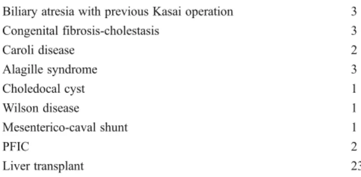
Table 2 Diagnoses in 39 children with liver disease included in the study
Biliary atresia with previous Kasai operation 3
Congenital fibrosis-cholestasis 3 Caroli disease 2 Alagille syndrome 3 Choledocal cyst 1 Wilson disease 1 Mesenterico-caval shunt 1 PFIC 2 Liver transplant 23
maximum, 4.70) in children with liver disease (P < 0.001). Table 3 provides the descriptive statistics for the healthy group and each METAVIR score group as well as tests comparing ARFI imaging scores between each consecutive METAVIR score.
Correlations between ARFI imaging values and META-VIR (F0-F4) are shown in Fig. 1. ARFI imaging mean scores (n, SD) were 1.47 (n 0 11, 0.47) for F0, 1.65 (n 0 11, 0.60) for F1, 2.30 (n 0 6, 1.36) for F2, 2.61 (n0 8, 1.25) for F3, and 2.81 (n 0 3, 0.61) for F4.
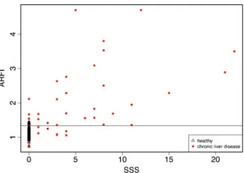
Association between ARFI values and SSS scores are shown in Fig.2. Mean SSS score in the children with liver disease was 5.33 (SD, 5.40; median, 4; minimum, 0; maximum, 22).
The box plots of ARFI imaging values for each category of biopsies with METAVIR scores show an increase in ARFI values evident in fibrosis stages F3 and F4. ARFI imaging values present a slight increase in the F2 stage. By contrast, it seems difficult to distinguish stage F0 from stage F1. We found the same conclusions concerning the association of ARFI and the SSS. ROC curves (Fig. 3) show that sensitivity rapidly drops for both METAVIR and SSS scores when comparing
F0 (SSS of 0–2) and ≥ F1 (SSS ≥ 3). The threshold to detect all children with some liver fibrosis according to the biopsy (100% sensitivity threshold) would be 1.0, and the specificity at this threshold would be very low. The Youden index threshold (maximising both sensitivity and specificity) is 1.34 (Table4), yielding a sensitivity of 0.85 for SSS and 0.82 for METAVIR and specificity of 0.46 for SSS and 0.45 for METAVIR. By using this threshold for screening purposes, five children with liver disease out of 39 would have been missed: four children with a positive SSS and METAVIR scores and one child with a positive METAVIR. Among the four children with a positive SSS score, two had a score of 3 and two had a score of 4. Among the five children with a positive METAVIR score, four had F1 scores and one had an F2 score. Thus, according to this cutoff, children not detected by the ARFI method would be in the initial stages of fibrosis.
If ARFI measurement is used to compare children with mild fibrosis against children with severe fibrosis (i.e. METAVIR≤ F2 vs. ≥ F3 or SSS score < 5 vs. ≥ 5), ROC curves show that sensitivity remains at 100% (children correctly identified: 16 of 16 with an SSS score≥ 5, 11 of
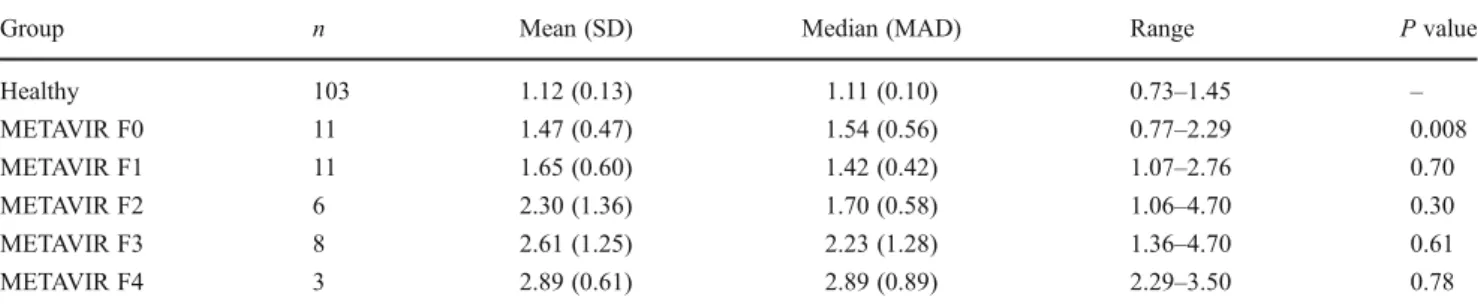
Table 3 Descriptive statistics of ARFI scores in healthy children and in children with liver disease by METAVIR scores
Group n Mean (SD) Median (MAD) Range P value
Healthy 103 1.12 (0.13) 1.11 (0.10) 0.73–1.45 – METAVIR F0 11 1.47 (0.47) 1.54 (0.56) 0.77–2.29 0.008 METAVIR F1 11 1.65 (0.60) 1.42 (0.42) 1.07–2.76 0.70 METAVIR F2 6 2.30 (1.36) 1.70 (0.58) 1.06–4.70 0.30 METAVIR F3 8 2.61 (1.25) 2.23 (1.28) 1.36–4.70 0.61 METAVIR F4 3 2.89 (0.61) 2.89 (0.89) 2.29–3.50 0.78
P values are given by the Mann–Whitney U test between the previous group and the group considered SD standard deviation, MAD median absolute deviation (measure of variability)
Fig. 1 ARFI and METAVIR. Box plots of ARFI imaging scores for each METAVIR category. Number of children per category is indicated on top of the figure. The ref. group is the normal population; F0 to F4 represent children with liver disease with histologically increasing levels of liver fibrosis
11 with a METAVIR score≥ F3, with a specificity of 39.1% for SSS and 32.1% for METAVIR). In other words, 9 of 23 children with an SSS score < 5 were correctly ruled out, and 9 of 28 children with a METAVIR score≤ F2 were correctly ruled out. The 100% sensitivity threshold between mild and severe is 2.0 m/s.
Discussion
The grading of liver fibrosis is essential for the monitoring and appropriate treatment of children with chronic liver disease. Even if biopsy is considered the gold standard for assessing liver fibrosis [14], it is unthinkable to use it as the only tool in monitoring these pathologies in children. De-velopment of noninvasive techniques is essential [15].
FibroScan and serum markers are among the most current noninvasive techniques used in adults in predicting fibrosis [16] and recently the ARFI imaging technique has emerged. In children, the FibroScan has several disadvantages: fixed depth measurement and absence of real-time US guidance, and it cannot be performed in obese children or children with ascites [6, 17–19]. Further, blood markers of liver fibrosis (Fibrotest, APRI) are poorly evaluated and impre-cise in children in current literature.
ARFI imaging elastography is a noninvasive technology to evaluate hepatic fibrosis (Fig.4) that has proved advan-tageous in adults. To assess its ability to reliably identify and quantify liver fibrosis, ARFI values in adults have been compared to METAVIR or Ludwig scoring [20–23].
We report our ARFI imaging elastography experience in assessing the degree of hepatic fibrosis in children with chronic liver disease and liver transplant with pathological correlation based on two histological scoring systems, METAVIR and the SSS.
In our study, ARFI imaging values were clearly in-creased in METAVIR fibrosis stages F3 and F4 (Fig. 1). ARFI values showed a slight increase in the F2 stage. By contrast, it was difficult to distinguish F0 from F1, confirm-ing studies in adults [24, 25]. The same applies to the
Fig. 2 ARFI and SSS. Scatter plot of SSS scores as a function of ARFI imaging scores for healthy children and for children with liver disease. The horizontal line indicates the cutoff value for ARFI to maximise sensitivity and specificity
Fig. 3 ROC curves of ARFI imaging scores for METAVIR and SSS scores
Table 4 SSS/METAVIR with ARFI scores: Youden’s index threshold (maximising both sensitivity and specificity) is 1.34
SSS METAVIR Total
3–28 0–2 F1–F4 F0
ARFI > 1.34 22 7 23 6 29
ARFI < 1.34 4 6 5 5 10
association between ARFI and the SSS (Fig.2). ROC curves show that ARFI imaging scores are predictive of both METAVIR and SSS scores (Fig.3). Based on the Youden index, a cutoff of 1.34 m/s maximises sensitivity and spec-ificity (Table2), yielding a sensitivity of 0.85 for the SSS and 0.82 for METAVIR.
A similar cutoff value of 1.31 m/s was reported in the only other paediatric study to date examining the relation-ship between ARFI imaging and liver fibrosis [26]. How-ever, that study differs from ours in four respects. First, the populations differed in that only ours includes children post liver transplant, which is a large share of any paediatric hepatology practice. Second, there were technical differen-ces: Noruegas et al. [26] used a 4-MHz probe, which is not optimal in young children in our experience. Third, there was a maximum interval of 1 year between biopsy and ARFI evaluation in the Noruegas et al. [26] study, whereas we selected children who had biopsy and ARFI within a 3-month interval [27,28]. The last point of contrast is that we used METAVIR and SSS, while Noruegas et al. [26] used the Batts and Ludwig score [29].
In paediatrics, there is no validated histological scoring system for studying liver fibrosis. In adults, the METAVIR and Ludwig scores are the most widely used but have mainly been validated for fibrosis secondary to hepatitis C. Because the interpretation of liver biopsies in children is difficult because of the diversity of diseases, we chose to apply the METAVIR score, which is considered the gold standard in adults, and the SSS described by Chevallier et al. [12], a more complicated but arguably more accurate scor-ing system with respect to tissue changes.
The analysis of ROC curves with both METAVIR scores and the SSS findings allowed us to establish a cutoff ARFI value of 1.34 m/s for fibrosis stage > F0 in our paediatric population. A cutoff of 2 m/s provided a sensitivity of 100% to detect SSS > 4 or METAVIR > F2 in our population. This is in
contrast with the meta-analysis in adults by Friedrich-Rust et al. [30], wherein there was a cutoff of 1.34 m/s for F≥ 2; 1.55 m/s for F≥ 3, and 1.8 m/s for F 0 4. Adult studies have the advantage of having a larger population with liver biop-sies, so there are cutoffs in adults corresponding to various degrees of fibrosis. Unfortunately, biopsies in children occur more seldom and therefore the numbers are too small in this study to allow for stratified cutoffs.
Nonetheless, it is safe to conclude that akin to what has been described in adults, early stages of fibrosis are difficult to detect by ARFI imaging because the measurements obtained in early fibrosis are similar to those obtained in normal liver. ARFI values should be interpreted with cau-tion because of the limited specificity. However, abnormal values should encourage frequent clinical follow-up and serial ARFI imaging elastography. ARFI is not a substitute for other means of detecting fibrosis, and correlation with fibrosis blood tests must be continued in parallel to elastog-raphy monitoring [30].
Although ours is the first study to include liver transplant recipients in the population, thus widening the scope, it is limited by the small number of children who met our crite-rion of having had a biopsy. This is because liver biopsies are less often performed in children with liver disease than in their adult counterparts. Nonetheless, our study joins that of Noruegas et al. [26] to confirm the feasibility and the utility of ARFI imaging as a noninvasive technique to measure liver elastography and assess liver fibrosis in children.
Conclusion
ARFI elastography is a recent and clinically promising non invasive technique to assess liver fibrosis in children with chronic liver disease or liver transplants and can be used in
Fig. 4 US ARFI measurements. a ARFI imaging in a 4-month-old, using a 9-MHz probe, shear-wave velocity 4.76 m/s at 2.5-cm depth. METAVIR score in this child is F4. b ARFI in a 15-year-old boy, using
a 4-MHz probe, shear-wave velocity 1.55 m/s at 4.8-cm depth. META-VIR score in this child is F2
clinical practice. ARFI values increase with increasing he-patic fibrosis but there are limitations in assessing initial stages of fibrosis. ARFI imaging might help to reduce or postpone the number of biopsies in some children. Given the diversity of liver diseases in children, studies using ARFI imaging in specific liver diseases in children might be warranted.
References
1. Jagadisan B, Srivastava A, Yachha SK et al (2012) Acute on chronic liver disease in children from the developing world: rec-ognition and prognosis. J Pediatr Gastroenterol Nutr 54:77–82 2. Mews C, Sinatra F (1993) Chronic liver disease in children. Pediatr
Rev 14:436–444
3. El-Shabrawi MH, Kamal NM (2011) Medical management of chronic liver diseases in children (part I): focus on curable or potentially curable diseases. Paediatr Drugs 13:357–370 4. El-Shabrawi MH, Kamal NM (2011) Medical management of
chronic liver diseases (CLD) in children (part II): focus on the complications of CLD, and CLD that require special considera-tions. Paediatr Drugs 13:371–383
5. Imbert-Bismut F, Ratziu V, Pieroni L et al (2001) Biochemical markers of liver fibrosis in patients with hepatitis C virus infection: a prospective study. Lancet 357:1069–1075
6. de Lédinghen V, Le Bail B, Rebouissoux L et al (2007) Liver stiffness measurement in children using FibroScan: feasibility study and com-parison with Fibrotest, aspartate transaminase to platelets ratio index, and liver biopsy. J Pediatr Gastroenterol Nutr 45:443–450
7. Breton E, Bridoux-Henno L, Guyader D et al (2009) Value of transient elastography in noninvasive assessment in children’s hepatic fibrosis. Arch Pediatr 16:1005–1010
8. Friedrich-Rust M, Ong MF, Martens S (2008) Performance of transient elastography for the staging of liver fibrosis: a meta-analysis. Gastroenterology 134:960–974
9. Engelmann G, Gebhardt C, Wenning D et al (2012) Feasibility study and control values of transient elastography in healthy chil-dren. Eur J Pediatr 171:353–360
10. Hanquinet S, Courvoisier D, Kanavaki A et al. (2012) Acoustic radi-ation force impulse imaging (ARFI): normal values of liver stiffness in healthy children. Pediatr Radiol. doi:10.1007/s00247-012-2553-5
11. The French METAVIR Cooperative Study Group (1994) Intraob-server and interobIntraob-server variations in liver biopsy interpretation in patients with chronic hepatitis C. Hepatology 20:15–20
12. Chevallier M, Guerret S, Chossegros P et al (1994) A histological semiquantitative scoring system for evaluation of hepatic fibrosis in needle liver biopsy specimens: comparison with morphometric studies. Hepatology 20:349–355
13. R Development Core Team (2012) R: a language and environment for statistical computing. R Foundation for Statistical Computing,
Vienna, Austria ISBN 3-900051-07-0, URL http://www.R-project.org
14. Bedossa P, Carrat F (2009) Liver biopsy: the best, not the gold standard. J Hepatol 50:1–3
15. Sporea I (2011) Liver biopsy in the era of elastography. Med Ultrason 13:185–186
16. Castera L, Pinzani M (2010) Biopsy and non-invasive methods for the diagnosis of liver fibrosis: does it take two to tango? Gut 59:861–866
17. Nobili V, Monti L, Alisi A et al (2011) Transient elastography for assessment of fibrosis in paediatric liver disease. Pediatr Radiol 41:1232–1238
18. Nobili V, Parkes J, Bottazzo G et al (2009) Performance of ELF serum markers in predicting fibrosis stage in pediatric non-alcoholic fatty liver disease. Gastroenterology 136:160–167 19. Hermeziu B, Messous D, Fabre M et al (2010) Evaluation of
FibroTest-ActiTest in children with chronic hepatitis C virus in-fection. Gastroenterol Clin Biol 34:16–22
20. Friedrich-Rust M, Wunder K, Kriener S et al (2009) Liver fibrosis in viral hepatitis: noninvasive assessment with acoustic radiation force impulse imaging versus transient elastography. Radiology 252:595–604
21. Lupsor M, Badea R, Stefanescu H et al (2009) Performance of a new elastographic method (ARFI technology) compared to unidi-mensional transient elastography in the noninvasive assessment of chronic hepatitis C. Preliminary results. J Gastrointestin Liver Dis 18:303–310
22. Boursier J, Isselin G, Fouchard-Hubert I et al (2010) Acoustic radiation force impulse: a new ultrasonographic technology for the widespread noninvasive diagnosis of liver fibrosis. Eur J Gas-troenterol Hepatol 22:1074–1084
23. Haque M, Robinson C, Owen D et al (2010) Comparison of acoustic radiation force impulse imaging (ARFI) to liver biopsy histologic scores in the evaluation of chronic liver disease: a pilot study. Ann Hepatol 9:289–293
24. Sporea I, Sirli R, Popescu A et al (2010) Acoustic radiation force impulse (ARFI)—a new modality for the evaluation of liver fibro-sis. Med Ultrason 12:26–31
25. Fierbinteanu-Braticevici C, Andronescu D, Usvat R et al (2009) Acoustic radiation force imaging sonoelastography for noninva-sive staging of liver fibrosis. World J Gastroenterol 15:5525 26. Noruegas MJ, Matos H, Goncalves I et al (2012) Acoustic
radia-tion force impulse-imaging in the assessment of liver fibrosis in children. Pediatr Radiol 42:201–204
27. Hubscher S (2009) What does the long-term liver allograft look like for the pediatric recipient? Liver Transpl 15(Suppl 2):S19–S24 28. Evans HM, Kelly DA, McKiernan PJ et al (2006) Progressive histological damage in liver allografts following pediatric liver transplantation. Hepatology 43:1109–1117
29. Batts KP, Ludwig J (1995) Chronic hepatitis. An update on termi-nology and reporting. Am J Surg Pathol 19:1409–1417
30. Friedrich-Rust M, Nierhoff J, Lupsor M et al (2012) Performance of acoustic radiation force impulse imaging for the staging of liver fibrosis: a pooled meta-analysis. J Viral Hepat 19:e212–e219