SCIENTIFIC ARTICLE
Occult fractures of the scaphoid: the role of ultrasonography
in the emergency department
Alexandra Platon&Pierre-Alexandre Poletti&Jan Van Aaken&Cesare Fusetti&
Dominique Della Santa&Jean-Yves Beaulieu&Christoph D. Becker
Received: 19 September 2010 / Revised: 6 November 2010 / Accepted: 13 December 2010 / Published online: 1 January 2011 # ISS 2010
Abstract
Objective To evaluate ultrasonography (US) performed by an emergency radiologist in patients with clinical suspicion of scaphoid fracture and normal radiographs.
Materials and methods Sixty-two consecutive adult patients admitted to our emergency department with clinical suspicion of scaphoid fracture and normal radiographs underwent US examination of the scaphoid prior to wrist computed tomography (CT), within 3 days following wrist trauma. US examination was performed by a board-certified emergency radiologist, non-specialized in musculoskeletal
imaging, using the linear probe (5-13 MHz) of the standard sonographic equipment of the emergency department. The radiologist evaluate for the presence of a cortical interrup-tion of the scaphoid along with a radio-carpal or scapho-trapezium-trapezoid effusion. A CT of the wrist (reference standard) was performed in every patient, immediately after ultrasonography. Fractures were classified into two groups according to their potential for complication: group 1 (high potential, proximal or waist), group 2 (low-potential, distal or tubercle).
Results A scaphoid fracture was demonstrated by CT in 13 (21%) patients: eight (62%) of them belonged to group 1 (three in the proximal pole, five in the waist), five (38%) to group 2 (three in the distal part, two in the tubercle). US was 92% sensitive (12/13) in demonstrating a scaphoid fracture. It was 100% sensitive (8/8) in demonstrating a fracture with a high potential of complication (group 1). Conclusions Our data show that, in emergency settings, US can be used for the triage to CT in patients with clinical suspicion of scaphoid fracture and normal radiographs.
Keywords Bone . Scaphoid . Fracture occult . Ultrasonography . CT
Introduction
Affecting all ages, but predominantly young patients, wrist traumas is a frequent cause of consultation in the emergency room. Mainly due to a fall on an outstretched hand, the scaphoid is one of the most frequently fractured carpal bones [1]. Scaphoid fractures remain feared lesions in wrist traumatology because of their high potential of complications, which include delayed union, non-union, or osteonecrosis [1–3]. Moreover, some
A. Platon (*)
:
P.-A. Poletti:
C. D. BeckerDepartment of Radiology, University Hospital of Geneva, 4, rue Gabrielle-Perret-Gentil, 1211, Geneva, Switzerland e-mail: alexandra.platon@hcuge.ch P.-A. Poletti e-mail: Pierre-Alexandre.Poletti@hcuge.ch C. D. Becker e-mail: Christoph.Becker@hcuge.ch
J. Van Aaken
:
D. Della Santa:
J.-Y. Beaulieu Hand Surgery Unit, Department of Orthopaedics and Traumatology, University Hospital of Geneva, Geneva, Switzerland J. Van Aaken e-mail: Jan.vanAaken@hcuge.ch D. Della Santa e-mail: Dominique.DellaSanta@hcuge.ch J.-Y. Beaulieu e-mail: Jean-Yves.Beaulieu@hcuge.ch C. FusettiHand Surgery Unit, Department of Orthopaedics and Traumatology, Ospedale San Giovanni,
Bellinzona, Switzerland e-mail: cfusetti@yahoo.com
fractures of the scaphoid may be occult at presentation, meaning not visible on the initial radiographs performed after trauma.
In emergency settings, the diagnosis of scaphoid fracture is based on clinical examination and conventional radio-graphs. Nevertheless, 20–25% of patients have positive signs for a fracture at clinical examination and normal radiographs [2]. In most centers, these patients are considered to have a potential occult scaphoid fracture and are treated with wrist immobilization until the diagnosis is confirmed, or ruled out on follow-up radiographs obtained 2 weeks after trauma [4, 5]. This diagnostic approach is nevertheless limited by the unnecessary wrist immobilization in patients without scaphoid fracture. On the other hand, if overlooked at initial presentation, non-treated scaphoid fractures may lead to unwanted long-lasting after-effects; thus, the initial correct diagnosis is of crucial importance, in order to immediately start the appropriate treatment [2].
To shorten the time to a correct diagnosis, different imaging methods have been proposed for patients with clinically suspected occult radiographic fractures of the carpal scaphoid. Computed tomography (CT) and magnetic resonance imaging (MRI) are highly reliable diagnostic modalities for scaphoid fracture detection; both methods still remain limited in terms of immediate availability and costs [6–8].
Previous studies have already shown that ultrasonogra-phy (US) of the wrist performed by musculoskeletal radiologists could be useful for the diagnosis of scaphoid fractures [2, 9, 10]. However, the presence of a trained musculoskeletal radiologist in the emergency room is rarely possible. Therefore, in the current study we wanted to evaluate the usefulness of US, performed in emergency settings by a board-certified radiologist without subspecial-ty training in musculoskeletal imaging, in the diagnosis of the occult fractures of the scaphoid. In the settings of our study, we compared the diagnostic performance of the US examination with the results of wrist CT, considered the reference standard.
Material and methods
Between January 2004 and January 2009, all consecutive adult patients (more than 18 years old) admitted during the daytime in our emergency department with clinical suspi-cion of scaphoid fracture and normal radiographs under-went US examination of the scaphoid prior to wrist CT (reference standard), within 3 days following wrist trauma. The study protocol was approved by the institutional review board of our institution (IRB 04-201) and informed consent was obtained from all the patients included in our study. All patients were admitted in the emergency room
after wrist trauma and in the settings of our study, were clinically evaluated by the hand surgery fellow in charge of the daytime consultations. The clinical criteria for a suspected scaphoid fracture were wrist pain, associated with tenderness on axial loading of the first ray and swelling and tenderness at palpation of the anatomic snuffbox. The presence of clinical signs positive for scaphoid fracture along with negative complete standard X-ray examination mandated referral for US examination, followed by CT of the wrist. The routine radiographic series for a suspected scaphoid fracture consisted of a neutral posteroanterior and a lateral projection of the wrist together with additional views for the scaphoid: a semipronated oblique scaphoid, and a postero-anterior view with the wrist in ulnar deviation. These radiographs were read both by the hand surgery fellow and by the radiology resident on call. Patients with positive radiographs for scaphoid or other wrist bones fracture were excluded from the study.
Immediately after the clinical examination, US was performed by a board-certified radiologist, without sub-speciality musculoskeletal training. Images were obtained with the standard equipment (Prosound SSD-5000SV; ALOKA, Tokyo, Japan), available in the emergency department, using the small compact linear (5-13 MHz) transducer for superficial structures. The examination took place in the emergency department in the dedicated room for sonography with the patient sitting in front of the radiologist with the injured hand resting on the examination table; the examination was made through transmission sonographic gel, and no stand-off pad was used. Using the anatomic landmarks of the wrist and starting the examination from the radius, the scaphoid was identified by its bilobed or peanut-shape, with a smooth and hyper-echoic bone surface contour [11]. For correctly elongating the scaphoid, the hand was positioned in ulnar deviation, both in frontal and sagittal planes; a neutral frontal plane, with the palm lying on the table, was also obtained to assess the dorsal aspect of the bone. Using these different wrist positions, US examination covered the volar, dorsal, and lateral aspect of the scaphoid, with images obtained in the longitudinal and axial planes of the bone. The radio-carpal and mid-carpal recesses were examined in the sagittal plane, both for the dorsal and volar planes of examination [10, 11]. Examination of both wrists was performed only when the radiologist wanted to elucidate an unclear anatomical pattern on the traumatic side; therefore, the contralateral examination was not systematically used in all patients.
The radiologist was asked to assess the continuity of the scaphoid cortex, seen as a smooth hyperechoic thin line and the presence of fluid effusion in the radio-scaphoid and scapho-trapezium-trapezoid space [2,11]. Signs associated with scaphoid fracture were either the cortical disruption of
the scaphoid contour producing a focal step-of deformity, or hemarthrosis, seen as hypoechoic fluid in the radio-scaphoid or scapho-trapezium-trapezoid spaces; hemarth-rosis may sometimes show a mixed echogenicity, depending on the stage of degradation of blood products; however, most post-traumatic joint effusions appear hypo-echoic on sonographic images [12, 13]. These findings have been reported in the literature as the most suggestive signs associated with scaphoid fractures [2, 9–11]. US examination was considered positive for a scaphoid fracture when at least one of the above-mentioned signs was present at the examination. The examination was interpreted immediately, without knowledge of the further imaging results.
CT scanning of the wrist was performed immediately after the completion of the sonography examination. All CT examinations were performed on a 16-detector row scanner (MX 8000 Philips Medical Systems, Best, The Nether-lands), using 120 kV, 150mAs, 0.8-mm collimation thickness, 0.4-mm increment, and a pitch of 0.3. The patient was installed prone on the examination table, with the hand positioned in full pronation. The acquisition covered the wrist from the distal metaphysis of the radius to the metacarpal base; no intravenous contrast was injected. Images were routinely reformatted with a 2-mm thickness in coronal and sagittal planes using the bone algorithm and stored in the picture archiving and commu-nication system (PACS). Images were interpreted on a separate workstation (Cedara I-Softview, version 6.1, Cedara Software Corporation, Mississauga, Canada) using both the raw data of the examination, and the multiplanar reformations (MPR). During the interpretation session, radiologists made additional multiplanar reformations in oblique planes, in order to obtain the reconstruction of the scaphoid bone along its long axis. Immediately after scanning, the CT reading was done together by the radiology resident and by the board-certified attending radiologist, both unaware of the sonography results; these CT examinations were not read by members of the musculoskeletal unit.
At CT, a fracture of the scaphoid was defined either as a radiolucent line traversing the bone, or a break in the continuity of the cortex or a sharp step in the cortex [7]. According to the anatomic location, the fractures were localized into proximal pole, middle third (waist), or distal third (including the tubercle) [3,14].
According to their potential for complication [1,3,14], fractures of the scaphoid were further classified into two groups. Group 1 included the fractures with a high potential for complications: these fractures were localized in the proximal or middle third of the scaphoid. Group 2 included the fractures localized distally or involving the tubercle and were considered as fractures with low-potential for complications.
The patients included in our study were treated accord-ing to the CT results and clinical follow-up, usaccord-ing the standard procedures of our institution.
Statistical analysis
The results of the US examination were compared to those of CT imaging, considered the reference standard. Data were recorded using statistical software (SPSS 15.0 for Windows, Chicago, Il), and sensitivity, specificity and positive and negative predictive values for US with regard to CT were calculated.
Results
Our study group was formed of 62 patients (33 women and 29 men), with an age range between 18 and 89 years, with a mean age of 41.2 years.
A scaphoid fracture was eventually demonstrated by CT in 13 (21%) patients, four women and nine men (age range 18-70, mean 33.7 years). CT examination displayed five fractures of the scaphoid waist, three fractures of the proximal pole, three fractures of the distal scaphoid and two fractures involving the scaphoid tubercle.
According to the potential for complications related to the fracture site, eight (12%) patients (six men, two women, mean age 35.5 years) had a scaphoid fracture with high potential for complications (three in the proximal pole, five in the waist) and belonged to group 1.
Five patients (8%) (three men, two women, mean age 30.8) had scaphoid fractures with low potential of compli-cations (three in the distal part, two in the tubercle) and belonged to group 2.
In seven (11%) patients of the study, CT examination showed fractures in other wrist bones than the scaphoid, which were not seen on the initial radiographs. Thus, two patients had a fracture of the distal radius; two patients had a fracture of the os triquetrum, one of the trapezium, one of the trapezoid, and one of the base of the first metacarpal.
US examination reported positive signs suggestive of scaphoid fracture in 12 (92%) of the 13 patients with a CT-proven fracture.
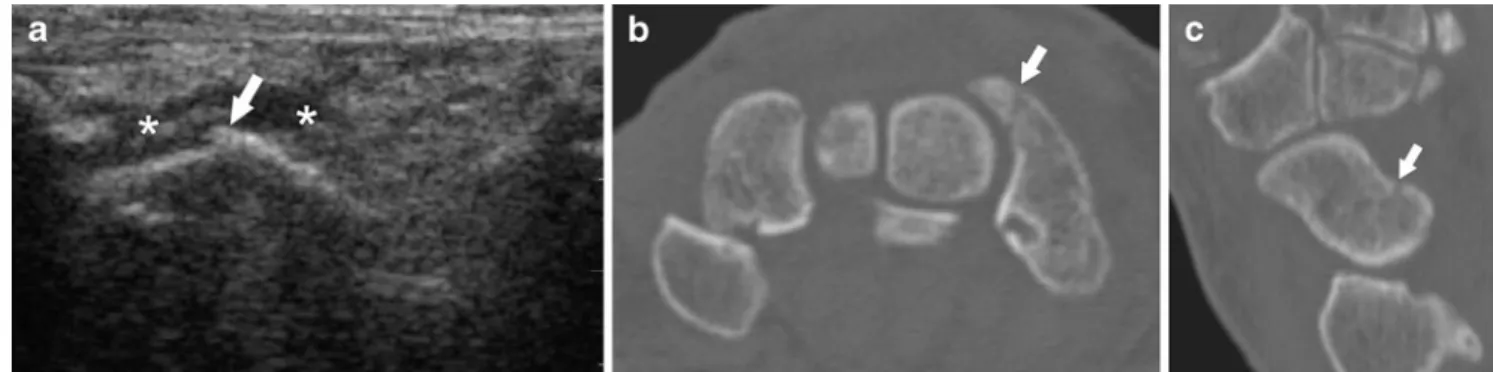
All patients belonging to group 1 (fractures with high potential of complications) had sonographic signs associat-ed with scaphoid fractures. Indeassociat-ed, radio-scaphoid effusion was present in all eight patients of group 1; three of them also had associated STT effusion; no patient had an isolated STT effusion. Associated cortical interruption of the scaphoid bone was described in six of the eight patients (Fig. 1).
In group 2 (fractures with low potential of complica-tions), US examination showed suggestive signs of scaph-oid fracture in 4 of the 5 patients (Fig.2). Radio-scaphoid or scapho-trapezoid effusion was reported in the 4 patients; three of them also had cortical discontinuity of the scaphoid bone. In the remaining patient, US did not show any signs suggestive of the diagnosis: this patient had a CT proven fracture of the scaphoid tubercle.
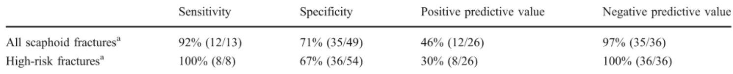
Overall, US examination was 92% sensitive (12/13) to detect a scaphoid fracture. It was 100% sensitive to detect a fracture of the scaphoid with a high potential of complica-tions. Sensitivity, specificity, positive predictive value (PPV), and negative predictive value (NPV) of the US examination for diagnosing a scaphoid fracture are reported in Table1.
Discussion
The early diagnosis of an occult scaphoid fracture still remains a challenging task in the emergency room. The difficulty is represented by a scaphoid bone that seems fractured based on the clinical examination, but appears normal on conventional radiographs [3].
Complications associated with a non-treated scaphoid fracture can be explained by the distribution of the vascular supply, which comes from the distal end of the bone. Therefore, the proximal pole of the scaphoid has the weakest blood supply, and this is the explanation for the increased risk of avascular necrosis, following fractures of the proximal portion or of the waist of the scaphoid [1,3,
14]. Reportedly, fractures of the scaphoid tubercle, which is
Fig. 1 A 36-year-old man admitted after wrist trauma, with positive clinical signs for scaphoid fracture and normal radiographs. a US of the scaphoid in the longitudinal plane shows cortical discontinuity (arrow), along with radio-carpal effusion and surrounding hematoma
(asterisk), suggestive of scaphoid fracture. b Axial CT image depicts the fracture as a radiolucent line (arrow), traversing the scaphoid. c Sagittal-oblique multiplanar reformation CT image demonstrates the proximal location of the scaphoid fracture (arrow)
Fig. 2 A 27-year-old woman admitted after wrist trauma, with positive clinical signs for scaph-oid fracture and normal radio-graphs. US of the scaphoid (S) in a longitudinal and b transverse plane shows a hypoechoic fluid collection (asterisk) anterior to the scaphoid, along with cortical discontinuity (arrow) of the bone, compatible with a fracture. Axial CT image in c and sagittal-oblique multiplanar reformation CT in d demonstrate the fracture as a radiolucent line (arrows) involving the distal pole of the scaphoid
non-articular and has a rich blood supply, are prone to heal well, regardless of the treatment [3].
In order to avoid the potential complications from a delayed diagnosis, a non-invasive, readily available imag-ing method in the emergency room for the early detection of occult scaphoid fractures is needed [15].
US has already been used by musculoskeletal radiol-ogists for the diagnosis of scaphoid fractures and cortical disruption or peri-scaphoid articular effusion or hematoma have been proven useful signs in the assessment of a scaphoid fracture [2,9,10]. Nevertheless, there is no data considering the role of US performed by a general radiologist in the diagnosis of an occult scaphoid fracture.
The results of our study are in keeping with data reported by musculoskeletal radiologists. Thus, we correct-ly classified 12 of the 13 CT-proved scaphoid fractures, which yielded a global sensitivity of 92% for sonography in our study. This result is in the range of previously reported sensitivities for US in depicting all types of scaphoid fractures, from 50% in the study of Munk et al. to 78% in the study of Herneth et al. [10,16].
Moreover, in our current study, US correctly assessed all eight scaphoid fractures situated in the waist or in the proximal pole of the scaphoid, yielding a sensitivity of 100% in this group of patients. These fractures have a high
potential for complications and their early detection is important in order to immediately start the proper treatment. In depicting these fractures with high potential of compli-cations, our results tally with those of Hauger et al. and Fusetti et al. [2,9], both of whom reported a sensitivity of 100% for US in depicting scaphoid waist fractures. These results are reproduced in our study; thus, US performed by the emergency radiologist, using the standard ultrasound machine, did not overlook any scaphoid fracture with high potential of complication, with accurate and good correla-tion with wrist CT.
The diagnostic performance of the US of the scaphoid is also reflected by the predictive values of this diagnostic method. Thus, the negative predictive value, which is the capacity of the test in predicting that a patient with a negative US of the scaphoid does not truly have a fracture, is very high. In our study, the negative predictive value of US was 97% for all types of scaphoid fractures; a negative US is therefore a good diagnostic test to rule out an occult scaphoid fracture.
A possible explanation of these results is related to the anatomical position of the scaphoid. The waist of the scaphoid has the easiest access to sonography examination; thus, the direct and indirect signs of fracture can be identified reliably [9]. Sonographic assessment of the distal
Table 1 Diagnostic performance of the US with regard to CT findings in the diagnosis of occult fractures of the scaphoid
Sensitivity Specificity Positive predictive value Negative predictive value All scaphoid fracturesa 92% (12/13) 71% (35/49) 46% (12/26) 97% (35/36)
High-risk fracturesa 100% (8/8) 67% (36/54) 30% (8/26) 100% (36/36)
a
Numbers in parenthesis correspond to number of patients
Fig. 3 Suggested algorithm in-tegrating US in the diagnostic work-up of patients with clini-cally suspected scaphoid frac-ture and negative radiographs
pole of the scaphoid is more technically challenging. Our study showed that an occult fracture of the scaphoid tubercle was missed at sonography examination.
US of the scaphoid seems to substantially contribute to the diagnosis of occult scaphoid fractures; nevertheless, this method has some inherent limitations. First, one should be aware that not all scaphoid fractures can be assessed at sonographic examination. Indeed, as different studies have previously shown [10], there are areas of the scaphoid that may be difficult to evaluate. This is due to the impossibility of evaluating the integrality of the bone contour. Reported-ly, the scapho-lunate area and the tubercle are the most difficult structures to image by ultrasonography [10]. Also, even when suspected by the US examination, the extent and the direction of the scaphoid fracture line cannot be described by sonography. This is a limitation with regard to the choice of treatment, either conservative or surgical. In these circumstances, a second-line imaging method, such as CT or MRI, should be proposed as a complementary tool [2,9].
A certain number of false-positive results of our study are related to the presence of fractures involving other wrist bones not seen on initial radiographs. Non-specific articular carpal effusion may be seen on US examination associated with other traumatic wrist lesions [2,9]. Indeed, scaphoid fractures are not the sole etiology for hemarthrosis after wrist trauma; ligament injuries or occult fractures of other wrist bones can also be associated with hemarthrosis. This is reflected by the low specificity of US examination in this setting and constitutes a limitation of our study. However, although an indirect sign of fracture, hemarthrosis still remains an important finding, and mandates further imaging examination in order to elucidate the exact origin of the articular fluid. On the other hand, absence of hemarthrosis at US examination was always associated with absence of high-risk fractures of the scaphoid bone, making sonography examination a highly sensitive method with a very good predictive value in excluding clinically relevant fractures of the scaphoid after wrist trauma. This is probably the major finding of this series.
Similarly, as previously reported, arthritic deformity of the wrist may create confusion with regard to cortical irregular-ities and simulate interruption of the scaphoid contour. Moreover, it is not possible to differentiate between fluid associated with degenerative changes and fluid related to fracture. False-positive cases would certainly be more frequent in this population and therefore US results in elderly patients should be interpreted cautiously [2].
Our study represents a comparison between US and CT in occult fractures of the scaphoid bone. We believe that US can provide a quick and reliable complement to CT or MRI in an emergency setting when a diagnosis of scaphoid fracture is made. As our results have shown, US of the
scaphoid did not require operator-specific skills or training in order to recognize typical signs associated with fracture of the scaphoid. Therefore, US could be integrated in a clinical and radiological algorithm (Fig.3) for the work-up of patients with clinically suspected scaphoid fracture. When an occult radiographic fracture is suspected based on US findings, the diagnostic strategy and the treatment can rapidly be initiated, decreasing thus the risk for potential delayed complications. In case of negative findings on conventional radiographs, scaphoid US could act as a bridge, orientating the patients toward second-line imaging methods.
Conclusions
Our study results indicate that the diagnosis performance of US performed by a board-certified radiologist without subspecialty training in musculoskeletal imaging is repro-ducible and is equal to those obtained by musculoskeletal radiologists in the diagnosis of occult fracture of the scaphoid. Therefore, ultrasonography could be used on a routine basis in emergency settings for the triage to CT in patients with clinical suspicion of scaphoid fracture and normal radiographs.
Conflict of interests None.
References
1. Haisman JM, Rohde RS, Weiland AJ. Acute fractures of the scaphoid. J Bone Joint Surg Am. 2006;88:2750–8.
2. Hauger O, Bonnefoy O, Moinard M, Bersani D, Diard F. Occult fractures of the waist of the scaphoid: early diagnosis by high-spatial-resolution sonography. AJR Am J Roentgenol. 2002;178:1239–45.
3. Schubert HE. Scaphoid fracture. Review of diagnostic tests and treatment. Can Fam Physician. 2000;46:1825–32.
4. Groves AM, Kayani I, Syed R, Hutton BF, Bearcroft PP, Dixon AK, et al. An international survey of hospital practice in the imaging of acute scaphoid trauma. AJR Am J Roentgenol. 2006;187:1453–6.
5. Ring D, Jupiter JB, Herndon JH. Acute fractures of the scaphoid. J Am Acad Orthop Surg. 2000;8:225–31.
6. Adey L, Souer JS, Lozano-Calderon S, Palmer W, Lee SG, Ring D. Computed tomography of suspected scaphoid fractures. J Hand Surg Am. 2007;32:61–6.
7. Memarsadeghi M, Breitenseher MJ, Schaefer-Prokop C, Weber M, Aldrian S, Gabler C, et al. Occult scaphoid fractures: comparison of multidetector CT and MR imaging–initial experi-ence. Radiology. 2006;240:169–76.
8. Ring D, Lozano-Calderon S. Imaging for suspected scaphoid fracture. J Hand Surg Am. 2008;33:954–7.
9. Fusetti C, Poletti PA, Pradel PH, Garavaglia G, Platon A, Della Santa DR, et al. Diagnosis of occult scaphoid fracture with
high-spatial-resolution sonography: a prospective blind study. J Trauma. 2005;59:677–81.
10. Herneth AM, Siegmeth A, Bader TR, Ba-Ssalamah A, Lechner G, Metz VM, et al. Scaphoid fractures: evaluation with high-spatial-resolution US initial results. Radiology. 2001;220:231–5. 11. Jacobson JA. Fundamentals of musculoskeletal ultrasound.
Phil-adelphia: Saunders Elsevier; 2007. p. 133–77.
12. Jelbert A, Vaidya S, Fotiadis N. Imaging and staging of haemophilic arthropathy. Clin Radiol. 2009;64:1119–28. 13. Zukotynski K, Jarrin J, Babyn PS, Carcao M, Pazmino-Canizares
J, Stain A, et al. Sonography for assessment of haemophilic
arthropathy in children: a systematic protocol. Haemophilia. 2007;13:293–304.
14. Kaewlai R, Avery LL, Asrani AV, Abujudeh HH, Sacknoff R, Novelline RA. Multidetector CT of carpal injuries: anatomy, fractures, and fracture-dislocations. Radiographics. 2008;28:1771–84. 15. Senall JA, Failla JM, Bouffard JA, van Holsbeeck M. Ultrasound
for the early diagnosis of clinically suspected scaphoid fracture. J Hand Surg Am. 2004;29:400–5.
16. Munk B, Bolvig L, Kroner K, Christiansen T, Borris L, Boe S. Ultrasound for diagnosis of scaphoid fractures. J Hand Surg Br. 2000;25:369–71.