ORIGINAL ARTICLE
Molecular and synaptic organization of GABA
Areceptors in the
cerebellum: Effects of targeted subunit gene deletions
JEAN-MARC FRITSCHY
1& PATRIZIA PANZANELLI
21
Institute of Pharmacology and Toxicology, University of Zurich, Zurich, Switzerland, and2Department of Anatomy, Pharmacology and Forensic Medicine, University of Turin, Turin, Italy
Abstract
GABAA receptors form heteromeric GABA-gated chloride channels assembled from a large family of subunit genes. In
cerebellum, distinct GABAAreceptor subtypes, differing in subunit composition, are segregated between cell types and
synaptic circuits. The cerebellum therefore represents a useful system to investigate the significance of GABAAreceptor
heterogeneity. For instance, studies of mice carrying targeted deletion of major GABAAreceptor subunit genes revealed the
role of a subunit variants for receptor assembly, synaptic targeting, and functional properties. In addition, these studies unraveled mandatory association between certain subunits and demonstrated distinct pharmacology of receptors mediating phasic and tonic inhibition. Although some of these mutants have a profound loss of GABAAreceptors, they exhibit only
minor impairment of motor function, suggesting activation of compensatory mechanisms to preserve inhibitory networks in the cerebellum. These adaptations include an altered balance between phasic and tonic inhibition, activation of voltage-independent K+conductances, and upregulation of GABAAreceptors in interneurons that are not affected directly by the
mutation. Deletion of thea1 subunit gene leads to complete loss of GABAAreceptors in Purkinje cells. A striking alteration
occurs in these mice, whereby presynaptic GABAergic terminals are preserved in the molecular layer but make heterologous synapses with spines, characterized by a glutamatergic-like postsynaptic density. During development of a10/0 mice,
GABAergic synapses are initially formed but are replaced upon spine maturation. These findings suggest that functional GABAAreceptors are required for long-term maintenance of GABAergic synapses in Purkinje cells.
Key words: Knockout mice, benzodiazepine, Purkinje cell, granule cell, GABAergic synapse, interneurons
Introduction
Owing to its apparent simplicity and stereotyped organization, with a clear segregation of cell types in distinct layers, the cerebellum represents an excel-lent structure to investigate the anatomical and functional organization of neuronal networks and major neurotransmitter systems. Most synaptic inhibition in the cerebellum is mediated by GABAA receptors, which are highly abundant
despite the overwhelming presence of glutamatergic synapses formed by parallel fibers on Purkinje cell spines. GABAA receptors belong to the superfamily
of ligand-gated ion channels, along with nicotinic acetylcholine receptors, glycine receptors and 5-HT3
receptors (1). They form heteropentameric chloride channels gated by GABA and modulated by several clinically relevant drugs, including benzodiazepines, barbiturates, neurosteroids, and ethanol (2–4). They are assembled for a large family of subunits (a1–6, b1–3, c1–3, d, e, p, and h in mammalian brain), of
which 13 are expressed in the cerebellum (5). Accordingly, the molecular organization of GABAA receptors is only partially understood and
the functional significance of this diversity in cerebellar inhibitory circuits is slowly starting to emerge.
Targeted deletion of GABAA receptor subunit
genes by homologous recombination has contribu-ted much to our understanding of GABAAreceptors.
Focusing on the cerebellum, numerous studies have investigated molecular, pharmacological and func-tional properties of GABAA receptors that are less
amenable for analysis in other brain regions (6). The present review gives a broad overview of recent reports on the molecular and synaptic organization of GABAA receptors in cerebellar cortex. It
high-lights major findings from knockout mice, with a particular focus on compensatory mechanisms allowing preservation of network organization and function despite the loss of a major constituent of synaptic transmission.
Correspondence: Jean-Marc Fritschy, Institute of Pharmacology and Toxicology, University of Zurich, Winterthurerstrasse 190, CH – 8057 Zurich, Switzerland. E-mail: [email protected]
ISSN 1473-4222 print/ISSN 1473-4230 online # 2006 Taylor & Francis DOI: 10.1080/14734220600962805
Organization of GABAAreceptors in the
cerebellar cortex
The heterogeneity of GABAA receptors in the
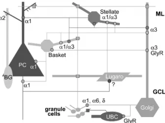
cerebellar cortex stands in striking contrast to the apparent simplicity of GABAergic circuits, which are formed by four types of interneurons (stellate and basket cells in the molecular layer, Golgi cells and Lugaro cells in the granule cell layer), in addition to Purkinje cells (PC). Each type of interneuron is activated by granule cells (parallel fiber synapses) and innervates specific targets, including other interneurons (Figure 1) (7). In total, 13 GABAA
receptor subunits have been detected immuno-chemically, including a1–a6, b1–3, c1–3, and d (5). The cellular distribution pattern has not yet been established for all these subunits but the major subunit combinations comprise a1bxc2, a6bxc2, (a1)a6bxd, a3bxc2, as well as a2bxc1 (x indicates that the b subunit variants have not been deter-mined). Despite this plethora of subunits, the majority of cell types express a limited repertoire of GABAAreceptor subtypes (Figure 1). For example,
PC express exclusively a1bxc2-GABAA receptors;
stellate/basket cells, a1bxc2 and a3bxc2; Golgi cells a3bxc2; Bergmann glia, a2/bx/c1 (8–14). The notable exception comes from granule cells, which express multiple receptor subtypes, despite receiving GABAergic input only from Golgi cells (15). Most
of these receptors are assembled from various combinations of a1, a6, b2, b3, c2 and d subunit (8,12,13,16). No information is available for Lugaro cells and unipolar brush cells (17), and conversely, several subunit expressed at low levels (a4, a5, c3) have been identified by Western blotting only (5,18). Depending on the cell type and subunit combina-tion, GABAAreceptors can be located
postsynapti-cally or extrasynaptipostsynapti-cally, where they mediate phasic and tonic inhibition, respectively (19). Postsynaptic GABAA receptors in cerebellar neurons largely are
co-localized with gephyrin, a cytoplasmic protein that is exclusively present in symmetric synapses (12). By immunofluorescence staining, they are recognized as bright clusters distributed on dendrites and somata, as well as on glomeruli of the granule cell layer (see Figure 3). Using this approach, the synaptic localization ofa1- and a3-GABAAreceptors
was shown in the molecular layer (12); in glomeruli, a1- and in part a6-GABAA receptors can also be
detected postsynaptically, confirming ultrastructural studies using postembedding immunogold electron microscopy (20–22). In whole cell patch clamp recordings of PC, spontaneous IPSCs of large amplitude predominate over EPSCs, indicating that PC are under powerful control from stellate and basket cells (23). These synaptic currents are mediated by GABAA receptors with no evidence
for a contribution by strychnine-sensitive glycine receptors (24). In addition, PC also carry extra-synaptic GABAA receptors diffusely distributed on
the cell surface and are modulated by tonic inhibi-tion, notably due to GABA release from basket cells (25). Similar, high frequency and high amplitude sIPSCs have been well characterized also in stellate cells (26,27), underscoring the major role played by postsynaptic GABAA receptors in the molecular
layer.
In granule cells, a major fraction of GABAA
receptors is associated with the d subunit (29,30), forming extrasynaptic receptors with a high affinity for GABA (31,32) and diffusely distributed on the soma and dendrites (12,22,28). The presence of the d subunit, which does not co-assemble with the c2 subunit, is determinant for the location of GABAA
receptors relative to postsynaptic sites and for their functional properties. Electrophysiologically, tonic inhibition in granule cells is independent of Golgi cell action potentials (33,34). Rather, it is produced mainly by non-vesicular release of GABA and is modulated by type 1 and type 3 GABA transporters (33). So far, it is not known whether this complex regulation of tonic inhibition is related to the heterogeneity of extrasynaptic GABAA receptors in
granule cells. However, it should be emphasized that the role of the d subunit is forming receptors mediating tonic inhibition is not restricted to the cerebellum, but also occurs in thalamus and hippocampus (35,36). Furthermore, these receptors Figure 1. Schematic organization of GABAergic circuits and
GABAA receptors in the cerebellar cortex. Each cell type is
indicated with a different color. Dendrites are shown in thick lines and axons in thin lines. GABAergic synapses (octogons) are shown with the color corresponding to the presynaptic cell and the main GABAA receptor subtype(s) or glycine receptors (GlyR)
present postsynaptically are indicated. Glutamatergic synapses are denoted with squares. Note that Lugaro cells innervate all cell types in the molecular layer except PC. In turn, the only inhibitory input they receive comes from PC. It is not known which GABAA
receptor subtype(s) they express. Basket/stellate cells express both a1- and a3-GABAAreceptor, but it is not established whether they
are segregated between afferents from other basket/stellate cells and from Lugaro cells. Bergman glial cells (BG) express an unusual GABAA receptor subtype, containing the a2 and c1
subunit, which is enriched at sites of contact with PC. Abbreviations: GCL, granule cell layer; ML, molecular layer; UBC, unipolar brush cell.
are a major target for neurosteroids, which reduce neuronal excitability by enhancing tonic inhibition mediated byd-GABAAreceptors (37).
A distinct population of ‘extrasynaptic’, diaze-pam-insensitive GABAA receptors, with a probably
unique subunit composition in brain (a2bxc1), is found in Bergman glia cells (10,11,14). These receptors are most abundant during early postnatal maturation and modulate K+conductances that are transiently expressed by immature Bergmann glia. As shown by immunoelectron microscopy in adult brain (10), GABAAreceptors are most concentrated
in glial processes wrapping PC somata and den-drites, in particular in the vicinity of symmetric synapses formed by basket cells. However, they are also concentrated in processes in contact with PC spines, in which GABAA receptors are occasionally
found. This highly regulated location suggested a role for sensing GABAergic synaptic function in PC (10).
Synaptic inhibition in the cerebellum is also mediated, to a lesser extent, by glycine receptors (38,39). A majority of Golgi and Lugaro cells have a dual GABAergic/glycinergic phenotype (40,41). The specificity of transmission in their postsynaptic targets is ensured by the differential expression of receptors. Thus, spontaneous IPSCs in granule cells are mediated selectively by GABAA receptors (39),
whereas Golgi cells, Lugaro cells, unipolar brush cells, and possibly other interneurons exhibit either glycinergic or mixed glycinergic/GABAergic currents (Figure 1) (42,43). At least in Golgi cells, GABAA
and glycine receptors can co-exist within a given postsynaptic site (42).
A distinctive feature of postsynaptic GABAA
receptors in the molecular layer is their colocaliza-tion with the dystrophin-glycoprotein complex (DCG), including b-dystroglycan, dystrophin, and a-/b-dystrobrevin (44–47). The DGC is required for proper maturation or maintenance of postsynaptic GABAA receptors in PC, as shown by a decreased
number and size of clusters in mutant mice lacking either dystrophin, or botha- and b-dystrobrevin. As a consequence, synaptic inhibitory input is reduced on PC of mice lacking dystrophin (48). While the relevance of the DGC in specific GABAergic synapses is unclear, it is required for proper expression of long-term depression in PC (49), suggesting a role in synaptic plasticity. A similar situation occurs in the cerebral cortex and hippo-campal formation, where large subsets of inhibitory synapses are associated with the DGC (45). In the hippocampus, absence of the DGC leads to altera-tions of short- and long-term synaptic plasticity (50,51). However, the DGC cannot be essential for the formation of GABAergic synapses or clustering of GABAAreceptors, since it is absent in many other
brain areas, such as thalamus, basal ganglia, tectum, etc. Furthermore, dystrophin expression occurs after
formation of GABAergic synapses during postnatal development (45).
The maturation of GABAA receptors during
ontogeny of the cerebellum has been investigated mainly in granule cells and in PC (52,53). The subunit composition and pharmacological profile of GABAA receptor in granule cells are
developmen-tally regulated and these changes are activity-dependent (54–57). A transient expression of the a2 and a3 subunit occurs at early postnatal stages (58–60), whereas the a6 and d subunit, which are expressed selectively in granule cells (61), is delayed (11,55,60) and parallels the formation of functional GABAergic synapses from Golgi cells (62–65). This maturation pattern contrasts with that of PC, which express a prominent a1 subunit immunoreactivity at birth, before having formed a monolayer and being innervated by basket and stellate cells (66,67).
Effects of targeted subunit gene deletion on cerebellar GABAAreceptors
Targeted deletions of eight GABAA receptor
sub-units (‘‘full knockout’’ mice) have been reported so far in the literature (a1, a3, a5, a6, b2, b3, c2, d). Among these only four have been analyzed for their effects on assembly of GABAA receptors and
function of the GABAergic system in the cerebellum (c2, a6, d, a1). The major alterations reported in these studies concern the function and pharmacol-ogy of the remaining GABAA receptors, which are
partially altered due to loss of an additional subunit that has an obligatory association with the deleted subunit (for example,d subunit in a60/0mice) or due to compensatory changes in expression of remaining subunit(s) (68,69). Unexpectedly, even targeted deletions affecting major GABAAreceptor subtypes,
such as a1-GABAA receptors, have only minor
behavioral consequences, suggesting that wide-spread adjustments take place within and outside the GABAergic system to ensure preserved function. Mostc20/0mice die shortly after birth and a small percentage survive up to the third postnatal week (70). In these mice, there is an almost complete loss of a1 subunit and gephyrin clusters in the cerebel-lum, in line with the requirement of thec2 subunit for postsynaptic aggregation of GABAA receptors
and gephyrin (71,72). No specific compensation with another c2 subunit variant was observed, indicating indirectly that thec1 and c3 subunits do not contribute significantly to neuronal GABAA
receptors in the mouse cerebellum.
Deletion of the a6 subunit gene, which is exclusively expressed in cerebellar granule cells, caused a 50% overall reduction of GABAAreceptors
in the cerebellum (73), underscoring the relative abundance of the a6 subunit in these cells.
Unexpectedly, this mutation caused the disappear-ance of thed subunit protein (74). However, since mRNA levels for thed subunit were not affected, this finding suggested that thed subunit protein requires the a6 subunit for assembly in a receptor complex and is degraded rapidly in the absence of this mandatory partner. As a consequence, deletion of a single subunit led to the loss of multiple GABAA
receptor subtypes in granule cells. This conclusion was confirmed by an autoradiographic analysis showing marked alterations of the pharmacological profile of GABAAreceptors in the granule cell layer
ofa60/0mice, best explained by a concurrent loss of a6bxc2- and a6bxd-GABAA receptors (69). No
compensation by the a1 subunit was detected in glomeruli or in the molecular layer of a60/0 mice (73), indicating that the a1 and a6 subunit are regulated independently, although their genes are present in the same chromosomal cluster (75). Interestingly, while a60/0 mice display no major spontaneous phenotype, a clear motor deficit could be shown upon treatment with diazepam, which could be reversed by flumazenil, indicating that it was mediated by the remaining, mainly postsynaptic, GABAAreceptors in these mice (76).
Deletion of thed subunit gene did not result in a loss of GABAA receptors in the cerebellum, as
shown by autoradiography and Western blot analy-sis. An increase in receptors containing the c2 subunit was noted, along witha6 and bx, suggesting that c2 and d compete for assembly with these subunits in granule cells from wildtype mice (77). Functionally, a loss of modulation of sIPSCs by the neurosteroid THDOC selectively occurs in granule cells, but not stellate cells of d0/0 mice (78), underscoring the importance of d-GABAA
receptors for the in vivo action of neurosteroids (79). Behaviorally, deletion of the d subunit gene produces no overt impairment in motor function or motor learning, similarly to thea6 subunit gene (76). This observation should not lead to the conclusion that tonic inhibition mediated by extrasynaptic GABAA receptors in granule cells is irrelevant.
Rather, the lack of phenotype is due to compensa-tory upregulation of voltage-independent K+ chan-nels (two-pore-domain K+ channels TASK-1 and TASK-3), which increases the membrane conduc-tance to the same extent as the tonic inhibition mediated by a6-GABAA receptors in wildtype mice
(80).
Like for thea6 subunit, targeted deletion of the a1 subunit gene has major consequences for GABAA
receptors in the cerebellum. A profound loss of binding sites for benzodiazepine ligands occurs (81). This decrease is mirrored in the 50% reduction of b2,3 and c2 subunit proteins in crude membrane extracts from the cerebellum (18,81), pointing to a decrease in the assembly of GABAAreceptors in the
absence of the a1 subunit. This phenotype is not
unique to the cerebellum but has been observed in other brain regions, as well (82). Despite the major reduction in the number of GABAAreceptors,a10/0
mice display no anatomical anomaly and no gross behavioral impairment (83–85). The most obvious deficits include a mild impairment of motor coordi-nation and a kinetic and postural tremor that is reminiscent of essential tremor in human; this tremor was responsive to drug therapies that alleviate symptoms of essential tremor in patients and is likely related to the loss of a1-GABAA
receptors in multiple stations of the motor system, including the cerebellum (86).
Compensatory changes in GABAAreceptor
subunit expression ina10/0mice
A major feature of a10/0 mice, despite the 50% overall loss of GABAA receptors, is the
compensa-tory up-regulation of subunits that are normally expressed at low levels, which might partially replace the missing a1-GABAA receptors. In particular,
expression of thea3, a4 and a6 subunit proteins is increased, as shown by two independent in vivo studies (18,81). In vitro, the loss of a1-GABAA
receptors induced an increase in a6bxd-receptors (and possibly a4bxd), suggesting that tonic inhibi-tion partially compensates for the loss of synaptic receptors (87). Functionally, these adaptations are reflected in the maintenance of postsynaptic currents in granule cells, but with altered kinetic properties. In particular, the decay time constants of remaining receptors are prolonged, reflecting the substitution of a1-GABAA receptors by another subtype. Since
a1-GABAAreceptors have faster kinetics than those
containing thea2 or a3 subunits (88,89), this factor was taken as an index for the sustained presence of these subunits in mature granule cells of mutant mice (87,90).
In contrast to granule cells, no compensation occurs in PC which lose all functional GABAA
receptors in mutant mice. Patch clamp recording analyses showed a complete loss of spontaneous and evoked IPSCs and no compensation by strychnine-sensitive glycine receptors (9,86). No evidence for tonic inhibition could be found either in PC ofa10/0 mice. A detailed immunohistochemical analysis was therefore undertaken to understand how this loss of GABAA receptors affects the synaptic organization
of the GABAergic system in the molecular layer (18). On the cellular level, this study confirmed the reduction ofb2,3 and c2 subunit expression, as well as the upregulation of the a3 subunit, mainly in interneurons of the molecular layer (Figure 2). Subcellularly, the loss ofa1 subunit causes a strong, but only partial decrease of gephyrin clusters in the molecular layer (Figure 3). Staining for the a3 subunit, which is normally present in a minor fraction of gephyrin clusters, revealed a prominent
increase in the number ofa3 subunit clusters in the molecular layer, most likely representing postsynap-tic receptors in stellate/basket cells and Golgi cell dendrites (Figure 3). These observations are in line with a study in which miniature IPSPs recorded from stellate cells in whole-cell patch clamp experi-ments had slower kinetics in a10/0 mice than in wildtype, compatible with the expression of a3-GABAA receptors (88). The changes in a3 subunit
expression were specific for this subunit, since no alteration of the a2 subunit (present in Bergman glia) was observed, whereas thea4 and a5 subunits were not detected immunohistochemically in the cerebellum of either wildtype or a10/0mice (18).
The comparison of compensatory changes in GABAA receptor expression between the granule
cell layer and the molecular layer in a10/0 mice highlights major features of the regulation of these
receptors: (i) In granule cells expressing multiple GABAA receptor subtypes, an increase of the
remaining subunit(s) might replace the missing subunit. However, this compensation is only partial, since postsynaptic GABAA receptors colocalized
with gephyrin and mediating mainly phasic inhibi-tion appear to be replaced by extrasynaptic receptors mediating tonic inhibition. This finding is supported by the decreased c2 subunit staining in the granule cell layer. A similar situation has been observed in neurons of the thalamic ventrobasal complex, in which postsynaptically clustered a1-GABAA
recep-tors are replaced by extrasynaptic a4-GABAA
receptors lacking thec2 subunit and gephyrin (18). Taken together, these observations suggest that GABAA receptor subtypes are not interchangeable
within a given type of neuron and that the lack of interaction of a4 or d-GABAA receptors with
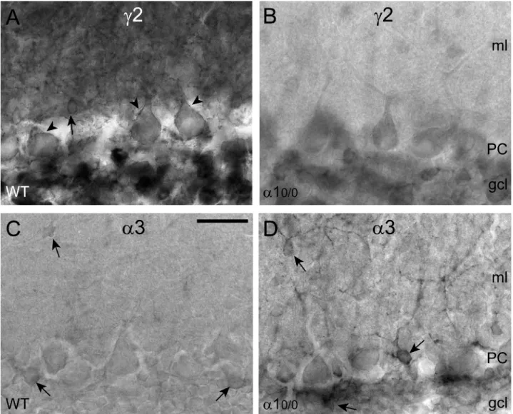
Figure 2. Differential regulation of GABAA receptor subtypes in the cerebellar cortex ofa10/0mice, as shown by immunoperoxidase
staining for thec2 and a3 subunit. (A) Strong and widespread expression of the c2 subunit in the molecular layer (ml), in PC and in the granule cell layer (gcl) of wildtype (WT) mice. The arrow points to a strongly labeled interneuron and arrowheads indicatec2 subunit immunoreactivity on PC, outlining the cell surface. (B) Marked reduction ofc2 subunit staining in a section from a mutant mouse. Note that PC are unlabeled. The granule cell layer (gcl) also appears weakly labeled, and interneurons in the ml are almost undetectable. (C–D) Striking up-regulation of thea3 subunit, which in wildtype is weakly expressed in Golgi cells and some ml interneurons (arrows). In a10/0 mice, thea3 subunit staining outlines individual interneurons in the ml and in the gcl (arrows), whereas PC remain unlabeled. Scale bar, 50mm.
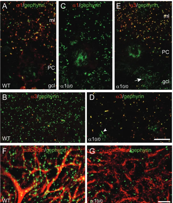
Figure 3. Partial disruption of gephyrin clustering and up-regulation of thea3 subunit in the molecular layer (ml) of a10/0
mice. Double immunofluorescence staining with the markers indicated in each panel. (A) Colocalization of thea1 subunit (red) and gephyrin (green) in discrete clusters (yellow) in the ml. Note that the soma of the PC is not labeled for gephyrin, whereas a few gephyrin clusters lacking thea1 subunit are evident in the ml. (B) Staining for thea3 subunit (red) in the ml reveals very few clusters, which are colocalized (yellow) with gephyrin (green). (C) Complete loss ofa1 subunit staining and decreased density of gephyrin clusters (green) in the molecular layer of an a10/0mouse. The weak labeling of the nucleus in non-specific and unrelated to the genotype. (D–E) Upregulation ofa3 subunit-positive
clusters (red) in the ml ofa10/0mice, which are associated with gephyrin (green) in virtually all remaining clusters. In panel D, the nuclei of
two interneurons appear weakly stained in green. The arrowhead points to a large gephyrin aggregate, presumably located intracellularly in the interneuron. In panel E, note that noa3 subunit staining becomes apparent in the numerous small gephyrin clusters in the granule cell layer (gcl; arrow). (F–G) Loss of gephyrin clusters (green) on the dendrites of PC (labeled with calbindin, red) ina10/0mice; the remaining
gephyrin-positive clusters are mainly distributed on non-labeled structures between PC dendrites. Scale bars, A–D: 20mm; F–G: 10 mm. See (18) for details.
gephyrin is not due to a competitive disadvantage with a1-GABAA receptors. (ii) In PC expressing
only the a1-GABAA receptors, no compensatory
expression of another a subunit variant occurs. However, overexpression of other GABAAreceptor
subtypes takes place in neighboring cells, such as Golgi and stellate cells, presumably to preserve the function of the cerebellar network. It is unclear how this up-regulation of a3-GABAA receptors is
trig-gered, but stellate and Golgi cells are linked synaptically with PC via Lugaro cells (Figure 1). This observation strongly suggests that expression of GABAAreceptors is regulated by synaptic activity in
the network. (iii) The upregulation of ‘minor’ GABAA receptor subunits in the cerebellum of
a10/0 mice occurs at the posttranslational level, as it is not accompanied by a change in mRNA transcription (81). Therefore, it appears that the turnover of GABAA receptor subunits can be
regulated dynamically, allowing neurons to adjust the use of specific GABAA receptor subtypes for
proper function.
The most likely implication of these changes is a reorganization of inhibitory circuits in the molecular layer of a10/0 mice to compensate for the loss of phasic and tonic inhibition in PCs. This conclusion, if confirmed by morphological analysis, implies that GABAA receptors control, at least indirectly, the
number of inhibitory synapses formed in a given circuit to set its overall level of activity. As shown in Figure 1, there are two major sources of inhibitory input on Golgi cells and molecular layer interneur-ons: Lugaro cells, which provide a dual glycinergic/ GABAergic input onto Golgi cells and stellate/basket cells, which innervate each other and Golgi cells. This synaptic reorganization suggests growth of interneuron axonal arbors in the molecular layer of mutant mice and/or change in synaptic target specificity. A possible mechanism underlying this change in connectivity is discussed in the next section.
Requirement of GABAAreceptors for
long-term maintenance of GABAergic synapses The observation that PC in a10/0 mice are com-pletely devoid of functional GABAA receptors
provided a unique incentive to investigate the effect of such a deletion on GABAergic synapses (9,86). At the light microscopy level, the morphology of PCs, as revealed by staining for calbindin or parvalbumin is unaffected in a10/0mice and they are present in normal numbers (86). Likewise, the distribution of climbing fibers is undistinguishable from that seen in wildtype mice (9). GABAergic terminals in the molecular are readily detectable, but are larger and less regularly distributed than in wildtype. However, striking alterations in the organization of GABAergic synapses were evident at the ultrastructural level (9),
as summarized in Figure 4. In sections processed for GABA immunogold labeling, the vast majority of GABAergic presynaptic profiles formed synapses with PC spines, characterized by an asymmetric postsynaptic specialization indistinguishable from that formed in glutamatergic synapses (Figure 4A). These aberrant presynaptic terminals nevertheless were GABAergic, as shown by the presence of vesicular GABA transporter and the absence of vesicular glutamate transporters type 1 and type 2, which were readily detected in parallel (Figure 4C) and climbing fibers, respectively. Furthermore, most aberrant GABAergic terminals originated from stellate or basket cells, as seen by labeling for parvalbumin. On the dendrites of PC, the number of symmetric, type II synapses was reduced by about 75%, indicating a failure to form or to maintain GABAergic synapses in the absence of postsynaptic GABAAreceptors (9).
Unexpectedly, GABAergic synapses on PC somata, which are formed selectively by basket cell terminals, were not affected in a10/0 mice and retained their morphology, neurochemical pheno-type, and content of presynaptic vesicles (Figure 4B). Therefore, the synaptic deficit observed in the molecular layer is selective for axo-dendritic synapses. Morphological and functional examination of the cerebellum during postnatal development revealed no transient expression of GABAA receptors in PC of mutant mice.
GABAergic axons contacting PC dendrites and somata were seen from P7 onwards. Ultrastructurally, the first synapses formed by terminals immunolabeled for GABA were symmetric on dendritic shafts. However, aberrant terminals, forming heterologous synapses with spines appeared first at P10 and their number increased rapidly thereafter (Figure 4D). These results were taken as evidence that initial steps of GABAergic synapse formation are not affected in a10/0 mice (9). However, a clear deficit for the maintenance of GABAergic synapses on PC dendrites occurs in the absence of functional GABAergic transmission.
Altogether, these observations confirm that neu-rotransmitter-mediated activity is not essential for synapse formation (91–94) and indicate that differ-ential mechanisms ensure long-term stability of synapses in various cell compartments. While the normal morphology and content of presynaptic vesicles of basket cell terminals suggest that these terminals remain competent for GABA synthesis and release, activation of postsynaptic GABAA
receptors is not required for maintenance of this synapse. A different situation prevails in dendrites, where most GABAergic synapses are lost in adult animals. It is possible that spines exert a synapto-genic action of GABAergic terminals, which is sufficient to attract them when they form ‘silent’ contacts with their normal postsynaptic targets. It is
also conceivable that the presence of GABAA
receptors and/or gephyrin is required for synaptic anchoring of a molecule mediating transsynaptic homo- or heterophilic interaction with a presynaptic partner, thereby explaining the profound reduction of symmetric synapses on dendritic shafts. It is of note that the formation of basket cell synapses on the axon initial segment of PC depends on specific interactions with extracellular molecules (95) and that gephyrin clusters are not detectable on PC somata, despite the prominent clustering of the a1 subunit (Figure 3), indicating a distinct mechanism for GABAA receptor clustering compared to
den-drites. In any case, the lack of long-term stability of stellate cell synapses onto PC dendrites might contribute to the formation of supernumerary synapses between interneurons, as discussed in the previous section.
Conclusions
The analysis of targeted deletion of GABAAreceptor
subunit genes expressed in the cerebellum has unraveled seemingly contradictory findings: GABAA receptor subtypes are functionally not
interchangeable and do not replace a missing subunit, as seen best by the loss ofd subunit protein in a60/0 mice and by the increase in extrasynaptic receptors in granule cells of a10/0 mice. However, wide ranging compensations contribute to the maintenance of function, in particular when the mutation concerns a major GABAA receptor
sub-type. Adaptations can even occur in cell types that are not affected by the mutation, as witnessed by the increase in a3-GABAA receptors in Golgi cells of
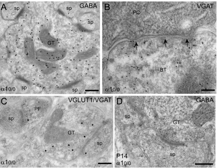
a10/0 mice. As a consequence, behavioral deficits are minimal compared to the strong impairment produced by acute pharmacological blockade or Figure 4. Formation of heterologous synapses in the molecular layer ofa10/0mice, as visualized by immunoelectron microscopy. Each
panel depicts a section from a mutant mouse (A–C) adult, (D) P14. Immunogold labeling of mitochondria is non-specific and should be disregarded. (A) Example of a GABAergic terminal (GT) labeled for GABA using a secondary antibody coupled to 20 nm gold particles, which forms asymmetric synapses with 6 spine profiles (sp) within a single ultrathin section. (B) A basket cell terminal (BT) labeled for vesicular GABA transporter (VGAT; 10 nm immunogold particles) makes symmetric synapses (arrows) with the soma of a PC. Such terminals were indistinguishable from those seen in wildtype mice. (C) Segregation of type 1 vesicular glutamate transporter (VGLUT1; 10 nm gold particles) and VGAT (20 nm gold particles) between parallel fiber terminals (PF) and GABAergic terminals, even when both make asymmetric synapses with spines. (D) Example of heterologous synapses formed during postnatal development between a GABAergic terminal and two spines (GABA labeling with 10 nm gold particles). Scale bars, A, D: 200 nm; B, C: 100 nm. For details, see (9).
stimulation of GABAA receptors in vivo. While the
stereotyped organization of the cerebellum facilitates these investigations, additional studies will be required to determine to which extent these findings can be generalized to other brain regions and other GABAAreceptor subtypes.
Acknowledgements
We would like to thank Dr G. Homanics (University of Pittsburgh) for the gift ofa10/0mice, Dr J. Kralic and Dr M. Sassoe`-Pognetto (University of Turin) for their contribution to the analysis of these mutant mice, Dr W. Sieghart (University of Vienna) for antibodies against the a4 and d subunit, and C. Sidler and F. Parpan for excellent technical help.
References
1. Barnard EA, Skolnick P, Olsen RW, Mohler H, Sieghart W, Biggio G, et al. International Union of Pharmacology. XV. Subtypes ofc-aminobutyric acidAreceptors: Classification on
the basis of subunit structure and function. Pharmacol Rev. 1998;50:291–313.
2. Fritschy JM, Bru¨ nig I. Formation and plasticity of GABAergic synapses: physiological mechanisms and pathophysiological implications. Pharmacol Ther. 2003;98:299–323.
3. Sieghart W, Ernst M. Heterogeneity of GABAA receptors:
Revived interest in the development of subtype-selective drugs. Curr Med Chem. 2005;5:217–242.
4. Rudolph U, Mohler H. GABA-based therapeutic approaches: GABAAreceptor subtype functions. Curr Opin Pharmacol.
2006;6:18–23.
5. Poltl A, Hauer B, Fuchs K, Tretter V, Sieghart W. Subunit composition and quantitative importance of GABAAreceptor
subtypes in the cerebellum of mouse and rat. J Neurochem. 2003;87:1444–1455.
6. Wisden W, Korpi ER, Bahn S. The cerebellum: A model system for studying GABAA receptor diversity.
Neuropharmacol. 1996;35:1139–1160.
7. Palay SL, Chan-Palay V. Cerebellar cortex: Cytology and organization. Berlin, Heidelberg, New York: Springer Verlag, 1974.
8. Fritschy JM, Mohler H. GABAA-receptor heterogeneity in the
adult rat brain: Differential regional and cellular distribution of seven major subunits. J Comp Neurol. 1995;359:154–194. 9. Fritschy JM, Panzanelli P, Kralic JE, Vogt KE, Sassoe`-Pognetto M. Differential dependence of axo-dendritic and axo-somatic GABAergic synapses on GABAA receptors
containing the a1 subunit in Purkinje cells. J Neurosci. 2006;26:3245–3255.
10. Riquelme R, Miralles CP, De Blas AL. Bergmann glia GABAA receptors concentrate on the glial processes that
wrap inhibitory synapses. J Neurosci. 2002;22:10720–10730. 11. Muller T, Fritschy JM, Grosche J, Pratt GD, Mohler H, Kettenmann H. Developmental regulation of voltage-gated K+-channel and GABAA-receptor expression in Bergmann
glial cells. J Neurosci. 1994;14:2503–2514.
12. Sassoe`-Pognetto M, Panzanelli P, Sieghart W, Fritschy JM. Co-localization of multiple GABAA receptor subtypes with
gephyrin at postsynaptic sites. J Comp Neurol. 2000;420:481–498.
13. Laurie DJ, Seeburg PH, Wisden W. The distribution of 13 GABAA receptor subunit mRNAs in the rat brain. II.
Olfactory bulb and cerebellum. J Neurosci. 1992;12:1063–1076.
14. Wisden W, McNaughton LA, Darlison MG, Hunt SP, Barnard EA. Differential distribution of GABAA receptor
mRNAs in bovine cerebellum-localization ofa2 mRNA in Bergmann glia layer. Neurosci Lett. 1989;106:7–12. 15. Hevers W, Luddens H. Pharmacological heterogeneity of
c-aminobutyric acid receptors during development suggests distinct classes of rat cerebellar granule cells in situ. Neuropharmacol. 2002;42:34–47.
16. Pirker S, Schwarzer C, Wieselthaler A, Sieghart W, Sperk G. GABAA receptors: Immunocytochemical distribution of 13
subunits in the adult rat brain. Neuroscience. 2000;101:815–850.
17. Mugnaini E, Floris A. The unipolar brush cell: a neglected neuron of the mammalian cerebral cortex. J Comp Neurol. 1994;339:174–180.
18. Kralic JE, Sidler C, Parpan F, Homanics G, Morrow AL, Fritschy JM. Compensatory alteration of inhibitory synaptic circuits in thalamus and cerebellum of GABAAreceptora1
subunit knockout mice. J Comp Neurol. 2006;495:408–421. 19. Farrant M, Nusser Z. Variations on an inhibitory theme: Phasic and tonic activation of GABAAreceptors. Nature Rev
Neurosci. 2005;6:215–229.
20. Nusser Z, Sieghart W, Stephenson FA, Somogyi P. Thea6 subunit of the GABAA receptor is concentrated in both
inhibitory and excitatory synapses on cerebellar granule cells. J Neurosci. 1996;16:103–114.
21. Somogyi P, Fritschy JM, Benke D, Roberts JDB, Sieghart W. The c2 subunit of the GABAA-receptor is concentrated in
synaptic junctions containing the a1 and b2/3 subunits in hippocampus, cerebellum and globus pallidus. Neuropharmacol. 1996;35:1425–1444.
22. Nusser Z, Roberts JDB, Baude A, Richards JG, Somogyi P. Relative densities of synaptic and extrasynaptic GABAA
receptors on cerebellar granule cells as determined by a quantitative immunogold method. J Neurosci. 1995;15:2948–2960.
23. Hausser M, Clark BA. Tonic synaptic inhibition modulates neuronal output pattern and spatiotemporal synaptic integra-tion. Neuron. 1997;19:665–678.
24. Konnerth A, Llano I, Armstrong CM. Synaptic currents in cerebellar Purkinje cells. Proc Natl Acad Sci USA. 1990;87:2662–2665.
25. Zhang CL, Messing A, Chiu SY. Specific alteration of spontaneous GABAergic inhibition in cerebellar Purkinje cells in mice lacking the potassium channel Kv1.1. J Neurosci. 1999;19:2852–2864.
26. Llano I, Gerschenfeld HM. Inhibitory synaptic currents in stellate cells of rat cerebellar slices. J Physiol (Lond.). 1993;468:177–200.
27. Nusser Z, Cull-Candy S, Farrant M. Differences in synaptic GABAA receptor number underlie variation in GABA mini
amplitude. Neuron. 1997;19:697–709.
28. Nusser Z, Sieghart W, Somogyi P. Segregation of different GABAAreceptors to synaptic and extrasynaptic membranes
of cerebellar granule cells. J Neurosci. 1998;18:1693–1703. 29. Santhakumar V, Hanchar HJ, Wallner M, Olsen RW,
Otis TS. Contributions of the GABAA receptora6 subunit
to phasic and tonic inhibition revealed by a naturally occurring polymorphism in the a6 gene. J Neurosci. 2006;26:3357–3364.
30. Brickley SG, Cull-Candy S, Farrant M. Single-channel properties of synaptic and extrasynaptic GABAA receptors
suggest differential targeting of receptor subtypes. J Neurosci. 1999;19:2960–2973.
31. Hevers W, Korpi ER, Luddens H. Assembly of functional a6b3c2dGABAA receptors in vitro. Neuroreport.
2000;11:4103–4106.
32. Saxena NC, Macdonald RL. Properties of putative cerebellar c-aminobutyric acidA receptor isoforms. Mol Pharmacol.
33. Rossi DJ, Hamann M, Attwell D. Multiple modes of GABAergic inhibition of rat cerebellar granule cells. J Physiol. 2003;548:97–110.
34. Rossi DJ, Hamann M. Spillover-mediated transmission at inhibitory synapses promoted by high affinity a6 subunit GABAA receptors and glomerular geometry. Neuron.
1998;20:783–795.
35. Nusser Z, Mody I. Selective modulation of tonic and phasic inhibitions in dentate gyrus granule cells. J Neurophysiol. 2002;87:2624–2628.
36. Cope DW, Hughes SW, Crunelli V. GABAA
receptor-mediated tonic inhibition in thalamic neurons. J Neurosci. 2005;25:11533–11563.
37. Stell BM, Brickley SG, Tang CY, Farrant M, Mody I. Neuroactive steroids reduce neuronal excitability by selec-tively enhancing tonic inhibition mediated by d subunit-containing GABAA receptors. Proc Natl Acad Sci USA.
2003;100(24):14439–14444.
38. Dieudonne S. Glycinergic synaptic currents in Golgi cells of the rat cerebellum. Proc Natl Acad Sci USA. 1995;92:1441–1445.
39. Kaneda M, Farrant M, Cull-Candy SG. Whole-cell and single-channel currents activated by GABA and glycine in granule cells of the rat cerebellum. J Physiol (Lond.). 1995;485:419–435.
40. Aoki E, Semba R, Kashiwamata S. New candidates for GABAergic neurons in the rat cerebellum: an immunocyto-chemical study with anti-GABA antibody. Neurosci Lett. 1986;68:267–271.
41. Ottersen OP, Storm-Mathisen J, Somogyi P. Colocalization of glycine-like and GABA-like immunoreactivities in Golgi cell terminals in the rat cerebellum: A postembedding light and electron microscopic study. Brain Res. 1988;450:342–353.
42. Dumoulin A, Triller A, Dieudonne S. IPSC kinetics at identified GABAergic and mixed GABAergic and glycinergic synapses onto cerebellar Golgi cells. J Neurosci. 2001;21:6045–6057.
43. Dugue GP, Dumoulin A, Triller A, Dieudonne S. Target-dependent use of coreleased inhibitory transmitters at central synapses. J Neurosci. 2005;25:6490–6498.
44. Knuesel I, Mastrocola M, Zuellig RA, Bornhauser B, Schaub MC, Fritschy JM. Altered synaptic clustering of GABAA-receptors in mice lacking dystrophin (mdx mice).
Eur J Neurosci. 1999;11:4457–4462.
45. Knuesel I, Zu¨ llig RA, Bornhauser B, Schaub MC, Fritschy JM. Differential expression of utrophin and dystro-phin in CNS neurons: An in situ hybridization and immunohistochemical study. J Comp Neurol. 2000;422:594–611.
46. Bru¨ nig I, Suter A, Knuesel I, Luscher B, Fritschy JM. GABAergic presynaptic terminals are required for postsynap-tic clustering of dystrophin, but not of GABAAreceptors and
gephyrin. J Neurosci. 2002;22:4805–4813.
47. Grady RM, Wozniak DF, Ohlemiller KK, Sanes JR. Cerebellar synaptic defects and abnormal motor behavior in mice lacking a- and b-dystrobrevin. J Neurosci. 2006;26:2841–2851.
48. Anderson JL, Head SI, Morley JW. Altered inhibitory input to Purkinje cells of dystrophin-deficient mice. Brain Res. 2003;982:280–283.
49. Anderson JL, Head SI, Morley JW. Long-term depression is reduced in cerebellar Purkinje cells of dystrophin-deficient mdx mice. Brain Res. 2004;1019:289–292.
50. Vaillend C, Billard JM. Facilitated CA1 hippocampal synaptic plasticity in dystrophin-deficient mice: role for GABAAreceptors? Hippocampus. 2002;12:713–717.
51. Vaillend C, Billard JM, Laroche S. Impaired long-term spatial and recognition memory and enhanced CA1 hippocampal LTP in the dystrophin-deficient DMD (mdx) mouse. Neurobiol Disease. 2004;17:10–20.
52. Wall MJ. Alterations in GABAA receptor occupancy occur
during the postnatal development of rat Purkinje cell but not granule cell synapses. Neuropharmacol. 2005;49:596–609. 53. Zempel JM, Steinbach JH. Neonatal rat cerebellar granule
and Purkinje neurons in culture express different GABAA
receptors. Eur J Neurosci. 1995;7:1895–1905.
54. Gault LM, Siegel RE. Expression of the GABAA receptor
d subunit is selectively modulated by depolarization in cultured rat cerebellar granule neurons. J Neurosci. 1997;17:2391–2399.
55. Behringer KA, Gault LM, Siegel RE. Differential regulation of GABAAreceptor subunit mRNAs in rat cerebellar granule
neurons – importance of environmental cues. J Neurochem. 1996;66:1347–1353.
56. Zhu WJ, Vicini S, Harris BT, Grayson DR. NMDA-mediated modulation ofc-aminobutyric acid type A receptor function in cerebellar granule neurons. J Neurosci. 1995;15:7692–7701.
57. Thompson CL, Pollard S, Stephenson FA. Developmental regulation of expression of GABAA receptor a1 and a6
subunits in cultured rat cerebellar granule cells. Neuropharmacol. 1996;35:1337–1346.
58. Takayama C, Inoue Y. Transient expression of GABAA
receptor a2 and a3 subunits in differentiating cerebellar neurons. Dev Brain Res. 2004;148:169–177.
59. Laurie DJ, Wisden W, Seeburg PH. The distribution of thirteen GABAA receptor subunit mRNAs in the rat brain.
III. Embryonic and postnatal development. J Neurosci. 1992;12:4151–4172.
60. Gao B, Fritschy JM. Cerebellar granule cells in vitro recapitulate the in vivo pattern of GABAA-receptor subunit
expression. Dev Brain Res. 1995;88:1–16.
61. Bahn S, Harvey RJ, Darlison MG, Wisden W. Conservation of c-aminobutyric acid type A receptor a6 subunit gene expression in cerebellar granule cells. J Neurochem. 1996;66:1810–1818.
62. Takayama C, Inoue Y. Morphological development and maturation of the GABAergic synapses in the mouse cerebellar layer. Dev Brain Res. 2004;150:177–190. 63. Mellor JR, Merlo D, Jones A, Wisden W, Randall AD. Mouse
cerebellar granule cell differentiation: electrical activity regulates the GABAAreceptora6 subunit gene. J Neurosci.
1998;18:2822–2833.
64. Farrant M, Brickley SG. Properties of GABAA
receptor-mediated transmission at newly formed Golgi-granule cell synapses in the cerebellum. Neuropharmacol. 2003;44:181–189.
65. Hevers W, Luddens H. Pharmacological heterogeneity of c-aminobutyric acid receptors during development suggests distinct classes of rat cerebellar granule cells in situ. Neuropharmacol. 2002;42:34–47.
66. Paysan J, Fritschy JM. GABAA-receptor subtypes in
devel-oping brain: Actors or spectators? Perspect Dev Neurobiol. 1998;5:179–192.
67. Fritschy JM, Paysan J, Enna A, Mohler H. Switch in the expression of rat GABAA-receptor subtypes during postnatal
development: an immunohistochemical study. J Neurosci. 1994;14:5302–5324.
68. Sinkkonen ST, Mihalek RM, Homanics GE, Lu¨ ddens H, Korpi ER. Altered atypical coupling ofc-aminobutyrate type A receptor agonist and convulsant binding sites in subunit-deficient mouse lines. Mol Brain Res. 2001;86:179–183. 69. Makela R, Uusi-Oukari M, Homanics GE, Quinlan JJ,
Firestone LL, Wisden W, et al. Cerebellar c-aminobutyric acid type A receptors: Pharmacological subtypes revealed by mutant mouse lines. Mol Pharmacol. 1997;52:380–388. 70. Gunther U, Benson J, Benke D, Fritschy JM, Reyes GH,
Knoflach F, et al. Benzodiazepine-insensitive mice generated by targeted disruption of thec2-subunit gene of c-aminobu-tyric acid type A receptors. Proc Natl Acad Sci USA. 1995;92:7749–7753.
71. Essrich C, Lorez M, Benson JA, Fritschy JM, Luscher B. Postsynaptic clustering of major GABAA receptor subtypes
requires the c2 subunit and gephyrin. Nature Neurosci. 1998;1:563–571.
72. Li RW, Yu W, Christie SB, Miralles CP, Bai J, Loturco JJ, et al. Disruption of postsynaptic GABA receptor clusters leads to decreased GABAergic innervation of pyramidal neurons. J Neurochem. 2005;95:756–770.
73. Nusser Z, Ahmad Z, Tretter V, Fuchs K, Wisden W, Sieghart W, et al. Alterations in the expression of GABAA
receptor subunits in cerebellar granule cells after the disruption of the a6 subunit gene. Eur J Neurosci. 1999;11:1685–1697.
74. Jones A, Korpi ER, McKernan RM, Pelz R, Nusser Z, Makela R, et al. Ligand-gated ion channel subunit partner-ships: GABAAreceptora6 subunit gene inactivation inhibits d
subunit expression. J Neurosci. 1997;17:1350–1362. 75. Uusi-Oukari M, Heikkila¨ J, Sinkkonen ST, Ma¨kela¨ R,
Hauer B, Homanics GE, et al. Long-range interactions in neuronal gene expression: evidence from gene targeting in the GABAAreceptorb2-a6-a1-c2 subunit gene cluster. Mol Cell
Neurosci. 2000;16:34–41.
76. Korpi ER, Koikkalainen P, Vekovischeva OY, Ma¨kela¨ R, Kleinz R, Uusi-Oukari M, et al. Cerebellar granule-cell-specific GABAAreceptors attenuate benzodiazepine-induced
ataxia:evidence from a6-subunit-deficient mice. Eur J Neurosci. 1998;11:233–240.
77. Tretter V, Hauer B, Nusser Z, Mihalek RM, Hoger H, Homanics GE, et al. Targeted disruption of the GABAA
receptor d subunit gene leads to an up-regulation of c2 subunit-containing receptors in cerebellar granule cells. J Biol Chem. 2001;276:10532–10538.
78. Vicini S, Losi G, Homanics GE. GABAAreceptord subunit
deletion prevents neurosteroid modulation of inhibitory synaptic currents in cerebellar neurons. Neuropharmacol. 2002;43:646–650.
79. Fodor L, Biro T, Maksay G. Nanomolar allopregnanolone potentiates rat cerebellar GABAAreceptors. Neurosci Lett.
2005;383:127–130.
80. Brickley SG, Revilla V, Cull-Candy SG, Wisden W, Farrant M. Adaptive regulation of neuronal excitability by a voltage-independent potassium conductance. Nature. 2001;409:88–92.
81. Ogris W, Lehner R, Fuchs K, Furtmuller B, Hoger H, Homanics GE, et al. Investigation of the abundance and subunit composition of GABAA receptor subtypes in the
cerebellum of a1-subunit-deficient mice. J Neurochem. 2006;96:136–147.
82. Kralic JE, O’Buckley TK, Khisti RT, Hodge CJ, Homanics GE, Morrow AL. GABAA receptor a1 subunit
deletion alters receptor subtype assembly, pharmacological
and behavioral responses to benzodiazepines and zolpidem. Neuropharmacol. 2002;43:685–694.
83. Kralic JE, Wheeler M, Renzi K, Ferguson C, O’Buckley TK, Grobin AC, et al. Deletion of GABAAreceptora1
subunit-containing receptors alters responses to ethanol and other anesthetics. J Pharmacol Exp Ther. 2003;305:600–607. 84. Kralic JE, Korpi ER, O’Buckley TK, Homanics GE,
Morrow A. Molecular and pharmacological characterization of GABAAreceptora1 subunit knockout mice. J Pharmacol
Exp Ther. 2002;302:1037–1045.
85. Sur C, Wafford Ka, Reynolds DS, Hadingham KL, Bromidge F, Macaulay A, et al. Loss of the major GABAA
receptor subtype in the brain is not lethal in mice. J Neurosci. 2001;21:3409–3418.
86. Kralic JE, Criswell HE, Ostermann JL, O’Buckley TK, Wilkie ME, Matthews DA, et al. Genetic essential tremor in c-aminobutyric acidA receptor a1 subunit knockout mice.
J Clin Invest. 2005;115:774–779.
87. Ortinski PI, Lu C, Takagaki K, Fu Z, Vicini S. Expression of distinct a subunits of GABAA receptor regulates inhibitory
synaptic strength. J Neurophysiol. 2004;92:1718–1727. 88. Vicini S, Ferguson C, Prybylowski K, Kralic JE, Morrow AL,
Homanics GE. GABAAreceptora1 subunit deletion prevents
developmental changes of inhibitory synaptic currents in cerebellar neurons. J Neurosci. 2001;21:3009–3016. 89. Bosman LWJ, Heinen K, Spijker S, Brussaard AB. Mice
lacking the major adult GABAAreceptor subtype have normal
number of synapses, but retain juvenile IPSC kinetics until adulthood. J Neurophysiol. 2005;94:338–346.
90. Barberis A, Lu C, Vicini S, Mozrzymas JW. Developmental changes of GABA synaptic transient in cerebellar granule cells. Mol Pharmacol. 2005;67:1221–1228.
91. Verhage M, Maia AS, Plomp JJ, Brussaard AB, Heeroma JH, Vermeer H, et al. Synaptic assembly of the brain in the absence of neurotransmitter secretion. Science. 2000;287:864–869.
92. Varoqueaux F, Sigler A, Rhee JS, Brose N, Enk C, Reim K, et al. Total arrest of spontaneous and evoked synaptic transmission but normal synaptogenesis in the absence of Munc13-mediated vesicle priming. Proc Natl Acad Sci USA. 2002;99:9037–9042.
93. Gally C, Bessereau JL. GABA is dispensable for the formation of junctional GABA receptor clusters in Caenorhabditis elegans. J Neurosci. 2003;23:2591–2599.
94. Harms KJ, Craig AM. Synapse composition and organization following chronic activity blockade in cultured hippocampal neurons. J Comp Neurol. 2005;490:72–84.
95. Ango F, Di Cristo G, Higashiyama H, Bennett V, Wu P, Huang ZJ. Ankyrin-based subcellular gradient of neurofascin, an immunoglobulin family protein, directs GABAergic innervation at Purkinje axon initial segment. Cell. 2004;119:257–272.