Introduction
Alphaviruses are enveloped (+)-strand RNA viruses of the togavirus family that are increas-ingly being applied as expression vectors. The most widely used alphaviral vectors are based
on Semliki Forest virus (SFV), Sindbis virus (SIN), and Venezuelan equine encephalitis virus (VEE), which infect cells via the major histocompatibility class I molecule, laminin, and other receptors (1). The original SFV iso-late (strain L10) was obtained from mosquitoes from the Semliki Forest, Uganda, in 1944 (2), while the original SIN isolate (strain AR339) stems from mosquitoes collected near Sindbis, Egypt, in 1952 (3); VEE was originally isolated from the brain of horses in the Guajira region All rights of any nature whatsoever reserved.
ISSN0893-7648/02/26(2–3): 183–201/$20.00
Alphaviral Vectors for Gene Transfer into Neurons
Markus U. Ehrengruber*
Brain Research Institute, University of Zurich, Winterthurerstrasse 190, CH-8057 Zurich, SwitzerlandAbstract
Alphaviruses are small, enveloped positive-strand RNA viruses that have been successfully transformed into expression vectors in the case of Semliki Forest virus (SFV), Sindbis virus (SIN), and Venezuelan equine encephalitis virus. Compared to other viral vectors, their advantages are easy and fast generation of recombinant viral particles, rapid onset, and high-level transgene expression. When applied to neuronal tissue, SFV and SIN vectors possess the additional advan-tage of efficiently and preferentially transducing neurons rather than non-neuronal cells. This arti-cle gives an overview of the biology of SFV and SIN, their generation into expression vectors, and their application in neurobiology, with particular emphasis on the transduction of hippocampal neurons. In addition, it describes the more recent development of alphaviral vectors with decreased or absent cytotoxicity and lowered transgene expression, temperature-controllable gene expression, and altered host-cell specificity in the central nervous system (CNS). Finally, the review evaluates the use of SFV and SIN vectors in hippocampal tissue cultures vs recombinant lentivirus, adenovirus type 5, adeno-associated virus type 2, and measles virus.
Index Entries:Semliki Forest virus; Sindbis virus; RNA virus; expression vector; green fluores-cent protein; hippocampus.
* Author to whom all correspondence and reprint requests should be addressed. E-mail: ehrengru@ hifo.unizh.ch
of Venezuela (4). Since their initial identifica-tion, SFV and SIN have become the model alphaviruses, providing detailed knowledge of their molecular biology and replication in host cells. While initial viral vectors were based on DNA viruses (such as vaccinia virus, aden-ovirus, and adeno-associated virus), the molec-ular cloning of SFV and SIN as cDNAs has enabled the subsequent development of these RNA viruses into efficient, high-level expres-sion systems.
In nature, alphaviruses cycle predominantly between mosquitoes and small mammals and birds. Wild-type SFV and SIN target neurons in the CNS, induce neuronal apoptosis, and cause encephalitis in rodents, sometimes with lethal outcome (5). Data from studies in mice and rats with replication-competent SFV and SIN indicate that neurotropism and neuronal survival are determined by both the viral strain and the age of the injected animals. While all strains cause fatal disease in newborn and suckling rodents, several strains (includ-ing SFV A7, its derivative SFV A7 [74] and SIN [AR339]) are avirulent for adult animals and cause limited CNS infection, which is cleared within 7–10 ds post-infection (6–9).
Alphaviral Replication
The first two thirds of the 11.8 kb long, capped RNA genome serve as mRNA for the polyprotein containing nonstructural proteins 1–4 (nsP1–4), which is post-translationally processed into the four proteins that are required for transcription and replication of the alphaviral RNA. NsP1 has methyl- and guanyltransferase activity; nsP2 is involved in the proteolytic processing of nsP1–4 and regu-lates viral RNA synthesis; and nsP4 is the viral RNA polymerase (1). NsP3 is a phosphoprotein (10) involved in alphaviral RNA replication (11), but its precise function remains unknown. Within the first hours of infection, the nsP123 polyprotein and nsP4 generate the negative-strand RNA that is complementary to the viral genome. Once the nsP123 polyprotein has been
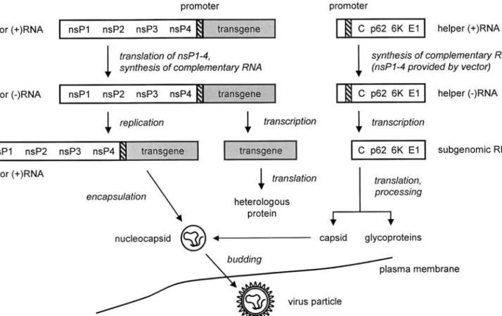
cleaved (through nsP2), the negative-strand RNA serves as a template for efficient synthe-sis of both new positive-strand genomic RNA, and subgenomic 26S RNA that encodes the viral structural proteins. The subgenomic RNA is translated into the polyprotein that is then processed into the capsid and spike proteins, p62 and E1. Owing to an encapsulation signal in the nsP1 gene for SIN and nsP2 gene for SFV, capsid proteins assemble with genomic RNA molecules to form nucleocapsids, which then bud from the cell membrane where processed spike proteins have been inserted (1). Figure 1 gives a schematic overview of the alphaviral replication and virus assembly.
Alphaviral Vectors: The
Replicon System
Vectors for SFV and SIN as well as the closely related VEE have been developed to express high levels of foreign genes in vitro and in vivo (12–15). Owing to their promiscu-ous usage of cell surface receptors for attach-ment, an extremely wide range of host cells, including most cell lines and many primary cultures from insect to mammalian cells, can be infected and successfully transduced (16,17).
While fully replication-proficient alphaviral vectors have been used (18), a more common approach is the replicon system (see Fig. 2) in which the transgene in the “vector RNA” replaces the viral structural protein genes (13–15). Vector RNAs are self-replicating and are referred to as “replicons.” They must be co-transfected with defective “helper RNA” to be packaged into infectious particles. Both vector and helper RNAs are obtained by in vitro tran-scription from plasmids containing their respective cDNAs. A vector RNA-encoding plasmid of the SFV system, pSFV2gen (19), is shown in Figure 3. It is based on pSFV1 (13) but contains a multiple-cloning site with addi-tional unique restriction sites.
Defective helper RNAs have a large deletion in nsP1–4 and are not self-replicating, but can be replicated and transcribed by the nsPs
pro-vided by the replicons. Helper subgenomic RNAs are translated to provide the capsid and spike proteins for replicon assembly under conditions in which helper RNAs are not pack-aged. Alternatively, the capsid and spike pro-teins can be supplied from stably transfected cells line (20). Packaged replicons derived by
this approach have been termed “suicide vec-tors” (21) because they infect target cells with-out the capability of forming new infectious particles (as the introduced replicon genome is lacking the structural protein genes).
A fully DNA-based SFV expression system employs vector and helper plasmids with the
Fig. 1. Alphaviral replication and assembly. Upon attachment of the viral particle to the cell and entry via endocytosis, the viral nucleocapsid is released into the cytoplasm. Binding of the nucleocapsid to ribosomes then triggers the uncoating process that releases the genomic (+)-strand RNA into the cytoplasm. The genomic RNA is then translated into the nsP1–4 preprotein that synthesizes the complementary (–)-strand genome. When nsP1–4 is cleaved into the separate proteins, many copies of both (+)-strand genomic and subgenomic RNA are formed. The subgenomic RNA is translated into the capsid protein (C) and the envelope proteins (p62, the pre-cursor of the E2 and E3 spike proteins; the 6K protein; and E1). Owing to the packaging signal in the nsP region of the genome, the genomic RNA is bound to capsid proteins, thereby forming the nucleocapsid. Interaction of the nucleocapsid with envelope glycoproteins (spikes), which have penetrated into the host cell membrane, lib-erates the virus particle from the host cell. (Modification of the figure kindly provided by Dr. Sondra Schlesinger, Washington University School of Medicine, St. Louis, USA.)
prokaryotic SP6 RNA polymerase promoter (for in vitro transcription) replaced by an RNA polymerase II-dependent promoter, permitting expression in eukaryotic cells (22). The viral titers obtained by co-transfection of these plas-mids into baby hamster kidney 21 (BHK) and COS-1 cells, however, are significantly lower than the titers obtained by co-electroporation of vector and helper RNAs.
In a different approach, the helper RNA, in addition to the vector RNA, similarly contains a packaging signal, resulting in the co-packag-ing of both vector and helper RNA into infec-tious particles. Virus particles derived by this method have been successfully used as an effi-cient expression cloning system for receptors and other membrane proteins, as well as for secreted and intracellular proteins (23).
Fig. 2. Packaging of SFV and SIN replicons. Vector RNA encoding nonstructural proteins 1–4 (nsP1–4) and the transgene (shaded) under the control of the 26S subgenomic RNA promoter (hatched), and defective helper RNA (encoding the structural alphavirus proteins downstream of the subgenomic RNA promoter) are obtained by in vitro transcription from their respective cDNAs and co-transfected into BHK cells. Only the vector RNA contains the packaging signal required for encapsidation into the nucleocapsid. Within the cytoplasm, vector RNA replication occurs through the action of nsP1–4. In parallel, the defective helper RNA is replicated and transcribed by nsP1–4, and the capsid protein C, 6K protein, glycoprotein E1, and precursor protein p62 (which is later cleaved into the E2 and E3 spike proteins) are translated. Capsid proteins package only vector RNA, while spike proteins are incorporated into the cell membrane. Nucleocapsids dock to the cell membrane where spike proteins have been incorporated, thus allowing the budding of virus particles.
Biosafety
While other alphaviruses such as VEE and rubella virus are human pathogens, wild-type SFV and SIN normally cause less severe infec-tions in humans, with SIN leading to mild fever, joint-pain, and rash (5). For SFV, human infec-tion is relatively common, but this has been linked to disease in only two occasions (5). This explains why SFV is classified as biosafety level 3 in the USA, but with the condition that most activities (including the work with SFV vectors) can be carried out at level 2, which is also the biosafety level for SIN (US HHS Publication no. CDC 93-8395, 1993) (24). In the European Union and in Switzerland, SFV and SIN are classified as biosafety level 2 (EC Council Directive 93/88/EEC, 1993; statement of the Swiss expert
committee for biosafety on the classification of work with genetically modified viral vectors, August 2001).
SFV and SIN expression systems using repli-cons (cf, above) repli-consist of two components: the expression vector and the helper plasmid. When the original SFV helper plasmid is used, fully replication-proficient viral particles may form by homologous recombination in BHK cells. In co-transfections of SFV expression vec-tors with the pSFV-Helper2 plasmid (91), how-ever, no infectious virus particles can arise due to point mutations in the structural genes, which prevent cleavage of the p62 precursor protein into functional E2 and E3 envelope gly-coproteins. The resulting SFV particles are non-infectious until activated with α-chymotrypsin. Therefore, viral vectors packaged with pSFV-Helper2 have been grouped into biosafety level 1 classifications in several European countries, including Germany and Switzerland (http://www.rki.de/GENTEC/ZKBS/ZKBS. HTM; statement of the Swiss expert committee for biosafety on the classification of work with genetically modified viral vectors, August 2001).
Applications in Neurobiology
Transduction of Neurons
Both SFV and SIN vectors have been used successfully to transduce neurons in primary and tissue cultures (see Fig. 4) and in vivo (e.g., refs. 25–32). Organotypic cultures of hip-pocampal slices (33,34) represent a highly applicable preparation that permits rapid tests for recombinant proteins in a system that is more similar to the in vivo situation than cul-tures of dissociated neurons. SFV and SIN vec-tors deliver genes efficiently to neurons in organotypic hippocampal slices prepared from neonatal and postnatal rats (see Fig. 4A,B; refs. 27,28,35). In collaboration with my colleague Kenneth Lundstrom, I successfully established a standardized protocol for the application of alphaviral vectors in this test system (36,37).
Fig. 3. Topological map of the pSFV2gen vector plasmid. Nucleotides are numbered with the first nucleotide of the cDNA derived from SFV4 genomic RNA defined as 1. The locations of the nsP1–4 genes encoding the nonstructural proteins, the ampicillin resistance gene, the SP6 promoter for in vitro tran-scription, the alphaviral 26S subgenomic RNA pro-moter (SG), and the poly-A sequence are indicated. The graph contains the unique restriction sites of the multiple cloning site for inserting genes of interest, as well as the linearization sites (underlined) for in vitro transcription.
Fig. 4. Transduction of cultured rat hippocampal slices (A,B) and primary rat hippocampal neurons (C) by using SFV and SIN vectors. (A) Rat hippocampal slices from P0 and P6 rats were cultured for 1 and 45 d (P0 vs P6, respectively) and then infected with 100- to 10,000-diluted SFV and SIN encoding enhanced green fluores-cent protein (GFP) or E. coliβ-galactosidase (LacZ). Dark field (top left) and fluorescence micrographs of living slices were taken at 2 d post-infection, whereas bright field images were obtained from slices fixed and stained for LacZ expression at 1 d post-infection. Transduced cells appear in green and blue for GFP and LacZ, respec-tively. Propidium iodide staining was performed on living slices at 5 d post-infection; an FITC filter set with a long-pass emission filter revealed both infected cells and propidium iodide-positive (red), non-viable cells (bot-tom middle), whereas a rhodamine filter set revealed only propidium iodide fluorescence (bot(bot-tom right). Note that most infected cells exclude propidium iodide, and therefore, appear to be intact. Abbreviations: DG, den-tate gyrus; so, stratum oriens; sp, stratum pyramidale; sr, stratum radiatum. Bar: 330 µm (left and middle panels of top and middle row) and 80 µm (remaining panels). Magnifications are from CA1 regions, except for the bot-tom left panel which shows infected CA3 pyramidal cells. (B) Confocal image of the CA1 region from a rat hip-pocampal slice culture at 2 d after infection with an SFV expressing GFP. The color code indicates the depth of the location of GFP-positive cells within the slice. Note that most GFP-positive cells are pyramidal cells (image kindly provided by Dr. R. Anne McKinney, University of Zurich, Switzerland). (C) Confocal image of a rat hip-pocampal neuron in culture, 1 d after infection with an SFV vector expressing GFP. GFP fluorescence is shown in green, while in vivo labeling of active boutons with an anti-synaptotagmin-I antibody is shown in red (image kindly provided by Dr. Antonio Malgaroli, Scientific Institute San Raffaele, Milano, Italy).
Table 1
Advantages and Disadvantages of Conventional SFV and SIN Vectors Compared to Other Viral and Non-Viral Transduction Techniques
Advantages Disadvantages
Rapid production Inhibition of host cell protein synthesis and cytotoxicity Extremely high viral titers obtained (stocks Not applicable for long-term gene transfer
are therefore used at ≤10,000-fold dilution)
Preferential and efficient infection of neurons No choice of promoter Fast gene expression (within hours)
High transgene expression levels
Compared to other viral vectors, biolistic and lipid-based transfection methods, SFV, and SIN vectors cause rapid, high-level, and efficient transduction (>95% in primary neu-rons, see Fig. 4C). It is also beneficial for many studies that conventional SFV and SIN vec-tors preferentially transduce neurons rather than non-neuronal cells, causing pyramidal cells and interneurons, in hippocampal slice cultures, to constitute approx 90% of all cells
positive for green fluorescent protein (GFP; see Fig. 4B) (27,28).
Aside from their advantages (see Table 1), wild-type alphaviral vectors (13,15) retain the disadvantage of inhibiting host cell protein synthesis, which eventually causes cell death. While neurotoxic effects appear relatively fast (within 1–2 d) and are readily observed in cul-tures of dissociated hippocampal neurons (25,38,39), they are delayed by several days in
Table 2
Proteins that Have Been Functionally (Over)expressed with SFV and SIN Vectors in Neurons
Recombinant protein Comments Refs.
Intracellular proteins
β-galactosidase Reporter molecule (27,29,30,62)
GFP Reporter molecule (27,30,32,63)
NEDD-2 Cysteine protease (64)
Aspartylglucosaminidase Enzyme (65)
p16ink4,p21waf/cip,p27kip1 Cyclin-dependent kinase inhibitors (66)
Cdk4, Cdk6 (dominant-negative) G1cyclin-dependent kinases (66)
CaMKII (truncated) Kinase (67)
Munc13-1, Munc 18-1 Synaptic vesicle priming proteins (68,69)
Snapin Binds synaptic protein SNAP-25 (70)
Syntaphilin SNARE assembly regulator (71)
Homer-1a and 1c Postsynaptic scaffolding protein a
Membrane proteins
GABAAreceptor α1 and β2 subunits Ligand-gated ion channel (48,72)
Serotonin 5-HT3receptor Ligand-gated ion channel (73)
P2X2receptor Ligand-gated ion channel (74)
Calcium channel β4 subunit Calcium channel regulator (26)
GluR1, GluR2, GluR4 AMPA receptor subunits (31,40,75–77)
GluR2 C-term Blocks Pick1-GluR2/3 interaction (78)
VGLUT1/BNP1, VGLUT2/DNPI Vesicular glutamate transporters (79,80)
Connexin 36 Increases intercellular coupling a
SNAP-25 Synaptic protein (81,82)
SGT Synaptic vesicle surface protein (83)
GFP-tagged pleckstrin homology domain Indicator of intracellular IP3dynamics (84)
Amyloid precursor protein (APP) Cleaved into β-amyloid peptide (85)
Presenilin-1 Associated with familial AD (86)
Catechol O-methyltransferase Membrane-bound enzyme (87)
Rab8 Membrane-associated GTPase (25)
Neprilysin Membrane-associated metalloendopeptidase (88)
VIP21 Integral membrane protein (25)
polymeric immunoglobulin receptor Binds immunoglobulins A and M (89)
F protein Measles virus glycoprotein a
hippocampal slice cultures. Based on propid-ium iodide exclusion experiments (cf, Fig. 4A, bottom middle and right panels) and electro-physiological tests of passive and active membrane properties (i.e., resting membrane potential and conductance, action potential generation, firing accommodation, and hyperpolarization-induced currents), we found pyramidal cells in this preparation to survive for 2–5 d post-infection (27,28). In any case, as alphaviral GFP expression in tissue cultures is fast (detectable within 4–6 h) and transient (peaking at 1 d post-infection) (28), the time window to examine transgene effects suffices for many physiological studies, including the microscopical and electrophysi-ological analysis of synaptic function (e.g., Fig. 4C; ref. 40), and the real-time investiga-tion of activity-dependent morphological changes (32).
Table 2 summarizes different transgenes that have been introduced into hippocampal and other neurons by using packaged SFV and SIN replicons. While it lists many intra-cellular and membrane proteins that could be expressed in a functional manner, our attempts to functionally overexpress the tran-scription factors EGR1 and EGR2 with the less cytopathic SFV(PD) mutant (38,39) were not successful. The analysis with an anti-EGR1 antibody revealed the presence of immunore-active protein in the cytoplasm rather than the nucleus of infected cells; and functional tests for transactivation of EGR1-dependent luciferase reporter gene expression were not successful (M.J. Fend, K. Lundstrom, and M.U. Ehrengruber, unpublished data). By contrast, when inserted into adenoviral vec-tors, exactly the same EGR1 cDNA was able to induce EGR1-dependent gene expression (41). These results indicate that the present alpha-viral vectors may not be applicable for the functional study of specific transcription fac-tors. Another report with a GFP-tagged tran-scription factor (ATF-4) overexpressed by SFV did not examine whether ATF-4 transactivat-ing function was retained in infected cells (42).
Heterologous Expression
of Neuronal Proteins
Owing to the intracellular amplification processes inherent in alphaviral genome repli-cation, SFV and SIN vectors generate extremely high transgene expression levels in infected cells. These levels can be problematic in cases where the transgene product has cyto-toxic effects (as in the human neurokinin-1 receptor [43]) or because it does not match physiological levels. On the other hand, alphaviral vectors are particularly advanta-geous when applied in cell lines for generating high amounts of recombinant protein required (e.g., for the drug screening of receptors and the purification of proteins for structural stud-ies and crystallization. Compared to the bac-ulovirus system, which generates similar recombinant protein levels, alphaviral vectors possess the advantage of functioning well in mammalian cells.
Alphaviral vector systems have been partic-ularly useful for the heterologous expression of receptors, including G protein-coupled tors such as the metabotropic glutamate recep-tors mGluR2, 3, 4, and 8 (19,44,45), the human neurokinin-1 receptor (43,46), and the α1b
-adrenergic receptor (47). Radioligand binding on intact cells and isolated cell membranes revealed extremely high expression levels, with 3–10 million receptors per cell and 50–200 pmol recombinant receptor per mg total pro-tein. An intact functional coupling of the over-expressed receptors to G proteins in response to agonists has been tested via measurements of, e.g., GTPγS binding (44), intracellular Ca2+
mobilization (43), and inositol phosphate accu-mulation (47).
In specific applications, the heterologous expression of distinct proteins, possibly at approximately equal ratios, within a given cell is required. For example, it has been shown for the GABAA receptor that α1 or β2 subunits,
when expressed alone, are retained in the endoplasmic reticulum, whereas co-expression of both subunits permits GABAA receptor
genes can be introduced into the same cell by co-infection with different viral vectors (47,48), the use of internal ribosomal entry site (IRES) sequences (67), and additional 26S subgenomic RNA promoters (18).
Identification of Infected Neurons
Using GFP
The function of transduced neurons is gener-ally compared to one of uninfected control neurons (as well as to one of neurons infected with a control virus expressing, e.g., GFP) to determine the effect of the expressed trans-gene. This comparison is done ideally in the same population of cells, i.e., the same slice culture or the same Petri dish. Electrophysio-logical recordings, as well as other measure-ments from infected cells, thus require the identification of (living) cells that have been genetically modified. This is normally achieved by using GFP as a reporter molecule.
Three GFP-based approaches have been employed in combination with alphaviral vec-tors. The most common method is the genera-tion of GFP fusion proteins where the gene of interest is fused to the GFP cDNA. This strat-egy, however, may be problematic as the attachment of the GFP domain can alter trans-port, localization, and function of the overex-pressed protein. A second approach is the use of an internal ribosomal entry site (IRES) sequence between the gene of interest and the GFP cDNA, permitting separate expression of both genes. Our experiments using IRES-dependent GFP expression, however, revealed relatively weak GFP fluorescence and allowed identification of infected pyramidal cells in organotypic hippocampal slices at only 3 d post-infection. A third method is to drive GFP expression separately under the control of an additional subgenomic alphaviral 26S pro-moter downstream of the gene of interest. This procedure, in our hands, resulted in suffi-ciently high GFP fluorescence levels to allow for electrophysiological recordings from GFP-positive primary neurons at 1 d post-infection. The second and third approaches, of course, can generally be used to express two or more
transgenes as separate proteins, which may be required, e.g., for the expression of heteromeric proteins such as ligand-gated ion channels.
Alphaviral Vector Development
As summarized in Table 1, wild-type alphaviral vectors may be problematic because of 1) cytotoxicity; 2) short-term expression due to cytotoxicity and/or transient nature of the viral RNA; 3) no choice of promoter; and 4) in certain cases, extremely high expression levels. The development of alternative SFV and SIN vectors focuses on these negative properties by introducing specific mutations in the nsP1–4 genes that control viral replication and expres-sion. Tables 3 and 4 give an overview of the modified alphaviral vectors and indicate their transduction characteristics in cell lines and neurons.
Non-Cytopathic Vectors
Spontaneous mutations discovered in the nonstructural genes, particularly in nsP2 and nsP4, have dramatic effects on the viral patho-genicity. Mutations in nsP2 of SIN have pro-duced viruses (49) and replicons (49–51) with greatly reduced cytotoxicity in cell lines (see Table 3). The majority of the mutants had a sin-gle change at amino acid 726. For example, a change at this site from Pro to Ser produced replicons that had reduced levels of RNA repli-cation, and therefore, also lower cytopatho-genicity (49). The change from Pro to Leu led to a further reduction in both RNA synthesis and cytotoxicity (50). Replication-persistent replicons with ongoing viral multiplication were generated by random mutagenesis of nsP genes in SIN and SFV vectors containing the neomycin resistance gene; once again they had either deletions or point mutations in nsP2 (51). All of these experiments were performed in cell lines (see Table 3), with mitosis of infected cells. In general, persistent infections appeared to be correlated with a lack of inter-feron production or the production of other potential cytokines.
With regard to neuronal infection (see Table 4), the introduction of known temperature-sensitive mutations into nsP2 and nsP4 reduced the cytotoxicity of SFV vectors in pri-mary hippocampal neurons (39). Further-more, studies on replication-competent SFV revealed a point mutation (Arg649Asp) in the nuclear localization signal of nsP2 to confer lower neurovirulence in mice (52,53). When
we combined a similar mutation in the nsP2 nuclear localization signal (Arg650Asp) with an additional mutation in nsP2 (Ser259Pro), we obtained a vector, SFV(PD), that was less cytopathic and caused increased transgene expression in mammalian cell lines, primary hippocampal and cortical neurons, and organotypic hippocampal slices (38,39) (K. Lundstrom, A. Abenavoli, A. Malgaroli, and
Table 3
Mutant SFV and SIN Replicons and Their Transduction Characteristics in Cell Lines Compared to the Conventional, Wild-Type Vectors SINrep5 (15) and SFV1 (13)
Vector Characteristics Refs.
SIN(nsP2A1E) Non-cytopathic in BHK cells, establishes (51)
persistent infectiona
SIN(nsP2P726S) Less cytopathic in BHK cells, noncytopathic (49)
in mouse and human cell lines, higher expression in BHK cells
SIN(nsP2P726L/T) Noncytopathic in all cell lines tested, establish (50,51,90)
persistent infections in BHK, Vero and CHO cellsa
SIN(nsP2P726SnsP4G153E), pCytTS Temperature-inducible, DNA-based expression (57)
system in BHK, CHO-K1, C2C12, COS-7, and HEK 293 cellsa
SFV(nsP2L10T), SF2A; SFV(nsP2∆D469), Noncytopathic in BHK cells, establish persistent (51)
SF1B; SFV(nsP2L713P), SF2C infection
SFV(nsP2S259P,R649D), SFV(PD) Less cytopathic in BHK, HEK 293, and (38,39)b
CHO-K1 cells, higher expression, reduced viral titers
SFV(nsP2S259P,R650D,L713P), SFV(PD713P) Noncytopathic in BHK cells, temperature- c
sensitive, expresses at 31°C but not at 37°, higher expression, low viral titers
SFV(nsP2S259P,R649DnsP4G153E), Temperature-sensitive in BHK cells, expresses (39)
SFV(PDE153) at 31°C but little at 37°
SFV(nsP2S259P,R649DnsP4G324E), Temperature-sensitive in BHK cells, expresses (39)
SFV(PDE324) at 31°C but little at 37°
SFV(nsP2S259P,R649D,M780TnsP4G153E), Temperature-sensitive in BHK cells, expresses (39)
SFV(PDTE) at 31°C but not at 37°, achieving higher expression
SFV(A774nsP) Temperature-sensitive in BHK cells,
transduces ~100-fold more cells at 31 vs 37°C d
SFV(26S-M5), SFV(26S-M1), Decreased transgene expression levels (1, 3,
SFV(26S-M3) and 30% of wild-type, respectively) (58) aThis mutant has only been used as a nonpackaged replicon.
bPlus unpublished data by K. Lundstrom, A. Abenavoli, A. Malgaroli, and M.U. Ehrengruber. cK. Lundstrom and M.U. Ehrengruber, unpublished data.
M.U. Ehrengruber, unpublished data). Fur-thermore, when a mutation in nsP2 known to confer replication-persistence (Leu713Pro; ref. 51) was inserted into SFV(PD), the resulting vector SFV(PD713P) was noncytopathic in BHK cells and still infected neurons in hip-pocampal slice cultures (K. Lundstrom, and M.U. Ehrengruber, unpublished data).
Taken together, the less cytopathic SFV and SIN mutants will permit the application of alphaviral vectors under more physiological conditions. In addition to general overexpres-sion of recombinant proteins, the vectors can
also be used to study expression kinetics and signal transduction events (54), suppress genes by antisense and ribozyme approaches, and perform gene therapy trials with prolonged gene expression.
Vectors with Temperature-Dependent
Regulation
In present SFV and SIN vectors, transgene expression relies on the endogenous 26S subgenomic RNA promoter. The minimal 26S subgenomic RNA promoter consists of 19 nucleotides upstream and 5 nucleotides
down-Table 4
Transduction Characteristics of SFV Replicons in Hippocampal and Other Neurons
Vector Characteristics Refs.
Conventional SFVa Infects primary rat hippocampal neurons, (25,26)
cytotoxic after >8–24 hs
Targets pyramidal cells and interneurons in rat (27,28)
hippocampal slices, neurons tolerate infection for 2–5 d
In vivo, infects striatal neurons in rat brain (29)
SFV(nsP2S259P,R650D), SFV(PD) Less cytopathic in primary rat hippocampal (39)b
and cortical neurons, higher expression
Targets pyramidal cells and interneurons in rat (28)
hippocampal slices
SFV(nsP2S259P,R650D,L713P), Transduces pyramidal cells in rat c
SFV(PD713P) hippocampal slices
SFV(nsP2S259P,R649DnsP4G153E), Targets interneurons at 31°C, but both pyramidal (39)
SFV(PDE153) cells and interneurons at 37°C in rat
hippocampal slices
SFV(nsP2S259P,R649DnsP4G324E), Temperature-sensitive in primary rat hippocampal (39)
SFV(PDE324) neurons, expresses at 31°C but little at 37°C
SFV(nsP2S259P,R649D,M780TnsP4G153E), Transduces both pyramidal cells and interneurons (39)
SFV(PDTE) of rat hippocampal slices at 31°C but not at 37°C
SFV(A774nsP) Transduces selectively glial cells at 37°C, and only d
neurons at 31°C in primary rat hippocampal cells
Transduces selectively glia and other non-neuronal d
cells at 37°C, but also neurons at 31°C in rat hippocampal slices
Note that SIN vectors also efficiently transduce hippocampal neurons in primary culture (30,31), in slices (27,32), and in vivo (30).
aThe conventional vectors SFV1 (13) and SFV2gen (19) are based upon the cDNA for SFV4 that is derived from the
wild-type, virulent L10 strain (5).
bPlus unpublished data by K. Lundstrom, A. Abenavoli, A. Malgaroli, and M.U. Ehrengruber. cK. Lundstrom, and M.U. Ehrengruber, unpublished data.
stream of the transcription start site for the subgenomic RNA, but its activity is enhanced when neighboring sequences are included (1). The 26S subgenomic RNA promoter functions in the cytoplasm of infected cells, as it is the compartment where the transcription of alphaviruses occurs (cf Fig. 1). In contrast, the nucleus is the compartment where “non-alphaviral” promoters are functional that can be regulated and transcribe RNA from DNA. Thus, other means to control SFV and SIN-mediated transgene expression have to be found. Temperature-dependence is one of the few ways to regulate alphavirus vectors that replicate and are transcribed in the cytoplasm.
Temperature-sensitive mutations have been described for SIN in each of the nsP genes (55,56), and those in nsP4 (14) have been intro-duced into SIN expression vectors (57). Combi-nation of the Pro726Ser change in nsP2 with a temperature-sensitive mutation in nsP4 led to the inducible expression of DNA-based vectors in a variety of cell lines (57). Several of the known temperature-sensitive point mutations in nsP2 and nsP4 have also been introduced into the SFV expression vector, causing tem-perature-sensitive transgene expression in cell lines, primary hippocampal neurons, and organotypic hippocampal slices (see Tables 3 and 4) (39). A quadruple-mutant vector, SFV(PDTE), with three point mutations in nsP2 and one in nsP4, permitted GFP expres-sion at 31°C but not at 37°C. Similarly, SFV(PD713P) permitted transgene expression at 31°C rather than 37°C (K. Lundstrom and M.U. Ehrengruber, unpublished data). Interest-ingly, the triple-mutant vector SFV(PDE153)
when used at 37°C transduced interneurons, rather than pyramidal cells in rat hippocampal slices, characterized by a ratio of approx 0.6 between GFP-positive pyramidal cells and interneurons (the corresponding ratio is >10 for the wild-type SFV vector) (28). When SFV(PDE153) was used at 31°C instead, a
wild-type phenowild-type was observed with a ratio of approx 10 (39). A different study revealed another SFV mutant, SFV(A774nsP) encoding GFP downstream of the nsP genes from the
avirulent SFV strain A7(74), to transduce glial cells, and not neurons, in cultured hippocam-pal cells and slices at 37°C, whereas this was reversed at 31°C (M.U. Ehrengruber, M. Reng-gli, M.J.V. Vähä-Koskela, and K. Lundstrom, unpublished data). These results show that mutant alphaviral vectors are useful, at least in hippocampal tissue, to target transgene expres-sion to a subset of neurons or even glial cells rather than principal neurons. At the non-per-missive temperature, the host cellular environ-ment presumably does not support alphaviral replication.
Vectors with Decreased Expression Levels
For many applications, the elevated expres-sion levels obtained with alphaviral vectors are normally favorable or non-problematic. On the other hand, they may be detrimental under specific conditions—in particular when the overexpressed protein has cytotoxic effects or when more physiological levels of the expressed protein are essential (the functional expression of G protein-coupled receptors, e.g., may require a more physiological ratio of receptors vs G proteins). To achieve lower expression levels, several point mutations were introduced into the 26S subgenomic RNA promoter of SFV (see Table 3) (58). The result-ing levels of reporter protein (β-galactosidase and luciferase) expressed in BHK cells were only 1, 3, and 30%, as compared to the quanti-ties expressed from wild-type SFV vectors. The down-regulated expression vectors will thus permit examination of recombinant protein function under conditions that are more simi-lar to the in situ situation. The use of the GFP reporter in combination with the more down-regulated SFV vectors, however, will no longer be practical (transduced cells will not be recog-nized due to low GFP fluorescence).
Comparison with Other Viral Vectors
Many viruses have been successfully employed to transfer genes into neurons, andeach viral vector has its advantages and disad-vantages. As each vector system has normally been established in a different laboratory and tested under distinct conditions, it is some-times difficult for a newcomer to choose a viral vector that is optimal for study. We have previ-ously characterized wild-type SFV and SFV(PD) vectors, recombinant adenovirus type 5, adeno-associated virus type 2 (AAV), lentivirus, and measles virus by their expres-sion of GFP in rat hippocampal slice cultures (28). Figure 5 illustrates the results obtained with a lentivirus expressing GFP under the
control of a cytomegalovirus (CMV) promoter. Both SFV vectors transduced more neurons (>90% of all GFP-positive cells) than AAV, lentivirus, and measles virus (71, 69, and 62%, respectively), while resting membrane poten-tial and conductance, action potenpoten-tials, firing accommodation, and H-current appeared nor-mal in all infected CA1 pyramidal cells. No infected neurons were identified with aden-ovirus type 5 (28). This was an unexpected finding because this virus is useful to trans-duce primary rat hippocampal neurons (59), although relatively high titers (approx 108
Fig. 5. Transduction of rat hippocampal slices with a lentiviral vector. P6 rat hippocampal slices at 28 d in culture were injected in the CA1 and CA3 pyramidal cell layer with two-fold diluted lentivirus expressing GFP under a CMV promoter. Slices were incubated at 37°C and fixed at 7 d post-infection. Fluorescence micrographs of a whole slice (A), CA1 neurons (B), arrows, CA3 pyramidal cells (C), arrows, and glial cells in the CA1 region
(D), arrowheads. Abbreviations: DG, dentate gyrus; so, stratum oriens; sp, stratum pyramidale; sr, stratum
plaque-forming units/mL) have to be applied to overcome the predominant glial cell infec-tion. AAV-mediated GFP expression was restricted to neurons when the neuron-specific PDGF promoter (for platelet-derived growth factorβ-chain) rather than the CMV promoter was used. Whereas transgene expression medi-ated by SFV was rapid but transient (as described above), it increased more slowly but remained stable with AAV and lentivirus, but was fast with measles virus. As no replication-impaired system is available for measles virus, this vector propagated in the hippocampal slices. It is noteworthy that spreading occurred to pyramidal cells rather than non-neuronal cells (60), which may facilitate the selective transduction of most pyramidal cells within a slice culture. In any case, SFV is useful for short-term and AAV and lentivirus for long-term transduction of hippocampal slices, while measles virus may permit both short-and long-term transduction.
A qualitative evaluation of the maximal GFP expression levels obtained in hippocam-pal slice cultures revealed the following order: measles virus ≥ SFV ≈ lentivirus > AAV. Com-pared to GFP-expressing lentivirus, the identi-fication of neurons transduced with SFV and SIN vectors encoding GFP was easier (cf Fig. 4A vs Fig. 5). We also estimated the transduc-tion efficiencies in hippocampal slices by com-paring the number of GFP positive cells vs the number of applied virus particles. The follow-ing order was found: wild-type SFV ≈ SFV(PD) > measles virus > lentivirus ≈ AAV-PDGF-GFP > AAV-CMV-GFP (28). For SFV and SFV(PD), nearly every virus particle resulted in a GFP-positive cell. This high infec-tion rate is due to the broad host range of the virus, and the fact that entry of one virus par-ticle suffices to initiate viral replication in a host cell (1,61). For SIN vectors in organotypic hippocampal slices, similar results as with SFV vectors are obtained (27). In addition to the high infection rate, the easy generation of high-titer virus stocks (36,37) that contain a high ratio of infectious to physical particles (61) makes SFV and SIN vectors favorable for
gene transfer into neurons. In several coun-tries, SFV vectors packaged with pSFV-Helper2 may be preferred as they are classified as biosafety level 1.
Acknowledgments
This review article is dedicated to the mem-ory of my former postdoctoral advisor Dr. Norman Davidson (April 5, 1916 to February 14, 2002). The author’s work is supported by grant no. 31-57125.99 from the Swiss National Science Foundation. Thanks to Drs. Antonio Malgaroli and R. Anne McKinney for provid-ing the confocal images of Figure 4; Martin Renggli for editing Figure 3; Roland Schöb for editing Figure 4; and Drs. Sondra Schlesinger and Kenneth Lundstrom for comments on the manuscript.
References
1. Schlesinger S. and Schlesinger M. J. (2001)
Togaviridae: the viruses and their replication, in Fields Virology. (Knipe D. M. and Howley P. M.,
eds.) Lippincott Williams & Wilkins, Philadel-phia, pp. 895–916.
2. Smithburn K. C. and Haddow A. J. (1944) Sem-liki Forest virus. I. Isolation and pathogenic properties. J. Immunol. 49, 141–147.
3. Taylor R. M., Hurlbut H. S., Work T. H., Kings-bury J. R., and Frothingham T. E. (1955) Sindbis virus: a newly recognized arthropod-transmit-ted virus. Am. J. Trop. Med. Hyg. 4, 844–846. 4. Kubes V. and Rios F. A. (1939) The causative
agent of infectious equine encephalomyelitis in Venezuela. Science 90, 21.
5. Griffin D. E. (2001) Alphaviruses in Fields
Virol-ogy. (Knipe D. M. and Howley P. M., eds.)
Lip-pincott Williams & Wilkins, Philadelphia, pp. 917–962.
6. Bradish C. J., Allner K., and Maber H. B. (1971) The virulence of original and derived strains of Semliki Forest virus for mice, guinea pigs, and rabbits. J. Gen. Virol. 12, 141–160.
7. Fazakerley J. K., Pathak S., Scallan M., Amor S., and Dyson H. (1993) Replication of the A7(74) strain of Semliki Forest virus is restricted in neurons. Virology 195, 627–637.
8. Oliver K. R., Scallan M. F., Dyson H., and Faza-kerley J. K. (1997) Susceptibility to a neu-rotropic virus and its changing distribution in the developing brain is a function of CNS matu-rity. J. Neurovirol. 3, 38–48.
9. Griffin D. E. (1998) A review of alphavirus repli-cation in neurons. Neurosci. Biobehav. Rev. 22, 721–723.
10. Peränen J., Takkinen K., Kalkkinen N., and Kääriäinen L. (1988) Semliki Forest virus-spe-cific non-structural protein nsP3 is a phospho-protein. J. Gen. Virol. 69, 2165–2178.
11. LaStarza M. W., Lemm J. A., and Rice C. M. (1994) Genetic analysis of the nsP3 region of Sindbis virus: evidence for roles in minus-strand and subgenomic RNA synthesis. J. Virol.
68,5781–5791.
12. Davis N. L., Willis L. V., Smith J. F., and John-ston R. E. (1989) In vitro synthesis of infectious venezuelan equine encephalitis virus RNA from a cDNA clone: analysis of a viable deletion mutant. Virology 171, 189–204.
13. Liljeström P. and Garoff H. (1991) A new gener-ation of animal cell expression vectors based on the Semliki Forest virus replicon. BioTechnology
9,1356–1361.
14. Xiong C., Levis R., Shen P., Schlesinger S., Rice C. M., and Huang H. V. (1989) Sindbis virus: an efficient, broad host range vector for gene expression in animal cells. Science 243,
1188–1191.
15. Bredenbeek P. J., Frolov I., Rice C. M., and Schlesinger S. (1993) Sindbis virus expression vectors: packaging of RNA replicons by using defective helper RNAs. J. Virol. 67, 6439–6446. 16. Lundstrom K. (1999) Alphaviruses as tools in
neurobiology and gene therapy. J. Recept. Signal
Transduct. Res. 19, 673–686.
17. Schlesinger S. (2001) Alphavirus vectors: devel-opment and potential therapeutic applications.
Expert Opin. Biol. Ther. 1, 177–191.
18. Hahn C. S., Hahn Y. S., Braciale T. J., and Rice C. M. (1992) Infectious Sindbis virus transient expression vectors for studying antigen pro-cessing and presentation. Proc. Natl. Acad. Sci.
USA 89, 2679–2683.
19. Malherbe P., Kratzeisen C., Lundstrom K., Richards J. G., Faull R. L. M., and Mutel V. (1999) Cloning and functional expression of alternative spliced variants of the human metabotropic glutamate receptor 8. Mol. Brain
Res. 67, 201–210.
20. Polo J. M., Belli B. A., Driver D. A., et al. (1999) Stable alphavirus packaging cell lines for Sind-bis virus and Semliki Forest virus-derived vec-tors. Proc. Natl. Acad. Sci. USA 96, 4598–4603. 21. Schlesinger S. (1993) Alphaviruses – vectors for
the expression of heterologous genes. Trends
Biotechnol. 11, 18–22.
22. DiCiommo D. P. and Bremner R. (1998) Rapid, high level protein production using DNA-based Semliki Forest virus vectors. J. Biol. Chem.
273,18,060–18,066.
23. Koller D., Ruedl C., Loetscher M., et al. (2001) A high-throughput alphavirus-based expression cloning system for mammalian cells. Nat.
Biotechnol. 19, 851–855.
24. Atkins G. J., Sheahan B. J., and Liljeström P. (1999) The molecular pathogenesis of Semliki Forest virus: a model virus made useful? J. Gen.
Virol. 80, 2287–2297.
25. Olkkonen V. M., Liljeström P., Garoff H., Simons K., and Dotti C. G. (1993) Expression of heterologous proteins in cultured rat hip-pocampal neurons using the Semliki Forest virus vector. J. Neurosci. Res. 35, 445–451. 26. Wittemann S., Mark M. D., Rettig J., and
Herl-itze S. (2000) Synaptic localization and presy-naptic function of calcium channel β4-subunits in cultured hippocampal neurons. J. Biol. Chem.
275,37,807–37,814.
27. Ehrengruber M. U., Lundstrom K., Schweitzer C., Heuss C., Schlesinger S., and Gähwiler B. H. (1999) Recombinant Semliki Forest virus and Sindbis virus efficiently infect neurons in hip-pocampal slice cultures. Proc. Natl. Acad. Sci.
USA 96, 7041–7046.
28. Ehrengruber M. U., Hennou S., Büeler H., Naim H. Y., Déglon N., and Lundstrom K. (2001) Gene transfer into neurons from hippocampal slices: comparison of recombinant Semliki For-est virus, adenovirus, adeno-associated virus, lentivirus, and measles virus. Mol. Cell.
Neu-rosci. 17, 855–871.
29. Lundstrom K., Richards J. G., Pink J. R., and Jenck F. (1999) Efficient in vivo expression of a reporter gene in rat brain after injection of recombinant replication-deficient Semliki For-est virus. Gene Ther. Mol. Biol. 3, 15–23.
30. Gwag B. J., Kim E. Y., Ryu B. R., et al. (1998) A neuron-specific gene transfer by a recombinant defective Sindbis virus. Mol. Brain Res. 63, 53–61.
31. Osten P., Khatri L., Perez J. L., et al. (2000) Mutagenesis reveals a role for ABP/GRIP bind-ing to GluR2 in synaptic surface accumulation of the AMPA receptor. Neuron 27, 313–325. 32. Maletic-Savatic M., Malinow R., and Svoboda
K. (1999) Rapid dendritic morphogenesis in CA1 hippocampal dendrites induced by synap-tic activity. Science 283, 1923–1927.
33. Gähwiler B. H. (1981) Organotypic monolayer cultures of nervous tissue. J. Neurosci. Methods
4,329–342.
34. Stoppini L., Buchs P.-A., and Muller D. (1991) A simple method for organotypic cultures of ner-vous tissue. J. Neurosci. Methods 37, 173–182. 35. Malinow R., Hayashi Y., Maletic-Savatic M.,
Zaman S. H., Poncer J.-C., Shi S.-H., and Este-ban J. A. (2000) Introduction of green fluores-cent protein into hippocampal neurons through viral infection, in Imaging Neurons: A
Laboratory Manual (Yuste R., Lanni F., and
Kon-nerth A., eds.) CSHL Press, Cold Spring Har-bor, pp. 58.1–58.8.
36. Ehrengruber M. U. and Lundstrom K. (2000) Alphavirus-mediated gene transfer into neu-rons, in Current Protocols in Neuroscience. (Craw-ley J. N., Gerfen C. R., Rogawski M. A., Sib(Craw-ley D. R., Skolnick P., and Wray S., eds.), John Wiley & Sons, New York, pp. 4.22.1–4.22.23.
37. Ehrengruber M. U. and Lundstrom K. (2002) Semliki Forest virus and Sindbis virus vectors, in
Current Protocols in Human Genetics. (Dracopoli
N. C., Haines J. L., Korf B. R., et al. eds.) John Wiley & Sons, New York, pp. 12.2.1–12.2.23. 38. Lundstrom K., Schweitzer C., Richards J. G.,
Ehrengruber M. U., Jenck F., and Mülhardt C. (1999) Semliki Forest virus vectors for in vitro and in vivo applications. Gene Ther. Mol. Biol. 4, 23–31.
39. Lundstrom K., Rotmann D., Hermann D., Schneider E. M., and Ehrengruber M. U. (2001) Novel mutant Semliki Forest virus vectors: gene expression and localization studies in neu-ronal cells. Histochem. Cell Biol. 115, 83–91. 40. Shi S.-H., Hayashi Y., Petralia R. S., Zaman S.
H., Wenthold R. J., Svoboda K., and Malinow R. (1999) Rapid spine delivery and redistribution of AMPA receptors after synaptic NMDA recep-tor activation. Science 284, 1811–1816.
41. Ehrengruber M. U., Muhlebach S. G., Söhrman S., Leutenegger C., M., Lester H. A., and David-son N. (2000) Modulation of early growth response (EGR) transcription factor-dependent
gene expression by using recombinant aden-ovirus. Gene 258, 63–69.
42. Nehring R. B., Horikawa R. P. M., El Far O., et al. (2000) The metabotropic GABAB receptor
directly interacts with the activating transcrip-tion factor 4. J. Biol. Chem. 275, 35,185–35,191. 43. Lundstrom K., Mills A., Buell G., Allet E.,
Adami N., and Liljeström P. (1994) High-level expression of the human neurokinin-1 receptor in mammalian cell lines using the Semliki For-est virus expression system. Eur. J. Biochem. 224, 917–921.
44. Monastyrskaia K., Lundstrom K., Plahl D., Acuna G., Schweitzer C., Malherbe P., and Mutel V. (1999) Effect of the umami peptides on the ligand binding and function of rat mGlu4a receptor might implicate this receptor in the monosodium glutamate taste transduction. Br.
J. Pharmacol. 128, 1027–1034.
45. Schweitzer C., Kratzeisen C., Adam G., et al. (2000) Characterization of [(3)H]-LY354740 binding to rat mGlu2 and mGlu3 receptors expressed in CHO cells using Semliki Forest virus vectors. Neuropharmacology 39, 1700–1706. 46. Lundstrom K., Vargas A., and Allet B. (1995) Functional activity of a biotinylated human neurokinin 1 receptor fusion expressed in the Semliki Forest virus system. Biochem. Biophys.
Res. Commun. 208, 260–266.
47. Scheer A., Björklöf K., Cotecchia S., and Lund-strom K. (1999) Expression of the α1b
-adrener-gic receptor and G protein subunits in mammalian cell lines using the Semliki Forest virus expression system. J. Recept. Signal
Trans-duct. Res. 19, 369–378.
48. Gorrie G. H., Vallis Y., Stephenson A., Whitfield J., Browning B., Smart T. G., and Moss S. J. (1997) Assembly of GABAA receptors
com-posed of α1 and β2 subunits in both cultured neurons and fibroblasts. J. Neurosci. 17,
6587–6596.
49. Dryga S. A., Dryga O. A., and Schlesinger S. (1997) Identification of mutations in a Sindbis virus variant able to establish persistent infec-tion in BHK cells: the importance of a mutainfec-tion in the nsP2 gene. Virology 228, 74–83.
50. Agapov E. V., Frolov I., Lindenbach B. D., Pra-gai B. M., Schlesinger S., and Rice C. M. (1998) Non-cytopathogenic Sindbis RNA vectors for heterologous gene expression. Proc. Natl. Acad.
Sci. USA 95, 12,989–12,994.
51. Perri S., Driver D. A., Gardner J. P., Sherrill S., Belli B. A., Dubensky T. W., Jr., and Polo J. M.
(2000) Replicon vectors derived from Sindbis virus and Semliki Forest virus that establish persistent replication in host cells. J. Virol. 74, 9802–9807.
52. Rikkonen M. (1996) Functional significance of the nuclear-targeting and NTP-binding motifs of Semliki Forest virus nonstructural protein nsP2. Virology 218, 352–361.
53. Fazakerley J. K., Boyd A., Mikkola M. L., and Kääriäinen L. (2002) A single amino acid change in the nuclear localization sequence of the nsP2 protein affects the neurovirulence of Semliki Forest virus. J. Virol. 76, 392–396.
54. Mazzucchelli C., Vantaggiato C., Ciamei A., et al. (2002) Knockout of ERK1 MAP kinase enchances synaptic plasticity in the striatum and facilitates striatal-mediated lerning and memory. Neuron 34, 807–820.
55. Hahn Y. S., Grakoui A., Rice C. M., Strauss E. G., and Strauss J. H. (1989) Mapping of RNA-temperature-sensitive mutants of Sindbis virus: complementation group F mutants have lesions in nsP4. J. Virol. 63, 1194–1202.
56. Hahn Y. S., Strauss E. G., and Strauss J. H. (1989) Mapping of RNA- temperature-sensitive mutants of Sindbis virus: assignment of com-plementation groups A, B, and G to nonstruc-tural proteins. J. Virol. 63, 3142–3150.
57. Boorsma M., Nieba L., Koller D., Bachmann M. F., Bailey J. E., and Renner W. A. (2000) A tem-perature-regulated replicon-based DNA expres-sion system. Nature Biotech. 18, 429–432.
58. Lundstrom K., Ziltener P., Hermann D., Schweitzer C., Richards J. G., and Jenck F. (2001) Improved Semliki Forest virus vectors for receptor research and gene therapy. J. Recept.
Signal Transduct. Res. 21, 55–70.
59. Ehrengruber M. U., Doupnik C. A., Xu Y., Gar-vey J., Jasek M. C., Lester H. A., and Davidson N. (1997) Activation of heteromeric G protein-gated inward rectifier K+ channels
overex-pressed by adenovirus gene transfer inhibits the excitability of hippocampal neurons. Proc.
Natl. Acad. Sci. USA 94, 7070–7075.
60. Ehrengruber M. U., Ehler E., Billeter M. A., and Naim H. Y. (2002) Measles virus spreads in rat hippocampal neurons by cell-to-cell contact and in a polarized fashion. J. Virol. 76, 5720–5728.
61. Strauss J. H. and Strauss E. G. (1994) The alphaviruses: gene expression, replication, and evolution. Microbiol. Rev. 58, 491–562.
62. Altman-Hamamdzic S., Groseclose C., Ma J.-X., Hamamdzic D., Vrindavanam N. S., Middaugh L. D., Parratto N. P., and Sallee F. R. (1997) Expression of β-galactosidase in mouse brain: utilization of a novel nonreplicative Sindbis virus vector as a neuronal gene delivery sys-tem. Gene Ther. 4, 815–822.
63. Knight D. E. (2000) Secreation from bovine chromaffin cells acutely expressing exogenous proteins using a recombinant Semliki Forest virus containing an EGFP reporter. Mol. Cell.
Neurosci. 14, 486–505.
64. Allet B., Hochmann A., Martinoiu I., Berger A., Missotten M., Antonsson B., et al. (1996) Dis-secting processing and apoptotic activity of a cysteine protease by mutant analysis. J. Cell
Biol. 135, 479–486.
65. Kyttälä A., Heinonen O., Peltonen L., and Jalanko A. (1998) Expression and endocytosis of lysosomal aspartylglucosaminidase in mouse primary neurons. J. Neurosci. 18, 7756.
66. Park D. S., Levine B., Ferrari G., and Greene L. A. (1997) Cyclin dependent kinase inhibitors and dominant negative cyclin dependent kinase 4 and 6 promote survival of NGF-deprived sympathetic neurons. J. Neurosci. 17, 8975–8983.
67. Hayashi Y., Shi S.-H., Esteban J. A., Piccini A., Poncer J.-C., and Malinow R. (2000) Driving AMPA receptors into synapses by LTP and CaMKII: requirement for GluR1 and PDZ domain interaction. Science 287, 2262–2267. 68. Voets T., Toonen R. F., Brian E. C., de Wit H.,
Moser T., Rettig J., Südhof T. C., Neher E., and Verhage M. (2001) Munc18-1 promotes large dense-core vesicle docking. Neuron 31,
581–591.
69. Ashery U., Varoqueaux F., Voets T., Betz A., Thakur P., Koch H., Neher E., Brose N., and Ret-tig J. (2000) Munc13-1 acts as a priming factor for large dense-core vesicles in bovine chromaf-fin cells. EMBO J. 19, 3586–3596.
70. Chheda M. G., Ashery U., Thakur P., Rettig J., and Sheng Z. H. (2002) Phosphorylation of Snapin by PKA modulates its interaction with the SNARE complex. Nat. Cell Biol. 3, 331–338. 71. Lao G., Scheuss V., Gerwin C. M., Su Q.,
Mochida S., Rettig J., and Sheng Z. H. (2000) Syntaphilin: a syntaxin-1 clamp that controls SNARE assembly. Neuron 25, 191–201.
72. Okada M., Onodera K., Van Renterghem C., Sieghart W., and Takahashi T. (2000) Functional correlation of GABAA receptor α subunits
expression with the properties of IPSCs in the developing thalamus. J. Neurosci. 20, 2202–2208. 73. Werner P., Kawashima E., Reid J., Hussy N., Lundstrom K., Buell G., Humbert Y., and Jones K. A. (1994) Organization of the mouse 5-HT3 receptor gene and functional expression of two splice variants. Mol. Brain Res. 26, 233–241. 74. Khakh B. S., Smith W. B., Chiu C. S., Ju D.,
Davidson N., and Lester H. A. (2001) Activa-tion-dependent changes in receptor distribution and dendritic morphology in hippocampal neurons expressing P2X2-green fluorescent
pro-tein receptors. Proc. Natl. Acad. Sci. USA 98, 5288–5293.
75. Shi S.-H., Hayashi Y., Esteban J., and Malinow R. (2001) Subunit-specific rules governing AMPA receptor trafficking to synapses in hip-pocampal pyramidal neurons. Cell 105,
331–343.
76. Zhu J. J., Esteban J. A., Hayashi Y., and Malinow R. (2000) Postnatal synaptic potentiation: deliv-ery of GluR4-containing AMPA receptors by spontaneous activity. Nat. Neurosci. 3,
1098–1106.
77. Okada T., Yamada N., Kakegawa W., Tsuzuki T., Kawamura M., Nawa H., Iino M., and Ozawa S. (2001) Sindbis viral-mediated expression of Ca2+-permeable AMPA receptors at
hippocam-pal CA1 synapses and induction of NMDA receptor-independent long-term potentiation.
Eur. J. Neurosci. 13, 1635–1643.
78. Iwakura Y., Nagano T., Kawamura M., Horikawa H., Ibaraki K., Takei N., and Nawa H. (2001) N-methyl-D-aspartate-induced
α- amino-3-hydroxy-5-methyl-4-isoxazolepropri-onic acid (AMPA) receptor down-regulation involves interaction of the carboxyl terminus of GluR2/3 with Pick1: ligand-binding studies using Sindbis vectors carrying AMPA receptor decoys. J. Biol. Chem. 276, 40,025–40,032.
79. Takamori S., Rhee J. S., Rosenmund C., and Jahn R. (2001) Identification of differentiation-associated brain-specific phosphate transporter as a second vesicular glutamate transporter (VGLUT2). J. Neurosci. 21, RC182:1–6.
80. Takamori S., Rhee J. S., Rosenmund C., and Jahn R. (2000) Identification of a vesicular glu-tamate transporter that defines a gluglu-tamatergic phenotype in neurons. Nature 407, 189–194. 81. Owe-Larsson B., Berglund M., Kristensson K.,
Garoff H., Larhammar D., and Low P. (1999) Perturbation of the synaptic release machinery in hippocampal neurons by overexpression of
SNAP-25 with the Semliki Forest virus vector.
Eur. J. Neurosci. 11, 1981–1987.
82. Wei S., Xu T., Ashery U., Kollewe A., Matti U., Antonin W., Rettig J., and Neher E. (2000) Exo-cytotic mechanism studied by truncated and zero layer mutants of the C-terminus of SNAP-25. EMBO J. 19, 1279–1289.
83. Tobaben S., Thakur P., Fernandez-Chacon R., Südhof T. C., Rettig J., and Stahl B. (2001) A trimeric protein complex functions as a synap-tic chaperone machine. Neuron 31, 687–699. 84. Okubo Y., Kakizawa S., Hirose K., and Iino M.
(2001) Visualization of IP3 dynamics reveals a
novel AMPA receptor-triggered IP3 production
pathway mediated by voltage-dependent Ca2+
influx in Purkinje cells. Neuron 32, 113–122. 85. Simons M., De Strooper B., Multhaup G.,
Tien-ari P. J., Dotti C. G., and Beyreuther K. (2001) Amyloidogenic processing of the human amy-loid precursor protein in primary cultures of rat hippocampal neurons. J. Neurosci. 16, 899–908. 86. Cook D. G., Sung J. C., Golde T. E., et al. (1996)
Expression and analysis of presenilin 1 in a human neuronal system: localization in cell bodies and dendrites. Proc. Natl. Acad. Sci. USA
93,9223–9228.
87. Ulmanen I., Peranen J., Tenhunen J., Tilgmann C., Karhunen T., Panula P., Bernasconi L., Aubry J. P., and Lundstrom K. (1997) Expresion and intracellular localization of catechol O-methyltransferase in transfected mammalian cells. Eur. J. Biochem. 243, 452–459.
88. Hama E., Shirotani K., Masumoto H., Sekine-Aizawa Y., Sekine-Aizawa H., and Saido T. C. (2001) Clearance of extracellular and cell-associated amyloid beta peptide through viral expression of neprilysin in primary neurons (Tokyo). J.
Biochem 130, 721–726.
89. de Hoop M., von Poser C., Lange C., Ikonen E., Hunziker W., and Dotti C. G. (1995) Intracellu-lar routing of wild-type and mutated polymeric immunoglobulin receptor in hippocampal neu-rons in culture. J. Cell Biol. 130, 1447–1459. 90. Frolov I., Agapov E., Hoffman T. A., Prágai B.
M., Lippa M., Schlesinger S., and Rice C. M. (1999) Selection of RNA replicons capable of persistent noncytopathic replication in mam-malian cells. J. Virol. 73, 3854–3865.
91. Berglund P., Sjöberg M., Garoff H., et al. (1993) Semliki Forest virus expression system: produc-tion of condiproduc-tionally infectious recombinant particles. BioTechnology 11, 916–920.