ORIGINAL ARTICLE
Transarterial embolization in acute colonic bleeding: review
of 11 years of experience and long-term results
Andrea Rossetti&Nicolas C. Buchs&Romain Breguet&
Pascal Bucher&Sylvain Terraz&Philippe Morel
Accepted: 21 November 2012 / Published online: 4 December 2012 # Springer-Verlag Berlin Heidelberg 2012
Abstract
Background Lower gastrointestinal bleeding represents 20 % of all gastrointestinal bleedings. Interventional radiol-ogy has transformed the treatment of this patholradiol-ogy, but the long-term outcome after selective embolization has been poorly evaluated. The aim of this study is thus to evaluate the short-term and long-term outcomes after selective em-bolization for colonic bleeding.
Methods From November 1998 to December 2010, all acute colonic embolizations for hemorrhage were retrospectively reviewed and analyzed. The risk factors for post-embolization ischemia were also assessed.
Results Twenty-four patients underwent colonic emboliza-tion. There were 6 men and 18 women with a median age of 80 years (range, 42–94 years). The underlying etiologies included diverticular disease (41.9 %), post-polypectomy bleeding (16.7 %), malignancy (8.2 %), hemorrhoid (4.1 %), and angiodysplasia (4.1 %). In 23 patients, bleeding stopped (95.8 %) after selective embolization. One patient presented a recurrence of bleeding with hemorrhagic shock and required urgent hemorrhoidal ligature. Four patients required an emer-gent surgical procedure because of an ischemic event (16.7 %). One patient died of ileal ischemia (mortality, 4.1 %). The level of embolization and the length of hypoper-fused colon after embolization were the only risk factors for
emergent operation. Mean hospital stay was 18 days (range, 9–44 days). After a mean follow-up of 28.6 months (range, 4– 108 months), no other ischemic events occurred.
Conclusion In our series, selective transarterial emboliza-tion for acute colonic bleeding was clinically effective with a 21 % risk of bowel ischemia. The level of embolization and the length of the hypoperfused colon after embolization should be taken into consideration for emergent operation.
Keywords Colonic hemorrhage . Arteriography . Embolization . Ischemia . Surgery
Introduction
Traditionally, lower gastrointestinal bleeding (LGIB) is de-fined as bleeding occurring distal to the ligament of Treitz, and it accounts for 20 % of all gastrointestinal hemorrhage [1]. Concerning the etiology, colonic diverticula seems to be the most frequent source of hematochezia, followed by angiodysplasia, inflammatory bowel disease, and post-polypectomy bleeding [2]. Acute LGIB stops spontaneously in 80–85 % of cases, and the overall mortality rate is around 10 % [3]. Localization is mostly performed by colonoscopy, but endoscopic therapeutic intervention is successful in only a minority of patients [1–7].
Given that emergency surgery typically results in signif-icant morbidity and even death [8], angiography and embo-lization transformed the management of LGIB, in particular with the introduction of super-selective embolization, min-imizing the risk of ischemia [9]. Several recent reports describe the safety and efficacy of super-selective angiobolization, but the long-term outcomes after selective em-bolization remain poorly evaluated [10–13].
The present study is a retrospective analysis of 24 patients treated for LGIB of colonic origin with embolization. The aim
Presented at the 99th Congress of the Swiss Surgical Society, Davos, Switzerland, 2012.
A. Rossetti
:
N. C. Buchs (*):
P. Bucher:
P. MorelClinic for Visceral Surgery and Transplantation, Department of Surgery, University Hospital of Geneva, Rue Gabrielle-Perret-Gentil 4, 1211 Geneva, Switzerland e-mail: Nicolas.c.buchs@hcuge.ch
R. Breguet
:
S. TerrazDepartment of Radiology, University Hospital of Geneva, Geneva, Switzerland
of the study is to evaluate the short-term and long-term clinical success of embolization as a primary therapeutic modality in the control of LGIB.
Methods
Study population
From November 1998 to December 2010, all colonic embo-lizations for acute bleeding were retrospectively analyzed. Patients who underwent only a diagnostic angiography without any radiological intervention were excluded from this study. The data collected included age, gender, locali-zation and cause of bleeding, other treatments after emboli-zation, length of stay, and long-term follow-up.
Twenty-four patients formed the study group. The medi-an age was 80 years (rmedi-ange, 42–94 years). There were 18 women and 6 men. All 24 patients underwent angiography and embolization. Of note, these patients were considered as poor surgical candidates and/or the preoperative computed tomography (CT) scan did not localize the bleeding source. Long-term results were analyzed with a mean follow-up of 28.6 months (range, 4–108 months).
Technique of embolization
All patients were transferred to the angiography room with the assistance of an anesthesia team for monitoring and resuscitation. After local anesthesia, the right or left femoral artery was punctured and a 5-Fr introducer sheath was inserted. Superior and inferior mesenteric angiography was performed in each patient, using digital subtraction imaging and standard 5-Fr catheters. After identifying a contrast media extravasation, a 2.7-Fr (Progreat, Terumo, Tokyo, Japan) or a 1.4-Fr (Excel 10, Boston, MA, USA) coaxial microcatheter system was positioned as close as possible to the bleeding site. Then, super-selective embolization was achieved through the microcatheter using different embolic agents at the operator’s discretion.
Data were analyzed according to the guidelines for trans-catheter embolization [14]. Technical success was defined as the immediate cessation of contrast media extravasation as identified by a post-procedural angiography, while clinical success was defined as the termination of bleeding per rectum and the stabilization of hemoglobin levels that required no more than 2 U of packed red blood cells within 30 days of the procedure. Two interventional radiologists (ST and RB) reviewed the procedure reports and imaging of all patients to register the embolic materials and to evaluate the level of arterial embolization (first-order or second-order branch, mar-ginal artery, or vasa recta) and the length of hypoperfused bowel on post-procedural angiography (Fig.1).
Statistical analysis
The results of parametric and nonparametric data were expressed as the mean ± standard deviation and median (range), respectively. GraphPad Software (GraphPad, La Jolla, CA, USA) was used for all statistical analyses. Confidence intervals were set at 95 %. A two-sided P value of≤0.05 was considered statistically significant. Comparisons between both groups were determined using Fisher’s exact test for discrete variables and Student’s t test for continuous variables.
Results
From November 1998 to December 2010, more than 500 patients presented with LGIB, and among them, a total of 24 patients underwent colonic embolization for this indication. All patients were hospitalized for passing fresh blood through the rectum. Twenty patients (83.3 %) required blood transfusions. On initial angiography, contrast media extravasation was confirmed in 22 patients (92 %), whereas 2 patients did not show active bleeding but were super-selectively embolized according to CT angiography.
The site of bleeding was the ileocolic artery in 11 cases (46 %), the left colic artery in 5 cases (21 %), the right colic artery in 3 cases (13 %), the middle colic artery in 2 cases (8 %), the sigmoid artery in 2 cases (8 %), and the superior rectal artery in 1 case (4 %). The most common agents used for embolization were gelatin sponge (Gelfoam, Upjohn) in eight patients (33 %) and microcoils (number range, 1–5; diameter range, 2–4 mm) in seven patients (29 %). Polyvi-nyl alcohol microparticles (Contour, Boston) were used in five cases (21 %), and silk threads were used in four cases (17 %). The site of embolization was the vasa recta in 11 cases (45.8 %), the marginal artery in 8 cases (33.3 %), and the lower-order branches of the superior and inferior mes-enteric arteries in 5 cases (20.8 %). The causes of bleeding included diverticular disease (41.9 %), spontaneous bleed-ing (25 %), post-polypectomy hemorrhage (16.7 %), malig-nancy (8.2 %), angiodysplasia (4.1 %), and hemorrhoid (4.1 %). Table1summarizes the patients’ characteristics.
The technical success rate was 100 %, while clinical suc-cess was achieved in 23 of 24 patients (95.8 %). One patient presented a recurrence of the bleeding after the embolization procedure, and the final therapy was the ligature of bleeding hemorrhoids that were not observed on initial endoscopy.
One patient died of ileal ischemia 11 days after em-bolization (mortality rate, 4.1 %). Four patients (16.7 %) required an emergent surgical operation for ischemic events. Two patients presented ischemia of the left colon after embolization of bleeding diverticula: one patient underwent a sigmoidectomy with direct anastomosis and
one patient underwent a Hartmann procedure. While pa-tient who underwent the Hartmann procedure did well in the postoperative course, the patient who underwent sig-moidectomy developed an anastomotic leak, which ne-cessitated a Hartmann procedure as well. The other two cases presented as right colon ischemia and as cecum ischemia; these two patients were treated surgically with emergent right colectomy.
When comparing the group of patients with ischemic events to patients without, the former group stayed in the hospital for longer (p00.003). However, there were no statistical differences between those both groups in terms of age, gender, indications, location, degree of atheromato-sis, and follow-up (p>0.05). On the other hand, when ana-lyzing the patients who presented an ischemic event requiring an emergent operation, several risk factors were found. The level of embolization was seen more proximal for those patients (third-order branches of mesenteric arter-ies in three cases and marginal artery in one case), while for patients without an ischemic event, 55 % had an emboliza-tion of the vasa recta and 35 % of the marginal artery. Only two patients without ischemia had more proximal emboli-zation (third-order and fourth-order branches). In addition, there was a statistically significant difference in terms of the length of devascularized bowel on post-procedural angiog-raphy between both groups (13.4 cm for the ischemic group vs. 3.2 cm; p00.0001).
Adverse events included a groin hematoma at the puncture site in one patient and acute renal failure related to contrast nephropathy in another patient, which was treated medically with full recovery of renal func-tion after 10 days.
Concerning the long-term results after an average follow-up of 28.6 months (range, 4–108 months), one patient pre-sented a new episode of bleeding after 84 months. This patient was known for heart transplantation and for important vascu-lar comorbidity. It was decided not to perform a second angiography because of the major risk of ischemia in a patient who had already been embolized once. The final treatment consisted of a right hemicolectomy. The final diagnosis was a right colonic diverticula. The postoperative course was un-eventful. Lastly, no ischemic event occurred during the follow-up period. Table2summarizes the patients’ outcomes.
Fig. 1 In these images, we present the case of a patient who presented an acute LGIB. This patient was treated with acetylsalicylic acid and clopi-dogrel for a recent cardiac ischemic event. We perform an axial CT for localization of the bleeding and after an angiography with super-selective embolization. a Axial CT image during the arterial phase shows contrast extravasation in the cecal lumen (arrow). b Superior mesenteric
angiography with subtraction confirms a large bleeding at this location (arrow). c A 1.9-Fr microcatheter is positioned in the arterial branch close to the extravasation site. After embolization of the feeding vessel with a 2-mm-diameter microcoil (arrowhead), the control angiogram shows ces-sation of bleeding and preservation of an acceptable bowel perfusion. The clinical outcome of this patient was uneventful
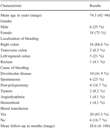
Table 1 Characteristic of the 24 patients with LGIB treated by embolization
Characteristic Results
Mean age in years (range) 74.3 (42–94)
Gender Male 6 (25 %) Female 18 (75 %) Localization of bleeding Right colon 16 (66.6 %) Transverse colon 2 (8.3 %) Left/sigmoid colon 5 (21 %) Rectum 1 (4.1 %) Cause of bleeding Diverticular disease 10 (41.9 %) Spontaneous 6 (25 %) Post-polypectomy 4 (16.7 %) Tumors 2 (8.2 %) Angiodysplasia 1 (4.1 %) Hemorrhoid 1 (4.1 %) Blood transfusion Yes 20 (83.3 %) No 4 (16.7 %)
Discussion
The first description of angiographic diagnosis and treat-ment of gastrointestinal hemorrhage was published in 1974 [15]. The results of the initial attempts were associated with high recurrence rates and complications [15,16].
Significant advances in the embolization technique and devices (super-selective, microcatheters) and increased tech-nical expertise have improved the adoption of this procedure for the treatment of lower gastrointestinal hemorrhage. Nu-merous studies reported that using multiphasic contrast-enhanced CT in the diagnosis and localization of acute LGIB could quickly determine who may benefit from super-selective embolization [17–21]. In the meantime, flex-ible endoscopy is still considered the mainstay for the eval-uation of LGIB [9]. Usually, it is recommended as the first diagnostic step in a stable patient, with the possibility to locally treat bleeding. Additionally, identifying the source of bleeding has been demonstrated to be very useful to show the radiologist the target artery before the angiography. Recent reports have demonstrated the safety and efficacy of this procedure [22–24].
Different methods exist to treat an ongoing bleeding discovered during an arteriography: microcoils, Gelfoam, and particles [9]. The success rate is usually higher than 70 % [9]. The short-term results in our study are very close to other experiences reported in the literature. For example, Lipof et al. [13] reported immediate hemostasis in 97 % of the patients, with short-term rebleeding in 16 % of patients and acute ischemia in 7 % of patients. Table3resumes the short-term results reported in the literature.
Concerning the risk factors, advanced age, intestinal ische-mia, and comorbidity are the strongest predictors of mortality. Conversely, the presence of colorectal polyps or hemorrhoids is associated with a lower risk of mortality [24].
In fact, LGIB is common in elderly patients, as confirmed by our series in which the median age was 80 years old. Frequently, these patients present several associated comor-bidities, such as coronary artery disease and peripheral vascular disease, which decrease their collateral blood flow. The main problem in these patients is that the peripheral colonic vessels are necessary for the blood supply and embolization may cause ischemia [24].
Our series demonstrated an important immediate clinical efficacy, given that 95.8 % of bleeding was stopped after embolization. Yet, the short-term outcomes showed 21 % of post-embolization ischemia (five patients) that is relatively high. Four patients were surgically treated with success, and one died. These results compare favorably to the outcomes after emergent colectomy in general (mortality, 14 %; mor-bidity, 36 %) [25] and specifically for LGIB with a mortality rate that can be as high as 27 % [8].
As we have seen in our study, the level of embolization is one of the main risk factors for post-embolization ischemia, as well as the length of hypoperfused bowel at the end of the embolization procedure. These factors should be considered for prompt surgical intervention in those high-risk patients.
Table 2 Short-term and long-term outcomes
Parameters Results
Short-term outcomes
Clinical success 23 (95.8 %)
Failure with urgent surgery 1 (4.1 %)
Hematoma at puncture site 1 (4.1 %)
Urgent surgery for post-embolization ischemia 4 (16.7 %)
Right colectomy 2 (8.2 %) Left colectomy 1 (4.1 %) Hartmann procedure 1 (4.1 %) Mortality 1 (4.1 %) Long-term outcomes Colonic ischemia 0
Late surgeryafor recurrence of bleeding 1 (4.1 %)
a
After 84 months
Table 3 Short-term outcomes in the literature
Authors Results
Bandi et al. [11] 35 patients
Immediate hemostasis 94 %
Ischemia rate 24 %
Rebleeding rate 34 %
Burgess et al. [21] 15 patients
Immediate hemostasis 93 %
Ischemia rate 60 %
Rebleeding rate 53 %
Silver et al. [23] 11 patients
Immediate hemostasis 91 %
Ischemia rate 63 %
Lipof et al. [13] 71 patients
Immediate hemostasis 97 %
Ischemia rate 7 %
Rebleeding rate 16 %
Tan et al. [24] 32 patients
Immediate hemostasis 97 %
Ischemia rate 1 %
Gillespie et al. [27] 38 patients
Immediate hemostasis 100 %
Ischemia rate 8 %
Rebleeding rate 24 %
Koganemaru et al. [28] 4 patients
Immediate hemostasis 100 %
Ischemia rate 0 %
Finally, ischemia remains the main problem, and the risk decreased with better embolization technique. Re-cently, Tan et al. [26] described only one patient (3 %) suffering from ischemic complications that required im-mediate surgical intervention. However, the procedure resulted in an anastomotic leak, similar to what we report here. The leak rate in their series was 22 %, with two of nine patients that required an operation. The rebleeding rate in this study was 22 % [26].
The risk of ischemia has been decreased with the intro-duction of new materials and techniques, but this risk still remains and could require immediate surgical intervention with high risk of an anastomotic leak, as was the case in one patient in our series. Perhaps, it would be wiser not to perform primary anastomosis in patients that present an ischemia after an embolization because of the possi-bility of compromising bowel blood supply. However, there are several techniques to check the vascularization of an anastomosis (Doppler, fluorescein, or indocyanine green), and these tools could help to decide whether to perform anastomosis.
While not new, the data reported herein show the analysis of long-term follow-up with only one patient presenting a rebleeding after 84 months. It also demonstrates the efficacy of this procedure as a definitive treatment for LGIB. In the literature, Lipof et al. [13] reported 15 % of recurrences of hemorrhage (8 patients out of 71) with a mean follow-up of 32 months. Tan et al. [25] reported 13 % of rebleeding at 30 days or more after the first episode (4 of 32 patients). These data finally confirm the safety of the procedure, even after long-term follow-up.
However, some limitations of this study deserve to be mentioned. First, the decision to perform an embolization or a surgical exploration remains at least debatable. Today, clear indications for a transarterial embolization are still under evaluation in most centers. In our series, we have evaluated retrospectively this approach for poor surgical candidates in whom the localization of the bleeding source was not always done preoperatively by angio-CT. We con-sider that a good risk surgical patient, in whom the source of bleeding was localized, should undergo a surgical explora-tion. On the other hand, a blind segmental resection carries a high mortality rate and is associated with significantly higher rebleeding rates [8]. This type of approach should be reserved for critically unstable patients in whom all the diagnostic methods have failed to localize the bleeding source.
Secondly, this is a retrospective series, and thus, by the nature of the study, the selection criteria were not strict and could render the interpretation of the results more difficult. A large prospective study with a defined algorithm, as proposed by others [8], is required before drawing definitive conclusions.
Conclusion
In our series, selective transarterial embolization for colonic bleeding was highly effective during the short-term and long-term follow-up. However, the risk of ischemia is sig-nificant (21 %) and is related mainly on the level of embo-lization and the length of the resulting hypoperfused colon. This risk should be taken into consideration for emergent operation.
References
1. Zuccaro G Jr (1998) Management of the adult patient with acute lower gastrointestinal bleeding. American College of Gastroenter-ology. Practice Parameters Committee. Am J Gastroenterol
93:1202–1208
2. Barnert J, Messmann H (2009) Diagnosis and management of lower gastrointestinal bleeding. Nat Rev Gastroenterol Hepatol 6
(11):637–646
3. Peter DJ, Dougherty JM (1999) Evaluation of the patient with gastrointestinal bleeding: an evidence based approach. Emerg
Med Clin North Am 17:239–261
4. Jenson DM, Machicado GA, Jutabha R et al (2000) Urgent colo-noscopy for the diagnosis and treatment of severe diverticular
hemorrhage. N Engl J Med 342:78–82
5. Angtuaco TL, Reddy SK, Drapkin S et al (2001) The utility of urgent colonoscopy in the evaluation of acute lower gastrointesti-nal tract bleeding: a 2-year experience from a single center. Am J Gastroenterol 96:1782–1785
6. Bloomfield RD, Rockey DC, Shetzline MA (2001) Endoscopic therapy of acute diverticular hemorrhage. Am J Gastroenterol 96:2367–2372
7. Eisen GM, Dominitz JA, Faigel D et al (2001) An annotated algorithmic approach to acute lower gastrointestinal bleeding.
Gastrointest Endosc 53:859–863
8. Schuetz A, Jauch KW (2001) Lower gastrointestinal bleeding: therapeutic strategies, surgical techniques and results.
Langen-becks Arch Surg 386(1):17–25
9. Luchtefeld MA, Senagore AJ, Szomstein M et al (2000) Evalua-tion of transarterial embolizaEvalua-tion for lower gastrointestinal
bleed-ing. Dis Colon Rectum 43:532–534
10. Nicholson AA, Ettles DF, Hartley JE et al (1998) Transcatheter coil embolotherapy: a safe and effective option for major colonic
haemorrhage. Gut 43:4–5
11. Bandi R, Shetty PC, Sharma RP, Burke TH, Burke MW, Kastan D (2001) Superselective arterial embolization for the treatment of
lower gastrointestinal hemorrhage. J Vasc Interv Radiol 12:1399–
1405
12. Peck DJ, McLoughlin RF, Hughson MN et al (1998) Percutaneous embolotherapy of lower gastrointestinal hemorrhage. J Vasc Interv Radiol 9:747–751
13. Lipof T, Sardella WV, Bartus CM, Johnson KH, Vignati PV, Cohen JL (2008) The efficacy and durability of super-selective emboliza-tion in the treatment of lower gastrointestinal bleeding. Dis Colon
Rectum 51(3):301–305
14. Drooz AT, Lewis CA, Allen TE, Citron SJ, Cole PE, Freeman NJ, Husted JW, Malloy PC, Martin LG, Van Moore A, Neithamer CD, Roberts AC, Sacks D, Sanchez O, Venbrux AC, Bakal CW (1997) Quality improvement guidelines for percutaneous transcatheter embolization. SCVIR Standards of Practice Committee. Society
of Cardiovascular & Interventional Radiology. J Vasc Interv
Radiol 8(5):889–895
15. Bookstein JJ, Chlosta EM, Foley D, Walter JF (1974) Transcath-eter hemostasis of gastrointestinal bleeding using modified autog-enous clot. Radiology 113:277–285
16. Funaki B, Kostelic JK, Lorenz J, Ha TV, Yip DL, Rosenblum JD, Leef JA, Straus C, Zaleski GX (2001) Superselective microcoil embolization of colonic hemorrhage. AJR Am J Roentgenol 177:829–836
17. Junquera F, Quiroga S, Saperas E et al (2000) Accuracy of helical computed tomographic angiography for the diagnosis of colonic
angiodysplasia. Gastroenterology 119:293–299
18. Rajan R, Dhar P, Praseedom RK, Sudhindran S, Moorthy S (2004) Role of contrast CT in acute lower gastrointestinal bleeding. Dig
Surg 21:293–296
19. Kuhle WG, Sheiman RG (2003) Detection of active colonic hem-orrhage with use of helical CT: findings in a swine model.
Radi-ology 228:743–752
20. Danzer D, Gervaz P, Platon A, Poletti PA (2003) Bleeding Meckel’s
diverticulum diagnosis: an unusual indication for computed
tomog-raphy. Abdom Imaging 28:631–633
21. Burgess AN, Evans PM (2004) Lower gastrointestinal haemor-rhage and superselective angiographic embolization. ANZ J Surg 74(8):635–638
22. Gordon RL, Ahl KL, Kerlan RK, Wilson MW, LaBerge JM, Sandhu JS, Ring EJ, Welton ML (1997) Selective arterial emboli-zation for the control of lower gastrointestinal bleeding. Am J Surg
174:24–28
23. Silver A, Bendick P, Wasvary H (2005) Safety and efficacy of superselective angioembolization in control of lower gastrointesti-nal hemorrhage. Am J Surg 189:361–363
24. Tan KK, Wong D, Sim R (2008) Superselective embolization for lower gastrointestinal hemorrhage: an institutional review over
7 years. World J Surg 32:2707–2715
25. Skala K, Gervaz P, Buchs N, Inan I, Secic M, Mugnier-Konrad B,
Morel P (2009) Risk factors for mortality–morbidity after
emergen-cy–urgent colorectal surgery. Int J Colorectal Dis 24(3):311–316
26. Tan KK, Nallathamby V, Wong D, Sim R (2010) Can superselective embolization be definitive for colonic diverticular hemorrhage? An
institution’s experience over 9 years. J Gastrointest Surg 14:112–118
27. Gillespie CJ, Sutherland AD, Mossop PJ, Woods RJ, Keck JO, Heriot AG (2010) Mesenteric embolization for lower
gastrointes-tinal bleeding. Dis Colon Rectum 53(9):1258–1264
28. Koganemaru M, Abe T, Iwamoto R, Kusumoto M, Suenaga M, Saga T, Hayabuchi N (2012) Ultraselective arterial embolization of vasa recta using 1.7-French microcatheter with small-sized detach-able coils in acute colonic hemorrhage after failed endoscopic treatment. AJR Am J Roentgenol 198(4):W370–W372