HAL Id: pasteur-03107143
https://hal-pasteur.archives-ouvertes.fr/pasteur-03107143
Preprint submitted on 12 Jan 2021
HAL is a multi-disciplinary open access
archive for the deposit and dissemination of sci-entific research documents, whether they are pub-lished or not. The documents may come from teaching and research institutions in France or abroad, or from public or private research centers.
L’archive ouverte pluridisciplinaire HAL, est destinée au dépôt et à la diffusion de documents scientifiques de niveau recherche, publiés ou non, émanant des établissements d’enseignement et de recherche français ou étrangers, des laboratoires publics ou privés.
Copyright
RAD50 promotes DNA repair by homologous
recombination and restrains antigenic variation in
African trypanosomes
Ann-Kathrin Mehnert, Marco Prorocic, Annick Dujeancourt-Henry, Sebastian
Hutchinson, Richard Mcculloch, Lucy Glover
To cite this version:
Ann-Kathrin Mehnert, Marco Prorocic, Annick Dujeancourt-Henry, Sebastian Hutchinson, Richard Mcculloch, et al.. RAD50 promotes DNA repair by homologous recombination and restrains antigenic variation in African trypanosomes. 2021. �pasteur-03107143�
RAD50 promotes DNA repair by homologous recombination and
1
restrains antigenic variation in African trypanosomes
2
3
Ann-Kathrin Mehnert1, Marco Prorocic2, Annick Dujeancourt-Henry1, Sebastian Hutchinson3, Richard
4
McCulloch2, Lucy Glover1*
5
6
1 Trypanosome Molecular Biology, Department of Parasites and Insect Vectors, Institut Pasteur, 25-28
7
Rue du Docteur Roux 75015, Paris, France.
8
9
2 Wellcome Center for Integrative Parasitology, Sir Graeme Davis Building, 120 University Place,
10
Glasgow, G12 8TA, UK.
11
12
3 Trypanosome Cell Biology, Department of Parasites and Insect Vectors, Institut Pasteur, 25-28 Rue
13
du Docteur Roux 75015, Paris, France.
14
15
Present address: : Centre for Infectious Diseases, Virology, Heidelberg University Hospital, Im
16
Neuenheimer Feld 344, 69120 Heidelberg, Germany
17
18
19
* To whom correspondence should be addressed lucy.glover@pasteur.fr; Tel: +33 140613425
20
21
KEYWORDS: VSG, RAD50, DNA damage.
22
Abstract 147 words23
24
ABSTRACT25
Homologous recombination dominates as the major form of DNA repair in Trypanosoma brucei, and is
26
especially important for recombination of the subtelomeric variant surface glycoprotein during
27
antigenic variation. RAD50, a component of the MRN complex (MRE11, RAD50, NBS1), is central to
28
homologous recombination through facilitating resection and governing the DNA damage response.
29
The function of RAD50 in trypanosomes is untested. Here we report that RAD50 is required for
30
RAD51-dependent homologous recombination, phosphorylation of histone H2A and controlled
31
resection following a DNA double strand break (DSB). Perhaps surprisingly, DSB resection in the
32
rad50 nulls was not impaired and appeared to peak earlier than in the parental strains. Finally, we
33
show that RAD50 suppresses DNA repair using donors with short stretches of homology at a
34
subtelomeric locus, with null strains producing a greater diversity of expressed VSG variants following
35
DSB repair. We conclude that RAD50 promotes stringent homologous recombination at subtelomeric
36
loci and restrains antigenic variation.
37
38
(which was not certified by peer review) is the author/funder. All rights reserved. No reuse allowed without permission.
The copyright holder for this preprint this version posted March 17, 2020.
;
https://doi.org/10.1101/2020.03.17.994905
doi: bioRxiv preprint
(which was not certified by peer review) is the author/funder. All rights reserved. No reuse allowed without permission.
The copyright holder for this preprint this version posted March 17, 2020.
;
https://doi.org/10.1101/2020.03.17.994905
doi: bioRxiv preprint
(which was not certified by peer review) is the author/funder. All rights reserved. No reuse allowed without permission.
The copyright holder for this preprint this version posted March 17, 2020.
;
https://doi.org/10.1101/2020.03.17.994905
doi: bioRxiv preprint
(which was not certified by peer review) is the author/funder. All rights reserved. No reuse allowed without permission.
The copyright holder for this preprint this version posted March 17, 2020.
;
https://doi.org/10.1101/2020.03.17.994905
doi: bioRxiv preprint
(which was not certified by peer review) is the author/funder. All rights reserved. No reuse allowed without permission.
The copyright holder for this preprint this version posted March 17, 2020.
;
https://doi.org/10.1101/2020.03.17.994905
doi: bioRxiv preprint
(which was not certified by peer review) is the author/funder. All rights reserved. No reuse allowed without permission.
The copyright holder for this preprint this version posted March 17, 2020.
;
https://doi.org/10.1101/2020.03.17.994905
doi: bioRxiv preprint
(which was not certified by peer review) is the author/funder. All rights reserved. No reuse allowed without permission.
The copyright holder for this preprint this version posted March 17, 2020.
;
https://doi.org/10.1101/2020.03.17.994905
doi: bioRxiv preprint
(which was not certified by peer review) is the author/funder. All rights reserved. No reuse allowed without permission.
The copyright holder for this preprint this version posted March 17, 2020.
;
https://doi.org/10.1101/2020.03.17.994905
doi: bioRxiv preprint
(which was not certified by peer review) is the author/funder. All rights reserved. No reuse allowed without permission.
The copyright holder for this preprint this version posted March 17, 2020.
;
https://doi.org/10.1101/2020.03.17.994905
doi: bioRxiv preprint
(which was not certified by peer review) is the author/funder. All rights reserved. No reuse allowed without permission.
The copyright holder for this preprint this version posted March 17, 2020.
;
https://doi.org/10.1101/2020.03.17.994905
doi: bioRxiv preprint
(which was not certified by peer review) is the author/funder. All rights reserved. No reuse allowed without permission.
The copyright holder for this preprint this version posted March 17, 2020.
;
https://doi.org/10.1101/2020.03.17.994905
doi: bioRxiv preprint
(which was not certified by peer review) is the author/funder. All rights reserved. No reuse allowed without permission.
The copyright holder for this preprint this version posted March 17, 2020.
;
https://doi.org/10.1101/2020.03.17.994905
doi: bioRxiv preprint
(which was not certified by peer review) is the author/funder. All rights reserved. No reuse allowed without permission.
The copyright holder for this preprint this version posted March 17, 2020.
;
https://doi.org/10.1101/2020.03.17.994905
doi: bioRxiv preprint
(which was not certified by peer review) is the author/funder. All rights reserved. No reuse allowed without permission.
The copyright holder for this preprint this version posted March 17, 2020.
;
https://doi.org/10.1101/2020.03.17.994905
doi: bioRxiv preprint
(which was not certified by peer review) is the author/funder. All rights reserved. No reuse allowed without permission.
The copyright holder for this preprint this version posted March 17, 2020.
;
https://doi.org/10.1101/2020.03.17.994905
doi: bioRxiv preprint
(which was not certified by peer review) is the author/funder. All rights reserved. No reuse allowed without permission.
The copyright holder for this preprint this version posted March 17, 2020.
;
https://doi.org/10.1101/2020.03.17.994905
doi: bioRxiv preprint
(which was not certified by peer review) is the author/funder. All rights reserved. No reuse allowed without permission.
The copyright holder for this preprint this version posted March 17, 2020.
;
https://doi.org/10.1101/2020.03.17.994905
doi: bioRxiv preprint
(which was not certified by peer review) is the author/funder. All rights reserved. No reuse allowed without permission.
The copyright holder for this preprint this version posted March 17, 2020.
;
https://doi.org/10.1101/2020.03.17.994905
doi: bioRxiv preprint
(which was not certified by peer review) is the author/funder. All rights reserved. No reuse allowed without permission.
The copyright holder for this preprint this version posted March 17, 2020.
;
https://doi.org/10.1101/2020.03.17.994905
doi: bioRxiv preprint
(which was not certified by peer review) is the author/funder. All rights reserved. No reuse allowed without permission.
The copyright holder for this preprint this version posted March 17, 2020.
;
https://doi.org/10.1101/2020.03.17.994905
doi: bioRxiv preprint
(which was not certified by peer review) is the author/funder. All rights reserved. No reuse allowed without permission.
The copyright holder for this preprint this version posted March 17, 2020.
;
https://doi.org/10.1101/2020.03.17.994905
doi: bioRxiv preprint
(which was not certified by peer review) is the author/funder. All rights reserved. No reuse allowed without permission.
The copyright holder for this preprint this version posted March 17, 2020.
;
https://doi.org/10.1101/2020.03.17.994905
doi: bioRxiv preprint
(which was not certified by peer review) is the author/funder. All rights reserved. No reuse allowed without permission.
The copyright holder for this preprint this version posted March 17, 2020.
;
https://doi.org/10.1101/2020.03.17.994905
doi: bioRxiv preprint
(which was not certified by peer review) is the author/funder. All rights reserved. No reuse allowed without permission.
The copyright holder for this preprint this version posted March 17, 2020.
;
https://doi.org/10.1101/2020.03.17.994905
doi: bioRxiv preprint
(which was not certified by peer review) is the author/funder. All rights reserved. No reuse allowed without permission.
The copyright holder for this preprint this version posted March 17, 2020.
;
https://doi.org/10.1101/2020.03.17.994905
doi: bioRxiv preprint
(which was not certified by peer review) is the author/funder. All rights reserved. No reuse allowed without permission.
The copyright holder for this preprint this version posted March 17, 2020.
;
https://doi.org/10.1101/2020.03.17.994905
doi: bioRxiv preprint
(which was not certified by peer review) is the author/funder. All rights reserved. No reuse allowed without permission.
The copyright holder for this preprint this version posted March 17, 2020.
;
https://doi.org/10.1101/2020.03.17.994905
doi: bioRxiv preprint
(which was not certified by peer review) is the author/funder. All rights reserved. No reuse allowed without permission.
The copyright holder for this preprint this version posted March 17, 2020.
;
https://doi.org/10.1101/2020.03.17.994905
doi: bioRxiv preprint
(which was not certified by peer review) is the author/funder. All rights reserved. No reuse allowed without permission.
The copyright holder for this preprint this version posted March 17, 2020.
;
https://doi.org/10.1101/2020.03.17.994905
doi: bioRxiv preprint
(which was not certified by peer review) is the author/funder. All rights reserved. No reuse allowed without permission.
The copyright holder for this preprint this version posted March 17, 2020.
;
https://doi.org/10.1101/2020.03.17.994905
doi: bioRxiv preprint
INTRODUCTION
39
40
Trypanosoma brucei (T. brucei) is a protozoan parasite and the causative agent of human African
41
trypanosomiasis, or sleeping sickness, and nagana in cattle. Trypanosomes cycle between their insect
42
vector, the tsetse fly, and mammalian hosts, where they colonise the blood, fat 1 and skin 2 and
43
eventually cross the blood brain barrier in late stage infection. If left untreated, trypanosomiasis is
44
normally fatal3. In the mammalian host, each trypanosome cell is covered in a dense layer of a single
45
species of variant surface glycoprotein (VSG). The highly immunogenic VSG layer4,5 acts as an
46
barrier, concealing other surface components from the host immune response 6. Trypanosomes
47
maintain a persistent infection by continuously escaping the host’s immune response though antigenic
48
variation7. Central to this survival strategy is monoallelic expression of the VSG from a subtelomeric
49
locus, known as an expression site (VSG-ES), and stochastic VSG switching. The ~ 15 VSG-ESs in
50
the trypanosome genome share a high degree of sequence and structure conservation 8, each being
51
an RNA polymerase-I (RNA Pol-I) polycistronic transcription unit with a single VSG gene found
52
adjacent to the telomere, up to 60 kb downstream of the promoter 8. The VSG gene is flanked by two
53
sets of repetitive sequence: downstream is the telomere, and upstream is a block of repetitive
54
sequence, known as the 70-bp repeats, which modulates VSG switching 8,9. Characteristic of a
55
trypanosome infection are recrudescent waves of parasitemia, each of which is composed of a diverse
56
VSG expressing population, with between 7 – 79 VSGs detected in each peak of parasitemia 10-12.
57
VSG diversity arises through altering the single VSG-ES that is transcribed or, more commonly, by
58
recombination of silent VSGs into the active VSG-ES. The seemingly unrestricted use of VSG genes
59
might be expected to result in a rapid exhaustion of the VSG gene repertoire. However, the parasite’s
60
ability to sustain an infection appears to lie in an enormous repertoire of >2000 VSG genes and
61
pseudogenes13-15, mainly found in subtelomeric VSG arrays, and a capacity for generation of novel
62
‘mosaic’ VSG genes through segmental gene conversion of multiple (pseudo) VSGs, in particular late
63
in infection 10,11,14. Importantly, almost all of the array VSGs are associated with upstream tracts of
70-64
bp repeats, providing the necessary substrate needed for homologous recombination mediated
65
antigenic variation 16.
66
67
A DNA double-strand break (DSB) is an extremely toxic lesion in any cell, which if left unrepaired can
68
lead to cell death. In T. brucei RAD51-dependent homologous recombination (HR) dominates as the
69
major DNA repair and recombination pathway, with microhomology mediated end-joining (MMEJ)
70
playing a minor role 17-20. HR is important for VSG switching, and though it is not clear how MMEJ acts
71
in this reaction, repair of induced DSBs can occur by coupled HR and MMEJ, and MMEJ is more
72
frequently used for repair of DSBs induced within the VSG-ES21. Unrepaired DSBs appear to persist
73
throughout the cell cycle without inhibiting the trypanosomes ability to replicate their DNA22, but
74
whether HR or MMEJ are regulated is unknown. In addition, non-homologous end-joining (NHEJ)
75
appears to be absent in trypanosomes 21,23,24. These features of trypanosome DSB repair contrast with
76
mammalian cells, where NHEJ is highly active, HR predominates in S and G2 phase cells and MMEJ
77
is considered a minor reaction 25. In trypanosomes both transcriptionally active and silent
78
(which was not certified by peer review) is the author/funder. All rights reserved. No reuse allowed without permission.
The copyright holder for this preprint this version posted March 17, 2020.
;
https://doi.org/10.1101/2020.03.17.994905
doi: bioRxiv preprint
subtelomeres are fragile 26,27, and accumulate natural breaks. Within the active VSG-ES specifically, a
79
DSB between the VSG and 70-bp repeats acts as a potent driver of antigenic variation and
80
precipitates VSG switching 27. Several DNA repair and recombination proteins have been shown to be
81
important for antigenic variation in trypanosomes, thus linking VSG switching with this process: loss of
82
RAD51 18, the RAD51-3 paralogue 28, or the RAD51-interacting protein BRCA2 29,30 results in
83
impaired VSG switching, while loss of RECQ2 31, TOPO3α or RMI1 increases VSG switching 32,33 , as
84
does loss of the histone variants H3.V and H4.V 15. Loss of ATR, which is involved in DNA damage
85
signalling, impairs monoallelic VSG expression and increases VSG switching through localized DNA
86
damage 34. Histone Acetyltransferase (HAT3) is required for recombination repair of a
chromosome-87
internal DSB, but suppresses DSB repair within the VSG-ES which suggests repair is
88
compartmentalised in trypanosomes 35.
89
90
The DNA damage response (DDR) is an orchestrated cellular response to many different genome
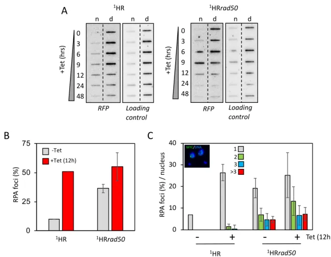
91
lesions, including DSBs, which most commonly form via stalled replication forks 36. Critical to DSB
92
repair is the MRE11 – RAD50 – NBS1 (MRN) complex (in yeast MRE11 – RAD50 – XRS1, MRX),
93
which acts as a DNA damage sensing complex and is responsible for recognizing the free DNA ends,
94
where it is one of the first complexes to bind and initiate HR 37,38. MRE11 – RAD50 forms the core of
95
this complex and is conserved across all domains of life, whereas NBS1 only forms part of the
96
complex in eukaryotes 37. MRN consists of two molecules of each component protein, and diffuses
97
along homoduplex DNA searching for free DNA ends – a process that is driven by RAD50 39. The
98
MRE11 subunit is a nuclease with both 5′ flap endonuclease activity and 3′→5′ exonuclease activity
99
and catalyses resection through cleaving the 5’ strand, internal to the DSB, which is then resected
100
using its exonuclease function to generate the short 3’ single-strand (ss) DNA overhangs 40. These
101
overhangs are further resected byExonuclease 1 (EXO1), forming long tracts of 3 ssDNA on either
102
side of the DSB 39. NBS1, the eukaryote specific component, is responsible for binding multiple
103
phosphorylated proteins and recruiting MRE11 and RAD50 to DSB sites 41 through its interaction
104
with MRE11, CtIP, which is also required for initiating resection, and the ATM kinase 42. End
105
recognition and DSB processing by MRN is an ATP dependent process: here, ATP binding to
106
RAD50 acts to switch the complex from an open to a closed conformation 43, which facilitates DSB
107
recognition, tethering and ATM activation 43. In yeast the MRX complex also acts in telomere
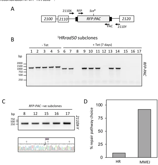
108
maintenance by binding the end of short telomeres and recruiting TEL1, which then recruits
109
telomerase to extend the telomere 44. Conversely, in mammalian cells, MRN regulates an ATM
110
dependent response at dysfunctional telomeres 45.
111
112
RAD50 (Tb.927.11.8210), MRE11 (Tb927.2.4390) 46,47 and NBS1 (Tb 927.8.5710) homologues are
113
present in the trypanosome genome and previous studies have shown that MRE11 is required for HR
114
but its inactivation did not lead to telomere shortening or changes in VSG switching 46,47, despite the
115
dominance of HR in repair in trypanosomes and requirement for the reaction in antigenic variation.
116
What roles these proteins play in the trypanosome DDR is largely unexplored. In addition, though we
117
know that DSBs accumulate at the subtelomeres 26,27, it is unclear how they are sensed or how they
118
(which was not certified by peer review) is the author/funder. All rights reserved. No reuse allowed without permission.
The copyright holder for this preprint this version posted March 17, 2020.
;
https://doi.org/10.1101/2020.03.17.994905
doi: bioRxiv preprint
contribute to antigenic variation. Given the central, early role of the MRN complex in DSB recognition
119
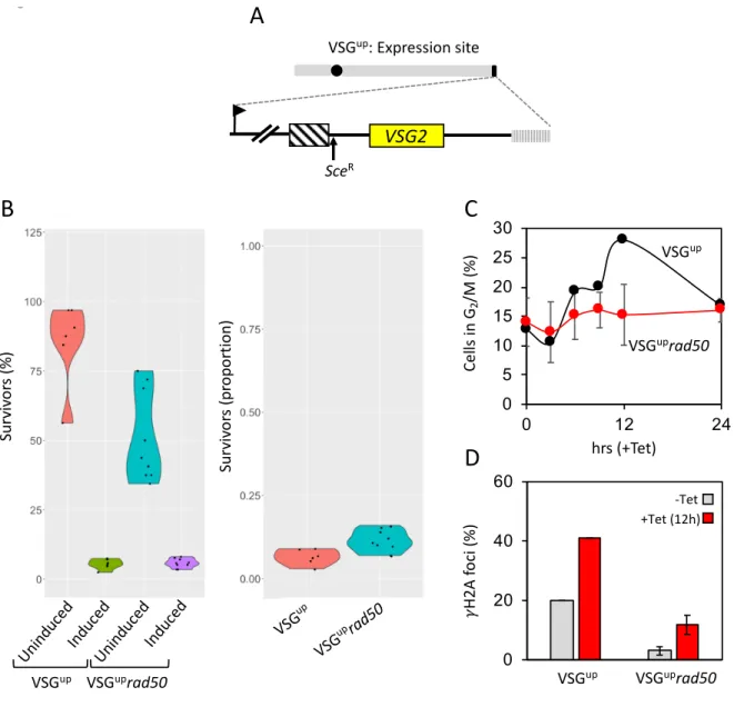
and in telomere maintenance we set out to characterise its role in HR and VSG switching in
120
trypanosomes. We found that RAD50, like MRE11, is required for efficient HR, and in its absence
121
MMEJ dominated as the major form of repair. RAD50 also plays a perhaps surprising role in VSG
122
switching, where it restricts HR substrate selection in the VSG repertoire and so may act to preserve
123
the VSG archive during long-term infections.
124
125
MATERIALS AND METHODS
126
Trypanosoma brucei growth and manipulation. Lister 427, MITat1.2 (clone 221a), bloodstream stage
127
cells were cultured in HMI-11 medium 79 at 37.4 °C with 5 % CO2. Cell density was determined using a
128
haemocytometer. For transformation, 2.5 x 10⁷ cells were spun for 10 minutes at 1000g at room
129
temperature and the supernatant discarded. The cell pellet was resuspend in prewarmed cytomix
130
solution 80 with 10 µg linearised DNA and place in a 0.2 cm gap cuvette, and nucleofected (Lonza)
131
using the X-001 program. The transfected cells were placed into one 25 cm2 culture flask per
132
transfection with 36 ml warmed HMI-11 medium only and place in an incubator to allow the cells to
133
recover for approximately 6 hours. After 6 hours, the media distributed into 48-well plates with the
134
appropriate drug selection. Strains expressing TetR and I-SceI with I-SceI recognition-sites at a
135
chromosome-internal locus 17 and an active VSG-ESs 27 have been described previously. G418, and
136
blasticidin were selected at 10 µg.ml-1 and 2 µg.ml-1 respectively. Puromycin, phleomycin, G418,
137
hygromycin and blasticidin and tetracycline were maintained at 1 µg.ml-1. Clonogenic assay were
138
plated out at either 32 cells per plate under both inducing and non-inducting conditions for 1HR and
139
VSGup strains and 480 cells per plate for VSGup strains under inducing conditions. Plates were
140
counted 5-6 days later and subclones selected for further analysis.
141
To generate the RAD50 nulls in the 1HR strain we employed multi-step transfection strategy 81
142
that recycled a Neomycin phosphotransferase gene (NEO) in order to rescue one marker (here
143
Blasticidin - BLA). Briefly, an I-SceI recognition sites was inserted into the pRAD50-BLA knock-out
144
cassette between the 5’UTR and BLA ORF (Figure 1C) in the 2T1 cell line 82 with a tetracycline
145
inducible Sce ORF. Induction of Sce induces a break in the BLA cassette and subsequent repair,
146
using homology in the NEO modified allele, replaces BLA with NEO.
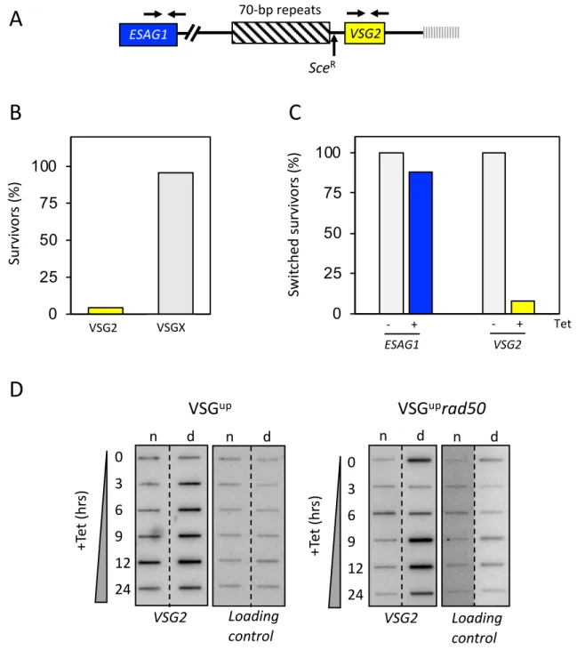
147
Clonogenic assays were plated out at either 32 cells per plate under both inducing and
non-148
inducing conditions for 1HR and VSGup strains and 480 cells per plate for VSGup strains under inducing
149
conditions. Plates were counted 5-6 days later and subclones selected for further analysis.
150
151
Plasmid construction. For native C-terminal epitope tagging of Tb927.5.1700 / RPA2 a 765-bp
152
fragment was amplified using primers RPA28F:GATCAAGCTTATGGAAGGAAGTGGAAGTAA; and
153
RPA28R:GATCTCTAGAAATGCCAAACTTACAATCATG and cloned in pNATxTAG83 using the HindIII
154
and XbaI sites (underlined). The construct was linearized with XhoI prior to transfection. MRE11F5
155
(GATCgcggccgcATGGCCGAGAGGGCATC), MRE11R5 (GATCtctagaCAACGAAGATGTATGCCC),
156
MRE11F3 (GATCgggcccCGATGGATAGTGGTAAT) and
157
(which was not certified by peer review) is the author/funder. All rights reserved. No reuse allowed without permission.
The copyright holder for this preprint this version posted March 17, 2020.
;
https://doi.org/10.1101/2020.03.17.994905
doi: bioRxiv preprint
MRE11R3 (GATCggtaccCTAATAGTTATCTGGCA) were used to clone in target regions to generate
158
pMRE11KOBLA and pMRE11KONEO. For transfection, 20 μg pMRE11KO Blasticidin (BLA) and
159
Neomycin (NEO) plasmids were sequentially digested with Acc65I and NotI and cleaned by phenol-
160
chloroform extraction and ethanol precipitation after each digestion. Strains were validated using
161
MRE11F5 and MRE11R3 in a PCR assay. Heterozygous (+/-) and homozygous (-/-) knockout mutants
162
of RAD50 were generated by deleting most of replacing most of the gene’s open reading frame with
163
either BLA and NEO. The strategy used is as described in 84; briefly, two modified versions of the
164
plasmid pmtl23 were used to allow PCR-amplified 5’ and 3’ flanking untranslated regions of RAD50 to
165
be inserted around BLA and NEO cassettes (where the antibiotic resistance genes’ ORF were flanked
166
by tubulin and actin intergenic regions). The selective drug markers, flanked by RAD50 5’ and 3’
167
untranslated regions, were then excised using NotI and transfected into T. brucei, and clones selected
168
using 10 μg.ml-1 blasticidin or 5 μg.mL-1 G418.
169
170
Immunofluorescence microscopy. Immunofluorescence analysis was carried out using standard
171
protocols as described previously 85. Mouse a-Myc was used at 1:400 and rabbit a-gH2A 55 was used
172
at 1:250. Fluorescein-conjugated goat a-rabbit and goat a-mouse secondary antibodies (Pierce) were
173
used at 1:2000. Samples weremounted in Vectashield (Vector Laboratories) containing 4,
6-174
diamidino-2-phenylindole(DAPI). In T. brucei, DAPI-stained nuclear and mitochondrial DNA can be
175
used as cytological markers for cell cycle stage 86; one nucleus and one kinetoplast (1N:1K) indicate
176
G1, one nucleus and an elongated kinetoplast (1N:eK) indicate S phase, one nucleus and two
177
kinetoplasts (1N:2K) indicate G2/M and two nuclei and two kinetoplasts (2N:2K) indicate post-mitosis.
178
Images were captured using a ZEISS Imager 72 epifluorescence microscope with an Axiocam 506
179
mono camera and images were processed and in ImageJ.
180
181
DNA analysis. Slot blots for detection of ssDNA were carried as described previously 17. ImageJ was
182
used to generate linear density plots. The VSG probe was a 750 bp fragment VSG2 fragment from a
183
Pst1 digest of pNEG. The RFP probe was a 687-bp HindIII/NotI fragment encompassing the full ORF.
184
Loading control was a 226 – bp product from Tb427.01.570 (Dot1bKOF:
185
TGGTCGGAAGTTGGATGTGA Dot1bKOR: CTTCCATGCATAACACGCGA).
186
187
PCR analysis of RAD50 nulls to confirms knock-out were done using standard PCR conditions with
188
the following primers; a 402 bp product for RAD50 using RAD50KOF
189
(CGTGAGAAACAGGAACAGCA) and RAD50KOR (AACACGTTTTTCCAACTCGG); a 399 bp product
190
for Blasticidin ORF using BlaF (GATCGAATTCATGGCCAAGCCTTTGTCT) and BlaR
191
(GATCCCATGGTTAGCCCTCCCACACATAA); and a 795 bp product for Neomycin
192
Phosphotransferase ORF using NPTF (ATGATTGAACAAGATGGATTG) and NPTR
193
(TCAGAAGAACTCGTCAAGAA). Analysis of subclones was previously described21,27,35 and used the
194
following primers VSG221F (CTTCCAATCAGGAGGC), VSG221R (CGGCGACAACTGCAG), RFP
195
(ATGGTGCGCTCCTCCAAGAAC), PAC (TCAGGCACCGGGCTTGC), ESAG1F
196
(which was not certified by peer review) is the author/funder. All rights reserved. No reuse allowed without permission.
The copyright holder for this preprint this version posted March 17, 2020.
;
https://doi.org/10.1101/2020.03.17.994905
doi: bioRxiv preprint
(AATGGAAGAGCAAACTGATAGGTTGG), ESAG1R (GGCGGCCACTCCATTGTCTG), 2110X
197
(GGGGTGAATGTTGGCTGTG), 2110Y (GGGATTCCCAGACCAATGA)
198
199
VSG sequencing analysis. For the RT-PCR, the reaction mix were as following; 1 µg of cDNA, 1 x
200
PCR buffer, 0.2 mM dNTPs, 1 µl each of SL (ACAGTTTCTGTACTATATTG) and SP6-14mer
201
(GATTTAGGTGACACTATAGTGTTAAAATATATC) primers, H2O to 50 µl and 0.5 µl Phusion
202
polymerase (New England Biolabs). For the PCR conditions. Five cycles were carried out at 94 °C for
203
30s, 50 °C for 30s and 72 °C for 2 min; followed by 18 cycles at 94 °C for 30s, 55 °C for 30s and 72 °C
204
for 2 min. DNA concentration was measured using a Nanodrop. Libraries were prepared from VSG
205
PCR products and sequenced on a BGI-Seq (BGI500) with a 150 bp paired-end read length with BGI
206
Genomics Hong Kong.
207
Replicate libraries for WT uninduced, WT induced, VSGUP uninduced and VSGUP induced ,
208
VSGUPrad50 uninduced and VSGUPrad50 induced were sequenced on the BGIseq500 platform
209
producing 8.03, 9.01, 7.60, 7.22, 6.66, 7.07, 6.90, 6.98 million reads per library, respectively. Reads
210
were aligned to the T. brucei Lister 427 genome 15with the cohort of minichromosomal VSGs added
211
from the Lister 427 VSGnome 13 using bowtie2 87 with the parameters --very-sensitive and BAM files
212
created with samtools 88, aligning (WT uninduced) 97.76, 98.42, (WT induced) 97.69, (VSGUPrad50
213
uninduced) 97.57, 98.60, (VSGUPrad50 induced) 97.63, 97.80 percent of reads successfully. Reads
214
counts per transcript were obtained using featureCounts89. Differential expression analysis was
215
performed using EdgeR90on all genes, followed by filtering for VSG genes (1848 VSG sequences in
216
total). An R script (https://github.com/LGloverTMB/DNA-repair-mutant-VSG-seq ) was used to perform
217
differential expression analysis, and generate Volcano and genome scale plots. BLAST analysis was
218
performed locally using a database containing significantly up-regulated VSG genes from both
219
conditions, including 2 kb of sequence upstream and downstream of the start and stop codons,
220
respectively (except where sequences in the contigs 5’ or 3’ to the CDS were shorter than 2 kb
221
excluding). For this analysis, minichromosomal VSG genes were excluded as the VSGnome does not
222
contain any sequence beyond the CDS. The resulting database of 131 VSGs was queried using the
223
VSG2 sequence including 2 kb of sequence upstream of the CDS and all sequence between the stop
224
codon and end of contig (1,272 nt). The BLASTn algorithm was used query the database using default
225
parameters except allowing up-to 20 hits per subject sequence, and outputting up to 2,000 alignments.
226
Alignments were filtered to remove overlapping hits from the same subject sequence using Microsoft
227
Excel, retaining the one with the higher alignment score. Non-overlapping alignments were plotted
228
using a custom R script (https://github.com/LGloverTMB/DNA-repair-mutant-VSG-seq). Lengths of
229
average alignments were calculated for cohorts of VSGs up-regulated in both VSGup and VSGuprad50
230
or VSGuprad50 only.
231
232
(which was not certified by peer review) is the author/funder. All rights reserved. No reuse allowed without permission.
The copyright holder for this preprint this version posted March 17, 2020.
;
https://doi.org/10.1101/2020.03.17.994905
doi: bioRxiv preprint
RESULTS
233
234
RAD50 is required for normal cell growth and DSB repair.
235
236
RAD50, the largest component of the MRN complex, belongs to the structural maintenance of
237
chromosomes (SMC) family of proteins 48 and has not been examined in T. brucei, though the gene
238
has been reported to be essential in Leishmania infantum 49. The domain architecture of RAD50 is
239
approximately palindromic (Figure 1A) and characterized by the presence of ATP-binding cassette
240
(ABC)-ATPase domains at the N- and C- termini, each followed by an MRE11 binding site (MBS), and
241
then by anti-parallel coiled-coil regions, which form linker structures that enable the MRN complex to
242
act as a tethering scaffold to hold broken chromosomes together for repair 50. Between the antiparallel
243
coiled-coils, a central Zn hook, a CxxC motif, facilitates Zn2+ dependent RAD50-RAD50 subunit
244
interactions and is presumed to be important for tethering 51. A conformational change is invoked
245
through binding of RAD50 to two ATP molecules, which then allows for binding to DNA43. Primary
246
sequence comparison suggested all RAD50 domains are recognisably conserved in the putative T.
247
brucei RAD50 homologue (Tb.927.11.8210; Supplementary Figure 1). Within the ATPase domains,
248
the ABC nucleotide binding domain is defined by the conserved presence of Walker A, Q-loop,
249
Signature, Walker B, D-loop, and H-loop motifs required to form the active ATPase site 52.
250
Furthermore, structure prediction using Phyre253 modelled 503 residues (37 % of the sequence) of the
251
T. brucei protein, revealing a SMC head domain and antiparallel coiled coil regions (Figure 1A).
252
To test the function of RAD50 in DSB repair, we used a previously validated T. brucei cell line,
253
referred to as 1HR (Figure 1B), where a single I-SceI meganuclease DSB can be induced in an
RFP-254
PAC (red fluorescent protein – puromycin N-acetyltransferase) fusion cassette in the core region on
255
chromosome 11 17 (Figure 1B). We generated rad50 null mutants (referred to as 1HRrad50) in these
256
cells by sequentially replacing the two gene alleles with neomycin phosphotransferase (NEO) and
257
blasticidin (BLA) resistance cassettes: PCR analysis of double antibiotic resistant clones confirmed
258
RAD50 loss and replacement (Supplementary Figure 2A and B) and demonstrates RAD50 is not
259
essential in T. brucei. To determine the role RAD50 plays in DNA repair, we set up clonogenic assays.
260
Cells were distributed across 96-wells plates under both I-SceI non-inducing and inducing conditions,
261
and wells with live cells scored after 5 -7 days. This revealed a significant growth defect in the
262
1HRrad50 null cells in unperturbed cells (Figure 1C Left panel and Table 1): 95 % of the WT 1HR cells
263
survived compared with ~ 35 % of the 1HRrad50 cells, revealing a 2.6 - fold decrease in cell survival.
264
This growth impairment is likely due to the inability to repair spontaneous DSBs. Induction of the I-SceI
265
meganuclease results in ~ 95 % cutting and repair mainly by homologous recombination 17. Consistent
266
with previous findings, in the WT 1HR strain ~ 48 % of cells are able to repair the DSB and survive
267
(Figure 1C Left panel and 17 and Table 1), whereas in the 1HRrad50 cells, a severe growth defect was
268
seen following a DSB, with less than 3 % survival (a 16 – fold reduction), suggesting a significant
269
defect in DSB repair (Figure 1C Left panel and Table 1). This was recapitulated when assessing the
270
normalised survival efficiency (compared to uninduced survival) following an I-SceI break (Figure 1C
271
(which was not certified by peer review) is the author/funder. All rights reserved. No reuse allowed without permission.
The copyright holder for this preprint this version posted March 17, 2020.
;
https://doi.org/10.1101/2020.03.17.994905
doi: bioRxiv preprint
right panel and Table 1), indicating that a DSB is more lethal in the null mutant cells. In parallel we
272
also tested the function of MRE11, as it forms a complex with RAD50, by generating null mutants
273
through sequentially replacing the two gene alleles with NEO and BLA resistance cassettes; PCR
274
analysis confirmed MRE11 loss and replacement (Supplementary Figure 2C). The mre11 nulls
275
(referred to as 1HRmre11) also showed a growth defect cells in the unperturbed cells, with only 47 %
276
of cells surviving cloning. Induction of the I-SceI meganuclease resulted in a severe growth defect,
277
with less than 2 % survival, suggesting a significant defect in DSB repair (Supplementary Figure 4A
278
and Table 1) whose magnitude was very similar to 1HRrad50 cells, which is expected given they have
279
been shown to act in complex in other systems37,38.
280
281
282
283
Figure 1: RAD50 is essential for DSB response and repair at a chromosome-internal locus. (A) Upper
284
panel: Schematic of TbRAD50 with protein domains. Amino acid position of conserved domains are:
285
ATPase - N, 4 – 170; MRE11 binding site (MBS), 182 – 205; Zn hook, 690 – 693; MRE11 binding site,
286
1158 – 1181; ATPase - C, 1243 – 1333. Lower panel: The structure of the T. brucei RAD50 was
287
modelled using Phyre2 showing the SMC head domain with a coiled coil. (B) Schematic of the
288
chromosome-internal DSB cell line with the I-SceI recognition site, SceR, highlighted. (C) A clonogenic
289
assay reveals survivors following a DSB at a chromosome-internal locus in the parental and 1HRrad50
290
cell lines. Cells were plated out into media with or without tetracyline. The proportion of survivors was
291
calculated by dividing the number of induced survivors by uninduced. R:P, red fluorescent protein:
292
puromycin fusion gene. 1HR technical replicates; n=2, and with 1HRrad50 biological replicates for the
293
strains; n=2.294
295
B
RFP:PACSce
R 1HR: Chromosome-internalC
Figure 1
A
SM C he ad Coiled coil TbRAD50 MBS Zn hook MBS ATPase-N ATPase-C Coiled-coil Coiled-coil 1HRrad50 1HR Su rv iv or s (% ) Su rv iv or s (p ro po rt io n) Unind uced Unind uced Induc ed Induc ed 1HRrad 50 1HRWe next asked what effect of the loss of RAD50 had on the mechanisms by which
296
trypanosomes recognize a DSB lesion and initiate a signalling cascade resulting in DNA repair 54. In
297
T. brucei, the DDR after an I-SceI induced DSB has been characterized thus far to include an increase
298
of cells in G2/M 17, phosphorylation of histone H2A 55, break resection and accumulation of RAD51 foci
299
at the site of the DSB 17. The cell cycle distribution of WT 1HR and 1HRrad50 cells was assessed
300
following induction of a DSB. In WT 1HR cells ~ 28% were in G2 12 hours after I-SceI induction and
301
this returned to background levels (~15% of the population) by 24 hours. In contrast, no increase in G2
302
cells was seen after DSB induction in the 1HRrad50 cells (Figure 2A), suggesting RAD50 is required
303
for eliciting the G2/M checkpoint. In mammals the MRN complex recruits the ATM kinase to a DSB,
304
where it phosphorylates H2AX 37.
305
306
307
Figure 2: DNA damage response is compromised in 1HRrad50 cells. (A) The number of cells in G2/M
308
phase cells was counted by DAPI staining at several points following induction of an I-SceI break in.
309
G2 cells contain one nucleus and two kinetoplasts. (B) Immunofluorescence assay to monitoring gH2A
310
foci. The number of positive nuclei were counted in uninduced cells and 12 hours post DSB. Inset
311
showing a nucleus with a gH2A focus. n = 200 for each time point in the 1HR cell line and n= 400 for
312
the 1HRrad50 strain. Error bars, SD, for 1HRrad50 biological replicates for the strains; n=2. (C)
313
Immunofluorescence assay to monitoring RAD51 foci. The number of positive nuclei were counted in
314
uninduced cells and 12 hours post DSB. Inset showing a nucleus, with a single RAD51 focus. n = 200
315
for each time point in the 1HR cell line and n= 400 for the 1HRrad50 strain. Error bars, SD, for
316
1HRrad50 biological replicates for the strains; n=2.
317
318
Using an antibody specific to the Thr130 phosphorylated form of T. brucei H2A, gH2A 55, we
319
saw the expected background staining of ~15 – 20% of nuclei with foci in unperturbed WT 1HR cells,
320
which increased to ~ 60% at 12 hours post I-SceI induction (Figure 2B). In the 1HRrad50 cells, the
321
background level of gH2A foci was reduced to 5%, and the DSB-induced increase was drastically
322
impaired (Figure 2B), with only 8% of cells containing gH2A foci. Repair at this locus is predominately
323
via RAD51-dependent homologous recombination 17, and so we next assessed RAD51 foci assembly
324
1HRrad50 1HR Ce lls in G2 /M (% ) Hrs (+Tet) 5 10 15 20 25 30 0 12 24A
Figure 2
0 20 40 60 80 1 2 +Tet (12h) -Tet 1HRrad50 1HR !H 2A fo ci (% )B
RA D 51 fo ci (% )C
1HRrad50 1HR Hrs (+Tet) 0 10 20 30 40 50 0 3 6 9 12 24following DSB induction. In the WT 1HR strain, the number of detectable foci increased from 0 to 27%
325
within 9 – 12 hours after I-SceI induction. In contrast, in 1HRrad50 cells the background level of
326
RAD51 foci, before I-SceI induction, was higher at 4%, and only increased to 10 % (~3 fold reduced)
327
in response to a DSB (Figure 2C). Like the 1HRrad50 cells, we detected fewer gH2A foci in 1HRmre11
328
cells following a DSB (no foci detected, Supplementary Figure 4B) and a significant reduction in the
329
number of RAD51 foci (14% compared with 36% in WT, Supplementary Figure 4B) and a loss of the
330
G2/M checkpoint (8.5% compared with 28% in WT, Supplementary Figure 4B). These results reveal an
331
important role for RAD50 and MRE11 in the DDR to a DSB in trypanosomes at a single copy locus
332
and suggest wider roles in tackling spontaneous DNA damage.
333
RAD50 restricts resection during allelic recombination.
334
335
Figure 3: RAD50 directs resection at chromosome-internal locus. (A) Accumulation of ssDNA was
336
monitored using slot-blots. Genomic DNA was extracted as indicated following I-SceI induction. Ninety
337
percent of the sample was ‘native’ (n; ssDNA) and ten percent was denatured (d). Probe RFP and the
338
loading control are described in the materials and methods. (B) Immunofluorescence assay to
339
monitoring RPA foci. The numbers of positive nuclei were counted in uninduced cells and 12 hours
340
post DSB. n = 200 for each time point in the 1HR cell line and n= 400 for the 1HRrad50 strain. Error
341
bars, SD, for 1HRrad50 biological replicates for the strains; n=2. (C) Immunofluorescence assay to
342
monitor the number of RPA foci per nucleus. The number of RPA foci was counted in uninduced cells
343
and 12 hours post DSB. n = 200 nuclei for each time point in the 1HR cell line and n= 400 nuclei for
344
B
RFP 1HR n d n d Loading control 0 3 6 9 12 24 48Figure 3
A
RP A fo ci (% ) +Tet (12h) -Tet 0 25 50 75 1 1HRrad502 1HR MYC/DNA RP A fo ci (% ) / n uc le us 12 3 >3-
+
-
+
Tet (12h) 1HRrad50 1HR +Te t( hr s) 1HRrad50 n d n d RFP Loading control 0 3 6 9 12 24 48 +Te t( hr s) 0 10 20 30 40C
the 1HRrad50 cells. Inset showing representative nuclei, with RPA foci. Error bars, SD, for 1HRrad50
345
biological replicates for the strains; n=2.
346
347
An early step in the DSB repair cycle is the formation of extensive 3’ ssDNA overhangs,
348
initiated by MRE11 3’ – 5’ nuclease activity, which are a substrate for RAD51 nucleoprotein filament
349
formation and act as a template for homology-directed repair 37. In light of the reduced accumulation of
350
RAD51 foci after DSB induction in the absence of RAD50, we sought to determine whether the
351
formation of ssDNA at the I-SceI target locus was compromised, using slot blots. In the WT 1HR cells,
352
ssDNA accumulated up to 12 h after I-SceI induction and declined thereafter (Figure 3A), mirroring the
353
phosphorylation of H2A and accumulation of RAD51 (Figure 2 B and C)17. Processing of the DSB in
354
the 1HRrad50 cells appeared to be accelerated, with ssDNA signal peaking at 9 hours and declining
355
thereafter (Figure 3A and Supplementary Figure 3). We conclude that DNA resection is not lost in the
356
1HRrad50 cells but the timing is affected, though we cannot say if the extent of resection is changed.
357
Prior to RAD51 loading on to ssDNA, the trimeric RPA (replication protein A) complex binds the
358
ssDNA and is subsequently displaced by RAD51 56. Rescue of the BLA selectable marker in this strain
359
(Supplementary Figure 2) allowed tagging of RPA2 with the myc epitope and subsequent localization.
360
In WT 1HR cells the number of nuclei with RPA foci increased 5-fold (from 10% to 50%) following an
I-361
SceI break (Figure 3B). The 1HRrad50 cells showed a pronounced increase in RPA foci prior to
362
induction of a DSB, and only a marginal increase at 12 hours post DSB (~30% - 55%; Figure 3B). In
363
the WT 1HR cells, a single RPA focus is most commonly seen in response to an I-SceI break 22.
364
However, we observed multiple RPA foci in both induced and uninduced 1HRrad50 null cells (Figure
365
3C). We therefore tentatively conclude that most RPA signal in the 1HRrad50 cells22 represents
366
persistent, widespread damage, meaning it is unclear if loss of RAD50 alters the accumulation of RPA
367
at the I-SceI induced DSB in chromosome 1.
368
RAD50 is crucial for homologous recombination in T. brucei
369
370
To explore how trypanosomes repair a DSB in the 1HRrad50 nulls, DSB-survivors from the clonogenic
371
assay were scored for repair by homologous recombination or MMEJ by a PCR assay (Figure 4A)
372
using sets of primers that flanked the RFP-PAC cassette. In the surviving subclones, sensitivity to
373
puromycin is indicative of cleavage by I-SceI 17. All twelve of the surviving subclones were sensitive to
374
puromycin, indicating cleavage by I-SceI 17 and disruption of the RFP:PAC cassette (data not shown).
375
Eleven of these clones showed repair by MMEJ, as seen by the reduction in the size of the PCR
376
product as compared to the controls (Figure 4B, RFP and PAC primer pair17), or loss of the entire
377
cassette (Figure 4C, 2110X and 2110Y primer pair 57). Sequencing revealed repair by MMEJ using
378
the Xcm1 sites that flanked the RFP-PAC cassette in clones 12, 15, 16 and 17 (Figure 4C lower
379
panel). Sequencing of the 2110X-Y product in clone 8 revealed repair using the homologous template,
380
suggesting homologous recombination can still occur, although is significantly impaired. In the mre11
381
nulls, 14 out of 15 clones repaired by MMEJ (Supplementary Figure 4C, clone 10, 17 and 19 show a
382
PCR product of reduced size and for the remaining clones the 2110XY PCR product was sequenced
383
revealing 11 clones had repaired by MMEJ). These data show a significant shift in the pathway used
to repair a DSB in the 1sHRrad50 and 1HRmre11 null cells at a chromosome-internal locus, with repair
385
by MMEJ dominating (Figure 4D), compared with the pronounced predominance of homologous
386
recombination in WT 1HR cells 17.
387
388
Figure 4: RAD50 is required for homologous recombination. PCR analysis of 1HR repaired
389
subclones. (A) Schematic showing the 2110 locus and position of the Sce recognition site (SceR).
390
Position of primers indicated by arrows. Primer sequence detailed in materials and methods. (B). PCR
391
assay of repaired subclones showing RFP:PAC presence or absence. (C) Upper panel: PCR assay of
392
repaired subclones that were negative for RFP:PAC. Lower panel: Sanger Sequence trace showing
393
XcmI site. (D) Percentage of survivors for each repair pathway choice. n= 12 clones. Arrows indicate
394
position of primers. White box, genes; Grey box, RFP – PAC fusion gene; black box, UTRs.
395
Loss of RAD50 increases survival following a DSB at the active VSG-ES.
396
397
1 2 3 4 5 6 7 8 9 10 11 12 13 14 15 16 17
Figure 4
MMEJ
HR
%
re
pa
ir
pa
th
w
ay
ch
oi
ce
0
25
50
75
100
1000 750 500 250 1500 2000 1HRrad50 subclones
- Tet + Tet (7 days)
RF
P-PA
C
750 500 2508 12 15 16 17
RFP-PAC –ve subclones2110X
-Y
RFP:PAC
2100
2110
2120
2110X 2110Y PAC RFPA
B
C
SceR bp bpD
Trypanosomes rely on homologous recombination to facilitate antigenic variation. We therefore
398
wanted to test the role of RAD50 in VSG switching. We generated RAD50 nulls in a cell line where the
399
I-SceI recognition site is fused to a puromycin selectable marker and inserted immediately
400
downstream of the major block of 70-bp repeats and upstream of VSG2 in Bloodstream form
401
Expression site 1 (BES1) (on chromosome 6a), the active VSG-ES in this strain (Figure 5A). The
402
resulting cell line is known as VSGup27. Cell survival following a DSB at this position is contingent
403
upon VSG switching, most commonly using the 70-bp repeats and replacing the active VSG via
break-404
induced replication 26,27. We generated rad50 null VSGup strains (VSGuprad50), by replacing the two
405
gene alleles with NEO and BLA resistance cassettes: PCR analysis of double antibiotic resistant
406
clones confirmed RAD50 loss and replacement (Supplementary Figure 2A and B). Using a clonogenic
407
assay we found that, like in the 1HR strain, there was a growth defect (1.6-fold reduction) in the
408
VSGuprad50 nulls compared with VSGup WT cells in the absence of I-SceI induced damage (Figure
409
5B; left panel and Table 1). This effect again suggests an impaired ability to repair spontaneous
410
damage within the mutant cell. However, quite differently to 1HR cells, following induction of an I-SceI
411
DSB we observed an increase in survival in the VSGuprad50 nulls compared with induced VSGup WT
412
cells (Figure 5B; left panel and Table 1). These data indicate that RAD50 suppresses DSB repair at a
413
VSG-ES, the opposite of its role at a chromosome-internal DSB. In contrast, survival is reduced in the
414
VSGup mre11 nulls relative to VSGup WT, with only 1.75% able to survive a DSB (Supplementary
415
Figure 6A and Table 1). We then assessed the DDR in the VSGup cell line. As in the 1HR cells,
416
following a DSB, the number of cells that accumulate in G2/M increases to ~ 30 % in the VSGup WT
417
cell line 27, and this cell cycle checkpoint was lost in the VSGuprad50 cells (Figure 5C) and in the
418
VSGupmre11 cells (Supplementary Figure 6B). In the VSGup WT cell line, the number of gH2A foci
419
increased from 20% to 41% after I-SceI induction, as has been previously reported 27. In the
420
VSGuprad50 nulls, gH2A foci were only detected in 3% of uninduced cells, and this increased to 11%
421
following a DSB (Figure 5D). A similar phenotype was seen in the VSGupmre11 cells, with the number
422
of gH2A foci increasing from 1.3% to 7% (Supplementary Figure 6B). Thus, while it appears that loss
423
of RAD50 or MRE11 diminishes the capacity of cells to phosphorylate H2A in response to a DSB at
424
this locus, the increased survival in the VSGuprad50 cells suggests that while it is required for an
425
efficient DDR, in its absence the cells are more adept at repair.
426
Using a series of assays (Figure 6A and Supplementary figure 5) we next looked at DNA
427
rearrangements in the VSG-ES to determine how the VSGuprad50 cells repair an induced DSB. 25
428
subclones were selected from the clonogenic assay and tested for sensitivity to puromycin, asking
429
about the frequency of loss of the puromycin gene from the VSG-ES. 24 out of 25 subclones were
430
sensitive to 1 µg.ml-1 puromycin, indicating that the majority of the population had been subject to a
431
DSB and had deleted the resistance cassette. Immunofluorescence using antibodies against VSG2,
432
the VSG expressed from the modified VSG-ES, showed that 23 out of 24 puromycin sensitive
433
subclones had switched VSG (Figure 6B). This is comparable to what is seen in the VSGup WT strain,
434
suggesting loss of RAD50 does not affect the cell’s ability to undergo VSG switching. The single
435
puromycin resistant clone was VSG2 positive, suggesting I-SceI did not cut (Supplementary Figure 5,
436
subclone 14).
438
439
Figure 5: RAD50 suppresses repair at a subtelomeric locus. (A) A schematic of the active expression
440
site DSB cell line with the I-SceI recognition site, SceR, highlighted. (B) A clonogenic assay reveals the
441
survivors following a DSB at the active expression site in the parental and VSGUPrad50 cell lines. Cells
442
were plated out into media with or without tetracyline and counted after seven days. Other details as in
443
Figure 1. (C) The number of cells in G2/M phase cells was counted by DAPI staining at 0 hours and
444
12 hours following induction of an I-SceI break in. G2 cells contain one nucleus and two kinetoplasts.
445
(D) Immunofluorescence assay to monitoring gH2A foci. The nuclei with gH2A foci were counted in
446
uninduced cells and 12 hours post DSB. n = 200 for each time point in the VSGup cell line and n= 400
447
for the RAD50 null. Error bars, SD, for VSGuprad50 biological replicates for the strains; n=2. Arrow,
448
RNA Pol 1 promoter; box with diagonal lines, 70-bp repeats; vertical lines, telomere.
449
450
We then looked at DNA rearrangements in the ES using primers specific to VSG2 and ESAG1.
451
ESAG1 is found upstream of a block of 70-bp repeats in the active VSG-ES and cells that retain
452
ESAG1 are presumed to have repaired by gene conversion using the 70-bp repeats. Both VSG2 and
453
ESAG1 were retained in five out of five uninduced control subclones (Figure 6C and Supplementary
454
Su rv iv or s (p ro po rt io n) Su rv iv or s (% ) SceRVSG2
VSGup: Expression siteFigure 5
A
B
Ce lls in G2 /M (% ) hrs (+Tet)C
0 20 40 60 1 2 !H 2A fo ci (% ) +Tet (12h) -Tet VSGuprad50 VSGupD
0 5 10 15 20 25 30 0 12 24 VSGup VSGuprad50 Unind uced Unind uced Induc ed Induc ed VSG up VSG uprad50 VSGup VSGuprad50Figure 3). Of the 24 puromycin sensitive subclones (i.e. cleaved by I-SceI), ESAG1 was retained in 23
455
and VSG2 was lost in 23 (Figure 6C and Supplementary Figure 5).
456
457
Figure 6: RAD50 is not required for VSG switching . (A) A schematic map shows the primer position
458
at the active expression site. (B) Immunofluorescence assay for VSG2, showing the percentage of
459
switched survivors in the VSGuprad50 cell line. (C) PCR analysis shows the percentage of switched
460
survivors that kept ESAG1 and VSG2. n= 24 clones. Arrows indicate position of primers; box with
461
diagonal lines, 70-bp repeats; vertical lines, telomere. (D) Accumulation of ssDNA was monitored
462
using slot-blots. Genomic DNA was extracted as indicated following I-SceI induction. Ninety percent
463
the sample was ‘native’ (n; ssDNA) and ten percent was denatured (d). Probe VSG2 and the loading
464
control are described in the materials and methods.
465
0
25
50
75
100
1 2 ESAG1 VSG2 - + TetSce
R VSG2 ESAG1Sw
itc
he
d
su
rv
iv
or
s
(%
)
VSG20
25
50
75
100
1 VSGXSu
rv
iv
or
s
(%
)
- +Figure 6
A
B
C
70-bp repeatsn d
+Te
t(
hr
s)
n d
VSG2
VSG2
0
3
6
9
12
24
+Te
t(
hr
s)
0
3
6
9
12
24
Loading
control
n d
D
Loading
control
n d
VSG
upVSG
uprad50
One subclone was found to be VSG2 negative by immunofluorescence, puromycin sensitive,
466
and ESAG1 positive and VSG2 positive by PCR. This suggests that this single clone had switched by
467
in situ transcriptional activation of another VSG-ES (Figure 6B and C and Supplementary Figure 3),
468
whereas all other clones had undergone VSG switching by recombination. In the VSGupmre11 cells, all
469
the puromycin sensitive subclones had switched VSG and lost the VSG2 gene as seen by PCR
470
analysis (Supplementary Figure 4C). 18 out of the 20 subclones had also retained ESAG1. As with
471
the VSGuprad50 cells, these clones are presumed to have repaired by gene conversion using the
70-472
bp repeats. We then assessed the formation of ssDNA and found that as in the 1HRrad50 nulls, the
473
formation of ssDNA was not abolished but seemed to accumulate earlier - here at 6 hours post DSB
474
induction (Figure 6D). These data suggest that loss of RAD50 or MRE11 does not impair the cell’s
475
ability to undergo switching by DNA recombination.
476
477
RAD50 promotes recombination using long stretches of homology.
478
479
Increased survival of the VSGuprad50 nulls after induction of a DSB suggested a
hyper-480
recombinogenic mechanism in this locus, through which the cells are able to repair and switch at a
481
higher rate than in the parental cell line. One explanation for such enhanced repair could be through
482
greater access to the silent VSG repertoire. In the T. brucei genome there are in excess of 2000 VSG
483
genes found at the subtelomeric arrays 1613,15,58, 90% of which are associated with a stretch of 70-bp
484
repeat sequence14 that can be used for homology during repair9. To ask if differences in VSG
485
repertoire access explains increased survival of VSGuprad50 cells after DSB induction, we used
VSG-486
seq 10. In the VSGup WT cell line, 83 VSG gene transcripts were significantly enriched in the induced
487
cells compared with uninduced (Figure 7A) (greater than 2 log2 fold change and p value of less than
488
0.05). In the VSGuprad50 cell line, a greater number of VSGs were detected: here 225 VSG transcripts
489
were significantly enriched after I-SceI induction (Figure 7A). To understand this increase, we then
490
looked at the genomic position of the VSG cohorts. In the VSGup cells, we found that approximately
491
equal numbers of enriched VSGs mapped to the VSG-ES and minichromosomes relative the
492
megabase arrays, despite the much greater number of genes in the latter component of the archive
493
(Figure 7B, Supplementary Figure 7A). These data appear consistent with VSG switching having
494
followed a loose hierarchy, as previously published 59, with telomeric VSG preferred as donors. In the
495
VSGuprad50 cell line, enriched VSG genes also mapped to the VSG-ESs, minichromosomes and
496
megabase arrays, but a significantly higher proportion (67% compared with 34% in WT) were from
497
subtelomeric arrays (Figure 7B, Supplementary Figure 7A). This suggests that the VSGuprad50 cells
498
are able to access a greater proportion of the silent VSG archive for repair and VSG switching.
499
To ask if increased VSG switching in the absence of RAD50 could be explained by changes in the
500
mechanism of recombination, we looked at the length of homology used for repair. Using BLAST
501
analysis, we queried the significantly enriched VSGs against the telomeric end of BES1, searching for
502
regions of homology (Figure 7C and Supplementary Figure 7B). This analysis revealed that, compared
503
with the VSGup WT cells, VSG genes activated in the VSGuprad50 cells shared shorter stretches of
504
homology with the active VSG2 locus.
506
507
Figure 7: RAD50 restricts antigenic variation. (A) Volcano plots of VSG-seq showing log2 fold change
508
vs. log10 P value for VSGup and VSGuprad50 strains. Red genes are significantly up-regulated (P value
509
< 0.05 and log2 FC > 2. (B) T. brucei 427 Genome map showing all 11 megabase chromosomes in
510
Figure 7
A
VSGup VSGuprad50 0 10 20 30 40 50 60 0-100 100-200 200-300 300-400 >400 % of B LA ST h itsLength of BLAST hits (nucleotides)
VSGuprad50