Characterization of the two major merlin isoforms and
merlin regulation of YAP
by
Jeffrey W. Schindler
AfcHIVES
MASSACHUSETTS INSTITUTE OF rECHNOLOLGYMAY 2 82015
LIBRARIES
B.Sc., Life Sciences
Hebrew University, Jerusalem, Israel, 2002
SUBMITTED TO THE DEPARTMENT OF BIOLOGY IN PARTIAL
FULFILLMENT OF THE REQUIREMENTS FOR THE DEGREE OF
DOCTOR OF PHILOSOPHY IN BIOLOGY
AT THE
MASSACHUSETTS INSTITUTE OF TECHNOLOGY
JUNE 2015
Massachusetts Institute of Technology. All rights reserved.
Signature of Author Certified By: Accepted By:
_Signature
/.' //Signature
Signature
red acted___
Department of Biology May 22, 2015red acted
_ _ _ Richard 0. Hynes Professor of Biology Thesis supervisorred acted
Michael Hemann Associate Professor of Biology Chairman, Committee for Graduate StudiesCharacterization of the two major merlin isoforms and merlin
regulation of YAP
by
Jeffrey W. Schindler
Submitted to the Department of Biology on May 2 2nd 2015, in Partial Fulfillment
of the Requirements for the Degree of Doctor of Philosophy in Biology
ABSTRACT
Merlin is the protein encoded by the tumor suppressor gene NF2. Deletion or loss-of-function of NF2 leads to neurofibromatosis type 2, a disease characterized by the formation of multiple benign tumors of the nervous system. In addition to the genetic disorder, loss of merlin expression has been found in sporadically occurring
schwannomas and meningiomas, as well as in mesothelioma. Merlin has two major isoforms that differ in only one exon at the C-terminal. Previous work hypothesized that isoform 11 is unable to suppress growth. In this thesis, I show that both of the major merlin isoforms are able to suppress growth in multiple cell lines, including
mesothelioma. Merlin has been shown to suppress growth through multiple
mechanisms, including upstream regulation of the oncogene YAP through stabilization of the Hippo-pathway kinase Lats, allowing Lats to phosphorylate and inhibit YAP. In this thesis I identify an additional mechanism for merlin regulation of YAP, which occurs independently of the Hippo-pathway-based regulation. I show that mesothelioma cells expressing merlin have lower YAP-driven transcriptional activity and that expression of merlin leads to cytoplasmic localization of YAP. Furthermore, I show that this regulation of YAP is not dependent on the major Lats phosphorylation sites, but does require the YAP WW domains. This thesis provides additional insight into how merlin controls cell growth, and into YAP regulation by merlin.
Thesis supervisor: Richard 0. Hynes Professor of Biology
Acknowledgments
I would like to thank my advisor, Richard Hynes, for his constant support,
encouragement and guidance. I could not have chosen a better lab in which to do this work and my experience has been invaluable. I thank my thesis committee members,
Frank Gertler and Mike Hemann, for their time and advice throughout the many years of my graduate work. I would also like to thank Andrea McClatchey, whose work I've admired, for serving on my thesis defense committee.
I have enjoyed working in the Hynes lab for the past seven years, and would particularly
like to acknowledge Jane Trevithick, the former lab manager, who aside from keeping the lab running smoothly would bake custom birthday cakes and Christmas cookies yearly. All the Hynes lab members have been helpful, friendly, and supportive, serving as a second family to me during my time in the lab. In particular I'd like to thank
Alexandra Naba for her extremely helpful advice and criticism over the years, especially for our conversations about FERM domain proteins.
I am grateful to John Lamar, who has been a steadfast friend and mentor, constantly
providing guidance, stimulating conversation and valuable suggestions.
I would like to thank my dear friends Sara Fleming and Carmelle Amar, and my sister
Chevi Schindler, for their interest and feedback, offering insight and perspective when needed. My father, Daniel Schindler, stimulated my interest in science and taught me how to run a gel. Thank you Abba, that came in handy.
I would like to acknowledge to Koch Institute facilities that made these experiments
possible, and the scientists who provided reagents I used in this work- Ian Farrance, Jonathan Fletcher, Kun-Liang Guan, Fred Miller, and Bob Weinberg.
Finally, I would like to thank my wife, Caryn-Amy Rose, who has supported this endeavor with commitment, patience and love.
Table of Contents Title page...1 Abstract...3 Acknowledgem ents... 4 Table of contents... 5 List of figures...8 Chapter 1: Introduction... 9
1. 4.1 protein superfam ily ... 10
2. ERM proteins: Characterization and regulation ... 11
3. 4.1 fam ily proteins in cancer and m etastasis ... 12
4. Merlin: A well characterized tum or suppressor ... 15
5. Merlin regulation through phosphorylation ... 16
6. Multiple mechanisms for merlin-derived suppression of growth...19
7. The Hippo pathway in flies and m am m als ... 21
Regulation of the Hippo pathway in Drosophila...21
Regulation of the Hippo pathway in mammals ... 22
8. Merlin regulation of YAP ... 24
Merlin regulation of YAP in flies... 24
Merlin regulation of YAP in m am m als ... 25
Merlin can stabilize Lats... 26
9. Sum m ary ... 27
References ... 28
Chapter 2: FERM domain proteins in tumorigenesis and metastasis...40
Introduction... 40
1. 4.1 family expression in murine mammary carcinoma cells ... 40
2. Barcode-containing knockdown of ERM proteins ... 44
3. Orthotopic and tail vein assays following ERM knockdown... 47
4. Migration and invasion in vitro following ERM knockdown ... 49
5. Compensation and redundancy of ERM proteins ... 51
6. Analysis of metastasis suppressor 4.1 B knockdown ... 53
Discussion ... 56
References ... 59
Chapter 3: Both major merlin isoforms regulate cell growth... 62
Introduction... 62
Results...63
1. Expression of two merlin isoforms in multiple cell lines ... 63
2. Both major merlin isoforms can suppress cell growth ... 65
3. Growth suppression requires contact inhibition and is regulated by phosphorylation ... 67
4. Isoform I and 11 are differentially regulated and differ in stability in cu ltu re ... . . 6 9 Discussion ... 73
References ... 76
Chapter 4: Merlin regulation of YAP...79
Introduction... 79
Results...81
1. Merlin expression decreases nuclear localization of YAP ... 81
2. Merlin expression decreases TEAD reporter activity... 83
3. Merlin can regulate a LATS-insensitive YAP mutant ... 85
4. Hippo-pathway-independent regulation of YAP by merlin requires the WW domains of YAP ... 86
5. Merlin binding of YAP requires the WW domains of YAP...92
Discussion ... 94
References ... 96
1. The role of FERM domain proteins in tumorigenesis and metastasis ... 100
2. Suppression of cell growth by both m ajor m erlin isoform s ... 101
3. Hippo-pathway-independent regulation of YAP by m erlin ... 103
References ... 109
M aterials and M ethods...114
Chapter 2...114
C h a p te r 3 ... 1 1 9 Chapter 4...122
List of Figures
Chapter 1
Figure 1: The ERM protein structure and activation ... 10
Figure 2: Multiple mechanisms for merlin regulation of growth... 19
Figure 3: The Hippo pathway in Drosophila and mammals ... 23
Chapter 2 Figure 1: FERM domain family protein expression in the 4T1 panels of cell lines... 43
Figure 2: Knockdown of FERM domain-containing proteins in the 4T1 cell line... 46
Figure 3: Metastasis assays for 4T1 cells... 48
Figure 4: In vitro assays for 4T1 cells ... 50
Figure 5: Knockdown of ERM proteins simultaneously and sequentially... 52
Figure 6: Tail vein and orthotopic metastasis assays for 4.1 B knockdown... 54
Chapter 3 Figure 1: Merlin isoform I and I expression using an MSCV-based retroviral vector... 64
Figure 2: Both major merlin isoforms suppress growth in multiple cell lines... 66
Figure 3: Both merlin isoforms regulate growth through a contact-dependent mechanism... 68
Figure 4: Isoform I and 11 of merlin differ in stability in culture... 71
Chapter 4 Figure 1: The canonical Hippo pathway... 80
Figure 2: Merlin expression decreases nuclear localization of YAP ... 82
Figure 3: Merlin expression decreases TEAD reporter activity... 84
Figure 4: Merlin expression decreases TEAD reporter activity and YAP nuclear localization in presence of LATS-insensitive YAP ... 86
Figure 5: The YAP WW domains are required for merlin regulation of YAP... 88
Figure 6: The YAP WW domains are required for YAP cytoplasmic relocation by merlin ... 90
Figure 7: Merlin expression decreases proliferation in presence of YAP but not YAP WW domain m u ta n ts ... 9 1 Figure 8: Merlin binds YAP and requires YAP WW domains... 93
Chapter 1: Introduction
The metastatic process consists of the successful completion of a number of steps, termed also the metastatic cascade. Initially, cells need to detach from the primary tumor and enter the blood stream in a process called intravasation. Once in the blood stream, the tumor cells need to survive, arrest, and extravasate from the blood vessel into a new location, where they must initiate and then maintain growth (1).
Since invasive behavior is required for metastasis, proteins which control cell polarity, adhesion, and cytoskeletal rearrangements, are an obvious target to examine when searching for proteins that determine the metastatic potential of a tumor.
The proteins of the 4.1 superfamily of proteins, which share a common domain termed the FERM domain, have been shown to be involved in these processes, and my research initially focused on developing a method to screen this family of proteins for enhancers and suppressors of metastasis. While studying the 4.1 family of proteins, I became interested in a well-characterized member of the family, merlin, a tumor suppressor.
Merlin was identified as the protein product of the Nf2 tumor suppressor gene that is lost in the familial neurofibromatosis type II (NF2) syndrome. In NF2, multiple tumors arise in the central nervous system (2). Mutations or biallelic inactivation of merlin has also been shown in other tumor types (3). Mice that are heterozygous for NF2 are prone to a wide variety of tumors and the tumors that arise exhibit a high rate of metastasis (4). Like other FERM domain family members, merlin can be found in a number of spliced
isoforms, and although merlin has been studied extensively, the functions of the various isoforms was not clear. I decided to investigate whether a commonly found isoform, isoform II, is able to function in growth suppression and whether there is differential
regulation between the major isoform, isoform I, and this splice variant. Finally, merlin has been shown to suppress growth through multiple mechanisms, one being regulation of the Hippo tumor suppressor pathway and its target, the oncogene YAP. Our lab is interested in the mechanisms by which YAP enhances tumorigenesis and metastasis (Lamar, Schindler et al. 2012), and the ways in which it can be regulated. In this context, my study of merlin as a FERM domain protein turned towards the mechanism by which merlin regulates YAP, and whether merlin can regulate YAP in a
Hippo-pathway-independent manner. This thesis will expand on these three points: (i) the role of FERM-domain proteins in metastasis, (ii) the growth suppressive capabilities of the two major merlin isoforms, and (iii) how merlin regulates the YAP.
1. The 4.1 Protein superfamily
The initial family member to be described, 4.1 R (6) was originally identified as a critical component of erthrocytes, localizing in the submembranous cytoskeleton and stabilizing the red blood cell's unique shape. Since 4.1R was identified, the family has grown into more than 50 proteins and is generally classified into five groups through sequence analysis (7), (i) the band 4.1 group; (ii) the ERM family; (iii) talin-related; (iv) the PTPH (protein tyrosine phosphatase) family; and (v) the NBL4 family. The 4.1 superfamily members are characterized by a common conserved N-terminal domain known as the FERM domain (Four-point-one, Ezrin, Radixin, Moesin), which is not only highly
conserved among all mammalian species, but is also conserved among eukaryotes, with the fly FERM domain protein ortholog, coracle, containing 65% homology to the human 4.1 R (8). The FERM domain has been shown to bind a wide variety of molecules, including phosphoinositols (9), the hyaluronate receptor CD44 (10, 11), ICAM (12), and glycophorins (13) as well as the C-terminal domain of the FERM-domain proteins (14). This variety of binding partners allows the FERM-domain family of proteins to act in
multiple roles that are necessary to the function and maintenance of a multi-cellular organism, explaining their diversity across metazoans. FERM-domain family proteins have been implicated in providing a cytoskeleton-membrane scaffold necessary for assembling multi-protein complexes required for cell signaling, cell junction
maintenance, and establishing cell membrane compartments.
2. ERM Proteins: Characterization and regulation
Among the different subgroups of the 4.1 protein superfamily, the ERM group, consisting of ezrin, radixin and moesin has been studied extensively. This subgroup is
characterized by a C-terminal actin-binding site in addition to the N-terminal FERM domain, and the protein activity is regulated through intermolecular associations between the N-terminal FERM domain and the C-terminal tail (15). Historically, ERM proteins were described as inactive when found in the "closed" formation, with the C-terminal tail masking other protein-binding regions in the FERM-domain head (16). Following phosphorylation of a C-terminal threonine, the interaction between the head and the tail is weakened enough to allow the FERM-domain head to bind phospholipids
(5, 17) and the now "open" conformation allows multiple protein interactions to occur
(18-20). The crystal structure of moesin strengthened this model, as it showed a FERM-domain head/tail interaction that could be weakened due to phosphorylation, and that the head/tail interaction masked actin-binding sites on the tail (21). Merlin is closely related to the ERM subfamily, although it has some differences; most importantly, the merlin C-terminal lacks the actin-binding site found in the other family members, ezrin, radixin and moesin, and sequence conservation in the tail is low, the fact that the FERM-domain/tail interface residues are conserved hints that merlin is also regulated in a similar manner through intermolecular interactions between the head and tail and is
110
M f
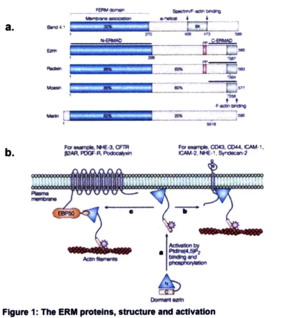
FIgure 1: The ERM proteins, structure and activation
a. A schematic showing the homology between ERM proteins and merlin.
ERM members have a C-terrminal actin binding site, whereas merdin does
not. Ezrln and radixin share a prolne rich region which moeuin and merlin
do not. The major
reuaoypopoyain(threonine
for ERM, swdne for
merln) Is indicated. C-ERMAD, carboxy-ERM domain;
N-ERMAD. amino-ERM association domain.
b. A model of ERM
activation.
ERM proteins can form IntermTOlecular
asscitinsbetWeen
the heed and tall. Association With PIP
2can recu
ERMs to the membrane, where they can be phosphorylated and persist In
an
*open"
form for additional protein-protein Interactions. (From Bretacher,
dependent on phosphorylation of a serine in the C-terminal tail, and indeed, that this structure is possibly a model for all 4.1 superfamily proteins (15) (Figure 1, from (22)).
3. 4.1 family proteins in cancer and metastasis
4.1 family proteins have been implicated in functions such as cell-cell adhesion, cell signaling, and cell migration that, when altered, can mediate the progression of tumor formation and metastasis (23). Following are a number of examples of 4.1 family proteins which have been shown to play roles in various cancer development (24). Ezrin: Ezrin expression was shown to be elevated in osteosarcomas (25, 26) and prostate cancer (27), as well as being a prognostic marker of metastasis in multiple cancers (28), including osteosarcoma (26) and pancreatic cancer (29). Mutation of the Src-phosphorylation target Y145, a tyrosine residue not conserved in ezrin's close family
members radixin and moesin, reduced cell migration (30). Mutation of an additional Src-phosphorylation target on ezrin, Y477, reduced in vivo invasion in mice (31). Ezrin expression was also increased in highly metastatic lung cancer cell lines when
compared with poorly metastatic lung adenocarcinoma cell lines and knockdown of ezrin in these highly metastatic lines led to a decrease in proliferation, migration and invasion
(32). Furthermore, abnormal ezrin localization from apical regions to the cytoplasm in
breast cancer patient samples correlated with poor patient prognosis (33).
Radixin: Knockdown of radixin in PANC1 human pancreatic carcinoma cells showed a decrease in proliferation, adhesion and invasiveness in vitro as well as a decrease in tumor growth in PANC1 tumor cells implanted in nude mice (34). In addition to role radixin plays in pancreatic cancer, radixin has also been shown to enhance migration of
PC3 prostate cancer cells (35).
Moesin: Moesin expression has been linked to an increase in tumor size and
a tail-vein assay (37). In addition, moesin has been described as a marker of epithelial-to-mesenchymal transition in pancreatic cancer (38) and breast cancer (39), and moesin was required for actin remodeling in a mouse mammary epithelial cell model of TGF-beta induced EMT (40).
In addition to the roles these 4.1 family members play as enhancers of tumorigenesis and metastasis, two 4.1 family members have been implicated as tumor suppressors. These include merlin, which is the protein product of the NF2 tumor suppressor gene and will be described more extensively below, and 4.1B/DAL, which was identified as a novel gene whose expression was decreased in non-small cell lung carcinoma
compared to normal lung tissue (41). Furthermore, 4.1B was downregulated in breast cancer (42) and in metastatic prostate cancer cell lines and shown to be a suppressor of metastasis (43).
Objectives: Considering the roles that these proteins play in tumor formation and
metastasis, we wanted to see whether other members of the 4.1 superfamily of proteins could also have roles in cancer and metastasis. The dual goal of this research would be to develop a multiplex in-vivo tumorigenesis and metastasis screen, allowing
investigation of the involvement of multiple proteins simultaneously through the use of barcoded knockdowns in the context of tumor formation and metastasis, while also discovering previously unidentified 4.1 family members which might also play a role in tumor development.
4. Merlin: A well-characterized tumor suppressor
As we began to investigate the role of FERM-domain proteins in tumorigenesis and
metastasis it became clear that there were still questions surrounding the function of merlin in tumor suppression. Merlin had already been studied extensively due to its involvement in human disease, yet questions remained regarding its regulation and its potential role in tumor suppression through the Hippo pathway.
Merlin is an outlier in the ERM subgroup of the 4.1 superfamily of proteins. Although it contains extensive sequence homology to ezrin, radixin and moesin in the FERM
domain N-terminal of the protein, it lacks the actin-binding domain that characterizes the ERM protein C-terminal.
Merlin was initially discovered through its role in human disease; genetic mapping of tumors from neurofibromatosis type 2 (NF2) patients revealed a deletion on
chromosome 22 (2, 44), which was mapped to the NF2 gene, also called merlin, due to its similarity to the ERM family of proteins (moesin-radixin-ezrin-like-protein).
Neurofibromatosis type 2 is characterized by the development of schwannomas, meningiomas and ependymomas, but merlin loss has also been identified in additional tumor types, including mesothelioma, melanoma, and thyroid cancer (3). NF2 is one of the most frequently mutated tumor suppressors in malignant mesothelioma, and is lost in multiple mesothelioma-derived cell lines (45).
In addition to the benign tumors found in human neurofibromatosis type 2, heterozygous merlin knock out in mice leads to highly metastatic disease (4), including
osteosarcomas, fibrosarcomas and hepatocellular carcinomas, suggesting that merlin plays a role in metastasis, and furthermore, that merlin serves as a tumor suppressor in a larger number of cell types.
The 4.1 family is extensively alternatively spliced, and merlin is not different in this regard (46, 47). The NF2 gene contains 17 exons, and the two major splice isoforms differ by inclusion or deletion of exon 16. While exon 16 is lacking in isoform I, it is included in isoform II, which leads to a frameshift involving an early stop codon. This creates an alternative C-terminal (3). Merlin isoform I contains 595 amino acids whereas isoform I contains 590 amino acids. Both share a molecular weight of about 70kDa. Initially, isoform II was described as an inactive form of merlin, due to limited ability of the C-terminal to bind the N-terminal and thus form the "closed" isoform, which appeared to be necessary for merlin activity (48-50). In addition, profiling the changes in splicing following epithelial-to-mesenchymal transition (EMT) showed a change in expression of merlin isoforms (Shapiro, personal communication). In these experiments, the splicing profile of HMLE cells was compared before and after induction of EMT through Twist expression with isoform I of merlin being present prior to EMT in epithelial cells and a switch to isoform 11 occurring in the mesenchymal cells following Twist expression and EMT (51). This could indicate that the switch to isoform 11 coincided with an inactivation of merlin during EMT. However, newer data (52) showed that merlin activity was not determined by a simple conformational switch, and furthermore, that the "open" form of merlin (in which there is no intermolecular interaction between the head and tail) was able to suppress growth, thus indicating that the isoform 11 might be active in
suppression of growth as well.
5. Merlin regulation through phosphorvlation
Like its close family members, the ERM proteins, merlin can be regulated through
phosphorylation at the C-terminal of the protein. This phosphorylation varies in response to growth conditions, with increased confluency, serum starvation, or loss of cell-cell
phosphorylated by p21-activated-kinase (PAK1/2)(54, 55), and by cAMP-dependent protein kinase A (PKA) (56). Following phosphorylation on Ser518, merlin can be phosphorylated on multiple other serine and threonines (57, 58). Relying on a similarity to other ERM proteins, and pull-down experiments showing that the unphosphorylated tail of merlin was able to bind the FERM-domain head (50), a canonical model was adopted where phosphorylated merlin was described as being in the "open" form, and unphosphorylated merlin was described as the "closed" form (5). Investigation of merlin's function determined that phosphorylated merlin was unable to suppress growth, and that mutating the predominant phosphorylation target Ser518 lead to constitutively active merlin (59). However, this model indicated that the "closed" form of merlin was the active state, which seemed paradoxical since the closed form would have less exposed sites available for binding mediators of merlin function. Further study showed that indeed the conformation of merlin was more fluid, and not altogether "open" or "closed". In this model, the conformationally "open" state of merlin was the growth suppressive state, and phosphorylation tipped the protein into adopting a more closed (but not entirely) closed state (52). In addition, FRET studies show that Merlin's N-terminal FERM-domain head and C-terminal tail are constitutively found in close proximity and only undergo subtle changes following phosphorylation (60).
Objectives: Recent research determining that a "closed" conformation isn't necessary
for merlin activity indicates that previous assumptions regarding the inability of isoform 11
to inhibit growth were mistaken. We decided to compare the function of the two major merlin isoforms, in order to determine whether both of the major merlin isoforms were able to regulate growth, and further, in what context does this happen. The role of isoform I in growth regulation during contact inhibition has been described, but the role of isoform II in growth inhibition remains unclear. In addition, the regulation of isoform I
through phosphorylation has been investigated, but it is unknown whether regulation of isoform II occurs in a manner similar to isoform 1.
6. Multiple mechanisms for merlin-derived suppression of growth
Merlin is able to suppress cell growth through many different mechanisms (Figure 2), due to the ability of merlin to bind numerous transmembrane and intercellular proteins
(reviewed by (61)). In some cases, contradictory evidence raises questions about the context and relevance of these different mechanisms to various biological models. For example, merlin has been implicated in suppressing proliferation through contact inhibition via control of actin organization. Although merlin lacks the C-terminal actin-binding domain found in the ERM proteins, it contains an actin-actin-binding site in the N-terminal (62). Schwannoma cells lacking merlin have been shown to have disorganized stress fibers, altered cell spreading, and increased membrane ruffling, all related to abnormal actin organization (63), which can be reversed by the expression of merlin (64). In addition, expression of merlin in mesothelioma cells lacking endogenous merlin leads to decrease of phospho-FAK (65). This decrease in FAK phosphorylation leads to a downstream decrease in Src activation, either directly, since merlin has been shown to form a complex with FAK (66), or indirectly, through inhibition of Rac/PAK signaling (67). Furthermore, merlin is able to block recruitment of Rac to the membrane and loss of merlin can lead to increased Rac activity, lamellipodia formation, and increased cell motility (68).
The mechanism behind merlin's ability to regulate Rac can be explained by Merlin's interaction with angiomotin. Angiomotin, a scaffolding protein that localizes to tight junctions, was shown to bind merlin. This interaction is competitive with angiomotin
binding Rich, which is then able to inactivate Rac (69). However, both unphosphorylated merlin (S518A) and a phosphomimetic form of merlin (S518D) were shown to bind
a
12
1
catenin J
4
6
mM
3
NUCLEUS
Figure 2: Multiple mechanisms for merlin regulation of growth
1. Negatively regulates CD44 (Bai et aL. 2007).
2. Regulates the
distribution,
aggregation and
availability
of receptor
tyrosine kinases (McClatchey et al. 2009).
3. Translocates to the nucleus and inhibits the ubiquitin ligase
CRL4DCAF(Li et aL 2012).
4. Stabilizes adherens junctions through an interaction with a-catenin
(Gladden et aL. 2010).
5. Interacts with angiomotin to suppress Rac activity (Yi et a/. 2011).
6. Recruits the Lats kinases to the plasma membrane and
coordinates their activation by Mstl, driving phosphorylation and
inhibition of YAP/TAZ (Yin et al. 2013).
5
I V
angiomotin, which raises the question of how merlin regulation through phosphorylation can be explained in this context, and in what contexts does this mechanism for Rac regulation through angiomotin confer biological relevance.
Merlin has also been shown to stabilize adherens junctions in mouse embryonic fibroblasts and keratinocytes (70), through an interaction with a-catenin (71).
Aside from mediating contact inhibition through stabilization of adherens junctions, merlin has also been shown to mediate contact inhibition through an interaction with CD44, the hyaluronan receptor. CD44 is upregulated in several cancers, and is an indicator of poor prognosis (reviewed by (72)). Using a rat schwannoma cell line, Morrison et al. found that at high cell densities, when contact inhibition was required, a hypophosphorylated form of merlin bound CD44, and that treatment with hyaluronic acid or with anti-CD44 antibody led to a merlin-dependent decrease in cell proliferation (10). In addition to its role in mediating contact inhibition, presumably through adherens junction and CD44 interactions, merlin can also regulate cell growth through control of the distribution, aggregation and availability of receptor tyrosine kinases (RTKs) in the
plasma membrane. This function of merlin as a regulator of cell growth has been shown both in mammals (73, 74) and in Drosophila (75).
Finally, merlin has also been suggested to localize to the nucleus and inhibit cell growth through an inhibitory interaction with the E3 ubiquitin ligase CRL4 DCAF1 (7 6).
CRL4 ligases have been implicated in histone remodeling and can mediate a generally cell-proliferative gene expression pattern, as well as directly ubiquitinating and inhibiting a major component of the canonical Hippo pathway, LATS1/2 (77), providing an
additional, nuclear and transcriptionally-mediated role for merlin-dependent growth suppression. However, since the merlin FERM-domain alone is able to suppress growth in this manner, and the literature supports a role for C-terminal domain being required to suppress growth as well (Lallemand et al. 2009), it is clear that merlin does not sustain
its major effects through nuclear localization and activity alone, and that the biological context and other participants in the system described must be carefully considered.
7. The Hippo pathway in flies and mammals Regulation of the Hippo pathway in Drosophila
Although multiple mechanisms and functions have been proposed for merlin as an explanation for its role as a tumor suppressor, there remains one pathway, the Hippo pathway in which merlin has been described as a member, and yet a mechanism for merlin's participation in this pathway remain unclear; this will be one topic of interest in this thesis. The Hippo pathway has recently been described as an important tumor
suppressor pathway in both flies and mammals, with mammalian orthologs for the canonical components of the Drosophila pathway. In Drosophila the Hippo pathway serves to control organ size through regulation of proliferation, cell growth and apoptosis
(78, 79), by limiting imaginal disc size. The core components of the pathway include the
serine/threonine kinases Warts (mammalian LATS1/2) and Hippo (mammalian MST1/2), and two adaptor proteins, Salvador (Sav), a scaffolding protein that interacts with Hippo, and Mats, which binds Warts. When the kinase cascade is activated, Hippo, in a
complex with Sav, phosphorylates and activates Warts, bound to its co-factor Mats. Activated Warts phosphorylates the transcriptional co-activator Yorkie (mammalian YAP/TAZ) at multiple sites, inhibiting its function (80). This inhibition occurs through cytoplasmic sequestration of Yorkie, as the phosphorylation on Serl68 (Ser127 in the
mammalian ortholog YAP) acts as a recognition site for 14-3-3 binding, which retains Yki in the cytoplasm and prevents its activity as a transcriptional co-activator. In the nucleus, Yki promotes a transcriptional profile that is growth-permissive and anti-apoptotic
flies, Hippo-independent regulation of Yki can occur through the FERM-domain protein Expanded, which can suppress Yki activity directly through binding and cytoplasmic retention (83-85) (Figure 3).
Mammalian regulation of the Hippo pathway
In mammals, multiple upstream regulators have been identified that can control the Hippo pathway or alternatively YAP activity directly, independently of the Hippo pathway.
A major component of YAP regulation involves mechanical stress (86), with the
extracellular matrix regulating YAP/TAZ activity as a function of varying stiffness. Dupont et al. found that on stiff ECM YAP localized to the nucleus regulated through Rho and actin cytoskeleton activity, whereas on a soft matrix YAP localized to the
cytoplasm. Furthermore, knockdown of LATS was unable to rescue cells plated on a soft matrix, suggesting that the stiffness-mediated regulation is independent of the Hippo pathway. However, contradictory evidence showed that LATS was activated by cell detachment and that LATS knockdown can prevent mechanical-stress-induced YAP phosphorylation (87, 88).
In addition to YAP regulation through matrix stiffness, G-protein receptors have also been identified as regulators of the Hippo pathway (89) through LATS inhibition by
G12/13 or Gq/1 1-coupled receptors, which leads to YAP activation, or LATS activation
through Gs-coupled receptors which leads to an inhibition of YAP.
The mammalian orthologs of Yki, YAP and TAZ, were discovered to be Yes-associated proteins, and were the first WW-domain containing proteins to be identified (90). YAP contains one or two WW domains, depending on the splice isoform involved, and TAZ contains one WW domain. These domains contain two conserved tryptophans (WW), and bind proline-rich motifs. In addition to the WW domains, YAP and TAZ also contain a coiled-coil domain and a PDZ-binding motif, which regulate YAP localization and function (91). There are five HXRXXS LATS target sequences in YAP/TAZ (92), with two
DROSOPHILA
I I I I I I I I'4
<1
I
Anti-apoptosis, pro-proliferation * a * I I I IL4
I
Anti-apoptosis, pro-proliferationFigure 3: The Hippo pathway In Drosophila and mammals
The corresponding proteins are indicated by matching colors. Direct biochemical interactions are shown with a solid line, genetic Interactions are shown with a dashed line. An hypothesized interaction is Indicated with a grey line.
M AMM ALS
of these sites appearing most important for regulation (80). Phosphorylation at Ser127 in YAP (Ser89 in TAZ) creates the 14-3-3 binding site which mediates YAP cytoplasmic sequestration, and phosphorylation at Ser381 mediates YAP ubiquitination and
degradation (93).
YAP is implicated in human disease as an oncogene amplified in multiple tumor types (94), and YAP expression and nuclear localization correlate with poor patient outcome in several cancers (80, 95-98). Furthermore, YAP overexpression in multiple cell lines, as well as transgenic activation in mice, has been shown to promote tumor growth (99,
100). Our lab has demonstrated an important role for YAP activity in metastasis (101).
8. Merlin regulation of YAP
Merlin regulation of the Hippo pathway in flies
Merlin has been implicated as an upstream regulator of the Hippo pathway in
Drosophila. Mutations in merlin and another FERM-domain family protein, expanded,
lead to overgrowth in multiple adult tissues in Drosophila, phenocopying mutations of Hippo pathway components. In addition, mer;ex double mutants had higher levels of the Yki target gene diapi, an anti-apoptotic gene. Interestingly, Hpo mutants could reverse the reduced-eye-size phenotype caused by loss of Expanded, placing Expanded upstream of Hippo (102), which is in contrast to later findings that Expanded is able to regulate Yki directly without relying on the kinase cascade for phosphorylation and regulation of Yki (83). However, the epistasis experiments could not rule out an
additional model of regulation in which the Hippo pathway is bypassed altogether, and the experiments in Drosophila that illustrate how loss of merlin leads to EGFR trafficking
defects (75) demonstrate that merlin may play multiple roles through various mechanisms depending on the cellular context and the developmental stage.
Since merlin and expanded appeared to synergize (103) and were assumed to be redundant to each other in Drosophila, these genetic experiments which placed
expanded upstream of the Hippo pathway were considered to place merlin upstream of the Hippo pathway as well. Due to the presence of a highly conserved Hippo kinase cascade in mammals, and the relevance of the pathway to human disease, there has been a focus on identifying the role of merlin in regulation of YAP, the Yki homolog, primarily as an upstream regulator of the Hippo pathway.
Merlin regulation of YAP in mammals
In mammalian cells, merlin has been shown to regulate the expression and localization of YAP in meningioma cells (104) and mesothelioma cells (105), although neither of these studies convincingly implicated the Hippo pathway in this regulation of YAP. In the search for a tractable mammalian model, that could be compared to the fly model of the Hippo pathway regulating organ size, the liver was selected as an appropriate example, since mammalian liver can undergo dramatic changes in organ size. When YAP was inducibly overexpressed in the adult liver this led to an increase in liver size, which was reversed when YAP overexpression was removed (99) Similar overgrowth
phenotypes were observed when the mammalian Hippo orthologs MST1 and MST2 were depleted in embryonic livers using a Cre-albumin promoter, which acts on
embryonic liver progenitors (106). These results show that YAP, as regulated by the Hippo pathway, appears to be an important player in liver regeneration. However, attempts to determine whether merlin plays a role in this regulation led to contradictory results. Work by Zhang et al. showed that elimination of NF2 using an albumin-cre model led to expanded liver progenitor growth, which could be suppressed by
heterozygous deletion of YAP, linking YAP regulation to merlin tumor-suppressor activity
(107). But research by Benhamouche et al. contradicted this experiment. When merlin
albumin-cre, there was an enlargment of the liver. However, there was no evidence of Hippo pathway activity or YAP activation in the in these experiments, and the phenotype observed following loss of merlin was explained through EGFR pathway regulation and could be suppressed by treatment with erlotinib, an EGFR inhibitor (108). Considering that there is crosstalk between YAP and downstream components of the EGFR signaling pathway, where Raf can prevent MST2 interaction with Latsl (reviewed by (109)), and on the other hand, the EGFR ligand amphiregulin is upregulated through YAP-mediated transcription (100), the intertwined nature of EGFR activity and YAP activity can further complicate the picture when trying to tease out regulatory mechanisms.
Merlin can stabilize Lats
One possible role for merlin in regulation of the Hippo pathway is through direct stablization of Lats. Merlin has been shown to bind Lats through the FERM-domain, recruiting Lats to the membrane, which allows it to be phosphorylated by MST1/2, activating it and allowing it to phosphorylate and inhibit YAP (110). Although this is a
mechanism that allows regulation of YAP through attenuation of the Hippo pathway, it does not rule out other mechanisms for merlin regulation of YAP. The question remains whether, in addition to the role of merlin in LATS stablilization, merlin is able to regulate YAP independently of the Hippo pathway.
Objective: examining Hippo pathway independent regulation of YAP by merlin
An example of Hippo pathway-independent regulation by merlin occurs in Drosophila. In flies, expanded can regulate Yki without Yki phosphorylation by the Hippo kinase
cascade, through binding of the PPxY motifs of expanded to the WW domains of Yki
(83). Could a similar mode of regulation be available in mammals? Mammalian
expanded is shortened and lacks the PPxY motif found in Drosophila, however one Hippo pathway-independent regulatory function involves the scaffolding protein angiomotin binding YAP through a WW domain/PPxY motif interaction (111, 112) and
sequestering it in the cytoplasm without prior phosphorylation of YAP by the Hippo kinase cascade. Since merlin has been shown to bind angiomotin through its role in Rac inhibition (69), could it play an additional function in YAP inhibition as well? And what are the cellular contexts that mediate merlin suppression of growth through the multiple avenues that appear to be available for this suppression? In order to attempt to address these questions we decided to investigate the ability of merlin to regulate YAP in a mesothelioma model, and determine whether the ability of merlin to regulate YAP was dependent upon the Hippo pathway.
9. Summary
This thesis aims to investigate multiple aspects of the FERM domain family of proteins. The subsequent chapters will describe the following:
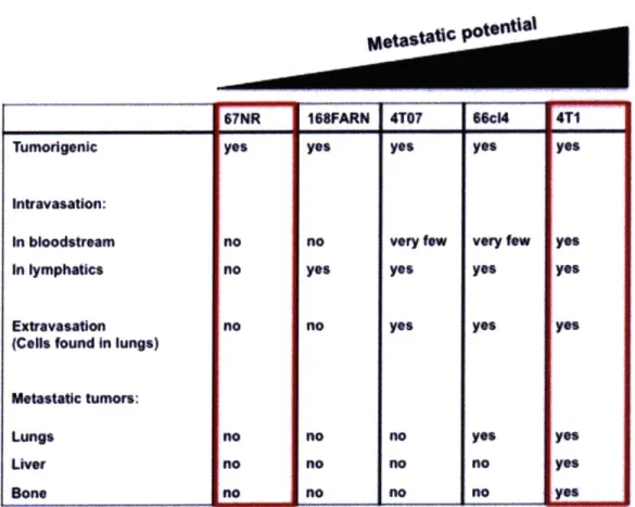
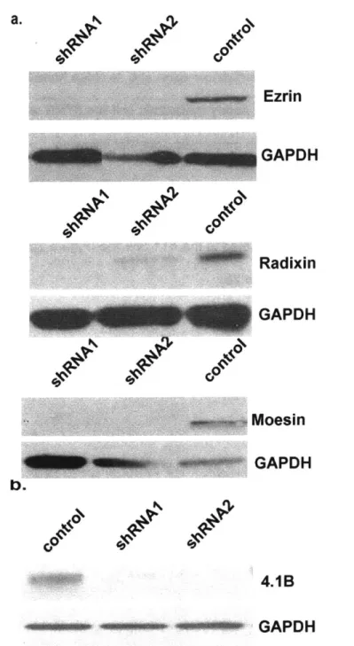
In Chapter 2 I attempt to establish an in vivo screen to determine whether members of the FERM domain family are enhancers or suppressors of metastasis. In order to do this we chose ezrin, a known enhancer of metastasis, as well as close family members radixin and moesin, and 4.1 B, a known metastasis suppressor, as preliminary targets for screening.
In Chapter 3 1 describe the FERM domain family member merlin's two major splice
isoforms, isoform I and isoform II, and establish that both of the isoforms are able to suppress growth in multiple cell lines. I further show that similarly to the well
characterized isoform I, isoform II also suppresses cell growth in a contact inhibition-dependent manner, and is regulated by phosphorylation. However, there appears to be differential regulation of the two isoforms, and isoform 11 not only is able to suppress growth, despite being described previously as an inactive form of merlin, but is also more stable when expressed in culture.
In Chapter 4 I investigate the mechanism by which merlin regulates the Hippo pathway
target YAP. I show that in addition to regulation through the Hippo pathway, which has been described previously, merlin is able to regulate YAP in a
Hippo-pathway-independent manner, which requires the WW domains of YAP.
In Chapter 5 I summarize the work described in this thesis, the unanswered questions that arose from this study, and possible avenues for future research into these
questions.
References
1. Chambers AF, Groom AC, MacDonald IC. Dissemination and growth of cancer cells in metastatic sites. Nat Rev Cancer 2002;2:563-72.
2. Trofatter JA, MacCollin MM, Rutter JL, et al. A novel moesin-, ezrin-, radixin-like gene is a candidate for the neurofibromatosis 2 tumor suppressor. Cell 1993;75:826.
3. Bianchi AB, Hara T, Ramesh V, et al. Mutations in transcript isoforms of the neurofibromatosis 2 gene in multiple human tumour types. Nat Genet 1994;6:185-92. 4. McClatchey Al, Saotome I, Mercer K, et al. Mice heterozygous for a mutation at the Nf2 tumor suppressor locus develop a range of highly metastatic tumors. Genes Dev
1998;12:1121-33.
5. Bretscher A, Edwards K, Fehon RG. ERM proteins and merlin: integrators at the cell cortex. Nat Rev Mol Cell Biol 2002;3:586-99.
6. Holzwarth G, Yu J, Steck TL. Heterogeneity in the conformation of different protein fractions from the human erythrocyte membrane. J Supramol Struct
1976;4:161-8.
7. Louvet-Vallee S. ERM proteins: from cellular architecture to cell signaling. Biol Cell 2000;92:305-16.
8. Baines AJ, Lu HC, Bennett PM. The Protein 4.1 family: hub proteins in animals for organizing membrane proteins. Biochim Biophys Acta 2014;1838:605-19.
9. Anderson RA, Marchesi VT. Regulation of the association of membrane skeletal
protein 4.1 with glycophorin by a polyphosphoinositide. Nature 1985;318:295-8.
10. Morrison H, Sherman LS, Legg J, et al. The NF2 tumor suppressor gene product,
merlin, mediates contact inhibition of growth through interactions with CD44. Genes Dev
2001;15:968-80.
11. Tsukita S, Yonemura S. ERM (ezrin/radixin/moesin) family: from cytoskeleton to
signal transduction. Curr Opin Cell Biol 1997;9:70-5.
12. Ramesh V. Merlin and the ERM proteins in Schwann cells, neurons and growth
cones. Nat Rev Neurosci 2004;5:462-70.
13. Kusunoki H, Kohno T. Solution structure and glycophorin C binding studies of the
protein 4.1R FERM alpha-lobe domain. Proteins 2009;76:255-60.
14. Gary R, Bretscher A. Ezrin self-association involves binding of an N-terminal
domain to a normally masked C-terminal domain that includes the F-actin binding site. Mol Biol Cell 1995;6:1061-75.
15. McClatchey Al, Fehon RG. Merlin and the ERM proteins--regulators of receptor
distribution and signaling at the cell cortex. Trends Cell Biol 2009;19:198-206.
16. Bretscher A, Gary R, Berryman M. Soluble ezrin purified from placenta exists as
stable monomers and elongated dimers with masked C-terminal ezrin-radixin-moesin
association domains. Biochemistry 1995;34:16830-7.
17. Fievet BT, Gautreau A, Roy C, et al. Phosphoinositide binding and
phosphorylation act sequentially in the activation mechanism of ezrin. J Cell Biol
18. Crepaldi T, Gautreau A, Comoglio PM, Louvard D, Arpin M. Ezrin is an effector of hepatocyte growth factor-mediated migration and morphogenesis in epithelial cells. J Cell Biol 1997;138:423-34.
19. Matsui T, Maeda M, Doi Y, et al. Rho-kinase phosphorylates COOH-terminal threonines of ezrin/radixin/moesin (ERM) proteins and regulates their head-to-tail association. J Cell Biol 1998;140:647-57.
20. Naba A, Reverdy C, Louvard D, Arpin M. Spatial recruitment and activation of the Fes kinase by ezrin promotes HGF-induced cell scattering. EMBO J 2008;27:38-50. 21. Pearson MA, Reczek D, Bretscher A, Karplus PA. Structure of the ERM protein moesin reveals the FERM domain fold masked by an extended actin binding tail domain. Cell 2000;101:259-70.
22. Bretscher A, Chambers D, Nguyen R, Reczek D. ERM-Merlin and EBP50 protein families in plasma membrane organization and function. Annu Rev Cell Dev Biol
2000;16:113-43.
23. McClatchey Al. Merlin and ERM proteins: unappreciated roles in cancer development? Nat Rev Cancer 2003;3:877-83.
24. Clucas J, Valderrama F. ERM proteins in cancer progression. J Cell Sci
2014;127:267-75.
25. Curto M, McClatchey Al. Ezrin... a metastatic detERMinant? Cancer Cell 2004;5:113-4.
26. Khanna C, Wan X, Bose S, et al. The membrane-cytoskeleton linker ezrin is necessary for osteosarcoma metastasis. Nat Med 2004;10:182-6.
27. Bruce B, Khanna G, Ren L, et al. Expression of the cytoskeleton linker protein ezrin in human cancers. Clin Exp Metastasis 2007;24:69-78.
28. Hunter KW. Ezrin, a key component in tumor metastasis. Trends Mol Med 2004;10:201-4.
29. Akisawa N, Nishimori 1, Iwamura T, Onishi S, Hollingsworth MA. High levels of ezrin expressed by human pancreatic adenocarcinoma cell lines with high metastatic potential. Biochem Biophys Res Commun 1999;258:395-400.
30. Srivastava J, Elliott BE, Louvard D, Arpin M. Src-dependent ezrin
phosphorylation in adhesion-mediated signaling. Mol Biol Cell 2005;16:1481-90.
31. Mak H, Naba A, Varma S, et al. Ezrin phosphorylation on tyrosine 477 regulates invasion and metastasis of breast cancer cells. BMC Cancer 2012;12:82.
32. Li Q, Gao H, Xu H, et al. Expression of ezrin correlates with malignant phenotype of lung cancer, and in vitro knockdown of ezrin reverses the aggressive biological
behavior of lung cancer cells. Tumour Biol 2012;33:1493-504.
33. Sarrio D, Rodriguez-Pinilla SM, Dotor A, Calero F, Hardisson D, Palacios J. Abnormal ezrin localization is associated with clinicopathological features in invasive
breast carcinomas. Breast Cancer Res Treat 2006;98:71-9.
34. Chen SD, Song MM, Zhong ZQ, et al. Knockdown of radixin by RNA interference suppresses the growth of human pancreatic cancer cells in vitro and in vivo. Asian Pac J Cancer Prev 2012;13:753-9.
35. Valderrama F, Thevapala S, Ridley AJ. Radixin regulates cell migration and cell-cell adhesion through Rac1. J Cell Sci 2012;125:3310-9.
36. Kobayashi H, Sagara J, Kurita H, et al. Clinical significance of cellular distribution of moesin in patients with oral squamous cell carcinoma. Clin Cancer Res
2004;10:572-80.
37. Estecha A, Sanchez-Martin L, Puig-Kroger A, et al. Moesin orchestrates cortical polarity of melanoma tumour cells to initiate 3D invasion. J Cell Sci 2009;122:3492-501.
38. Abiatari 1, Esposito I, Oliveira TD, et al. Moesin-dependent cytoskeleton
remodelling is associated with an anaplastic phenotype of pancreatic cancer. J Cell Mol Med 2010;14:1166-79.
39. Wang CC, Liau JY, Lu YS, Chen JW, Yao YT, Lien HC. Differential expression of moesin in breast cancers and its implication in epithelial-mesenchymal transition.
Histopathology 2012;61:78-87.
40. Haynes J, Srivastava J, Madson N, Wittmann T, Barber DL. Dynamic actin remodeling during epithelial-mesenchymal transition depends on increased moesin expression. Mol Biol Cell 2011;22:4750-64.
41. Tran YK, Bogler 0, Gorse KM, Wieland I, Green MR, Newsham IF. A novel member of the NF2/ERM/4.1 superfamily with growth suppressing properties in lung cancer. Cancer Res 1999;59:35-43.
42. Heller G, Geradts J, Ziegler B, et al. Downregulation of TSLC1 and DAL-1 expression occurs frequently in breast cancer. Breast Cancer Res Treat
2007;103:283-91.
43. Wong SY, Haack H, Kissil JL, et al. Protein 4.1B suppresses prostate cancer progression and metastasis. Proc Natl Acad Sci U S A 2007;104:12784-9.
44. Rouleau GA, Merel P, Lutchman M, et al. Alteration in a new gene encoding a putative membrane-organizing protein causes neuro-fibromatosis type 2. Nature
1993;363:515-21.
45. Sekido Y. Inactivation of Merlin in malignant mesothelioma cells and the Hippo signaling cascade dysregulation. Pathol Int 2011;61:331-44.
46. Arakawa H, Hayashi N, Nagase H, Ogawa M, Nakamura Y. Alternative splicing of the NF2 gene and its mutation analysis of breast and colorectal cancers. Hum Mol Genet 1994;3:565-8.
47. Pykett MJ, Murphy M, Harnish PR, George DL. The neurofibromatosis 2 (NF2) tumor suppressor gene encodes multiple alternatively spliced transcripts. Hum Mol Genet 1994;3:559-64.
48. Gonzalez-Agosti C, Wiederhold T, Herndon ME, Gusella J, Ramesh V.
Interdomain interaction of merlin isoforms and its influence on intermolecular binding to NHE-RF. J Biol Chem 1999;274:34438-42.
49. Gutmann DH, Haipek CA, Hoang Lu K. Neurofibromatosis 2 tumor suppressor
protein, merlin, forms two functionally important intramolecular associations. J Neurosci Res 1999;58:706-16.
50. Sherman L, Xu HM, Geist RT, et al. Interdomain binding mediates tumor growth
suppression by the NF2 gene product. Oncogene 1997;15:2505-9.
51. Shapiro IM, Cheng AW, Flytzanis NC, et al. An EMT-driven alternative splicing
program occurs in human breast cancer and modulates cellular phenotype. PLoS Genet 201 1;7:e1002218.
52. Sher I, Hanemann CO, Karplus PA, Bretscher A. The tumor suppressor merlin
controls growth in its open state, and phosphorylation converts it to a less-active more-closed state. Dev Cell 2012;22:703-5.
53. Shaw RJ, McClatchey Al, Jacks T. Regulation of the neurofibromatosis type 2
tumor suppressor protein, merlin, by adhesion and growth arrest stimuli. J Biol Chem
1998;273:7757-64.
54. Kissil JL, Johnson KC, Eckman MS, Jacks T. Merlin phosphorylation by
p21-activated kinase 2 and effects of phosphorylation on merlin localization. J Biol Chem
2002;277:10394-9.
55. Xiao GH, Beeser A, Chernoff J, Testa JR. p21-activated kinase links Rac/Cdc42
signaling to merlin. J Biol Chem 2002;277:883-6.
56. Alfthan K, Heiska L, Gronholm M, Renkema GH, Carpen 0. Cyclic
AMP-dependent protein kinase phosphorylates merlin at serine 518 inAMP-dependently of p21-activated kinase and promotes merlin-ezrin heterodimerization. J Biol Chem
57. Laulajainen M, Muranen T, Nyman TA, Carpen 0, Gronholm M. Multistep phosphorylation by oncogenic kinases enhances the degradation of the NF2 tumor suppressor merlin. Neoplasia 2011; 13:643-52.
58. Tang X, Jang SW, Wang X, et al. Akt phosphorylation regulates the tumour-suppressor merlin through ubiquitination and degradation. Nat Cell Biol
2007;9:1199-207.
59. Surace El, Haipek CA, Gutmann DH. Effect of merlin phosphorylation on neurofibromatosis 2 (NF2) gene function. Oncogene 2004;23:580-7.
60. Hennigan RF, Foster LA, Chaiken MF, et al. Fluorescence resonance energy transfer analysis of merlin conformational changes. Mol Cell Biol 2010;30:54-67.
61. Scoles DR. The merlin interacting proteins reveal multiple targets for NF2 therapy. Biochim Biophys Acta 2008;1785:32-54.
62. Xu HM, Gutmann DH. Merlin differentially associates with the microtubule and actin cytoskeleton. J Neurosci Res 1998;51:403-15.
63. Pelton PD, Sherman LS, Rizvi TA, et al. Ruffling membrane, stress fiber, cell spreading and proliferation abnormalities in human Schwannoma cells. Oncogene
1998;17:2195-209.
64. Bashour AM, Meng JJ, Ip W, MacCollin M, Ratner N. The neurofibromatosis type 2 gene product, merlin, reverses the F-actin cytoskeletal defects in primary human Schwannoma cells. Mol Cell Biol 2002;22:1150-7.
65. Poulikakos P1, Xiao GH, Gallagher R, Jablonski S, Jhanwar SC, Testa JR. Re-expression of the tumor suppressor NF2/merlin inhibits invasiveness in mesothelioma cells and negatively regulates FAK. Oncogene 2006;25:5960-8.
66. James MF, Beauchamp RL, Manchanda N, Kazlauskas A, Ramesh V. A NHERF binding site links the betaPDGFR to the cytoskeleton and regulates cell spreading and
67. Kissil JL, Wilker EW, Johnson KC, Eckman MS, Yaffe MB, Jacks T. Merlin, the product of the Nf2 tumor suppressor gene, is an inhibitor of the p21-activated kinase, Paki. Mol Cell 2003;12:841-9.
68. Shaw RJ, Paez JG, Curto M, et al. The Nf2 tumor suppressor, merlin, functions
in Rac-dependent signaling. Dev Cell 2001;1:63-72.
69. Yi C, Troutman S, Fera D, et al. A tight junction-associated Merlin-angiomotin
complex mediates Merlin's regulation of mitogenic signaling and tumor suppressive functions. Cancer Cell 2011; 19:527-40.
70. Lallemand D, Curto M, Saotome 1, Giovannini M, McClatchey Al. NF2 deficiency
promotes tumorigenesis and metastasis by destabilizing adherens junctions. Genes Dev
2003;17:1090-100.
71. Gladden AB, Hebert AM, Schneeberger EE, McClatchey Al. The NF2 tumor
suppressor, Merlin, regulates epidermal development through the establishment of a junctional polarity complex. Dev Cell 2010;19:727-39.
72. Toole BP. Hyaluronan-CD44 Interactions in Cancer: Paradoxes and Possibilities.
Clin Cancer Res 2009;15:7462-8.
73. Curto M, Cole BK, Lallemand D, Liu CH, McClatchey Al. Contact-dependent
inhibition of EGFR signaling by Nf2/Merlin. J Cell Biol 2007;177:893-903.
74. Lallemand D, Manent J, Couvelard A, et al. Merlin regulates transmembrane
receptor accumulation and signaling at the plasma membrane in primary mouse Schwann cells and in human schwannomas. Oncogene 2009;28:854-65.
75. Maitra S, Kulikauskas RM, Gavilan H, Fehon RG. The tumor suppressors Merlin
and Expanded function cooperatively to modulate receptor endocytosis and signaling. Curr Biol 2006;16:702-9.
76. Li W, You L, Cooper J, et al. Merlin/NF2 suppresses tumorigenesis by inhibiting
77. Li W, Cooper J, Zhou L, et al. Merlin/NF2 loss-driven tumorigenesis linked to CRL4(DCAF1)-mediated inhibition of the hippo pathway kinases Latsl and 2 in the nucleus. Cancer Cell 2014;26:48-60.
78. Halder G, Johnson RL. Hippo signaling: growth control and beyond. Development 2011;138:9-22.
79. Harvey K, Tapon N. The Salvador-Warts-Hippo pathway - an emerging tumour-suppressor network. Nat Rev Cancer 2007;7:182-91.
80. Zhao B, Wei X, Li W, et al. Inactivation of YAP oncoprotein by the Hippo pathway is involved in cell contact inhibition and tissue growth control. Genes Dev
2007;21:2747-61.
81. Harvey KF, Pfleger CM, Hariharan IK. The Drosophila Mst ortholog, hippo, restricts growth and cell proliferation and promotes apoptosis. Cell 2003;114:457-67.
82. Zhang L, Ren F, Zhang Q, Chen Y, Wang B, Jiang J. The TEAD/TEF family of transcription factor Scalloped mediates Hippo signaling in organ size control. Dev Cell
2008; 14:377-87.
83. Badouel C, Gardano L, Amin N, et al. The FERM-domain protein Expanded regulates Hippo pathway activity via direct interactions with the transcriptional activator Yorkie. Dev Cell 2009;16:411-20.
84. Oh H, Reddy BV, Irvine KD. Phosphorylation-independent repression of Yorkie in Fat-Hippo signaling. Dev Biol 2009;335:188-97.
85. Ren F, Zhang L, Jiang J. Hippo signaling regulates Yorkie nuclear localization and activity through 14-3-3 dependent and independent mechanisms. Dev Biol
2009;337:303-12.
86. Dupont S, Morsut L, Aragona M, et al. Role of YAP/TAZ in mechanotransduction. Nature 2011;474:179-83.
87. Wada K, Itoga K, Okano T, Yonemura S, Sasaki H. Hippo pathway regulation by cell morphology and stress fibers. Development 2011;138:3907-14.
88. Zhao B, Li L, Wang L, Wang CY, Yu J, Guan KL. Cell detachment activates the Hippo pathway via cytoskeleton reorganization to induce anoikis. Genes Dev
2012;26:54-68.
89. Yu FX, Zhao B, Panupinthu N, et al. Regulation of the Hippo-YAP pathway by G-protein-coupled receptor signaling. Cell 2012; 150:780-91.
90. Sudol M, Bork P, Einbond A, et al. Characterization of the mammalian YAP (Yes-associated protein) gene and its role in defining a novel protein module, the WW
domain. J Biol Chem 1995;270:14733-41.
91. Oka T, Sudol M. Nuclear localization and pro-apoptotic signaling of YAP2 require intact PDZ-binding motif. Genes Cells 2009;14:607-15.
92. Hao Y, Chun A, Cheung K, Rashidi B, Yang X. Tumor suppressor LATS1 is a negative regulator of oncogene YAP. J Biol Chem 2008;283:5496-509.
93. Zhao B, Li L, Lei Q, Guan KL. The Hippo-YAP pathway in organ size control and tumorigenesis: an updated version. Genes Dev 2010;24:862-74.
94. Overholtzer M, Zhang J, Smolen GA, et al. Transforming properties of YAP, a candidate oncogene on the chromosome 11q22 amplicon. Proc Natl Acad Sci U S A 2006;103:12405-10.
95. Dong J, Feldmann G, Huang J, et al. Elucidation of a universal size-control mechanism in Drosophila and mammals. Cell 2007;130:1120-33.
96. Steinhardt AA, Gayyed MF, Klein AP, et al. Expression of Yes-associated protein in common solid tumors. Hum Pathol 2008;39:1582-9.
97. Wang Y, Dong Q, Zhang Q, Li Z, Wang E, Qiu X. Overexpression of yes-associated protein contributes to progression and poor prognosis of non-small-cell lung cancer. Cancer Sci 2010;101:1279-85.
98. Xu MZ, Yao TJ, Lee NP, et al. Yes-associated protein is an independent prognostic marker in hepatocellular carcinoma. Cancer 2009;115:4576-85.
99. Camargo FD, Gokhale S, Johnnidis JB, et al. YAP1 increases organ size and expands undifferentiated progenitor cells. Curr Biol 2007;17:2054-60.
100. Zhang J, Ji JY, Yu M, et al. YAP-dependent induction of amphiregulin identifies a non-cell-autonomous component of the Hippo pathway. Nat Cell Biol 2009;1 1:1444-50.
101. Lamar JM, Stern P, Liu H, Schindler JW, Jiang ZG, Hynes RO. The Hippo pathway target, YAP, promotes metastasis through its TEAD-interaction domain. Proc Nati Acad Sci U S A 2012;109:E2441-50.
102. Hamaratoglu F, Willecke M, Kango-Singh M, et al. The tumour-suppressor genes NF2/Merlin and Expanded act through Hippo signalling to regulate cell proliferation and apoptosis. Nat Cell Biol 2006;8:27-36.
103. Yu J, Zheng Y, Dong J, Klusza S, Deng WM, Pan D. Kibra functions as a tumor suppressor protein that regulates Hippo signaling in conjunction with Merlin and
Expanded. Dev Cell 2010; 18:288-99.
104. Striedinger K, VandenBerg SR, Baia GS, McDermott MW, Gutmann DH, Lal A. The neurofibromatosis 2 tumor suppressor gene product, merlin, regulates human meningioma cell growth by signaling through YAP. Neoplasia 2008;10:1204-12.
105. Yokoyama T, Osada H, Murakami H, et al. YAP1 is involved in mesothelioma development and negatively regulated by Merlin through phosphorylation.
Carcinogenesis 2008;29:2139-46.
106. Lu L, Li Y, Kim SM, et al. Hippo signaling is a potent in vivo growth and tumor suppressor pathway in the mammalian liver. Proc Natl Acad Sci U S A 2010;107:1437-42.
107. Zhang N, Bai H, David KK, et al. The Merlin/NF2 tumor suppressor functions through the YAP oncoprotein to regulate tissue homeostasis in mammals. Dev Cell
2010; 19:27-38.
108. Benhamouche S, Curto M, Saotome 1, et al. Nf2/Merlin controls progenitor
homeostasis and tumorigenesis in the liver. Genes Dev 2010;24:1718-30.
109. Yi C, Kissil JL. Merlin in organ size control and tumorigenesis: Hippo versus EGFR? Genes Dev 2010;24:1673-9.
110. Yin F, Yu J, Zheng Y, Chen Q, Zhang N, Pan D. Spatial organization of Hippo signaling at the plasma membrane mediated by the tumor suppressor Merlin/NF2. Cell
2013; 154:1342-55.
111. Wang W, Huang J, Chen J. Angiomotin-like proteins associate with and negatively regulate YAP1. J Biol Chem 2011;286:4364-70.
112. Zhao B, Li L, Lu Q, et al. Angiomotin is a novel Hippo pathway component that