HAL Id: hal-02540226
https://hal.sorbonne-universite.fr/hal-02540226
Submitted on 10 Apr 2020
HAL is a multi-disciplinary open access
archive for the deposit and dissemination of
sci-entific research documents, whether they are
pub-lished or not. The documents may come from
teaching and research institutions in France or
abroad, or from public or private research centers.
L’archive ouverte pluridisciplinaire HAL, est
destinée au dépôt et à la diffusion de documents
scientifiques de niveau recherche, publiés ou non,
émanant des établissements d’enseignement et de
recherche français ou étrangers, des laboratoires
publics ou privés.
macrophages drives chronic Wnt activity for fibrotic skin
healing
Denise Gay, Giulia Ghinatti, Christian Guerrero-Juarez, Rubén Ferrer,
Federica Ferri, Chae Ho Lim, Shohei Murakami, Nathalie Gault, Vilma
Barroca, Isabelle Rombeau, et al.
To cite this version:
Denise Gay, Giulia Ghinatti, Christian Guerrero-Juarez, Rubén Ferrer, Federica Ferri, et al..
Phago-cytosis of Wnt inhibitor SFRP4 by late wound macrophages drives chronic Wnt activity for fibrotic
skin healing. Science Advances , American Association for the Advancement of Science (AAAS), 2020,
6 (12), pp.eaay3704. �10.1126/sciadv.aay3704�. �hal-02540226�
H E A L T H A N D M E D I C I N E
Phagocytosis of Wnt inhibitor SFRP4 by late wound
macrophages drives chronic Wnt activity for fibrotic
skin healing
Denise Gay1,2*†, Giulia Ghinatti1,2,3,4, Christian F. Guerrero-Juarez5, Rubén A. Ferrer6, Federica Ferri1,2,3,4, Chae Ho Lim7, Shohei Murakami1,2,3,4, Nathalie Gault1,2,3,4,
Vilma Barroca1,2,3,4, Isabelle Rombeau8, Philippe Mauffrey1,2,3, Lamya Irbah1,2,3, Elsa Treffeisen9, Sandra Franz6,10, Alexandre Boissonnas11, Christophe Combadière11, Mayumi Ito7,
Maksim V. Plikus5, Paul-Henri Romeo1,2,3,4†
Human and murine skin wounding commonly results in fibrotic scarring, but the murine wounding model wound-induced hair neogenesis (WIHN) can frequently result in a regenerative repair response. Here, we show in single-cell RNA sequencing comparisons of semi-regenerative and fibrotic WIHN wounds, increased expression of phagocytic/lysosomal genes in macrophages associated with predominance of fibrotic myofibroblasts in fibrotic wounds. Investigation revealed that macrophages in the late wound drive fibrosis by phagocytizing dermal Wnt inhibitor SFRP4 to establish persistent Wnt activity. In accordance, phagocytosis abrogation resulted in transient Wnt activity and a more regenerative healing. Phagocytosis of SFRP4 was integrin-mediated and dependent on the interaction of SFRP4 with the EDA splice variant of fibronectin. In the human skin condition hidradenitis suppurativa, phagocytosis of SFRP4 by macrophages correlated with fibrotic wound repair. These results reveal that macrophages can modulate a key signaling pathway via phagocytosis to alter the skin wound healing fate. INTRODUCTION
In many tissues, including lung, liver, kidney, muscle, and skin, injury promotes timed events that result in regeneration or fibrosis (1–5). Myofibroblasts are commonly invoked in response to damage, and they promote wound contraction, as well as closure, and produce extracellular matrix (ECM) components (6). Although considered as the key players in fibrosis, myofibroblasts also have important roles in regenerative repair: as a source of adipocyte progenitors in skin (7), patching of muscle (4), reconstruction of connective tissue including skin dermis (8, 9), and stem cell support in intestine and stomach (10, 11). Thus, the degree of myofibroblast induction and/or activa-tion might determine the ultimate healing fate as regenerative or fibrotic. At the molecular level, myofibroblast activity is regulated by mul-tiple signaling pathways, of which the transforming growth factor (TGF) pathway is the most studied. Target genes in this pathway include key ECM and contractile components such as collagens, elastin, fibronectin (FN), and –smooth muscle actin (SMA) (6, 12). Thus, controlled activity of TGF is essential for ECM restructuring
in regeneration; however, chronic escalated induction is considered a key component of fibrogenesis (13). The canonical Wnt pathway is also a keystone pathway for myofibroblast fibrogenic activity in numerous tissues (4, 14–20). High chronic Wnt activity in over-expression models leads to myofibroblast proliferation and de novo transcription of profibrotic ECM and matrix metalloproteinase genes (14–19) and, importantly, -catenin (-Cat) stabilization of lower- dermis myofibroblasts in young mice has been shown to prevent their transdifferentiation into adipocytes and to promote a fibrogenic environment in skin (20). Paradoxically, transient Wnt activity in myofibroblasts has been demonstrated as a criterion for hair neo-genesis in skin wound healing (21).
Macrophages contribute to all phases of tissue repair, providing essential inflammatory and debris clearance functions during early wounding, which then give way, during the regenerative phase, to functions supporting healing. These include roles not only in stem cell maintenance and differentiation in muscle and liver and angiogenesis and dermal reconstruction in skin but also fibrogenic programs in many tissues (22–26). Although two major populations, termed pro-inflammatory M1 and proregenerative M2, have been credited with these disparate roles, it has become increasingly apparent that macro-phages exhibit remarkable plasticity with regard to their micro-environment, thus complicating this simple categorization (27).
During late healing, macrophages secrete essential factors such as platelet-derived growth factor (PDGF), interleukin-10 (IL-10), TGF, FN, resistin-like molecule (RELM-), and vascular endothelial growth factor that drive tissue regeneration and/or fibrosis. Un-expectedly, another established macrophage function, phagocytosis, has received less attention as a potential driver of regeneration/ fibrosis, although it is recognized for impeding tumorigenesis and recurring tissue damage via ingestion of tumor and apoptotic cells and pathogens (28, 29), as well as remodeling of local microbiota and ECM components (30, 31).
1CEA/DRF/IBFJ/iRCM/LRTS, 92265 Fontenay-aux-Roses cedex, France. 2Inserm U1074,
92265 Fontenay-aux-Roses cedex, France. 3Université Paris-Diderot, Paris 7, France. 4Université Paris-Sud, Paris 11, France. 5Department of Developmental and Cell
Biology, Sue and Bill Gross Stem Cell Research Center, NSF-Simons Center for Multiscale Cell Fate Research, Center for Complex Biological Systems, University of California, Irvine, Irvine, CA 92697, USA. 6Department of Dermatology, University
Leipzig Medical Center, Leipzig, Germany. 7Ronald O. Perelman Department of
Dermatology and Cell Biology, School of Medicine, New York University, New York, NY 10016, USA. 8Charles River Laboratories, 169 Bois des Oncins, 69210 Saint-Germain-
Nuelles, France. 9Department of Pediatrics, Cohen Children's Medical Center Northwell
Health, New Hyde Park, NY 11040, USA. 10DFG-German Research Council Transregio
67, Leipzig-Dresden, Germany. 11Sorbonne Université, Inserm, CNRS, Centre
d’Immu-nologie et des Maladies Infectieuses, Cimi-Paris, F-75013, Paris, France.
*Present address: Ronald O. Perelman Department of Dermatology and Cell Biology, School of Medicine, New York University, New York, NY 10016, USA.
†Corresponding author. Email: denise.gay@nyulangone.org (D.G.); paul-henri.romeo@ cea.fr (P.-H.R.)
Copyright © 2020 The Authors, some rights reserved; exclusive licensee American Association for the Advancement of Science. No claim to original U.S. Government Works. Distributed under a Creative Commons Attribution NonCommercial License 4.0 (CC BY-NC). on April 10, 2020 http://advances.sciencemag.org/ Downloaded from
Skin wounding provides an ideal model to address roles for the innate immune system in regenerative versus fibrotic healing. Although wounding typically results in contraction, over-deposition of ECM components, and no adnexal structure regeneration, all hallmarks of fibrosis (5), a more regenerative response, with adnexal structure neogenesis, has been documented over the past century (32). This phenomenon can be recapitulated in the wound-induced hair neo-genesis (WIHN) model, in which transient Wnt activation in myo-fibroblasts during late healing promotes hair neogenesis (21, 33–35). Because the WIHN model can also result in fibrotic scarring, we compared these healing outcomes to interrogate potential roles for macrophages in healing fate determination.
RESULTS
Chronic Wnt activity is associated with fibrotic repair in WIHN− wounds
In the WIHN wounding model, large full thickness excisions (1 cm2)
can permit the regeneration of de novo hair follicles (termed WIHN+), suggesting a more regenerative environment (Fig. 1A) (33). However, large wounds can also result in hairless scars (termed WIHN−). Comparison of WIHN− and WIHN+ phenotypes during the
re-modeling phase confirmed that WIHN− wounds exhibited all the hallmarks of fibrotic repair including significant wound contraction (Fig. 1, A and B), increased collagen deposition (Fig. 1C), and in-creased transcription of profibrotic genes Tgfb1, Col1a1, and FN1 by wound day (WD) 22 (Fig. 1D).
Chronic canonical Wnt activity is a newly established hallmark of fibrotic repair, but transient dermal and then epidermal Wnt activities are known requisites for hair neogenesis (21, 33). To ask whether Wnt longevity correlated with wound phenotype, we compared WIHN+ and WIHN− wounds for transcription of known
dermal Wnt ligand Wnt2 and Wnt target Axin2 at WD16, a time point immediately after WIHN+ Wnt activity down-modulation,
and at WD22. Wnt activity was elevated in WIHN− wounds at both time points (Fig. 1D). Examination of dermal nuclear -Cat localiza-tion, a hallmark of Wnt activity, at WD22, confirmed that WIHN− wounds alone maintained substantial dermal Wnt activity at this late time point (Fig. 1E and fig. S1).
Between WD14 and WD16, all wounds lost their residual scab, signifying completion of reepithelialization (33), WIHN+ wounds down-modulated Wnt activity and ECM deposition and began hair neogenesis, while WIHN− wounds continued high ECM production and Wnt activity through WD22 at least (Fig. 1F). We focused around this transition period, the earliest point during which WIHN phenotype could be assessed, to ask about factors that might affect healing fate.
Single-cell RNA-seq of WD18 WIHN+ and WIHN− wound
dermis shows overrepresentation of fibrotic myofibroblasts in the WIHN− wound and reveals a phagocytic signature
in macrophages
To determine the transcriptome landscape in newly fibrotic and “regenerative” wounds (i.e., exhibiting hair neogenesis, reduced contrac-tion, reduced Wnt, TGF signaling activity, and reduced collagen production), we undertook single-cell RNA sequencing (RNA-seq) analysis of whole wound dermis several days after the transition period (WD18). Comparisons revealed significant differences in cellular composition. The WIHN+ RNA-seq analysis (approximately 10,587 cells)
comprised multiple highly populated myofibroblast clusters, a
Fig. 1. Chronic Wnt activity in the WIHN model is associated with fibrotic WIHN−
scarring. (A) Whole mounts of wild-type (WT; C57BL/6J) WIHN+ (left) and WIHN−
(right) wounds at WD22. Red rectangles define enlarged panels. Scale bar, 1 mm. Representative results from 25 experiments. (B) Pearson’s correlation between wound area (x axis) and numbers of new hair follicles (y axis) in WT wounds. Pearson correlation coefficient () = 0.775, P = 0.002. (C) Left: Sirius red staining of WD18 WIHN+ (left) and WIHN− (right) wounds. Scale bar, 500 m. White dotted line
rep-resents dermal:hypodermal border. Right: Bar graph shows Sirius red intensity in WT WIHN− (white) and WIHN+ (black) wound dermis. (D) Quantitative reverse
transcrip-tion polymerase chain reactranscrip-tion (qRT-PCR) of WIHN− (white) and WIHN+ (black)
wound dermis at WD16 and WD22 for specified genes. Experiments used the com-parative CT method (64), with complementary DNAs equalized for glyceraldehyde
phosphate dehydrogenase (GAPDH) or 18S. (E) Graph shows percent of cells with nuclear -Cat in WIHN− (white) and WIHN+ (black) wounds at WD18. See fig. S1
for details. (F) Schematic timeline highlighting the remodeling phase of healing in WIHN+ (top) and WIHN− (bottom) wounds. Red bar signifies “Transition” period, i.e.,
completion of reepithelialization (all wounds), down-modulation of ECM production and Wnt activity, and initiation of hair neogenesis (completed by WD22) in WIHN+
wounds. WIHN− wounds continue unabated production of ECM components and
maintain high Wnt activity. Data are expressed as means ± SEM. *P value is 0.05, **P value is 0.01, ***P value is 0.005. N = number of wounds analyzed. Wounds were analyzed between WD22 and WD28 for contraction, hair follicle number.
on April 10, 2020
http://advances.sciencemag.org/
modest immune cell compartment and a minor Prom1+ population, indicative of hair follicle dermal papillae (DP) (Fig. 2, A and B, and fig. S2, A and H). In contrast, in the WIHN− RNA-seq analysis (approximately 5199 cells), most myofibroblasts fell into a single cluster, no DP signatures were observed, and there was a remarkable
expansion of immune cells, including macrophages and dendritic cells (DCs; Fig. 2, A and B, and fig. S2A).
Detailed examination of myofibroblast clusters under WIHN+ and WIHN− conditions showed that both phenotypes contained three
basic myofibroblast groups. Groups I and II were broadly similar in
Fig. 2. Single-cell RNA-seq analysis reveals overrepresentation of fibrotic myofibroblasts and phagocytic macrophages in WD18 WIHN− dermis. (A) Integrated
whole dermis WIHN+ (approximately 10,587 cells, pink)/WIHN− (approximately 5199 cells, green) t-SNE plot. Blue dotted line outlines myofibroblast clusters. Red dotted
line outlines macrophage/DC clusters. (B) Pie charts show relative percentages of cell identities in WIHN+ and WIHN− wounds. (C) Integration analysis of myofibroblast
clusters only. (i) Integration t-SNE plot of WIHN+ and WIHN− myofibroblasts (WIHN+, pink; WIHN−, green). (ii) Color blocked t-SNE plot shows mapped distribution of
myofibroblast groups I to III. See fig. S2 for details. (D) Violin plots show distribution of selected genes within “merged” analysis. (E) Dot plots examine differential expression of selected genes in myofibroblast clusters (grouped I to III). Unless otherwise noted, expression “window” is 10 to 100. (F) Integration analysis of macrophage/DC clusters only. (i) Integration t-SNE plot of WIHN+ (pink) and WIHN− (green) macrophages. (ii) Color blocked t-SNE plot with mapped distribution of macrophages and DC populations.
(G) Violin plots showing cluster distribution of selected genes. (H) Dot plot shows expression of Fcgr1 (CD64) and MerTK in integrated macrophage clusters c0 and c2. (I) Dot plots examine differential expression of selected genes in WIHN+ and WIHN− macrophage clusters c0 and c2.
on April 10, 2020
http://advances.sciencemag.org/
gene signatures and strongly resembled previously described myo-fibroblast populations with adipocyte precursor (AP) characteristics (fig. S2, A and G) (36–38). Group II also showed differential up- regulation of a variety of genes including Cd44, Stmn1, etc. and included a proliferative cluster [see table S1 for groups I to III dif-ferentially expressed gene (DEG) signatures]. Both groups differed extensively from group III, the traditionally defined SMA+SM22+ myofibroblast group (38).
WIHN+ and WIHN− myofibroblast clusters were integrated and reclustered (Fig. 2, C to E). Featureplots identified groups I to III and the integrated t-distributed stochastic neighbor embedding (t-SNE) plot confirmed good representation of all three myofibroblast groups in WIHN+ wound dermis, whereas most myofibroblasts within WIHN− dermis fell into group III (Fig. 2, C and D). To ask how
myofibroblast population(s) might establish the fibrotic phenotype observed in WIHN− wounds, comparisons of gene expression were
undertaken to look for differences in fibrosis signature [including mechanotransduction (26)] and Wnt activity genes. Dot plots revealed that many fibrosis-related genes examined, including Wnt-related genes Wnt2, Axin2, Lef1, Sfrp4, and Tcf3, were up-regulated in WIHN−
clusters, (Fig. 2E), further supporting the conclusion that ongoing Wnt activity and up-regulation of fibrosis signature genes are char-acteristics of WIHN− wounds.
Late wound macrophages not only promote dermal remodeling and appropriate regenerative healing including hair neogenesis but also play important roles in fibrosis. Comparisons in whole WIHN+
and WIHN− transcriptomes revealed that they were most signifi-cantly represented in WIHN− wounds (12% versus 26% respectively;
Fig. 2B and fig. S2A). To identify macrophages under each condi-tion, macrophages/DCs were subclustered and examined (fig. S3, A and E). Comparisons to Immgen databases pulled out two macro-phage and three or four DC clusters from WIHN+ and WIHN−
wounds, respectively (fig. S3, C and G). Subclustering of WIHN+ macrophage/DC groups also pulled out a related myofibroblast cluster (fig. S3, B to D), supporting recent work that macrophages can engender myofibroblasts upon transdifferentiation (37).
Featureplots of WIHN+ macrophage clusters showed shared expression of Csf1r, Fcgr1 (CD64), Ccr2, Itgam (CD11b), Mrc1 (CD206), major histocompatibility complex (MHC) class II genes, and low Mgl2 (CD301b), suggesting high relatedness (fig. S3D). STRING (search tool for the retrieval of interacting genes/proteins) analyses of Gene Ontology (GO), Kyoto Encyclopedia of Genes and Genomes (KEGG) pathway, and Reactome databases showed high significance for macrophage-related functions in both clusters (table S2). In contrast to macrophage clusters, DCs had expression of known DC markers such as Cd209a, Cd209d, Itgax, high MHC class II, and high Mgl2 (fig. S3, D and H). Cluster 5 (c5) also had high Siglech and Bst expression, suggestive of a plasmacytoid DC phenotype.
Examination of macrophages and DCs in WIHN− wounds showed a very similar pattern of clustering except that representation of these subsets was greatly expanded compared to WIHN+ wounds (Fig. 2B and fig. S3, E to H). Comparison of macrophage DEG lists revealed significant overlap in macrophage gene expression (approximately 50%; fig. S3I) and functions (table S2), suggesting that similar subsets populated both wounds. However, STRING analysis also showed that WIHN− macrophages exhibited phagosomal functions (table S2).
Macrophages and DCs from WIHN+ and WIHN− wounds were integrated and reclustered for analysis (Fig. 2, F to I). The t-SNE plot and examination of conserved markers confirmed that clusters
from both wounds exhibited significantly overlapping gene expression (Fig. 2G and table S3). Dot plots further confirmed macrophage identity based on Fcgr1/MerTK coexpression (Fig. 2H). As previously observed (39), macrophages did not conform to a strong M1 or M2 phenotype (fig. S3J). Neither Nos2 nor Arg1 was observed in any WIHN+ or WIHN− macrophage cluster, although low expression of
Mrc2 (CD206) and Il4ra was common to macrophage clusters. Tran-scripts of known genes involved in small wound fibrosis, PDGFc, Igf1, and Retnla were essentially absent in all macrophage/DC (Mac/DC) populations at this time point (fig. S3K).
Because STRING analysis indicated a phagocytosis signature in WIHN− macrophages, integrated macrophage clusters (c0 and c2) were
compared for differences in expression of phagosomal, endosomal/ lysosomal, integrin activation, and phagocytosis downstream genes. WIHN− macrophage clusters showed significant increases in expres-sion of almost all genes examined including expresexpres-sion of integrin genes Itga5 and Itgb1 (Fig. 2I).
These combined results indicate that WD18 WIHN− wounds
maintain primarily group III myofibroblasts with exaggerated ex-pression of fibrotic genes and macrophages with elevated exex-pression of phagocytic/lysosomal machinery and ongoing phagocytosis.
Macrophage numbers are higher in WIHN− than
in WIHN+ wounds
To address the possibility that phagocytosis by macrophages might be involved in wound fibrosis, we first determined the macrophage number and their wound locations during the transition period (WD14 to WD16). Emerging WIHN+ and WIHN− phenotypes were
assessed on the basis of the correlative presence or absence of new hair placodes (lymphoid enhancer-binding factor 1 or LEF1+) and the
extent of wound contraction.
Marker expression analysis confirmed that CD64 and MERTK (tyrosine kinase protein MER) colocalized on late wound macrophages, which typically resided in the lower dermis (fig. S4). F4/80, also expressed by this population, was observed on additional cells that are likely DCs.
Macrophage numbers per wound area and total dermal percentages of macrophages were quantified by immunofluorescence and fluorescence-activated cell sorting (FACS) analysis, respectively, in wild-type (WT) WIHN+ and WIHN− wounds and in wounds from
Cx3cr1−/− mice, which lack the receptor for monocyte chemoattractant CX3CL1 (fractalkine) and are known to maintain reduced numbers of late wound macrophages (40). In agreement with RNA-seq data, wounds exhibiting a more regenerative healing response (WIHN+
WT and Cx3cr1−/−) maintained significantly fewer macrophages
(35 to 50%) than those in WIHN− WT mice (Fig. 3, A to C, and fig.
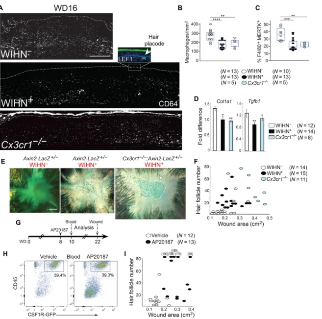
S5A). In accordance, Cx3cr1−/− wounds phenocopied WT WIHN+ wounds in that they maintained reduced collagen deposition (fig. S5B), reduced mRNA levels of profibrotic genes Col1a1 and Tgfb1 (Fig. 3D), and reduced Wnt activity as determined by AXIN2 ex-pression in Wnt reporter Axin2-Lacz+/− wounds (Fig. 3E) and ex-hibited significantly higher hair neogenesis associated with decreased wound contraction (Fig. 3F).
To specifically correlate late wound macrophage numbers with wound fate, we used the macrophage Fas-induced apoptosis (MAFIA) mouse model, which harbors a myeloid-specific colony-stimulating factor 1 receptor (Csf1r) promoter–driven AP20187 drug-inducible apoptosis cassette and green fluorescent protein (GFP) tag (41). A single injection of AP20187 at WD8 reduced blood monocyte numbers
on April 10, 2020
http://advances.sciencemag.org/
by 33% by WD10 (Fig. 3, G and H). Examination of wounds in the AP20187-treated mice showed that they phenocopied WIHN+ WT and
Cx3cr1−/− wounds with significantly increased hair follicle neogenesis and decreased contraction compared to vehicle-treated animals (Fig. 3I). Thus, decreased numbers of late wound macrophages promote a regenerative process rather than fibrogenesis, and this decision is implemented after initiation of the remodeling phase.
Late wound macrophages target Wnt inhibitor SFRP4 for phagocytosis and degradation
To ask whether macrophages might be linked to chronic Wnt activity in fibrotic wounds, we first compared their location with dermal Wnt activity in Axin2-LacZ+/− wounds at WD16. Significant -galactosidase (-Gal) expression, indicative of strong Wnt activity, was observed in the lower dermis of WIHN− wounds, the primary location of Fig. 3. Macrophage numbers in the late wound correlate inversely with regenerative WIHN+ repair. (A) CD64 (white), LEF1 (green) in WD16 WT WIHN− (top),
WIHN+ (middle), and Cx3cr1−/− (lower) wounds. Inset (middle) shows LEF1+ hair placode and dermal condensate (green, arrowhead). Scale bar, 500 m. Dashed line
denotes epidermal:dermal boundary. (B and C) Dot plots show macrophage numbers per mm2 lower dermis (B) or percent per whole wound dermis by FACS analysis (C)
in WD14 to WD16 WIHN− (white), WIHN+ (black), and Cx3cr1−/− (blue) wounds. See fig. S5A for corresponding representative cytometric dot plots. *P value is 0.05;
**P value is 0.01; ***P value is 0.005; ****P value is 0.001. (D) qRT-PCR of Col1a1 and Tgfb1 in WT WIHN− (white), WIHN+ (black), and Cx3cr1−/− (blue) wound dermis. See
Fig. 1 for details. (E) Whole mounts of Axin2-LacZ+/− WIHN− (left), WIHN+ (middle), and Axin2-LacZ+/−:Cx3cr1−/− (right) wounds stained for -galactosidase (-Gal) activity.
Circles outline regions of Axin2+ hair placodes and germs. Scale bar, 1 mm. Representative of four independent experiments. (F) Correlative comparison of wound areas
(cm2) and hair follicle numbers in WT WIHN− (white), WIHN+ (black), Cx3cr1−/− (blue) healed wounds. One-way MANOVA significance F (4,74) = 12.214, P (0.000) < 0.0005,
Pillai’s Trace = 0.795, 2 = 0.398. (G) Timeline for injections and analyses of MAFIA wounds. (H) Cytometric dot plots compare percent CD45+CSF1R+ blood monocytes from
WD10 vehicle-treated (left) and AP20187-treated (right) animals. Representative of four independent experiments. CD45+CSF1R− cells in both groups are T cells. (I)
Correla-tive comparison of wound area (cm2) and hair follicle numbers in vehicle-treated (white) and AP20187-treated (right) wounds. True hair follicle numbers for wounds
with >100 hair follicles are stated above dots. One-Way MANOVA F (2,22) = 21.386, P (0.000) < 0.0005, Pillai’s Trace = 0.660, 2 = 0.660.
on April 10, 2020
http://advances.sciencemag.org/
MERTK+ macrophages (Fig. 4A, left). More detailed examination of this region revealed that Wnt-activated cells frequently resided directly adjacent to macrophages (Fig. 4B; see also Fig. 4J), suggest-ing that macrophages exert their influence over a short distance.
Macrophages are known to secrete Wnt ligand WNT3a and exhibit Wnt activity after tissue damage, and macrophage-secreted
WNT3a is implicated as a player in both tissue regeneration and fibrosis (23). Examination of macrophage DEGs from both pheno-types revealed no Wnt ligand expression, and quantitative reverse transcription polymerase chain reaction (qRT-PCR) confirmed that macrophages neither expressed Wnt ligands Wnt3a and Wnt2 nor did they exhibit marked Wnt activity at WD16 (Fig. 4C). As
Fig. 4. Macrophages phagocytize and degrade Wnt inhibitor SFRP4 in the lower wound dermis. (A) -Gal (white, left) and MERTK (green, right) localization in WD16 Axin2-LacZ+/− WIHN− (left) and WIHN+ (right) wounds. Scale bar, 100 m. Representative results from six experiments. (B) Left: Close apposition of -Gal+ cells (pink) and
MERTK+ macrophages (green) in WD16 WIHN− Axin2-LacZ+/− dermis. DAPI (4′,6-diamidino-2-phenylindole) is blue. Scale bar, 10 m. Middle: Colocalization of macrophages (green) and -Gal+ cells (pink) in lower dermis. Scale bar, 100 m. Right (magnification): Arrowheads point to closely associated -Gal+ cells and macrophages. Bar graph indicates
percent of -Gal+ cells with closely apposing macrophages. (C) qRT-PCR for Wnt3a, Wnt2, Axin2, and Lef1 in sorted WT wound macrophages (yellow) and myofibroblasts
(pink). Combined results from three experiments. (D) Gene array comparison of major Wnt inhibitors between WD10 and WD12 (white) or WD10 and WD14 (black) wounds. Gene Expression Omnibus accession no. GSE46244 (29). (E) qRT-PCR of Sfrp4 in sorted wound populations. Combined results from three experiments. (F) qRT-PCR of Sfrp4 and Axin2 in cultured Wnt-activated wound myofibroblasts transduced with lentiviral control (white) or Sfrp4 (black) short hairpin RNAs (shRNAs). Combined results from three experiments. **P value is 0.01; ***P value is 0.005. (G) qRT-PCR of Sfrp4 in WD16 WT WIHN− (white) and WT WIHN+ (black) wounds. (H) SFRP4 (white) at
WD12 (left, top and bottom magnification) or WD16 (middle and right, respectively). WD16 wounds costained for MERTK (purple). Scale bar, 50 m. In magnified panels at far right, dotted lines represent macrophage boundaries. Representative results from six experiments. Stacked bar graph (right) shows percent of total WD16 WIHN− dermal
macrophages containing SFRP4 vesicles (black). (I) Left: MERTK (green), nuclear -Gal (pink), and SFRP4 (white) in WD16 Axin2-LacZ+/− lower dermis. Scale bar, 10 m. Right: Sequential slices (0.5-m difference) of color-merged magnified region (rectangle). Pink arrowheads point to -Gal+ cells, white arrowheads point to SFRP4.
Representa-tive results from eight experiments. (J) Top: SFRP4 (green) and LAMP1 (red) in WD16 WIHN− lower dermis. Dotted lines represent macrophage boundaries. Bottom (magnified):
Arrowsheads in left and middle panels correspond to regions of overlap. Scale bar, 10 m. Representative results from five experiments. In (A) and (H), dotted lines denote epidermal:dermal boundary. For (C) and (E) to (G), see Fig. 1 for details.
on April 10, 2020
http://advances.sciencemag.org/
previously shown, myofibroblasts were the primary producers of and responders to WNT2, the primary Wnt ligand in the late wound dermis (21).
To decipher how macrophages might promote chronic Wnt activity in WIHN− wounds, we first characterized the molecular player(s) that might down-regulate Wnt activity in WIHN+ wounds.
Comparison of known Wnt inhibitors from gene arrays of WD10, WD12, and WD14 wounds showed Sfrp4 to be the only member up-regulated after initiation of dermal Wnt activity in the late wound (Fig. 4D). qRT-PCR analysis of sorted myofibroblasts confirmed RNA-seq results that Sfrp4 is a myofibroblast product (Fig. 4E), and short hairpin RNA (shRNA) knockdown of Sfrp4 in Wnt-activated wound myofibroblasts in vitro showed a down-modulatory role for this protein in wound Wnt signaling (Fig. 4F). In agreement with recent evidence that Sfrp4 is a Wnt target gene (42), qRT-PCR con-firmed RNA-seq results that mRNA levels were significantly higher in Wnt-active WIHN− wounds during late healing (Figs. 2E and 4G). Together, these results indicate that Wnt activity in myofibroblasts is modulated by SFRP4 (secreted frizzled-related protein 4) in an autocrine fashion and that unabated Wnt activity in WIHN− wounds
persistently drives SFRP4 expression in an unsuccessful attempt to achieve Wnt down-modulation.
To determine SFRP4 protein localization in the healing wound, we first examined wounds at WD12, a time point when Sfrp4 mRNA levels are elevated (see Fig. 4D). Widespread SFRP4 expression was observed in all wounds surveyed (Fig. 4H, left), in accordance with widespread dermal Wnt activity observed at this time (22). SFRP4 expression was then examined at WD16, when Wnt activity was substantially down-modulated in WIHN+ wounds but maintained in WIHN− wounds. In accordance with Wnt activity, SFRP4 was
absent from the lower dermis of WIHN+ wounds but was observed in WIHN− wound dermis in Wnt-active macrophage-rich regions
(Fig. 4H, center, and fig. S6A for only extracellular localization of
SFRP4). Examination of SFRP4 in the WIHN− upper dermis, where residual levels could still be observed in both phenotypes, revealed a strand-like appearance along myofibroblasts and ECM, whereas in lower dermis, it showed a punctate pattern within many macro-phages (Fig. 4H, right). Detailed microscopic examination of SFRP4 in the WIHN− lower dermis confirmed that, in addition to some
localization within the ECM, it localized primarily to vesicles within CD64+MERTK+ macrophages, which adhered closely to adjacent
Wnt-active cells (Fig. 4I; fig. S6, B and C; and movie S1). These SFRP4-containing vesicles sometimes costained for lysosomal marker LAMP1 (lysosomal associated membrane protein 1), suggesting targeting of SFRP4 by lysosomes for degradation (Fig. 4J).
To ask whether excessive phagocytosis of SFRP4 by macrophages might lead to the WIHN− phenotype, we inhibited phagocytosis in late
wounds by injecting hemin into mice. Work by Martins et al. (43) has demonstrated that injected hemin specifically inhibits phago-cytosis by macrophages and resistance to introduced bacteria in vivo. Preliminary experiments showed that hemin efficiently inhibited the ability of bone marrow (BM)–derived IL-4–polarized (regenerative) macrophages to phagocytize FN-coupled beads in vitro (fig. S7A). We then injected hemin into wounded mice after initiation of the remodeling phase (see Fig. 5A for schedule). Both vehicle [phosphate- buffered saline (PBS)]– and hemin-injected wounds underwent normal reepithelialization by WD14, and examination of wounds at WD18 showed no differences in numbers of CD45+ cells, macro-phages, or vasculature (fig. S7, B to F). Wounds were then examined for phagocytosis of SFRP4. Immunofluorescence analyses showed that SFRP4 vesicles were essentially absent from hemin-injected wound macrophages compared with WT WIHN− wounds despite normal macrophage numbers (Fig. 5B). Consequently, SFRP4 protein levels were significantly elevated in the wound ECM of these mice, and hemin-injected wounds exhibited hallmarks of a more regenerative healing including reduced chronic Wnt activity, reduced collagen
Fig. 5. Inhibition of phagocytosis in wound macrophages promotes regenerative healing. (A) Timeline showing hemin injection and analysis schedule. (B) Top: Localization of SFRP4 (green) within macrophages (purple) in lower wound dermis from PBS-injected (left) and hemin-injected (right) mice. Scale bar, 50 m. Bottom: Magnified view of SFRP4 (green) and MERTK (purple) in PBS-injected (right) and hemin-injected (left) wound dermis. White denotes regions of colocalization. Scale bar, 25 m. Representative of six experiments. (C) Left: Threshold-matched comparison of SFRP4 (white) in wounds from PBS-injected (left) and hemin-injected (right) mice. Scale bar, 100 m. Right: Scatter plot comparing SFRP4 (corrected total cell fluorescence) in wound dermis from PBS-injected (white) and hemin-injected (black) mice. (D) Bar graph shows percent dermal cells with nuclear -Cat localization in PBS-injected (white) and hemin-injected (black) wounds. (E) Correlative comparison of healed wound surface areas (cm2) and hair follicle numbers in PBS-treated (white) and hemin-treated (black) wounds. One-way MANOVA significance F (2,28) = 10.658,
P (0.000) ≤ 0.005, Pillai’s Trace = 0.432, 2 = 0.432. Data are expressed as means ± SEM. ***P value is 0.005, ****P value is 0.001.
on April 10, 2020
http://advances.sciencemag.org/
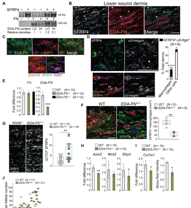
Fig. 6. Extracellular EDA-FN acts as a bridge for integrin-mediated phagocytosis of SFRP4. (A) Western blot of pull-down of cellular FN containing high (HI) or low (LO) EDA concentrations with SFRP4-bound beads. Protein molecular weights at the left. Relative densities are indicated. See fig. S8A for details. Representative of three independent experiments. (B) Localization of SFRP4 (left) on EDA-rich strands (middle) in ECM (*) and macrophages (arrowheads) in lower dermis. Scale bar, 10 m. Rep-resentative of six independent experiments. (C) Top: EDA-FN (green, left), LAMP1 (red, center), merge (right) in lower wound dermis. Scale bar, 10 m. Bottom: MERTK+
macrophages (dotted lines) stained for SFRP4 (green) and EDA-FN (red, left) or RAB7 (purple, right) in lower dermis. Scale bar, 5 m. Representative of nine independent experiments. (D) Top: SFRP4 (green) and 5-integrin (white) colocalization in WD16 lower dermis. See fig. S8D for details. Scale bar, 50 m. Stacking bar graph shows percent (above bar) of total macrophages (left) or other cells (right) positive for 5-integrin + SFRP4 (black). Four percent overlap in other cells was predominantly vascu-lature. Bottom: Colocalization of EDA-FN (red), SFRP4 (green, left), 5-integrin (5-integrin, white, right) in WD16 wound lower dermis. Arrowheads point to colocalization. Representative of six independent experiments. (E) qRT-PCR of WD16 WT (white), EDA-FN+/− (green), and EDA-FN−/− (black) wounds for total Fn1 (left) and Eda-FN (right). (F) Left: SFRP4 (green) and LAMP1 (red) colocalization in WT (left) and EDA-FN+/− (right) lower wound dermis. Scale bar, 10 m. (See fig. S9E for LAMP1 and MERTK colocalization in these images). Right: Scatter plot shows relative number of SFRP4 vesicle–bearing macrophages/mm2 in WD16 WT (white) versus EDA-FN+/− (green)
wounds. (B to D and F) Dotted lines represent macrophage boundaries. (G) Left: Threshold-matched comparisons for SFRP4 in WD16 WT WIHN+ (left), WT WIHN− (middle),
and EDA-FN+/− (right) wounds. Scale bar, 50 m. Right: Quantitation of SFRP4 (CTCF) in WT (white) and EDA-FN+/− (green) wounds. (H and I) qRT-PCR of WT (white) and EDA-FN+/− (green) wound dermis for indicated genes. (I) Right: Sirius red intensity in WT (white) and EDA-FN+/− (green) wounds. (J) Correlative comparison of wound areas
(cm2) and hair follicle numbers in WT (white) and EDA-FN+/− (green) wounds. One-way MANOVA significance F(2,36) = 6.383, P (0.004) < 0.005, Pillai’s Trace = 0.262, 2 = 0.131. For (E), (H), and (I), see Fig. 1 for details.
on April 10, 2020
http://advances.sciencemag.org/
deposition, and reduced contraction correlating with increased hair neogenesis (Fig. 5, C to E, and fig. S7G).
These combined results suggest that high numbers of macrophages can prevent Wnt down-modulation in the wound and promote a fibrotic WIHN− phenotype through persistent degradation of an important Wnt inhibitor, SFRP4, and that inhibition of this macro-phage function can reverse this phenotype.
Phagocytosis of SFRP4 by late wound macrophages is integrin dependent and requires association with EDA-FN
We next asked how macrophages phagocytize SFRP4. Related SFRP family member SFRP2 can associate with FN (44), a known mediator of bacterial or apoptotic cell phagocytosis by macrophages (29). We therefore asked whether SFRP4 might require association with FN for phagocytosis, and we focused on extra domain A (EDA)–FN, the predominant FN splice variant in wound dermis (45).
Protein pull-down experiments showed that SFRP4 associated preferentially with EDA-rich FN compared to cellular FN contain-ing other splice variants (Fig. 6A and fig. S8A for details). Analysis of frozen wound sections showed localization of SFRP4 to EDA-FN strands in the ECM (Fig. 6B) and many EDA-FN–containing vesicles within wound dermal macrophages, some of which localized to LAMP1+ lysosomes (Fig. 6, B and C). SFRP4 colocalized with EDA-FN within some vesicles in wound macrophages and both localized to RAB7+ late endosomes (Fig. 6C). In vitro phagocytosis studies showed that BM-derived IL-4–polarized macrophages could ingest and target SFRP4-bound EDA-FN–coupled beads to LAMP1+ lysosomes (fig. S8B). EDA-FN can also interact with Toll-like receptor 4 (TLR4), and this interaction, in association with integrins on fibroblasts, can drive fibrosis (46, 47). Tlr4 mRNA levels were reduced by WD12 (fig. S8C), and single-cell RNA-seq DEG lists of WIHN+ and WIHN− dermal populations did not bring up either Tlr4 or Elane (elastase-2), the enzyme required to expose the cryptic EDA domain for binding, in any wound population (48). Violin plots confirmed very low to no expression of Tlr4 in both myofibroblast and macrophage/DC populations, and dot plots of integrated myofibroblasts showed that when Tlr4 could be detected, its expression was often comparable or higher in WIHN+ clusters (fig. S8C). Together, these results indicate
that the SFRP4:EDA-FN complex can be phagocytized and targeted for degradation by wound macrophages and that TLR4:FN interactions do not play a role in fibrosis during this period in this model.
As integrins are FN receptors and are required for FN-mediated phagocytosis (29), we studied their role in phagocytosis of the SFRP4:EDA-FN complex. In vivo examination revealed frequent colocalization of 5-integrin, an established FN receptor (49) with SFRP4 specifically within wound macrophages (Fig. 6D, top, and fig. S8D), and detailed analyses confirmed colocalization of both SFRP4 and EDA-FN with either 5-integrin or its binding partner 1-integrin within wound macrophages (Fig. 6D, bottom, and fig. S8E). In vitro pretreatment of BM-derived IL-4–polarized macrophages with blocking anti–1-integrin antibody completely abrogated phagocytosis of EDA-FN beads (fig. S8F). Together, these observations suggest that EDA-FN acts as a bridge to target SFRP4 for integrin-mediated phagocytosis by macrophages.
We hypothesized that if EDA-FN targets SFRP4 for phagocytosis, then loss of this protein should result in a wound phenotype similar to that observed in phagocytosis-deficient hemin-treated mice (see Fig. 5). To address this, we examined wounds from EDA-FN−/− and
EDA-FN+/− mice. Wounds from EDA-FN−/− mice had no expression
of EDA-FN and maintained high levels of compensatory EDB-FN (Fig. 6E and fig. S9, A and B). Compared to WT, EDA-FN−/− wounds
revealed a distinct nonfibrous pattern of FN deposition and very little FN:SFRP4 colocalization, further confirming that SFRP4 pref-erentially binds EDA-FN (fig. S9B and see Fig. 6B for comparison).
EDA-FN−/− mice have been shown to exhibit some wound healing
defects (50), prompting us to use EDA-FN+/− mice for our studies. EDA-FN+/− wounds had significantly reduced EDA-FN mRNA and
protein levels (Fig. 6E and fig. S9A), although wounds healed completely, without significant delay and exhibited normal dermal remodeling (fig. S9, A to C). Further, EDA-FN+/− wound dermis maintained normal numbers of late wound macrophages with normal capacity to phago-cytize EDA-FN–coupled beads (fig. S9, C and D).
To ask whether reduced expression of EDA-FN might affect SFRP4 turnover by wound macrophages, comparisons of EDA-FN+/− and WT wounds were undertaken. In EDA-FN+/− wounds, SFRP4 localization
within macrophages, and specifically, within LAMP1+ lysosomes, was significantly reduced (Fig. 6F). In agreement with this finding, increased SFRP4 protein levels were found in the ECM of EDA-FN+/−
wound dermis (Fig. 6G). EDA-FN+/− wounds also exhibited reduced
Wnt activity (Fig. 6H), reduced collagen transcription and protein expression (Fig. 6I and fig. S9A), reduced wound contraction, and significantly higher hair neogenesis than WT littermates (Fig. 6J), all indicative of a more regenerative fate. Together, these results show that EDA-FN+/− wounds phenocopy phagocytosis-deficient wounds, indicating that EDA-FN is important for SFRP4 turnover by wound macrophages.
EDA-FN and SFRP4 expressed in human hidradenitis suppurativa wound dermis are targeted by macrophages for phagocytosis
Hidradenitis suppurativa (HS) or acne inversa is a common chronic skin disorder characterized by recurring follicular occlusions and in-flammation, leading to recurring localized wounding and subsequent fibrotic scarring (see fig. S10A for a representative diagram of these regions in HS skin) (51). Although macrophages are known important contributors to this disease, their role has not been established (52). In agreement with previous observations of granulation tissue formation in this disease, we found that regions i and ii (see fig. S10A) contained cells expressing EDA-FN, SFRP4, and SMA, whereas long-standing fibrotic regions from the same patient exhibited high levels of collagen but like normal skin, low levels of these proteins (figs. S10, B and C, and S11A). As observed in mouse wounds, EDA-FN frequently colocalized with SFRP4 in new wounds, indicating their close association (fig. S11B).
Immune infiltrates (region i) and adjacent dermis (region ii) were rich in CD68+ macrophages, many with vesicles containing SFRP4 (fig. S10D). Close examination of these vesicles showed occasional colocalization of SFRP4 with EDA-FN and LAMP1 (fig. S10E), sug-gesting that human wounds, such as mouse wounds, target SFRP4 for EDA-FN–mediated phagocytosis and subsequent degradation. Last, nuclear -Cat localization remained high in immune infiltrate (region i), adjacent dermis (region ii), and distant fibrotic regions (fig. S10, F and G), indicating chronic Wnt activity throughout HS- damaged skin. qRT-PCR comparisons of HS and normal dermis confirmed high Wnt activity and increased expression of fibrogenic genes (fig. S10H).
These results highlight the notable similarities between mouse and human wounds and suggest that phagocytosis of SFRP4 by
on April 10, 2020
http://advances.sciencemag.org/
macrophages may be a common mechanism for fibrotic skin wound healing in these species.
DISCUSSION
In this work, we show that late stage wound macrophages phagocytize and degrade wound Wnt inhibitor SFRP4 to drive chronic Wnt activity to promote fibrogenesis over regeneration. ECM component EDA-FN binds SFRP4 and acts as a bridge for its engulfment (model, see fig. S12). Obstruction of phagocytosis is alone sufficient to alter wound fate from fibrotic to a more regenerative response, indicating that this mechanism is a major contributor to skin fibrotic repair and may have broader relevance than to skin wound healing alone. Here, we show that the key component is an FN bridge, hypotheti-cally capable of binding many different substrates for phagocytosis. Thus, other environmental mediators might also be targets of this mechanism. In addition, ECM components such as collagen, which are subject to mannose receptor–mediated remodeling by macro-phages (31), might serve as alternative tethering components.
Several important questions remain. First, what earlier healing events determine the disparate macrophage density observed in WIHN+ and WIHN− wounds? Results by Kasuya et al. (35) suggest
that regenerative macrophages may be present by WD7. Inflamma-tion is an early event that sets the stage for all later healing, and the extent of inflammation likely affects the ultimate wound fate. How-ever, whether this impact is reflected through early recruitment of large numbers of macrophages that later contribute directly to re-modeling or to later events, which may drive de novo migration of macrophages into the wound, remains unknown. Second, RNA-seq analyses revealed not only the presence of high numbers of macro-phages in WIHN− wounds but also their increased transcription of phagosomal/lysosomal genes. Recent work has shown that phago-cytosis can augment subsequent phagophago-cytosis in a receptor-independent manner through translocation of transcription factor TFEB (transcrip-tion factor EB) to the nucleus and up-regula(transcrip-tion of lysosomal genes such as Ctsd (53). Here, we show that both TFEB and Ctsd tran-scripts are up-regulated in WIHN− macrophages. We postulate that large numbers of macro phages initiate widespread loss of SFRP4 to prolong Wnt activity. Once this cascade has begun, increased pro-duction of fibrosis signature protein FN1 would provide additional fodder for uptake by an increasingly efficient phagocytic population, thus further promoting an autofeedback loop for continued Wnt activity and fibrotic repair.
Multiple myofibroblast subsets, including PDGFRa+ AP-type
(groups I and II in this study) and PDGFRb+ classical myofibroblasts (group III in this study), have been identified in lung, skin, adipose tissue fibrosis (36–38). The importance of AP-type myofibroblasts to fibrosis in adipose tissue has been shown (38) and, in small skin wounds, which invariably heal in a scarring phenotype, AP-type myofibroblasts contribute to skin fibrotic repair (36). In unbiased transcriptome comparisons of bleomycin-induced fibrosis versus normal lung, Xie et al. (54) observed expansion of multiple PDGFRa+
and PDGFRb+ myofibroblast populations and described a previ-ously unidentified PDGFRb+ subset with elevated expression of
Notch3 and fibrosis signature genes, which they concluded might provide a true lung fibrogenic myofibroblast phenotype. Our single- cell RNA-seq comparisons also revealed fibrogenic PDGFRa+ (groups I and II) and PDGFRb+ (group III) subtypes in the WIHN− wound,
although there was preferential screwing toward group III. All groups
exhibited elevated fibrosis signature genes. These results support the hypothesis that both classical and AP-type myofibroblasts contribute to fibrosis in skin wound healing and that classical myofibroblasts may prove the most important for long-term fibrosis maintenance.
In small skin wounds, F4/80+CD206+CD301b+ myeloid cells were shown to express IGF1 and PDGFC. Further, these cytokines induced proliferation of APs, promoting their fibrogenic involvement (36). In other studies, alternatively activated macrophages have been shown to secrete RELM- promoting profibrotic collagen cross-linking (26). In our studies, WIHN− macrophage/DC populations had slightly
elevated Igf1 within a DC subset and no expression of either PDGFC or RELM-. These differences in results likely reflect the wound type (small versus large), timing, and/or the possibility that some of these proteins may be long lived in the ECM and require little new tran-scriptional activity for their continued functioning. In the WIHN wound model, we found up-regulated phagocytosis by wound CD64+MERTK+ macrophages in fibrotic wounds. Abrogation of this function resulted in a more regenerative repair, demonstrating that phagocytosis is a keystone mechanism of fibrosis in this model.
Expanding on the demonstration that macrophage-elicited tumor necrosis factor (TNF) promotes hair regeneration after plucking (55), Wang et al. (34) recently reported the same requirement for hair neogenesis after wounding. In support of this, Chen et al. (55) showed that macrophage-elicited TNF can promote Wnt ligand secretion by epidermal cells after hair plucking. As epidermal Wnt expression is a requisite for hair neogenesis (33), TNF production during reepithelialization could provide a potentially important pathway to placode induction. Our single-cell RNA-seq analysis revealed low expression of TNF by macrophages at WD18 in regenerative wounds and slightly increased expression in fibrotic wounds. Given the re-markable plasticity of macrophages in response to their environment, it is likely that their gene signatures change rapidly, as wound der-mis transforms during the healing process making it plausible that TNF levels 3 to 4 days earlier may indeed be elevated in WIHN+
wounds.
Wnt activity has been reported to affect the TGF pathway in a variety of scenarios, including wounding (13, 15). Our results suggest that elevated Wnt activity may directly affect TGF signaling for chronic activation of both and that sustained high Wnt activity precedes up-regulation of TGF and its targets by at least several days, thus making it an earlier benchmark for the cross-talk between Wnt and TGF pathways in fibrogenesis.
How fibrogenesis in wound healing precludes hair neogenesis remains unclear. Recently, Rognoni et al. (56) compared small wounds from young and adult mice, which result in hair neogenesis or scarring, respectively, and found that increased Wnt activity in older wounds correlated with loss of hair neogenesis. This finding supports the hypothesis that high chronic Wnt precludes hair neogenesis. High Wnt activity in fibrogenic wounds might promote fibrogenic over DP-forming programs in myofibroblasts, or because dermal Wnt activity precedes and is necessary for subsequent epidermal Wnt activation (21, 57), chronic dermal Wnt might indirectly affect placode initiation in the overlying epidermis. Because overexpres-sion of SHH can override some aspects of the fibrotic wound en-vironment to permit hair neogenesis suggests that it may also override chronic Wnt activity or its impact in small wounds (58). Last, hair neogenesis is absent from adult small wounds and only observed in the center of large wounds, suggesting a paracrine inhibitory effect by the surrounding unwounded environment. Therefore, wound
on April 10, 2020
http://advances.sciencemag.org/
contraction alone might be sufficient to place WIHN− epidermis and upper dermis closer to these external inhibitory influences, including Wnt ligands.
In mammals such as humans and rats, hair neogenesis is a very rare occurrence following wounding, likely reflecting rapid and sustained inflammatory responses to manage infection and augment rapid wound closure at the cost of a more regenerative response. In humans, SFRP4 expression has been associated with fibrotic skin diseases such as psoriasis and Dupuytren’s disease (59, 60). Here, we show that extensive degradation of SFRP4 by macrophages in immu-nologically active zones in HS skin correlates with chronic Wnt activity and fibrogenesis, conditions that are also common to Dupuytren’s syndrome (60). These results underscore the importance of modulating Wnt activity in wounding responses to chronic fibrotic skin disorders like HS and Dupuytren’s syndrome, surgery, radio-therapy, and possibly tumorigenesis and suggest that modulation of regenerative macrophage activity in particular may provide novel therapeutic avenues for wound treatment.
MATERIALS AND METHODS
Study design
The purpose of this study was to identify and understand the mech-anisms used by late wound macrophages to affect skin regeneration and fibrosis. All animal protocols were reviewed and approved by the French Research Ministry and Institutional Animal Care and Use Committee. Sample size was determined from previous experi-mentation, and end points for data collection were reached when statistical significance was achieved or for immunofluorescence ex-periments not dependent on statistical analyses, at least three com-parable examples were obtained. All animal studies were conducted on 3 to 30 biological replicates. To avoid bias, all animals were housed identically, and WT littermates were maintained with and compared to treated or genetically altered mice at ages ranging from 6 to 8 weeks. No formal randomization or blinding was performed, but results were confirmed by at least two independent experiments. See figure legends for replicate sizes. Control and HS skin was collected from male and female patients following consent of the individuals and approval by the Ethics Commission of the Medical Faculty of the University of Leipzig.
Human skin used in these studies
Human skin samples were obtained after informed consent from patients undergoing reconstructive surgery procedures (controls) and from patients receiving surgical removal of stage 2 and 3 HS lesions with the approval of the Ethics Commission of the Medical Faculty of the University of Leipzig (Az. 428/16-ek). HS material was collected from surgical excisions containing lesioned skin only. The samples were collected immediately after surgery and embedded in optimal cutting temperature compound (OCT), frozen, and kept at −80°C until cryosectioning or transferred to RNAlater (Sigma) and frozen at −80°C for later qRT-PCR analyses.
Mice used in these studies
The transgenic and knockout mice used in these studies include Cx3cr1−/− mice (JAX stock 005582), AXIN2-LacZ reporter mice
(JAX stock 009120), MAFIA mice (JAX stock 005070), EDA-FN−/−, and EDA-FN+/− mice (European Mouse Mutant Archive repository).
EDA-FN−/− mice were further backcrossed onto a C57BL/6J
back-ground to achieve an N of 10 to 11. C57BL/6J control mice were purchased from Charles River Laboratories, France.
Wounding protocol
Full thickness excision of skin was performed on the backs of adult 6- to 8-week-old mice under isoflurane anesthesia as previously described (35). Mice received 1.2 cm × 1.2 cm full thickness excision wounds on the lower back in all experiments. Animals received analgesic buprenorphine before surgery and 8 and 24 hours later to ameliorate pain. All animal protocols were reviewed and approved by the French Research Ministry and Institutional Animal Care and Use Committee (Bioproj APAFIS#2673-2015111317111206). Wounds typically healed completely within 14 days with extensive dermal remodeling and complete reepithelialization. Wounds from all trans-genic and knockout mouse lines healed completely and without ex-tensive delay (no more than 48 hours). In phagocytosis inhibition experiments, hemin (Sigma 51280) was prepared and injected as previously described (46) into wounded mice at WD10 and WD12. In all experiments, mice were maintained in microisolator cages to reduce the risk of infection.
Tissue treatment, hair neogenesis, and wound size analyses
Healed skin was taken 2 to 14 days (WD16 to WD28) after reepitheliali-zation, and epidermis and dermis were separated using 20 mM EDTA or dispase. To determine the number of hair placodes in WD16 skin, epidermis was stained with anti–LEF1-GFP antibody, and placodes were recorded throughout the entire epidermis using a Leica TCS SP8 confocal microscope for tiling acquisition, followed by analysis with ImageJ software. Dermis was dissociated as previously described (22), and cells were analyzed by FACS, qRT-PCR, or cell sorting and subsequent qRT-PCR analyses (see below). Wounds were photographed using a Leica MZ6 stereomicroscope fitted with a high-resolution digital camera, and digital images were analyzed for surface area using ImageJ software. New hairs were counted by direct microscopic visualization, and numbers were verified by dermal NBT/BCIP (nitro blue tetrazolium/5-bromo-4-chloro-3-indolyl phosphate) staining as previously described (35).
Whole-mount assays to detect -Gal activity
To detect -Gal activity in AXIN2-LacZ+/− wounds, tissue was treated
as previously described (22).
Antibodies used in these studies
For macrophage identification and dermal sorting, the following antibodies were used: anti-F4/80 (clone BMB, eBioscience), anti- CD64 allophycocyanin (APC; X54-5/7.1, BioLegend), anti-MERTK biotin (R&D Systems BAF591), anti-CD301b Alexa Fluor 647 (LOM-8.7, BioLegend), anti-CD206 APC (C068C2, BioLegend), anti- CD45 (clone 30-F11, BD Biosciences), anti-CD31 (clone MEC 13.3, BD Biosciences), anti–CD11b-APC (clone ICRF44, eBioscience), anti–CD209-AF647 (9E9AB, BioLegend), and anti–CD68-fluorescein isothiocyanate (Y1/82A, BioLegend). For hair placode identification, the following antibodies were used: LEF1 (Cell Signaling C12A5). To detect ECM proteins, the following antibodies were used: SFRP4 monoclonal (Abcam EPR9389) and SFRP4 polyclonal (PA5-52679, Thermo Fisher Scientific); FN: Rabbit polyclonal (Abcam ab23750), EP5 (MA1-12597, Thermo Fisher Scientific), EDA-FN clone IST-9 (Abcam ab6328), and anti-FN biotin (Abcam ab6584); Cola1a: Rabbit polyclonal (PA5-29569, Thermo Fisher Scientific). Anti–-Cat
on April 10, 2020
http://advances.sciencemag.org/
(Abcam ab6302) and anti–-GAL (Abcam ab9361) were used for Wnt activation analyses. To detect integrins and intracellular trafficking vesicles the following antibodies were used: 1-integrin (HM1-1, BioLegend), 5-integrin (5H10.27, Invitrogen), RAB7 (Abcam EPR7589), and LAMP1 (1D4B, BioLegend). LEAF Purified anti- mouse/rat CD29 antibody (HM1-1, BioLegend 102210) was used for phagocytosis assays.
Phagocytosis assays
Generation of BMDM (bone marrow-derived macrophage)–derived IL4-polarized (M2) macrophages: BM- derived monocytes from WT or EDA-FN+/− mice were obtained by flushing BM from adult WT
mouse femora and tibias, followed by removal of red blood cells with ammonium chloride (07850, STEMCELL), filtration through a 70-m-mesh filter (Becton Dickinson), and incubation for 3 hours in Iscove’s modified Dulbecco’s medium (IMDM) GlutaMAX (31980, Thermo Fisher Scientific) supplemented with 10% fetal bovine serum (Gibco), 1% penicillin-streptomycin (Life Technologies), and 150 M monothioglycerol (Sigma-Aldrich). Nonadherent cells, seeded at a density of 3 × 10⁴ cells/ml, were cultured in IMDM medium
containing mouse CSF (25 ng/ml; 130-101-705, Miltenyi Biotec) for 7 days, changing medium every 2 days. From days 7 to 8, medium was supplemented with mouse IL-4 (10 ng/ml; Miltenyi Biotech) to facilitate differentiation of monocytes into macrophages.
Coupling of EDA-FN to beads: FNnb (EDA-FN, see fig. S7A) was coupled to Fluoresbrite Carboxylate YG 1.0-m microspheres (Polysciences, catalog no. 15702) using the PolyLink Protein Coupling Kit (Polysciences, catalog no. 24350-1), according to the manufacturer’s instruction. The extent of association was determined by cytofluoro-graphic analyses. For studies examining SFRP4 phagocytosis, a 1:1 ratio of recombinant SFRP4 protein (1827-SF-025, R&D Systems) to FN bound to beads was incubated with FN-bound beads overnight and extent of association determined by cytofluorographic analyses.
Phagocytosis assay: Differentiated IL-4–polarized macrophages were preincubated with or without 3, 10, 15, and 30 M hemin (Sigma 51280) for 30 min as previously indicated (61) or with LEAF Purified anti-mouse/rat CD29 antibody (20 g/ml; HM1-1, BioLegend) or with concanamycin A (10 M, Sigma C9705; for colocalization of FN, SFRP4, LAMP1 studies) (61) for 1 hour, followed by addition of FN- coupled beads, SFRP4-bound FN-coupled beads, or uncoupled beads for 1 hour at 37°C. Cells were then fixed with 4% paraformaldehyde and treated with anti-FN antibody-biotin (Abcam ab6584), followed by secondary antibody and analyzed by cytofluorography (Fig. 5A). Loss of binding to anti-FN antibody indicated that beads were internalized (61). For SFRP4 studies (Fig. 5B), fixed cells were analyzed for intracellular SFRP4 and LAMP1 as described for other immunofluorescence analyses (see above).
Immunofluorescence, Sirius red staining, and intensity quantitations
Tissue, flash frozen in OCT at −80°C, was cryosectioned into 12- to 14-m-thick sections, and sections were fixed with 4% para-formaldehyde as previously described (62). For examination of extracellular and membrane proteins, sections were preblocked in PBS containing 1% bovine serum albumin (BSA), 1:10 anti-mouse CD16/CD32 Fc block (eBioscience), and then stained with the appropriate antibodies in 1% BSA/PBS/Fc block for 2 hours at 4°C followed by fixation. To further visualize internal antigens, sections were then incubated overnight with the appropriate antibodies in
PBS containing fish gelatin and saponin, followed by fixation (62). To identify nuclear localization of -Cat, fixed sections were first permeabilized with 0.3% Triton X-100, followed by incubation with anti–-Cat antibody (Abcam ab6302) in saponin block. All sections were mounted with Vectashield DAPI (4′,6-diamidino-2- phenylindole; Vector Laboratories, H-1200) and examined using a white light TSC SP8 Leica scanning confocal microscope. Sirius Red and fluorescence intensities (CTCF) in threshold-matched images were determined using ImageJ.
FACS analyses and cell sorting for qRT-PCR
The basic staining protocol for sorting of wound populations was undertaken as previously described. Briefly, dissociated wound dermal cells were stained for CD45, CD64, CD31, and propidium iodide to exclude dead cells. Gated populations were defined as CD45+CD64+
macrophages, CD45+CD64− lymphocytes (including T cells, B cells,
etc.), CD45−CD31+ endothelial cells, and all other CD31−CD45− cells as
myofibroblasts. FACS analyses were undertaken using a FACSCanto A or LSRii and cell sorting using FACSAria and FACSDiVa software. Data were analyzed using FlowJo software.
Single-cell RNA-seq of WIHN+ and WIHN− wound dermis
WD18 epidermis and dermis were separated, and epidermis were stained for LEF1 to assay for hair placodes. WIHN+ and WIHN–
wounds were defined on the basis of the appearance of placodes and on increased wound size. Approximately 7 to 10 wounds of each phenotype were combined, dermal cells were released by collagenase treatment (Worthington Labs) and filtered, and dead cells and debris were removed using the MACS Live-Dead kit (Miltenyi Biotec). Cells were subjected to 10× Genomics Chromium Single-Cell Platform manipulation, followed by sequencing using Illumina HiSeq 4000 system, and then results were run through Cell Ranger pipeline software for sequence alignment and basic filtering. The following expression matrices were used: WIHN+: capture of approximately 10,500 dermal cells
with average read depth of approximately 41,000 reads per cell across 19,000 genes with 3200 median unique molecular identifiers; WIHN−:
capture of approximately 5500 dermal cells with average read depth of approximately 70,000 reads per cell across more than 17,000 genes with over 2400 median unique molecular identifiers. Expression matrices underwent filtering, normalization, scaling, principal com-ponents analysis, and subsequent t-SNE analysis using Seurat packages (63). Groups with low gene expression were removed from analysis. Seurat packages were also used for CCA (canonical correlation analysis)–based integration studies (63). Seurat-based software was used to generate DEGs, feature plots, dot plots, and violin plots. Differ-ential expression for a specific cluster was determined by comparison against all other clusters.
Macrophage/DC DEG lists were queried against the Immgen gene expression database (www.immgen.org) using the interactive tool “My Gene Set” to identify functional cell types and against the Database for Annotation, Visualization and Integrated Discovery (DAVID) functional annotation tools (David.ncifrf.gov) and STRING- compiled databases (string-db.org) to generate functional enrichment lists from GO (biological process, molecular function, and cellular component) KEGG, and Reactome pathways.
PCR and qRT-PCR
RNA from whole tissue, sorted, or cultured cells was isolated using RNeasy kits (QIAGEN). RNA concentration was assessed using a
on April 10, 2020
http://advances.sciencemag.org/