HAL Id: hal-02271087
https://hal.archives-ouvertes.fr/hal-02271087
Submitted on 26 Nov 2020HAL is a multi-disciplinary open access archive for the deposit and dissemination of sci-entific research documents, whether they are pub-lished or not. The documents may come from teaching and research institutions in France or abroad, or from public or private research centers.
L’archive ouverte pluridisciplinaire HAL, est destinée au dépôt et à la diffusion de documents scientifiques de niveau recherche, publiés ou non, émanant des établissements d’enseignement et de recherche français ou étrangers, des laboratoires publics ou privés.
DDX6 Cause Intellectual Disability and Dysmorphic
Features and Lead to P-Body Defects and RNA
Dysregulation
Chris Balak, Marianne Bénard, Elise Schaefer, Sumaiya Iqbal, Keri Ramsey,
Michèle Ernoult-Lange, Francesca Mattioli, Lorida Llaci, Véronique Geoffroy,
Maïté Courel, et al.
To cite this version:
Chris Balak, Marianne Bénard, Elise Schaefer, Sumaiya Iqbal, Keri Ramsey, et al.. Rare De Novo Missense Variants in RNA Helicase DDX6 Cause Intellectual Disability and Dysmorphic Features and Lead to P-Body Defects and RNA Dysregulation. American Journal of Human Genetics, Elsevier (Cell Press), 2019, 105 (3), pp.509-525. �10.1016/j.ajhg.2019.07.010�. �hal-02271087�
Rare De Novo Missense Variants in the RNA Helicase DDX6 cause Intellectual Disability and Dysmorphic Features and Lead to P-Body Defects and RNA Dysregulation.
Chris Balak1,2,23,*, Marianne Benard3,23, Elise Schaefer4,11,23, Sumaiya Iqbal5,6, Keri Ramsey1,2, Michèle Ernoult-Lange3, Francesca Mattioli7,8,9,10, Lorida Llaci1,2, Véronique Geoffroy11,Maité Courel3, Marcus Naymik1,2, Kristine K. Bachman12, Rolph Pfundt13, Patrick Rump14, Johanna ter Beest13, Ingrid M. Wentzensen15, Kristin G. Monaghan15, Kirsty McWalter15, Ryan Richholt1, Antony Le Béchec16, Wayne Jepsen1,2, Matt De Both1,2, Newell Belnap2, Anne Boland17, Ignazio S. Piras1,2, Jean-François Deleuze17, Szabolcs Szelinger1,2, Hélène Dollfus4,11, Jamel Chelly7,8,9,10,18, Jean Muller11,18, Arthur Campbell5,6, Dennis
Lal5,6,19,20,21, Sampathkumar Rangasamy1,2, Jean-Louis Mandel7,8,9,10,22, Vinodh Narayanan1,2,24,
Matt Huentelman1,2,24, Dominique Weil3,24, Amélie Piton7,8,9,10,19,24,*
1 Translational Genomics Research Institute (TGen), Neurogenomics Division, Phoenix, AZ 85004, USA
2 TGen's Center for Rare Childhood Disorders (C4RCD), Phoenix, AZ 85012, USA
3 Sorbonne Université, CNRS, Institut de Biologie Paris-Seine (IBPS), Laboratoire de Biologie du Développement, F-75005 Paris, France
4 Medical Genetics Department, University Hospitals of Strasbourg, The Institute of Medical Genetics of Alsace, 67000 Strasbourg, France
5 Stanley Center for Psychiatric Research, Broad Institute of MIT and Harvard, Cambridge, MA 02142, USA
6 Analytic and Translational Genetics Unit, Massachusetts General Hospital, Boston, MA 02114, USA
7 Institute of Genetics and Molecular and Cellular Biology, Illkirch, France
8 French National Center for Scientific Research, UMR7104, 67400 Illkirch, France 9 National Institute of Health and Medical Research U964, 67400 Illkirch, France 10 University of Strasbourg, 67081 Illkirch, France
11 Laboratoire de Génétique Médicale, Institut de génétique médicale d’Alsace, INSERM U1112, Fédération de Médecine Translationnelle de Strasbourg (FMTS), Université de Strasbourg, 67081 Strasbourg, France
12 Geisinger Medical Center, Dansville, PA 17822, USA
13 Department of Genetics, University Medical Center Groningen, University of Groningen, 9713 GZ Groningen, The Netherlands
14 Radboud University Nijmegen Medical Center, Department of Human Genetics, Division of Genome Diagnostics, 6525 GA Nijmegen, The Netherlands
15 GeneDx, Gaithersburg, MD 20877, USA
16 Medical Bioinformatics Unit, UF7363, Strasbourg Universitary Hospital, 67000 Strasbourg, France
17 Centre National de Recherche en Génomique Humaine (CNRGH), Institut de Biologie François Jacob, CEA, Université Paris-Saclay, F-91057, Evry, France
18 Molecular Genetics Unit, Strasbourg University Hospital, 67000 Strasbourg, France 19 Epilepsy Center, Neurological Institute, Cleveland Clinic, Cleveland, OH 44195, USA
20 Genomic Medicine Institute, Lerner Research Institute Cleveland Clinic, Cleveland, OH 44195, USA
21 Cologne Center for Genomics (CCG), University of Cologne, 50931 Cologne, Germany 22 University of Strasbourg Institute of Advanced Studies, 67081 Strasbourg, France 23 These authors contributed equally to this work
24 These authors contributed equally to this work
*Correspondance: Amélie Piton, PhD
Laboratoire "Mécanismes génétiques des maladies neurodéveloppementales", IGBMC Illkirch, France
Tel: +33369551652 Email: piton@igbmc.fr
Chris Balak
Translational Genomics Research Institute (TGen) 445 N. 5th Street, Phoenix, AZ 85004
Email: cbalak@tgen.org or cbalak@ucsd.edu
Key words
Intellectual disability, RNA helicase, processing bodies, p-bodies, mRNA metabolism, missense variants, DDX6, DEAD-box, DExD/H-box, helicase, RecA domain, neurodevelopmental disorder
ABSTRACT
The human RNA helicase DDX6 is an essential component of membrane-less organelles called Processing Bodies (PBs). PBs are involved in mRNA metabolic processes including translational repression via coordinated storage of mRNAs. Previous studies in human cell lines have implicated altered DDX6 in molecular and cellular dysfunction but clinical consequences and pathogenesis in humans have yet to be described. Here we report the identification of five rare de
novo missense variants in DDX6 in probands presenting with intellectual disability,
developmental delay, and similar dysmorphic features including telecanthus, epicanthus, arched eyebrows, and low-set ears. All five missense variants (p.His372Arg, p.Arg373Gln, p.Cys390Arg, p.Thr391Ile and p.Thr391Pro) are located in two conserved motifs of the RecA-2 domain of DDX6 involved in RNA binding, helicase activity, and protein-partner binding. We use functional studies to demonstrate that the first variants identified (p.Arg373Gln and p.Cys390Arg) cause significant defects in PB assembly in primary fibroblast and model human cell lines. These variants’ interactions with several protein partners were also disrupted in immunoprecipitation assays. Further investigation using complementation assays included the additional variants p.Thr391Ile and p.Thr391Pro which, similarly to p.Arg373Gln and p.Cys390Arg, demonstrated significant defects in P-body assembly. Complementing these molecular findings, modeling of the variants on solved protein structures showed distinct spatial clustering near known protein binding regions. Collectively, our clinical and molecular data describe a neurodevelopmental syndrome associated with pathogenic missense variants in
DDX6. Additionally, we suggest DDX6 join the DExD/H-box genes DDX3X and DHX30 in an
INTRODUCTION
Intellectual disability (ID) is a result of abnormal neurodevelopment and affects at least 1% of the population. ID typically presents in the first few years of life and is characterized by impairments in cognition and adaptive behavior. It is often accompanied with further delays in language and motor skills (developmental delay, (DD)), as seen in many neurodevelopmental disorders (NDDs). Given the complexity of central nervous system development, the genetic contribution to ID is quite heterogeneous. Pathogenic variants in several hundred genes are now involved in monogenic forms of ID with each being responsible for a subset of individuals1.
The widespread use of high-throughput sequencing has allowed for a considerable increase in the identification of these pathogenic variants. Large-scale studies utilizing whole exome sequencing (WES) or whole genome sequencing (WGS) continue to be performed looking for pathogenic de novo variants involved in autosomal dominant forms of ID and NDD, but have focused more on loss-of-function (LoF) variants. Hence, we can expect that most genes sensitive to LoF events will be identified in the next few years. On the contrary, identification of missense variants which lead to ID/NDD will continue for some time due to the difficulty in defining pathogenicity from large-scale sequencing data. In silico analyses such as spatial clustering studies will help to identify novel genes with clustered de novo pathogenic missense variants2, but there remains a necessity for variant-specific functional studies to confidently demonstrate cellular dysfunction.
The hundreds of genes with pathogenic variants implicated in ID encode proteins involved in different neuron-specific or ubiquitous cellular processes: synaptic function and architecture, cytoskeleton remodeling, regulation of transcription/chromatin remodeling, or mRNA posttranscriptional regulation such as maturation, export, degradation, translation, etc1,3,4.
In eukaryotic cells, this post-transcriptional regulation of gene expression is essential to proper neurodevelopment. A large variety of RNA-binding proteins (RBPs) control mRNA localization, translation, storage and decay in the cytoplasm, thus enabling the spatio-temporal adjustment of protein synthesis depending on cellular needs. One of the most illustrative examples of how alterations of mRNA metabolism can lead to NDD is Fragile-X syndrome, caused by pathogenic variants in the gene FMR15, a gene encoding the FMRP protein, which is involved in regulation of mRNA transport and translation6.
In this study we have assembled a cohort of children with ID and other similar features who harbor rare de novo variants in a single exon of DEAD-box helicase 6 (DDX6, MIM: 600326), a gene involved in RNA metabolism. The DDX6 gene encodes a RNA helicase within the Helicase Superfamily 2 (SF2)7. SF2 helicases function on RNA in an ATP-dependent fashion. They are characterized by the presence of two RecA-like globular domains; ancient protein construction modules used in many motor-type proteins that transit on or remodel RNA/DNA, connected by a flexible linker that together forms a cleft for ATP and RNA binding. Within SF2, the DExD/H-box helicases (DEAD, DEAH, DExH and DExD) are a subfamily sharing at least eight conserved motifs and slight variations of the signature helicase motif II for which they are named. There are 50 DDX/DHX helicases in humans8. While most of them are functionally uncharacterized, the data available so far indicate that they have highly specific functions.
DDX6 in particular is a DEAD-box protein characterized in part by its Asp-Glu-Ala-Asp (DEAD)7 motif and is involved in the regulation of mRNA decay and translation9. It is also essential for forming cytosolic membrane-less ribonucleoprotein (RNP) granules called Processing Bodies (P-bodies, or PBs)10,11 which are involved in RNA metabolism through the
coordinated storage of mRNAs encoding regulatory functions12,13. P-bodies are observed in all eukaryotes. Like other RNP granules, such as stress granules, germ granules, and neuronal granules, their formation relies on RNA-protein networks, multivalent low-affinity interactions and liquid-liquid phase separation. Nevertheless, each RNP granule is distinct in its composition and function. Currently, no neurological diseases have been associated with P-body defects. We present here the clinical phenotypes of individuals harboring rare DDX6 missense variants and functional studies which indicate these variants affect DDX6 function in terms of mRNA decay and PB assembly, leading to a neurodevelopmental DDX6 syndrome.
SUBJECTS AND METHODS Subjects
Whole blood, saliva, buccal cells, skin fibroblasts, and/or photos were obtained from research participants subsequent to informed consent. The research on individuals within this study was performed according to research protocols approved by the institutional review boards or local ethics committees of the Translational Genomics Research Institute (TGen) / TGen’s Center for Rare Childhood Disorders (C4RCD) (Subjects 1 and 4), Strasbourg University Hospital (Subject 2) and Groningen University Medical Center / Radboud University Nijmegen Medical Center (Subject 5). All research participants and research groups, excluding Subject 5, were connected through GeneMatcher14.
DNA sequencing and bioinformatic processing
This study involves subjects and data from five unrelated families at different institutions in the US and Europe. Subject 1 and parents underwent WES at TGen. Genomic DNA was
extracted from peripheral blood then isolated in a CLIA lab using the DNeasy extraction kit (Qiagen). Libraries were prepared with the Hyper DNA Prep Kit for Illumina Platforms (Kapa Biosystems). Exome capture was performed with the SureSelectXT Target Enrichment Platform using Clinical Research Exome baits (Agilent Technologies). Sequencing was performed by 100 base pair (bp) paired-end sequencing on a HiSeq4000 instrument (Illumina, Inc.). Reads were aligned to the human genome (Hg19/GRCh37) using the Burrows-Wheeler Aligner (BWA mem v.0.7.8)15. PCR duplicates were identified using Picard MarkDuplicates v1.79. Base quality recalibration and indel realignment were performed using the Genome Analysis Toolkit (GATK v3.5-1)16. Variants were jointly called with HaplotypeCaller and recalibrated with GATK17 . Quality controls were conducted using FASTQC v0.11.5. Called variants were annotated with SnpEff v3.0a18 against Ensembl GRCh37.66 and filtered against dbSNP137 1000 Genomes Project (minor allele frequency <0.05), SnpEff Impact: High + Moderate, GATK quality score >300, and known genes. Prediction scores from dbNSFP (Database for Nonsynonymous SNP’s Functional Predictions) and an internal annotation tool were used for filtering. Subject 2 underwent a simplex WES (Dijon Hospital, Dijon, France) as previously described19. When no pathogenic variant was found, trio WGS was performed at the Centre National de Recherche en Génomique Humaine (CNRGH, Evry, France). Libraries were prepared and sequenced using the Illumina TruSeq DNA PCR-Free Library Preparation Kit and sequenced (3 lanes/genome, paired-end 100 bp) on a HiSeq2000 platform from Illumina (Illumina Inc., CA, USA) to obtain a depth of 30X for each sample. Reads were aligned on the human genome (GRCh37) using bwa software15, duplicate sequences were filtered out using Sambamba tools and an additional step of realignment was performed using GATK programs (IndelRealigner). Variants were identified using 4 programs (UnifiedGenotyper and HaplotypeCaller from GATK, Platypus20 and
Samtools). Single Nucleotide Variations (SNV) falling in a coding region or in a genomic region of a known ID gene were annotated using Varank21 including information about the inheritance (de novo vs inherited variants). Comparison of SNV identified genome-wide in the proband and his parents resulted in a list of potential de novo variants. Structural variants were detected using SoftSV22 and copy number variants using CANOES23 and then annotated by AnnotSV24. For
Subject 4, genomic DNA from the proband and respective parents was sequenced at GeneDx
(Gaithersburg, Maryland). The exonic regions and flanking splice junctions of the genome were captured using the IDT xGen Exome Research Panel v1.0. Massively parallel (NextGen) sequencing was done on an Illumina system with 100bp or greater paired-end reads. Reads were aligned to human genome build GRCh37/hg19 and analyzed for sequence variants using a custom-developed analysis tool. Additional sequencing technology and variant interpretation protocol has been previously described25. The general assertion criteria for variant classification
are publicly available on the GeneDx ClinVar submission page
(http://www.ncbi.nlm.nih.gov/clinvar/submitters/26957/). Subject 5 and parents underwent trio WES. Sanger sequencing was used to confirm DDX6 variants in all affected probands.
Variant prioritization and analysis
Candidate variants were categorized into inheritance patterns including de novo, recessive, compound heterozygous, and X-linked, and prioritized by various methods including variant frequency (less than 2% or absent) using the Genome Aggregation Database (gnomAD)26. Further filtering of candidate genes and variants was performed from prediction of damaging effects using in silico tools such as gnomAD v2.1.1 probability of loss-of-function intolerance score (O/E, pLI) and missense Z-score, Combined Annotation Dependent Depletion
(CADD)27, Genomic Evolutionary Rate Profiling (GERP)28, SIFT29, Polyphen 230, SNAP231, Envision32, biological relevance, and association with human disorders with a neurodevelopmental phenotype in the literature. Candidate variants were interpreted using the guidelines published by the American College of Medical Genetics and Genomics (ACMG)33.
RNA sequencing and gene expression analysis of fibroblasts
Experiments were performed in duplicate from two different fibroblast pellets obtained from S2. Total RNA was extracted using the RNeasy mini kit (Qiagen, Valencia, CA, USA) including a DNase treatment. RNA levels and quality were quantified using a Nanodrop spectrophotometer and a 2100 Bioanalyzer (Agilent, Santa Clara, CA, USA). mRNA libraries of template molecules suitable for high throughput DNA sequencing were created using TruSeq™ RNA Sample Preparation v2 Kit from 200 ng of total RNA. S2 was prepped with eight other individuals affected by Bardet Biedl syndrome or other causes of ID not related to DDX6 mutation, and the subsequent libraries were sequenced on one run with the Illumina Hiseq 4000 sequencer as paired-end 100 base reads. Image analysis and base calling were performed using RTA 1.18.61 and CASAVA 1.8.2. Reads (403,718,870 and 299,879,030) were mapped onto the hg19 assembly of the human genome using TopHat 2.0.14 and the Bowtie 2-2.1.0 aligner. The raw sequencing data generated in the course of this RNA study are not publicly available but more details are available on demand. Gene expression was quantified using HTSeq-0.6.134 and gene annotations from Ensembl release 75. Only uniquely-mapped and non-ambiguously assigned reads were retained for further analyses. Read counts were then normalized across libraries with the median-of-ratios method proposed by Anders and Huber35. To check if the normalization was correctly performed, Relative Log Expression (RLE) plots were drawn to
check that the distributions are centered around the zero line and as tight as possible. Comparisons to the eight aforementioned individuals with other neurodevelopmental or sensorineural conditions unrelated to DDX6 mutation were performed using the statistical method proposed by Anders and Huber35. The Wald test was used to estimate the p-values and they were adjusted for multiple testing with the Benjamini and Hochberg method36. Significant differentially-expressed (DE) genes were analyzed using the Database for Annotation, Visualization and Integrated Discovery (DAVID 6.7). Biological processes and molecular functions of Gene Ontology Consortium (GO), as well as pathways from KEGG were used for the functional annotations. The list of genes known or suspected to be involved in ID was extracted from the SysID database (http://sysid.cmbi.umcn.nl/). Genes with 10 or more reads detected (on average) were considered as expressed in fibroblasts.
Molecular modeling of DDX6 and missense variant impact prediction
High-resolution crystal structures for human DDX6 in an open conformation (PDB ID: 4CT5) and closed conformation in complex with the CNOT1 MIF4G and 4E-T CHD domains (PDB ID: 5ANR) were obtained from Protein Data Bank originally submitted by Mathys et al37 and Ozgur et al9. Three-dimensional (3D) mapping of gnomAD v2.1.1 missense variants and DDX6 variants p.His372Arg, p.Arg373Gln, p.Cys390Arg, p.Thr391Pro and p.Thr391Ile were performed using PyMOL v2.1.1 and the UCSF Chimera package38.
Cell Culture, siRNA, plasmids and transfection
Fibroblasts from Subject 2 and one sex-matched control individual, as well as fibroblasts from Subject 1 and her parents were maintained in 50% low glucose DMEM and 50% nutrient
mixture F10-Ham, supplemented with 10% fetal calf serum and 1% penicillin and streptomycin. Human embryonic kidney HEK293 and human epithelioid carcinoma HeLa cells were maintained in high glucose DMEM supplemented with 10% fetal calf serum and 1% penicillin and streptomycin.
To silence endogenous DDX6, HeLa cells were transfected using Lullaby (OZ Biosciences, Marseille, France) at the time of their plating with 0.7 µg (35 mm Petri dish) or 4.5 µg (100 mm Petri dish) of siRNA targeting the 3’UTR of DDX6 mRNA (Eurofins Genomics, Ebersberg, Germany). The sequence of the siRNA targeting the 3'UTR of DDX6 mRNA was: 5'-GGAACUAUGAAGACUUAAAdTdT-3' 11. 24h later, cells were transfected with 1 µg or 5 µg of plasmid DNA (35 mm or 100 mm Petri dish, respectively) using Genjet plus DNA (SignaGen Laboratories, Rockville, USA). The c.1118G>A (p.Arg373Gln), c.1168T>C (p.Cys390Arg), c.1172C>T (p.Thr391Ile) and c.1171A>C (p.Thr391Pro) variants were introduced in the previously described FLAG-DDX6-HA plasmid10 using the InFusion Advantage PCR cloning kit (Clontech, Saint-Germain en Laye, France) and the following oligonucleotides (Eurofins Genomics, Ebersberg, Germany):
Arg373Gln_F: 5’-GGAACATCAAAATCGTGTATTTCATGATTTCCGAAATGGCTTATG-3’ Arg373Gln_R: 5’-CGATTTTGATGTTCCTGCCTCATTTTAGCATGAATATAGAAGCAA-3’ Cys390Arg_F: 5’-ATCTTGTTCGCACTGATCTGTTTACCCGAGGTATTGATATACAAG-3’ Cys390Arg_R: 5’-CAGTGCGAACAAGATTGCGGCATAAGCCATTTCGGAAATCATGAA-3’ Thr391Ile_F: 5’-TGTTTGCATTGATCTGTTTACCCGAGGTATTGATATACAAGCTGT-3’ Thr391Ile_R: 5’-AGATCAATGCAAACAAGATTGCGGCATAAGCCATTTCGGAAATCA-3’
Thr391Pro_F: 5’-TTGTTTGCCCTGATCTGTTTACCCGAGGTATTGATATACAAGCTG-3’ Thr391Pro_R: 5’-GATCAGGGCAAACAAGATTGCGGCATAAGCCATTTCGGAAATCAT-3’. Cells were harvested 60 h after plating.
Immunofluorescence
Cells were grown on glass coverslips in 35 mm Petri dish and fixed in methanol for 3 min at -20°C. Cells were blocked 10 min in 2% BSA (bovine serum albumin) in PBS (phosphate-buffered saline), incubated 1 hour with the primary antibody, rinsed with PBS, incubated 1 hour with a fluorochome-conjugated secondary antibody and rinsed in PBS. Slides were mounted in Citifluor (Citifluor, London, UK). For complementation assays, microscopy was performed on a Leica DMR microscope (Leica) using a 63 × 1.32 oil immersion objective. Photographs were taken using a Micromax CCD camera13 (Princeton Instruments) driven by the Metamorph software. For fibroblast imaging, epifluorescence microscopy was performed on an inverted Zeiss Z1 microscope (Zeiss) equipped with a motorized stage using a 63x1.4 oil immersion objective and running under the Zen software. Photographs were obtained with an Axiocam 506 mono camera (Zeiss). Images were processed with ImageJ. To quantitate PBs, we used the plugin Spot Detector of the open bioimage informatics platform Icy (http://icy.bioimageanalysis.org39). Primary antibodies were rabbit DDX6 (Novus Biologicals, Bio-Techne, Lille, France), rabbit LSM14A (Merck-Millipore (Molsheim, France), mouse EDC4 (Santa Cruz Biotechnology, Heidelberg, Germany) and rabbit HA and mouse FLAG M2 (Sigma-Aldrich, Saint-Quentin Fallavier, France). Secondary antibodies were purchased from Jackson ImmunoResearch Laboratories (Suffolk, UK).
Immunoprecipitation and western blot analyses
For immunoprecipitation, HEK293 cells transfected with FLAG-DDX6-HA plasmids were grown in 100 mm Petri dish, washed twice in PBS, scrapped in 2 mL PBS and pelleted at 360 g. Pellets were resuspended in 0.5 mL lysis buffer (50 mM Tris pH 7.5, 125 mM NaCl, 0.5% NP40, 1mM EDTA, 1mM EGTA, 1mM DTT, 5% glycerol) supplemented with RNAseA (10 µg/mL) and 2x EDTA-free protease inhibitor cocktail (Roche Diagnostics, Meylan, France). After 30 min incubation at 4°C, nuclei were pelleted at 500 g for 10 min at 4°C, and proteins from the supernatant were immunoprecipitated as follows. 1–3 mg of the cytoplasmic extracts was incubated at 4°C for 2 h with anti-FLAG M2 magnetic beads (Sigma-Aldrich, Saint-Quentin Fallavier, France). After washing, bound proteins were eluted with SDS loading buffer. Immunoprecipitated proteins were migrated along with 30 µg of control cytoplasmic lysate and analyzed by western blot as described below. Signals were quantified from scanned X-ray films using ImageJ, normalized to the signal obtained with FLAG M2 antibody in the same experiment, and expressed as a relative percentage of binding proteins using the wild-type FLAG-tagged DDX6.
For western blot analyses, cytoplasmic extracts were prepared as described40, except that soluble and insoluble proteins were separated by centrifugation at 500 g for 10 min at 4°C. After immunoprecipitation, proteins were separated on a NuPage 4–12% gel (Invitrogen, Thermo Fischer Scientific, Carlsbad, CA, USA) and transferred to a PVDF (poly-vinylidene fluoride) membrane (Perkin Elmer, Courtaboeuf, France). After blocking in PBS containing 5% (wt/vol) nonfat dry milk for 1 h at room temperature, the membrane was incubated with the primary antibody overnight at 4°C, rinsed in PBS, and incubated with horseradish peroxidase-conjugated secondary antibody for 1 h at room temperature. After washing in PBS, proteins were detected
using Western lightning plus ECL kit (Perkin Elmer, Courtaboeuf, France) and visualized by exposure to CL-XPosure film (Pierce, Thermo Fischer Scientific, Carlsbad, CA, USA). Primary antibodies included rabbit DDX6 (Novus Biologicals, Bio-Techne, Lille, France), rabbit ribosomal S6 (Cell Signaling Technology, Danvers, MA, USA), mouse FLAG M2 (Sigma-Aldrich, Saint-Quentin Fallavier, France), rabbit 4E-T (Abcam, Paris, France), rabbit LSM14A (Merck-Millipore, Molsheim, France), rabbit LSM14B (Sigma-Aldrich, Saint-Quentin Fallavier, France), rabbit PAT1B (Cell Signaling Technology, Danvers, MA, USA), rabbit EDC3 (Abcam, Paris, France), and mouse EDC4 (Santa Cruz Biotechnology, Heidelberg, Germany). The secondary antibodies were purchased from Jackson ImmunoResearch Laboratories (Suffolk, UK).
RESULTS
Identification of rare de novo missense variants in DDX6 in individuals with ID
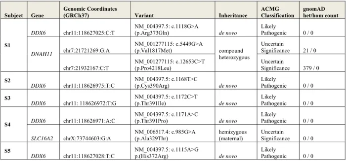
Individual analyses of the five families did not reveal any known pathogenic variants in genes with associated syndrome(s) closely overlapping the clinical features of the probands. Subject 1 (S1) WES study did reveal a de novo missense variant in a gene not previously associated to human disease, DDX6; NM_004397.5; c.1118G>A (p.Arg373Gln)(Table 1). Data also showed compound heterozygous missense variants of unknown significance (VUS) in
DNAH11 (MIM: 603339) (Table S1). DNAH11 is associated with autosomal recessive Ciliary
Dyskinesia, Primary, 7, with or without situs inversus (CILD7) (MIM: 611884) predominately through LoF mutations. Clinical features of CILD7 do not include ID or resemble S1’s phenotype. Further, DNAH11 possesses significantly more missense variation than expected in gnomAD (Z-score = -5.61) and ClinVar contains no confirmed pathogenic missense variants.
Thus, the VUS was excluded as a candidate. Subject 2 (S2) first underwent an ID gene panel sequencing, singleton WES, and finally WGS of the nuclear family. Among the 146 putative de
novo variants identified in the proband, only one occurred in a coding region: a missense change
c.1168T>C (p.Cys390Arg) in DDX6. No other candidate variant(s) were identified in coding or genomic regions of genes known to be involved in ID. Two additional de novo missense variants in DDX6, c.1172C>T (p.Thr391Ile)(Subject 3, S3) and c.1171A>C (p.Thr391Pro)(Subject 4, S4), were identified by trio WES in unrelated individuals with developmental disorders (GeneDx). Subject 4 carries an additional hemizygous missense VUS in the gene SLC16A2 (MIM: 30095) associated with Allan-Herndon-Dudley syndrome (AHDS, MIM: 300523), but this diagnosis does not fit with his clinical history (Table S1). A key feature of males with AHDS is abnormal triiodothyronine (T3) and thyroxine (T4) levels. Repeat thyroid studies in S4 including dosages of T3 and T4 (free, total, and reverse) in S4 were all normal. During the final stages of this study, a fifth individual (Subject 5, S5) with a single de novo missense variant c.1115A>G (p.His372Arg) in DDX6 was identified by WES. For all individuals, no other variants except the DDX6 missense changes described above were classified pathogenic or likely pathogenic according to ACMG guidelines.
Clinical features of individuals carrying de novo missense variants in DDX6 exon 11
We could retrieve clinical information for four of the individuals carrying de novo missense variants in DDX6. These probands are from unrelated families in both the USA and Europe. The affected individuals present with NDD of unknown etiology and range between the ages of 4 to 13 years at last clinical assessment. Parents are noted to be without any significant health problems. Phenotyping of the four affected probands showed overlap of clinical features
and minor variation in severity across individuals (Table 2). Clinical features include ID (4/4), DD (4/4), mild/moderate cardiac anomalies (3/4), hypotonia (3/4), gait instability (3/4), hand/feet and genitourinary abnormalities (3/4). Other features present include mild-moderate MRI anomalies, small head circumference (<-2 Standard Deviations SD), congenital hydronephrosis with vesicoureteral reflux, mild obesity, and autistic traits. The probands also share common facial dysmorphic features including a high forehead, a bulbous nasal tip, widely-spaced eyes, arched eyebrows and low-set ears (Figure 1). Expanded summaries can be found in Supplemental Data: Case Reports.
DDX6 missense variants found in affected individuals are absent from population databases,evolutionarily conserved, and spatially clustered within protein
All de novo missense variants identified in affected individuals are located in exon 11 of
DDX6. This exon is present in all DDX6 isoforms and codes for conserved motifs QxxR and V
within the second RecA-2 domain of the helicase core. The variants have not been observed in the gnomAD database, which contains genetic data from ~140 thousand individuals and is depleted of severe pediatric disorders. DDX6 is highly intolerant to LoF variants as indicated by gnomAD’s (v2.1.1) observed/expected LoF ratio of 0.04 (confident interval [0.01-0.17]), with only one heterozygous LoF change reported in the penultimate exon of its canonical isoform (NM_004397.5). DDX6 was also found to have significant intolerance to missense change indicated by a gnomAD Z-score of 3.93 (observed/expected ratio of 0.34 [0.29-0.41]) suggesting selection pressure against amino acid changes26. In particular, exon 11 (aa 371-391) was found highly depleted of nonsynonymous variations in gnomAD (Figure 2A). The specific variants identified in probands lead to changes at conserved positions and are predicted to be deleterious
by in silico analysis tools (Table 1, Figure 2B). The QxxR motif containing Arg373 is conserved in 33/35 DEAD-box family members, with the varying residues being structurally and chemically similar (Glutamine). In Motiv V, residue 390 maintains small, uncharged residues throughout the DEAD-box protein family (35/35), and Thr391 is conserved in every member (Figure S1, Figure S2). Further, mapping of the variants on a 3D structure of DDX6 in complex with the CNOT1 MIF4G and 4E-T CHD domains37 showed proband variants spatially cluster near or at the protein surface in a region depleted of missense variants in gnomAD populations (Figure 2C-D) and close to the 4E-T interaction surface, suggesting that this region is of functional importance for DDX6.
Mutant DDX6 proteins are defective in PB assembly
As DDX6 is known to be involved in the formation of cytoplasmic PBs, we first analyzed the presence of PBs in fibroblasts obtained from S2 carrying the p.Cys390Arg change and from an unrelated age-matched control subject. Cells were analyzed by immunofluorescence using antibodies against DDX6 and a second PB marker, EDC4 (Figure 3A). While most fibroblasts from the control subject showed numerous PBs detected by both DDX6 and EDC4 antibodies, only some of the fibroblasts obtained from S2 contained PBs. However, Western blot analyses indicated that similar amount of DDX6 protein was present in these cells (Figure S3A, 3C), suggesting a functional defect in this variant. Fibroblasts were also obtained from S1 carrying the p.Arg373Gln variant and from her parents. Again, Western blot analysis showed similar levels of DDX6 protein in the three cell samples (Figure S3A), while immunostaining indicated that the proband’s cells contained fewer PBs than the mother’s ones (Figure S3B-S3C). The father’s cells grew slowly however, and possibly related to that, had an intermediate number of PBs:
lower than the mother’s, but higher than S1’s cells (Figure S3B, S3C). To confirm the specific defect of PBs in S1, we therefore submitted the three cell cultures to a mild cold shock at 30°C for 2 hours, since we showed previously that such a treatment results in increased number and size of PBs in established cell lines10. With this treatment, 90% of the parents’ cells contained numerous PBs, while 70% of S1’s cells remained devoid of PBs (Figure 3B, 3C). Moreover, in the 30% of cells with PBs, their numbers remained low, thus confirming a specific PB defect in the proband cells.
We then investigated the ability of the different variant-containing DDX6 proteins to assemble PBs in a model human cell line11. Plasmids encoding the four DDX6 variants initially identified (p.Arg373Gln, p.Cys390Arg, p.Thr391Ile, and p.Thr391Pro) fused to FLAG and HA tags were transfected in HeLa cells (Figure S4A). The variant proteins localized in PBs, as assessed by immunofluorescence using LSM14A as a P-body marker (Figure S4B). However, they dramatically affect PB assembly as tested by the following complementation assay (Figure
4A). Briefly, cells were depleted of endogenous DDX6 by using siRNA targeting the 3’UTR of DDX6 mRNA (Figure 4B), which resulted as expected11 in PB disassembly (Figure 4A, 4C). 24
h later, cells were transfected with wild-type or mutant FLAG-DDX6-HA plasmids and analyzed 40 h later for the presence of PBs (Figure 4A, 4C). Whereas the wild-type DDX6 restored PB assembly, no or very few PBs were observed using the mutant proteins, with less than 5% efficiency for p.Arg373Gln, p.Cys390Arg and p.The391Ile variants as compared to the wild-type protein, and 17% for the p.Thr391Pro variant (Figures 4A, 4C). Similar results were obtained using EDC4 as a PB marker (Figure S4C). This indicated that the mutant DDX6 proteins were unable to replace the endogenous DDX6 for PB formation.
Mutant DDX6 proteins are defective in interactions with protein partners key to PB assembly.
Previously, we showed that three factors, DDX6, LSM14A and 4E-T, are required and their interactions essential for PB assembly10,11,41. Mutating the DDX6 FDF-binding pocket (Mut1: four substitutions introduced between residues 320 and 3319,42,43) prevents the binding of LSM14A and 4E-T and strongly reduces PB formation10. Since the variants identified in DDX6 are located close to the Mut1 mutations, we hypothesized that they could also alter interactions between DDX6 and these protein partners. To address this question, plasmids encoding wild-type and mutant DDX6 (p.Arg373Gln and p.Cys390Arg) fused to FLAG and HA were transfected in HEK293 cells after depletion of the endogenous DDX6 protein, immunoprecipitated 48 hours later with FLAG antibodies, and protein complexes were analyzed by western blot (Figure 4D). First, both mutant DDX6 proteins showed reduced binding to 4E-T and LSM14A, as compared to wild-type DDX6, and the defect was stronger for p.Arg373Gln than for the p.Cys390Arg mutant. This is consistent with the inability of these mutants to assemble PBs in the complementation assay. A binding defect was also observed for other DDX6 partners: LSM14B binding was as affected as LSM14A with both mutants, while other bindings were defective only in the case of the p.Arg373Gln mutant. This included a strong defect in PAT1B binding and some impairment of EDC3 and EDC4 binding as well. Overall, the p.Arg373Gln mutant protein was the most defective for interacting with its partners, especially those acting as translation repressors (LSM14A, 4E-T, PAT1B).
p.Cys390Arg variant cells accumulate mRNAs involved in translational regulation, DDX6 mRNA targets, and PB-excluded mRNAs.
Transcriptomic (RNAseq) analysis was performed from skin fibroblasts obtained from S2 (carrying the p.Cys390Arg variant) and compared to eight unrelated individuals with other neurodevelopmental or sensorineural conditions unrelated to DDX6 mutation. More than one thousand genes were found to be significantly (p-adjusted <0.05) differentially expressed (DE) (Figure 5A) including 493 up-regulated and 979 down-regulated protein-coding genes (Table
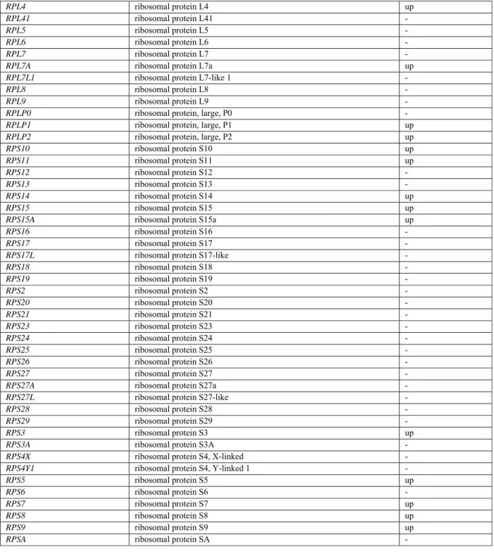
S2). Enrichment analysis using DAVID44 demonstrated that the set of genes significantly upregulated was enriched for Gene Ontology Biological Process and Molecular Function terms related to protein translation such as “SRP-dependent cotranslational membrane targeting” (GO:0006614, fold enrichment FE=12.5, adjusted p-value=2.0e-18) or “formation of translation preinitiation complex” (GO:0001731, FE=11.3, adjusted p-value=3.3e-02), or “structural component of the ribosome” (GO003735, FE=5.4, adjusted p-value=1.5e-9) (Figure 5B). This enrichment was confirmed by analyzing KEGG pathways (“ribosome”, map03010 FE=7.9, adjusted p-value=7.2e-15). More specifically, 4 out of the 14 expressed eIF3 translation initiation factor subunit mRNAs and 28 of the 85 expressed ribosomal protein mRNAs were significantly up-regulated in S2, whereas none were significantly depleted (Table S3). A subset of the DE genes encode RNA-binding proteins also known to be involved in ID (CNOT3, HNRNPH2,
DDX59, RPL10, etc.) (Table S4).
To investigate relationships between S2’s transcriptomic profile and DDX6 function, we took advantage of two datasets available from the ENCODE project, both from the human erythroid cell line K562: 1) a DDX6 cross-linking followed by immunoprecipitation (CLIP) experiment identifying mRNAs that bind to DDX6 and 2) a transcriptome after induction of a stably-transfected DDX6 shRNA for 48 hours identifying mRNAs differentially expressed after DDX6 silencing. If the DDX6-Cys390Arg variant leads to a defect in its function in mRNA
decay, we would expect an accumulation of these DDX6 mRNA targets. Indeed, we first observed that significantly enriched mRNAs in S2 fibroblasts were also enriched in ENCODE’s DDX6 CLIP experimental data (Figure 5C, left panel), indicating their direct binding to DDX6. Second, S2-enriched mRNAs also tended to be enriched in the DDX6-silencing dataset (Figure
5C, right panel). Thus, in spite of the differences between the two cell types under comparison
(dermal fibroblasts producing a p.Cys390Arg+/- variant DDX6 versus an immortalized erythroid cell line transiently depleted for DDX6); there was a common signature of enriched mRNAs between the two. Further, the enrichment was even stronger for the mRNA subset that was enriched in both the CLIP experiments and the DDX6 silencing experiment (Figure 5C, right
panel), indicating that the mRNAs enriched in the cells of S2 were likely decayed by a
DDX6-dependent mechanism that involved direct DDX6 binding.
We have recently shown that mRNAs regulated by DDX6 at the level of decay are localized out of PBs12. Accordingly, comparing S2’s transcriptome to the mRNA content of purified PBs showed that mRNAs which are enriched in S2’s fibroblasts are excluded from PBs (Figure 5D). In the same study we showed that the mRNAs targeted to PBs, in contrast, are regulated at the level of translation. While translation regulation is not accessible in our transcriptome analysis, the fact that PBs are drastically reduced in S2 strongly suggests that DDX6-dependent translation repression is also affected in these cells. Finally, the mRNAs accumulating in S2 have a high GC content compared to other human genes, whereas the mRNAs depleted are rather AU-rich (Figure 5E). This is consistent with our recent study showing that DDX6 is preferentially involved in the decay of GC-rich mRNAs. Such a high GC content increases the frequency of optimal codons and is therefore observed in actively translated mRNAs.
In this study, we describe five rare de novo missense variants in the RNA helicase DDX6, identified in individuals with intellectual disability (ID). Clinical, genetic, and functional data demonstrated that these missense variants, inducing deficits in DDX6 functions, cause an autosomal-dominant form of intellectual disability associated with mild dysmorphic features. These deficits include DDX6 ability to assemble PBs - constitutive cytoplasmic RNP granules which act in mRNA storage - resulting in their drastic reduction in primary cells of affected individuals as well as when modeled in human cell lines. Furthermore, co-immunoprecipitation assays showed the pathogenic variants affect DDX6 interactions with protein partners involved in PB assembly, such as LSM14A and 4E-T, suggesting a mechanism for PB dysfunction and subsequent deregulation of mRNA storage. Interactions with other DDX6 protein partners (PAT1B, EDC3, and EDC4) were differently impacted, depending on the variants. The surface of DDX6 that interacts with these different proteins was reported to be distinct, though overlapping. Therefore, we did not expect that different variants of DDX6 would alter all interactionsto the same extent. Additional defects included DDX6 dysfunction in the control of mRNA stability, since transcriptomic analysis of S2 revealed an enrichment of mRNAs that are regulated by DDX6 at the level of decay45 (Figure 5F). This includes in particular mRNA encoding subunits of the translation initiation factor eIF3 and ribosomal proteins. Many also contain a TOP motif in their 5’ UTR responsible for their control by the mTORC1 kinase (25 out of the 94 TOP mRNAs previously reported46 were enriched in S2 cells). Their deregulation could have consequences on the control of cell proliferation/function inside and outside of the
CNS46,47. Taken together, these findings highlight crucial roles for DDX6 and/or PBs in proper
The missense changes identified in affected individuals cluster in two pairs of amino acids in conserved motifs of the DDX6 C-terminal RecA domain: residues 372-373 which comprise the second half of the QxxR motif, and 390-391 which lie in motif V48 (Figure 2A,
Figure S1). The substitutions p.Arg373Gln (S1) and p.His372Arg (S5) cause significant
alterations to residue properties and likely the local structure/function. Electrically-charged surface residues like Arg373 are known to promote proper protein folding through solvent interactions, and reactive histidines are most common in protein active sites. The QxxR motif is also involved in interdomain interactions with motif Ia in vitro in the yeast DEAD-box protein Mss116p49, which was proposed to help form a continuous RNA binding surface and stabilize bound RNA. The substitution of Cys390 (S2) to a large electrically-charged arginine, and the conversions of Thr391 (S3,S4) from a polar threonine to non-polar, unreactive residues (proline/isoleucine), also suggest destabilizing regional changes to the RecA-2 domain. Our co-immunoprecipitation and complementation assays support these predictions with results showing dysfunction of the variant DDX6 proteins to bind partners through this region and to assemble PBs, respectively. Since the proband fibroblasts have a strong PB deficiency despite the presence of one wild-type allele, it raises the possibility that the DDX6 variants could have some negative effect on PB assembly. We have previously characterized a clearly dominant-negative DDX6 change in DDX6 (motif VI mutant HRIG) in an experiment similar to the one described here (Figure S4B) and found that its expression completely suppressed PBs in the presence of the endogenous DDX6 protein11, which is not the case for the probands’ variants. We would therefore favor the hypothesis that the low PB assembly results from haploinsufficiency of the wild-type allele.
DDX6 is not the first gene encoding a RNA helicase involved in monogenic forms of
NDD/ID. The involvement of DHX30 in ID was uncovered in 2017 with several de novo missense variants also in the RecA domains reported in twelve individuals with ID, speech impairment and gait abnormalities50. Pathogenic variants including de novo truncating and missense changes in RecA domains were have also been recently reported in the DDX3X gene51, located on the X-chromosome, in females with ID, hypotonia, movement disorders, and epilepsy.
DDX3X now represents one of the genes most frequently mutated in ID; involved in ~0.5% of ID
cases and ~2% of female cases52. Interestingly, after sequence alignment of DDX6 and DDX3X, the Arg373 position found mutated in our cohort corresponds to the Arg480 position recurrently mutated in the DDX3X cohort of affected individuals, reinforcing the functional importance of this amino acid and the QxxR motif in DEAD-box proteins. LoF function variants in another member of this helicase family, DDX59, have also been reported to cause an autosomal recessive form of ID with features of oral-facial-digital syndrome53–55.
Interestingly, the phenotypic traits observed in our DDX6 cohort align well with the syndromes described in individuals with pathogenic variants in DDX3X and DHX3050–52. These include ID/DD, movement/gait problems, cardiac anomalies, and corpus callosum hypoplasia as well as dysmorphic facial features (high forehead, bulbous nasal tip, hypertelorism, and arched eyebrows). Conversely, none of the individuals with DDX6 variants have reported seizures, a feature present in the DDX3X and DHX30 syndromes. This could be an interesting delineation of the DDX6 cohort and give specific biological insights into the specificity of each DExD/H-box protein. However, seizures are present in only a subset of the DDX3X and DHX30 cohorts (~16% and 25%, respectively), thus more individuals with DDX6 syndrome are needed to completely exclude seizures as an absent feature. Our cohort also exhibits a degree of variability in their
clinical presentations. Individuals S1 and S5 present with mild abnormalities on brain MRI (corpus callosum hypoplasia) and appear to be slightly more affected than the others. Their amino acid changes (p.His372Arg and p.Arg373Gln) lie in the QxxR motif suggesting variants in this motif elicit a slightly more severe phenotype than variants in the motif V region. In support of this, co-immunoprecipitation studies demonstrated more drastic defects of protein binding caused by the p.Arg373Gln variant. Since differences in the proband’s genetic backgrounds are also likely to contribute to phenotypic variability, more individuals with pathogenic DDX6 variation will be required to fully answer this question.
DDX6 is widely expressed throughout tissues with some of the highest levels in the
cerebellar hemispheres. Its expression is timely regulated in the mouse neocortex during development56, and it has been found to be expressed in both mouse57 and human neural stem cells (NSCs) (data extracted from previous works58). DDX6 is further involved in the CNS as it increases the activity of Let-7a, a microRNA important in neuronal differentiation. DDX6 overexpression in mouse NSCs leads to an increase in neuronal differentiation whereas loss of
DDX6 function inhibits the formation of new neurons57. DDX3X was also found to be essential
for neuronal differentiation and migration, and its inactivation in mice disturbs cortex development and leads to abnormal distribution of neurons in the cortical plate59. It is possible the brain anomalies present in DDX6 individuals S1 and S5 result from impaired processes in NSC specification. While some evidence supports this idea, the exceedingly heterogeneous nature of corpus callosum anomalies requires further in-vivo work to validate how these pathogenic variants would alter proliferation/differentiation/migration of neuronal precursors and synaptic functioning, and also to identify which mRNAs are deregulated in this cell type.
We have shown that our DDX6 variants result in failure to form PBs efficiently and lead to a decreased number of these RNP granules in fibroblasts of affected individuals. Lessel et al. recently showed that DHX30 missense variants, in addition to altering DHX30 ability to bind RNA or to exert its ATPase activity, increase the formation of another significant type of RNP granules, stress granules, which is associated with a decrease of global protein translation50. The missense variants identified in DDX3X were also shown to affect ATPase helicase activity and lead to protein accumulation into RNP granules not well characterized but harboring some translational activity59. RNP granule dysfunction in neurons specifically could relate to their polar, asymmetric nature. Indeed, there are a variety of neuronal RNP granules, mostly described in dendrites. Several types of dendritic granules contain PB components, and were initially called P-body-like structures60 or P-bodies61. They are important in regulating local translation at the synapse, which is essential for synaptic plasticity62. However, further attempts to define these granules with expression of DDX6 and decapping enzymes suggested that these structures were more diverse in neuronal processes than in other cells63. In the neuronal soma, RNP granules may be closer to fibroblast PBs in terms of composition and function, but they have not been well characterized. Repression by miRNAs also plays a major role in the control of signaling pathways regulating several steps of cortical development64, therefore we can speculate that the different cytoplasmic foci that play roles in the storage of miRNA targets are important for a proper development of the brain.
The present study has taken effort to demonstrate rare changes in DDX6 cause PB and mRNA metabolism dysfunction. These DDX6 dysfunctions were observed not only by overexpressing DDX6 in immortalized cell lines but also directly in affected individual’s primary fibroblasts (individuals S1 and S2). The use of these non-neuronal cells may only
Pdistantly reflect the actual disease pathogenesis in affected individual’s brains and other organs. Further transcriptomic and proteomic studies in neuronal cells derived from primary fibroblasts or in-vivo animal models will shine more light on the specific mechanisms of DDX6-associated pathogenesis in neurodevelopment.
In conclusion, our findings implicate rare de novo missense variants in the second RecA domain of DDX6 in a neurodevelopmental disorder (NDD) with ID. These variants significantly affect DDX6’s ability to assemble PBs, resulting in their drastic reduction in cells, and, in our affected individual with transcriptomic data, the significant alteration of the transcriptome landscape. We suggest rare variants in DDX6 be considered for pathogenicity in pediatric cases of NDDs with ID and this RNA helicase be added to the growing list of ID/NDD genes. Lastly, together with the recent involvement of DDX3X and DHX30 in ID and similar NDDs, these results also highlight an emerging class of NDD involving RNA helicases.
SUPPLEMENTAL DATA
Supplemental Data include four figures, four tables, and expanded clinical descriptions of the subjects.
ACKNOWLEDGEMENTS
The authors thank the families for their participation to the study. The authors also thank the Foundation Jerome Lejeune and Fondation Maladies Rares for their financial support, as well as the Association Paul and Liba Mandel and the CREGEMES. This study was also supported by the grant ANR-10-LABX-0030-INRT, a French State fund managed by the Agence Nationale de la Recherche under the frame program Investissements d’Avenir ANR-10-IDEX-0002-02, the
ANR grant 14-CE09-0013-01ANR, and the Laboratory of Excellence GENMED (Medical Genomics) grant no. ANR-10-LABX-0013 managed by the National Research Agency (ANR) part of the Investment for the Future program. The authors want also to thank all the people from IGBMC sequencing platform (Bernard Jost, Christelle Thibaut-Charpentier, Céline Keime, Damien Plassard) for their technical and bioinformatics support. The authors would like to thank all donors who have contributed and participated in TGen’s Center for Rare Childhood Disorders Center. Lastly, the authors thank all of the families for their participation in this study.
DECLARATION OF INTERESTS
IW, KGM, KM are employees of GeneDx, Inc.
WEB RESOURCES
The URLs for online tools and data presented herein are: Clinvar: http://www.ncbi.nlm.nih.gov/clinvar/
dbSNP: http://www.ncbi.nlm.nih.gov/projects/SNP/ Decipher: https://decipher.sanger.ac.uk/
ENCODE: https://www.encodeproject.org/
ExAC Browser (Beta) | Exome Aggregation Consortium: http://exac.broadinstitute.org/ GnomAD Exome Variant Server: http://gnomad.broadinstitute.org/
GeneMatcher: https://genematcher.org/
Integrative Genomics Viewer (IGV): http://www.broadinstitute.org/igv/ Mutation Nomenclature: http://www.hgvs.org/mutnomen/recs.html OMIM: http://www.omim/org/
UCSC: http://genome.ucsc.edu/
SNAP2: https://rostlab.org/services/snap2web/ Protein Data Bank (PDB) : http://www.pdb.org
REFERENCES
1. Vissers, L.E.L.M., Gilissen, C., and Veltman, J.A. (2016). Genetic studies in intellectual disability and related disorders. Nat. Rev. Genet. 17, 9–18.
2. Lelieveld, S.H., Wiel, L., Venselaar, H., Pfundt, R., Vriend, G., Veltman, J.A., Brunner, H.G., Vissers, L.E.L.M., and Gilissen, C. (2017). Spatial Clustering of de Novo Missense Mutations Identifies Candidate Neurodevelopmental Disorder-Associated Genes. Am. J. Hum. Genet. 101, 478–484.
3. Bardoni, B., Abekhoukh, S., Zongaro, S., and Melko, M. (2012). Intellectual disabilities, neuronal posttranscriptional RNA metabolism, and RNA-binding proteins: three actors for a complex scenario. Prog. Brain Res. 197, 29–51.
4. Sartor, F., Anderson, J., McCaig, C., Miedzybrodzka, Z., and Müller, B. (2015). Mutation of genes controlling mRNA metabolism and protein synthesis predisposes to neurodevelopmental disorders. Biochem. Soc. Trans. 43, 1259–1265.
5. Oberlé, I., Rousseau, F., Heitz, D., Kretz, C., Devys, D., Hanauer, A., Boué, J., Bertheas, M.F., and Mandel, J.L. (1991). Instability of a 550-base pair DNA segment and abnormal methylation in fragile X syndrome. Science 252, 1097–1102.
6. Hagerman, R.J., Berry-Kravis, E., Hazlett, H.C., Bailey, D.B., Moine, H., Kooy, R.F., Tassone, F., Gantois, I., Sonenberg, N., Mandel, J.L., et al. (2017). Fragile X syndrome. Nat Rev Dis Primers 3, 17065.
7. Jankowsky, E., and Fairman, M.E. (2007). RNA helicases--one fold for many functions. Curr. Opin. Struct. Biol. 17, 316–324.
8. Abdelhaleem, M., Maltais, L., and Wain, H. (2003). The human DDX and DHX gene families of putative RNA helicases. Genomics 81, 618–622.
9. Ozgur, S., Basquin, J., Kamenska, A., Filipowicz, W., Standart, N., and Conti, E. (2015). Structure of a Human 4E-T/DDX6/CNOT1 Complex Reveals the Different Interplay of DDX6-Binding Proteins with the CCR4-NOT Complex. Cell Rep 13, 703–711.
10. Ayache, J., Bénard, M., Ernoult-Lange, M., Minshall, N., Standart, N., Kress, M., and Weil, D. (2015). P-body assembly requires DDX6 repression complexes rather than decay or Ataxin2/2L complexes. Mol. Biol. Cell 26, 2579–2595.
11. Minshall, N., Kress, M., Weil, D., and Standart, N. (2009). Role of p54 RNA helicase activity and its C-terminal domain in translational repression, P-body localization and assembly. Mol. Biol. Cell 20, 2464– 2472.
12. Hubstenberger, A., Courel, M., Bénard, M., Souquere, S., Ernoult-Lange, M., Chouaib, R., Yi, Z., Morlot, J.-B., Munier, A., Fradet, M., et al. (2017). P-Body Purification Reveals the Condensation of Repressed mRNA Regulons. Mol. Cell 68, 144-157.e5.
13. Standart, N., and Weil, D. (2018). P-Bodies: Cytosolic Droplets for Coordinated mRNA Storage. Trends Genet. 34, 612–626.
14. Sobreira, N., Schiettecatte, F., Valle, D., and Hamosh, A. (2015). GeneMatcher: a matching tool for connecting investigators with an interest in the same gene. Hum. Mutat. 36, 928–930.
15. Li, H., and Durbin, R. (2009). Fast and accurate short read alignment with Burrows-Wheeler transform. Bioinformatics 25, 1754–1760.
16. McKenna, A., Hanna, M., Banks, E., Sivachenko, A., Cibulskis, K., Kernytsky, A., Garimella, K., Altshuler, D., Gabriel, S., Daly, M., et al. (2010). The Genome Analysis Toolkit: a MapReduce framework for analyzing next-generation DNA sequencing data. Genome Res. 20, 1297–1303.
17. Van der Auwera, G.A., Carneiro, M.O., Hartl, C., Poplin, R., Del Angel, G., Levy-Moonshine, A., Jordan, T., Shakir, K., Roazen, D., Thibault, J., et al. (2013). From FastQ data to high confidence variant calls: the Genome Analysis Toolkit best practices pipeline. Curr Protoc Bioinformatics 43, 11.10.1-33.
18. Cingolani, P., Platts, A., Wang, L.L., Coon, M., Nguyen, T., Wang, L., Land, S.J., Lu, X., and Ruden, D.M. (2012). A program for annotating and predicting the effects of single nucleotide polymorphisms, SnpEff: SNPs in the genome of Drosophila melanogaster strain w1118; iso-2; iso-3. Fly (Austin) 6, 80–92. 19. Thevenon, J., Duffourd, Y., Masurel-Paulet, A., Lefebvre, M., Feillet, F., El Chehadeh-Djebbar, S., St-Onge, J., Steinmetz, A., Huet, F., Chouchane, M., et al. (2016). Diagnostic odyssey in severe
neurodevelopmental disorders: toward clinical whole-exome sequencing as a first-line diagnostic test. Clin. Genet. 89, 700–707.
20. Rimmer, A., Phan, H., Mathieson, I., Iqbal, Z., Twigg, S.R.F., WGS500 Consortium, Wilkie, A.O.M., McVean, G., and Lunter, G. (2014). Integrating mapping-, assembly- and haplotype-based approaches for calling variants in clinical sequencing applications. Nat. Genet. 46, 912–918.
21. Geoffroy, V., Pizot, C., Redin, C., Piton, A., Vasli, N., Stoetzel, C., Blavier, A., Laporte, J., and Muller, J. (2015). VaRank: a simple and powerful tool for ranking genetic variants. PeerJ 3, e796.
22. Bartenhagen, C., and Dugas, M. (2016). Robust and exact structural variation detection with paired-end and soft-clipped alignments: SoftSV compared with eight algorithms. Brief. Bioinformatics 17, 51– 62.
23. Backenroth, D., Homsy, J., Murillo, L.R., Glessner, J., Lin, E., Brueckner, M., Lifton, R., Goldmuntz, E., Chung, W.K., and Shen, Y. (2014). CANOES: detecting rare copy number variants from whole exome sequencing data. Nucleic Acids Res. 42, e97.
24. Geoffroy, V., Herenger, Y., Kress, A., Stoetzel, C., Piton, A., Dollfus, H., and Muller, J. (2018). AnnotSV: an integrated tool for structural variations annotation. Bioinformatics 34, 3572–3574.
25. Tanaka, A.J., Cho, M.T., Retterer, K., Jones, J.R., Nowak, C., Douglas, J., Jiang, Y.-H., McConkie-Rosell, A., Schaefer, G.B., Kaylor, J., et al. (2016). De novo pathogenic variants in CHAMP1 are associated with global developmental delay, intellectual disability, and dysmorphic facial features. Cold Spring Harb Mol Case Stud 2, a000661.
26. Lek, M., Karczewski, K.J., Minikel, E.V., Samocha, K.E., Banks, E., Fennell, T., O’Donnell-Luria, A.H., Ware, J.S., Hill, A.J., Cummings, B.B., et al. (2016). Analysis of protein-coding genetic variation in 60,706 humans. Nature 536, 285–291.
27. Kircher, M., Witten, D.M., Jain, P., O’Roak, B.J., Cooper, G.M., and Shendure, J. (2014). A general framework for estimating the relative pathogenicity of human genetic variants. Nat. Genet. 46, 310–315. 28. Cooper, G.M., Stone, E.A., Asimenos, G., NISC Comparative Sequencing Program, Green, E.D.,
Batzoglou, S., and Sidow, A. (2005). Distribution and intensity of constraint in mammalian genomic sequence. Genome Res. 15, 901–913.
29. Ng, P.C., and Henikoff, S. (2003). SIFT: Predicting amino acid changes that affect protein function. Nucleic Acids Res. 31, 3812–3814.
30. Adzhubei, I.A., Schmidt, S., Peshkin, L., Ramensky, V.E., Gerasimova, A., Bork, P., Kondrashov, A.S., and Sunyaev, S.R. (2010). A method and server for predicting damaging missense mutations. Nat. Methods 7, 248–249.
31. Hecht, M., Bromberg, Y., and Rost, B. (2015). Better prediction of functional effects for sequence variants. BMC Genomics 16 Suppl 8, S1.
32. Gray, V.E., Hause, R.J., Luebeck, J., Shendure, J., and Fowler, D.M. (2018). Quantitative Missense Variant Effect Prediction Using Large-Scale Mutagenesis Data. Cell Syst 6, 116-124.e3.
33. Richards, S., Aziz, N., Bale, S., Bick, D., Das, S., Gastier-Foster, J., Grody, W.W., Hegde, M., Lyon, E., Spector, E., et al. (2015). Standards and guidelines for the interpretation of sequence variants: a joint consensus recommendation of the American College of Medical Genetics and Genomics and the Association for Molecular Pathology. Genet. Med. 17, 405–424.
34. Anders, S., Pyl, P.T., and Huber, W. (2015). HTSeq--a Python framework to work with high-throughput sequencing data. Bioinformatics 31, 166–169.
35. Anders, S., and Huber, W. (2010). Differential expression analysis for sequence count data. Genome Biol. 11, R106.
36. Benjamini, Y Controlling the False Discovery Rate: A Practical and Powerful Approach to Multiple Testing.
37. Mathys, H., Basquin, J., Ozgur, S., Czarnocki-Cieciura, M., Bonneau, F., Aartse, A., Dziembowski, A., Nowotny, M., Conti, E., and Filipowicz, W. (2014). Structural and biochemical insights to the role of the CCR4-NOT complex and DDX6 ATPase in microRNA repression. Mol. Cell 54, 751–765.
38. Pettersen, E.F., Goddard, T.D., Huang, C.C., Couch, G.S., Greenblatt, D.M., Meng, E.C., and Ferrin, T.E. (2004). UCSF Chimera--a visualization system for exploratory research and analysis. J Comput Chem 25, 1605–1612.
39. de Chaumont, F., Dallongeville, S., Chenouard, N., Hervé, N., Pop, S., Provoost, T., Meas-Yedid, V., Pankajakshan, P., Lecomte, T., Le Montagner, Y., et al. (2012). Icy: an open bioimage informatics platform for extended reproducible research. Nat. Methods 9, 690–696.
40. Ernoult-Lange, M., Baconnais, S., Harper, M., Minshall, N., Souquere, S., Boudier, T., Bénard, M., Andrey, P., Pierron, G., Kress, M., et al. (2012). Multiple binding of repressed mRNAs by the P-body protein Rck/p54. RNA 18, 1702–1715.
41. Kamenska, A., Simpson, C., Vindry, C., Broomhead, H., Bénard, M., Ernoult-Lange, M., Lee, B.P., Harries, L.W., Weil, D., and Standart, N. (2016). The DDX6-4E-T interaction mediates translational repression and P-body assembly. Nucleic Acids Res. 44, 6318–6334.
42. Tritschler, F., Eulalio, A., Helms, S., Schmidt, S., Coles, M., Weichenrieder, O., Izaurralde, E., and Truffault, V. (2008). Similar modes of interaction enable Trailer Hitch and EDC3 to associate with DCP1 and Me31B in distinct protein complexes. Mol. Cell. Biol. 28, 6695–6708.
43. Sharif, H., Ozgur, S., Sharma, K., Basquin, C., Urlaub, H., and Conti, E. (2013). Structural analysis of the yeast Dhh1-Pat1 complex reveals how Dhh1 engages Pat1, Edc3 and RNA in mutually exclusive interactions. Nucleic Acids Res. 41, 8377–8390.
44. Huang, D.W., Sherman, B.T., Tan, Q., Kir, J., Liu, D., Bryant, D., Guo, Y., Stephens, R., Baseler, M.W., Lane, H.C., et al. (2007). DAVID Bioinformatics Resources: expanded annotation database and novel algorithms to better extract biology from large gene lists. Nucleic Acids Res. 35, W169-175.
45. Courel, M., Clement, Y., Foretek, D., Vidal, O., Yi, Z., Kress, M., Vindry, C., Benard, M., Bossevain, C., Antoniewski, C., et al. (2018). GC content shapes mRNA decay and storage in human cells. BioRxiv. 46. Thoreen, C.C., Chantranupong, L., Keys, H.R., Wang, T., Gray, N.S., and Sabatini, D.M. (2012). A unifying model for mTORC1-mediated regulation of mRNA translation. Nature 485, 109–113. 47. Costa-Mattioli, M., and Monteggia, L.M. (2013). mTOR complexes in neurodevelopmental and neuropsychiatric disorders. Nat. Neurosci. 16, 1537–1543.
48. Sengoku, T., Nureki, O., Nakamura, A., Kobayashi, S., and Yokoyama, S. (2006). Structural basis for RNA unwinding by the DEAD-box protein Drosophila Vasa. Cell 125, 287–300.
49. Bifano, A.L., Turk, E.M., and Caprara, M.G. (2010). Structure-guided mutational analysis of a yeast DEAD-box protein involved in mitochondrial RNA splicing. J. Mol. Biol. 398, 429–443.
50. Lessel, D., Schob, C., Küry, S., Reijnders, M.R.F., Harel, T., Eldomery, M.K., Coban-Akdemir, Z., Denecke, J., Edvardson, S., Colin, E., et al. (2017). De Novo Missense Mutations in DHX30 Impair Global Translation and Cause a Neurodevelopmental Disorder. Am. J. Hum. Genet. 101, 716–724.
51. Snijders Blok, L., Madsen, E., Juusola, J., Gilissen, C., Baralle, D., Reijnders, M.R.F., Venselaar, H., Helsmoortel, C., Cho, M.T., Hoischen, A., et al. (2015). Mutations in DDX3X Are a Common Cause of