Transcatheter aortic valve implantation using anatomically oriented,
marrow stromal cell-based, stented, tissue-engineered heart valves:
technical considerations and implications for translational
cell-based heart valve concepts
Maximilian Y. Emmert
a,b,c,†, Benedikt Weber
a,b,†, Luc Behr
d, Sebastien Sammut
d, Thomas Frauenfelder
e,
Petra Wolint
a,b, Jacques Scherman
c, Dominique Bettex
f, Jürg Grünenfelder
b,c,
Volkmar Falk
cand Simon P. Hoerstrup
a,b,c,*
a Swiss Center for Regenerative Medicine, Zurich, Switzerland
b Department of Surgical Research, University Hospital of Zurich, Zurich, Switzerland c
Clinic for Cardiovascular Surgery, University Hospital of Zurich, Zurich, Switzerland
d IMM RECHERCHE, Institut Mutualiste Montsouris, Paris, France e Department of Radiology, University Hospital Zurich, Zurich, Switzerland f Institute for Anesthesiology, University Hospital Zurich, Zurich, Switzerland
* Corresponding author. Swiss Center for Regenerative Medicine and Clinic for Cardiovascular Surgery, University Hospital Zurich, Raemistrasse 100, CH-8091 Zürich, Switzerland. Tel: +41-44-2553644; fax: +41-44-2554369; e-mail: [email protected] (S.P. Hoerstrup).
Received 12 February 2013; received in revised form 25 March 2013; accepted 27 March 2013
Abstract
OBJECTIVES: While transcatheter aortic valve implantation (TAVI) has rapidly evolved for the treatment of aortic valve disease, the current-ly used bioprostheses are prone to continuous calcific degeneration. Thus, autologous, cell-based, living, tissue-engineered heart valves (TEHVs) with regeneration potential have been suggested to overcome these limitations. We investigate the technical feasibility of combin-ing the concept of TEHV with transapical implantation technology uscombin-ing a state-of-the-art transcatheter delivery system facilitatcombin-ing the exact anatomical position in the systemic circulation.
METHODS: Trileaflet TEHVs fabricated from biodegradable synthetic scaffolds were sewn onto self-expanding Nitinol stents seeded with autologous marrow stromal cells, crimped and transapically delivered into the orthotopic aortic valve position of adult sheep (n = 4) using the JenaValve transapical TAVI System ( JenaValve, Munich, Germany). Delivery, positioning and functionality were assessed by angiog-raphy and echocardiogangiog-raphy before the TEHV underwent post-mortem gross examination. For three-dimensional reconstruction of the stent position of the anatomically oriented system, a computed tomography analysis was performed post-mortem.
RESULTS: Anatomically oriented, transapical delivery of marrow stromal cell-based TEHV into the orthotopic aortic valve position was suc-cessful in all animals (n = 4), with a duration from cell harvest to TEHV implantation of 101 ± 6 min. Fluoroscopy and echocardiography dis-played sufficient positioning, thereby entirely excluding the native leaflets. There were no signs of coronary obstruction. All TEHV tolerated the loading pressure of the systemic circulation and no acute ruptures occurred. Animals displayed intact and mobile leaflets with an ad-equate functionality. The mean transvalvular gradient was 7.8 ± 0.9 mmHg, and the mean effective orifice area was 1.73 ± 0.02 cm². Paravalvular leakage was present in two animals, and central aortic regurgitation due to a single-leaflet prolapse was detected in two, which was primarily related to the leaflet design. No stent dislocation, migration or affection of the mitral valve was observed.
CONCLUSIONS: For thefirst time, we demonstrate the technical feasibility of a transapical TEHV delivery into the aortic valve position using a commercially available and clinically applied transapical implantation system that allows for exact anatomical positioning. Our data indicate that the combination of TEHV and a state-of-the-art transapical delivery system is feasible, representing an important step towards translational, transcatheter-based TEHV concepts.
Keywords:Aortic valve• Transcatheter • Transapical • TAVI • Tissue-engineered heart valves • Marrow stromal cells
†Both authors contributed equally to this paper.
© The Author 2013. Published by Oxford University Press on behalf of the European Association for Cardio-Thoracic Surgery. All rights reserved.
BA
SIC
S
CIENCE
INTRODUCTION
Transcatheter aortic valve implantation (TAVI) represents an ef fi-cient alternative for the treatment of valvular heart disease in the elderly [1–4], and numerous transfemoral and transapical TAVI devices are commercially available [1–9]. The concept of anatom-ically oriented devices has been suggested to be beneficial with regard to delivery, positioning and the avoidance of coronary obstruction (CO) [2,5–7,10].
However, despite these rapid technological advances, the cur-rently utilized prostheses for TAVI are bioprosthetic, containing bovine or porcine tissue, and are thus well known to be accom-panied by continuous and progressive degeneration, which may be even accelerated due to structural damage in response to crimping procedures [11]. To overcome such limitations, heart valve tissue-engineering (HVTE) technologies comprising repair, regeneration and growth capacities have been repeatedly sug-gested. However, a translational, clinically applicable HVTE concept requires a minimally invasive approach for both cell isola-tion and valve delivery, ideally completely circumventing highly technical, logistical andfinancial efforts [12–14]. In this regard, and based on previously described techniques in the setting of tissue-engineered vascular grafts [15–18], we have recently intro-duced a novel, clinically relevant concept of a single-step inter-ventional approach to implant autologous bone-marrow mononuclear cell (BMMC)-based tissue-engineered heart valves (TEHVs) into the pulmonary and systemic circulation of primates and adult sheep [12–14]. However, while these studies were pri-marily performed using self-made or generic, non-clinical concept stent and delivery systems, a translational, clinically HVTE approach requires the use of state-of-the-art devices, in particular when addressing the systemic circulation [12,13].
Table 1: Animal data and preoperative assessment
Parameter Mean ± SD
Body weight (kg) 57.7 ± 2.5 Diameter aortic annulus (mm) 22.9 ± 0.3 Distance to brachiocephalic trunk (mm) 42.1 ± 2.4 Diameter sinus portion (mm) 29.0 ± 1.0 Diameter sinotubular junction (mm) 24.3 ± 1.6 Height of sinus portion (mm) 14.5 ± 0.7
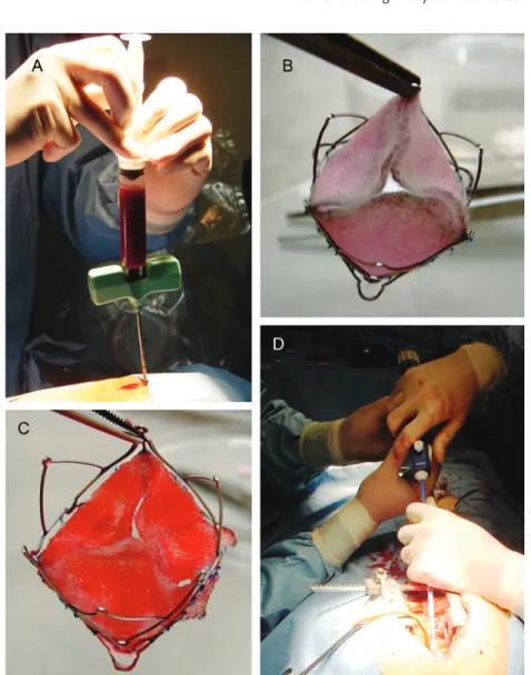
Figure 1:Bone marrow mononuclear cell isolation and TEHV preparation. Sternal bone marrow puncture (A) was performed before the prefabricated, stent-based polyglycolic-acid coated with 1.75% poly-4-hydroxybutyrate composite matrices (B) were seeded with isolated bone marrow mononuclear cells usingfibrin as a cell carrier (C) prior to transapical delivery (D).
Therefore, the present study investigates the acute technical feasibility of combining the BMMC-based single-step HVTE ap-proach with a clinically used, anatomically oriented state-of-the-art transapical stent delivery system in an adult sheep model with particular regard to TEHVin vitro fabrication, delivery, positioning and acute functionality.
MATERIALS AND METHODS
Experimentation approval
All animals received humane care in compliance with the ‘Principles of Laboratory Animal-Care’ as well as with the ‘Guide for the care and use of laboratory animals’ published by the National Institutes of Health ( publication no. 85-23/revised 1985). All procedures were approved by the Institutional Ethics Committee (Approval-11–15).
Scaffold fabrication and integration into the
JenaValve stent
Trileaflet heart valve scaffolds were fabricated from non-woven polyglycolic-acid meshes (PGA; Cellon, Luxembourg), coated with 1.75% poly-4-hydroxybutyrate (P4HB; TEPHA Inc., USA) as previ-ously described [12–14]. Thereafter, the scaffolds were sewn onto self-expanding Nitinol stents. The scaffolds were attached to the inside of the Nitinol stent frames by single interrupted sutures.
Isolation and seeding of ovine autologous BMMCs
Bone marrow mononuclear cells (BMMCs) were isolated via sternal puncture as previously described and characterized in accordance with the established protocols [12–14]. Usingfibrin as a cell-carrier (Sigma Chemical, USA), BMMCs were seeded onto the stented heart valve scaffolds (1.08 ± 0.5 × 10⁶cells/cm2 valve leaflets), before the stented TEHV was crimped and inserted onto the JenaValve transapical delivery system ( JenaValve, Munich, Germany).
Preoperative assessment, planning and sizing
Before implantation, all animals underwent transthoracic echocar-diography in order to assess the optimal stent size. Prior to the im-plantation, intraoperative angiography was done to re-confirm the stent size taking into consideration the following parameters: diameters of the aortic annulus (AA), sinus portion (SP), the sino-tubular junction (STJ) and the brachiocephalic trunk (BCT) as well as the distance to the BCT and the height of the SP (Table 1). Three animals received a 25-mm stented TEHV (n = 3), while one animal received a 27-mm stented TEHV (n = 1).
Transcatheter implantation of BMMC-based TEHV
using the JenaValve transapical delivery system
Using the JenaValve transapical delivery system, TEHVs were transa-pically delivered into the aortic valve position. The valves were
crimped and loaded onto a custom-made, over-the-wire JenaValve delivery system (outer diameter = 8 mm). Following a mini-sternotomy and placement of 5/0 Prolene pledgeted, purse string sutures, the apex of the left ventricle was punctured, and the TEHVs were delivered into the aortic valve position under fluoro-scopic control (OECW 9900 Elite GE, Fairfield, USA) applying the previously described and anatomically oriented JenaValve transapi-cal delivery technology. After positioning the feelers, the JenaValve stent was opened in a stepwise fashion underfluoroscopic control. After full delivery of the TEHV, the appropriate positioning, thereby fully excluding the native valve, as well as sufficient functionality of the TEHV, was confirmed by contrast angiography (CT), before the JenaValve delivery device was carefully removed.
Positioning and functionality
JenaValve stent positioning was assessed using intraoperative CT (Siemens, Munich, Germany), and in vivo functionality was evaluated using two-dimensional (2D) and three-dimensional (3D) echocardiography (Philips Healthcare iE33W xMATRIX Ultrasound, Netherlands).
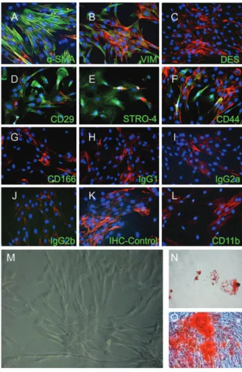
Figure 2:Cell characterization and functionality assessment. A representative patch of ovine bone marrow mononuclear cells (BMMCs) was plated and deriv-ing ovine bone marrow derived mesenchymal stem cells (oMSCs) were charac-terized using immunohistochemistry for common oMSC markers (A–L). oMSCs displayed a characteristic spindle-shaped fibroblastic morphology (M) and functionally was proven by differentiation of a representative oMSC patch into the adipogenic and osteogenic lineage (N and O).
BA
SIC
S
Computed tomography and gross examination
After harvest, cardiac computed tomography was performed to confirm appropriate stent positioning before the hearts were pro-cessed for gross examination.
Statistical analysis
Quantitative data are presented as mean ± standard deviation (SPSS 17.0, IBM, Somers, USA).
RESULTS
Scaffold fabrication, cell isolation and TEHV
preparation
Sternal bone marrow puncture (Fig. 1A) was successful in all animals with a mean amount of 52 ± 4 ml. polyglycolic-acid coated with 1.75% poly-4-hydroxybutyrate (PGA-P4HB) compos-ite matrices could be successfully integrated into the anatomically oriented stent system using single interrupted sutures (Fig. 1B). Bone marrow mononuclear cells (BMMCs) were isolated and seeded using fibrin as a cell carrier (Fig.1C) before they were
transapically implanted (Fig.1D). A representative patch of ovine bone marrow mononuclear cells (BMMCs) was plated and deriv-ing ovine bone marrow derived mesenchymal stem cells (oMSCs) were characterized using immunohistochemistry for common ovine MSC markers (Fig.2A–L). Ovine MSCs displayed a charac-teristic spindle-shaped fibroblastic morphology (Fig. 2M) and functionality was proven by differentiation into the adipogenic and osteogenic lineage (Fig.2N and O).
Transcatheter delivery of BMMC-based TEHV using
the JenaValve transapical delivery system
Delivery and positioning intraoperative complications.
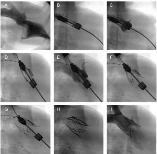
Transapical aortic valve delivery and deployment of BMMC-based TEHV utilizing the JenaValve system could be performed successfully in all animals (n = 4; Fig.3,Supplementary Video 1). In brief, the aortic root was visualized (Fig. 2A) before the loaded JenaValve device was introduced into the left ventricle and advanced into the aortic root (Fig. 3B and C). Under constant angiography guidance, the delivery process of the JenaValve delivery technology was started by positioning of the feeler arms into the deepest points of the sinuses (Step 1), which was then followed by the controlled deployment of the distal stent part (Fig.3D–F) and completed by deployment of the proximal part
Figure 3:Fluoroscopy-guided, anatomically oriented transapical delivery of marrow stromal cell-based TEHVs into the aortic valve position. The aortic root was visua-lized (A), before the TEHV loaded JenaValve Stent System was inserted and positioned (B and C). After positioning, the feelers (D and E), the JenaValve stent was opened in a stepwise fashion underfluoroscopic control (F and G). After full delivery of the TEHV (H), the appropriate positioning thereby fully excluding the native valve as well as sufficient functionality of the TEHV was confirmed by CT (I), before the JenaValve delivery device was carefully removed.
(Fig. 3G and H). Immediately after complete deployment, appropriate perfusion of the coronaries was assessed and sufficient valve functionality was evaluated (Fig.3I,Supplementary
Video 1). During the implantation procedure, all animals were
haemodynamically stable and no major complications such as bleeding or cardiac arrhythmia occurred during device removal. As in angiography, TEHV could be successfully positioned in the orthotopic aortic valve position, thereby fully excluding the native leaflets. The mean duration of the entire procedure, beginning with cell harvest until TEHV delivery was 101 ± 6 min (Table2).
Echocardiographic assessment of TEHV.
TEHV in vivoperformance was controlled viafluoroscopy (Fig.3,Supplementary
Video 1) and echocardiography (Fig.4,Supplementary Video 2).
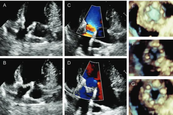
After implantation, all TEHV (n = 4) tolerated the loading pressure of the systemic circulation, and no leaflet tears or acute ruptures were detected. Sufficient leaflet mobility was present in all animals, and a sufficient opening and closing pattern along with an appropriate coaptation area was observed in two animals (Fig.4A–F,
Supplementary Video 2), while the two others displayed a
single-leaflet prolapse that was related to the leaflet/valve design of the TEHV. The mean transvalvular gradient was 7.8 ± 0.9 mmHg, and the mean effective orifice area (EOA) was 1.73 ± 0.02 cm² (Table2). Two animals displayed mild (n = 1)/moderate (n = 1) paravalvular leakage (PVL), and the first two animals showed moderate central aortic regurgitation (AR;n = 2) that was related with the scaffold design and geometry within the stent (Table2). As a response to the central AR, the scaffold design was changed to shorter radial leaflet extensions and smaller wall-belly angles to ensure fully coaptation after delivery. No stent dislocation, migration or affection of the mitral valve was observed.
Post-mortem computed tomography assessment
and gross examination
Optimal TEHV positioning was confirmed on post-mortem com-puted tomography of the excised hearts (Fig.5A–F). Gross examin-ation displayed the JenaValve stent with the TEHV to be well integrated into the aortic root and the left ventricular outflow tract. The leaflet structures were intact, with well-identifiable cusps, while there were no signs of leaflet thickening, shrinking or thrombus formation (Fig. 5G). Minor structural damage of one leaflet was observed in two TEHV and was either related to the procedural damage during animal harvest or related to the crimp-ing procedures involved. In thefirst two animals, the leaflets were positioned in a leaflet opening angle of 45–60°, which resulted
Figure 4:Echocardiographic assessment of TEHV functionality. Following implantation, TEHV tolerated the loading pressure of the systemic circulation adequately, and TEHV functionality was controlled via 2D (A and B), 2D color (C and D) and 3D echocardiography (E and F). TEHV displayed sufficient leaflet mobility and a suffi-cient opening and closing pattern along with an appropriate coaptation area. No stent dislocation, migration or affection of the mitral valve was observed.
Table 2: Intraoperative data and echo findings
Parameter Mean ± SD
Crimping time until TEHV delivery (min) 10 ± 4 Duration of the entire one-step procedure (min) 101 ± 6 TVG mean (mmHg) 7.8 ± 0.9 TVG peak (mmHg) 17.2 ± 2.2 EOA (cm²) 1.73 ± 0.02 Central aortic regurgitationa(n) n = 2 Paravalvular leakageb(n) n = 2
Regurgitation of mitral valve None Leaflet motionc Normal
Cardiac output (l/min) 5.7 ± 0.3 TEHV: tissue-engineered heart valve; TVG: transvalvular gradient; EOA: effective orifice area.
a
Two animals presented with moderate central aortic regurgitation.
bOne animal with mild and one animal with moderate paravalvular
leakage.
cTwo animals presented with a single-leaflet prolapse.
BA
SIC
S
central coaptation gaps due to excess of tissue in the coaptation area. Therefore, in the following TEHV constructs, the leaflet opening angle was changed to 15–30°, which resulted in reduc-tion of central regurgitareduc-tion and improved leaflet mobility.
DISCUSSION
This is thefirst report elucidating the principal technical feasibility of combining a clinically relevant HVTE approach with a clinically applied, anatomically oriented, latest-generation transapical stent delivery system. In the setting of an adult sheep model, we were able to demonstrate that BMMC-based TEHVs can be successfully implanted into the orthotopic aortic valve position with the JenaValve stent and delivery system. The implanted TEHV dis-played sufficient in vivo functionality, and importantly, an optimal delivery and positioning without any coronary compromise was achieved when using an anatomically oriented stent and delivery system.
Transcatheter aortic valve implantation (TAVI) is a valid and accepted treatment option for the elderly, high-risk patient suffer-ing from symptomatic aortic valve stenosis [1,19–22]. Numerous reports indicate the efficacy of transcatheter approaches along with the established benefits comprising the avoidance of sternot-omy and extracorporeal circulation. However, the utilized pros-thesis of the current TAVI concepts are bioprosthetic and therefore prone to continuous and generalized degeneration. In addition, and despite considerable mid-term clinical data [19], nothing is known about potential crimping-associated structural damage and its impact on long-term clinical outcome and dur-ability of these prostheses.
HVTE concepts generating living, autologous heart valve con-structs have been repeatedly proposed as a potential solution due to their regeneration and growth capacity and may extend the in-dication for TAVI to younger patient populations. On the other hand, and in the light of clinical relevance, previous HVTE con-cepts have been criticized for their high logistical andfinancial demands limiting a broad clinical applicability. Ideally, a
Figure 5:Computed tomography analysis and gross examination of TEHV. Post-mortem computed tomography (A and B) and 3D reconstruction analysis (C–E) dis-played sufficient stent positioning after anatomically oriented transapical delivery of the TEHV. Gross examination of the TEHV disdis-played intact leaflet structures with well-identifiable cusps, while there were no signs of leaflet thickening, shrinking or thrombus formation (F).
translational, clinically relevant HVTE concept comprises minimal-ly invasive techniques for both cell harvest and valve delivery [14]. Therefore, and based on previously reported technologies in the setting of tissue-engineered vascular grafts [16–18,23,24], we have recently introduced the novel concept of generating and implant-ing autologous BMMC-derived TEHV in a simplant-ingle-step intervention. In a preclinical setting, we were able to demonstrate the feasibility of this novel technology by replacing pulmonary and aortic valves of primates [14] and adult sheep [12, 13]. However, in these studies, only self-made or generic, non-clinical concept stent and delivery systems were used for transapical delivery, while a transla-tional, clinically HVTE approach also requires the implementation of state-of-the-art stent and delivery systems, in particular when addressing the systemic circulation [12,13].
Although numerous transcatheter devices have been estab-lished in the clinical setting [12–14,25], major concerns associated with current TAVI systems include on the one hand, mal-positioning leading to central AR or PVL and on the other hand, the important risk of CO. In this regard, the utilization of next-generation, anatomically oriented devices has been suggested to be beneficial, thereby minimizing the risk of the occurrence of AR, PVL and CO [14].
Therefore, in this study, we successfully combined our one-step BMMC-based approach with a state-of-the-art, anatomically oriented transapical delivery system as a further step towards translational, clinically relevant HVTE concepts. Importantly, our results show that the occurrence of mal-positioning and import-antly, stent dislocation as it was observed in our previous studies [13, 14], can be successfully minimized when using state-of-the-art, anatomically oriented transapical systems with feeler-guided positioning and an active clipping mechanism. Moreover, the safe and effective JenaValve delivery technology further simplified our procedural steps significantly when com-pared with the devices used for valve delivery in our previous studies [12–14,25].
In general, the transapical delivery system used in this study has proven its safety in clinical pilot studies [5,6] and in particular, its effectiveness with regard to the minimization of the occurrence of PVL [5, 6] and although the occurrence of PVL and AR was encountered in this study, it was primarily related to the scaffold design and integration within the stent, but not to the Jena Valve stent or the delivery system itself. After delivery in thefirst two animals, the TEHV systems revealed moderate central AR, which was most likely related to the steep belly-wall angle of the con-structs. Therefore, in the following animals, the radial leaflet exten-sion was decreased before implantation to ensure optimal leaflet coaptationin situ and to prevent central AR. After the delivery of these two constructs, less regurgitation and higher improved leaflet mobility were observed, suggesting that (i) TEHV native analogous leaflet geometry is indispensable for the optimal func-tionality of anatomically oriented TEHV systems and (ii) that too-steep belly-wall angles of TEHV leaflets lead to central AR due to the lack of sufficient leaflet coaptation.
Limitations
There are several limitations that need to be mentioned:first, the animal numbers are low, and consecutive studies will need larger cohorts, particularly when assessing the issue of optimized scaf-fold designs. In addition, this was an acute study with the primary aim of assessing the principal technical feasibility of combining
our BMMC-based one-step approach with a state-of-the-art, ana-tomically oriented transapical stent and delivery system. Further data are necessary to evaluate the long-term safety and efficacy of the presented technology.
CONCLUSION
For thefirst time, we demonstrate the technical feasibility of a transapical marrow stromal cell-based TEHV delivery into the aortic valve position using an anatomically oriented, clinically applied transapical implantation system. Our data indicate that the combination of TEHV and a state-of-the-art transapical deliv-ery system is feasible, representing an important step towards translational, transcatheter-based TEHV concepts that may broaden the indication for TAVI to younger patients. To minimize central regurgitation and the occurrence of PVL, further scaffold design and valve geometry optimization are necessary.
SUPPLEMENTARY MATERIAL
Supplementary material (Video 1 and 2) is available atEJCTS online. Video 1: Fluoroscopy shows functionality of the tissue-engineered heart valve after anatomically oriented, transapical delivery.
Video 2: Two-dimensional echocardiography after anatomically oriented, transapical delivery of the tissue-engineered heart valve.
Funding
This project was supported by the Center for Clinical Research, University Hospital and University of Zurich, Switzerland.
Conflict of interest: Luc Behr and Sebastien Sammut are employ-ees of IMM Recherche, Paris, France.
REFERENCES
[1] Leon MB, Smith CR, Mack M, Miller DC, Moses JW, Svensson LGet al. Transcatheter aortic-valve implantation for aortic stenosis in patients who cannot undergo surgery. N Engl J Med 2010;363:1597–607.
[2] Falk V, Walther T, Schwammenthal E, Strauch J, Aicher D, Wahlers Tet al. Transapical aortic valve implantation with a self-expanding anatomically oriented valve. Eur Heart J 2011;32:878–87.
[3] Smith CR, Leon MB, Mack MJ, Miller DC, Moses JW, Svensson LGet al. Transcatheter versus surgical aortic-valve replacement in high-risk patients. N Engl J Med 2011;364:2187–98.
[4] Walther T, Schuler G, Borger MA, Kempfert J, Seeburger J, Ruckert Yet al. Transapical aortic valve implantation in 100 consecutive patients: com-parison to propensity-matched conventional aortic valve replacement. Eur Heart J 2010;31:1398–403.
[5] Kempfert J, Rastan AJ, Mohr FW, Walther T. A new self-expanding trans-catheter aortic valve for transapical implantation—first in man implant-ation of the JenaValve. Eur J Cardiothorac Surg 2011;40:761–3.
[6] Treede H, Mohr FW, Baldus S, Rastan A, Ensminger S, Arnold Met al. Transapical transcatheter aortic valve implantation using the JenaValve system: acute and 30-day results of the multicentre CE-mark study. Eur J Cardiothorac Surg 2012;41:e131–8.
[7] Sundermann SH, Grunenfelder J, Corti R, Rastan AJ, Linke A, Lange Ret al. Feasibility of the Engager aortic transcatheter valve system using aflexible over-the-wire design. Eur J Cardiothorac Surg 2012;42:e48–52.
[8] Kempfert J, Rastan AJ, Beyersdorf F, Schonburg M, Schuler G, Sorg Set al. Trans-apical aortic valve implantation using a new self-expandable bio-prosthesis: initial outcomes. Eur J Cardiothorac Surg 2011;40:1114–9.
BA
SIC
S
[9] Kempfert J, Treede H, Rastan AJ, Schonburg M, Thielmann M, Sorg Set al. Transapical aortic valve implantation using a new self-expandable bio-prosthesis (ACURATE TA): 6-month outcomes. Eur J Cardiothorac Surg 2013;43:52–7.
[10] Falk V, Schwammenthal EE, Kempfert J, Linke A, Schuler G, Mohr FWet al. New anatomically oriented transapical aortic valve implantation. Ann Thorac Surg 2009;87:925–6.
[11] Kiefer P, Gruenwald F, Kempfert J, Aupperle H, Seeburger J, Mohr FWet al. Crimping may affect the durability of transcatheter valves: an experimental analysis. Ann Thorac Surg 2011;92:155–60.
[12] Emmert MY, Weber B, Behr L, Frauenfelder T, Brokopp CE, Grunenfelder J et al. Transapical aortic implantation of autologous marrow stromal cell-based tissue-engineered heart valves:first experiences in the systemic cir-culation. JACC Cardiovasc Interv 2011;4:822–3.
[13] Emmert MY, Weber B, Wolint P, Behr L, Sammut S, Frauenfelder Tet al. Stem cell-based transcatheter aortic valve implantation:first experiences in a pre-clinical model. JACC Cardiovasc Interv 2012;5:874–83.
[14] Weber B, Scherman J, Emmert MY, Gruenenfelder J, Verbeek R, Bracher M et al. Injectable living marrow stromal cell-based autologous tissue engi-neered heart valves:first experiences with a one-step intervention in pri-mates. Eur Heart J 2011;32:2830–40.
[15] Dolgin E. Taking tissue engineering to heart. Nat Med 2011;17:1032–5. [16] Roh JD, Sawh-Martinez R, Brennan MP, Jay SM, Devine L, Rao DAet al.
Tissue-engineered vascular grafts transform into mature blood vessels via an inflammation-mediated process of vascular remodeling. Proc Natl Acad Sci USA 2010;107:4669–74.
[17] Shin’oka T, Imai Y, Ikada Y. Transplantation of a tissue-engineered pulmon-ary artery. N Engl J Med 2001;344:532–3.
[18] Shin’oka T, Matsumura G, Hibino N, Naito Y, Watanabe M, Konuma T et al. Midterm clinical result of tissue-engineered vascular autografts seeded with autologous bone marrow cells. J Thorac Cardiovasc Surg 2005;129:1330–8. [19] Genereux P, Head SJ, Wood DA, Kodali SK, Williams MR, Paradis JMet al.
Transcatheter aortic valve implantation: 10-year anniversary part II: clinical implications. Eur Heart J 2012;33:2399–402.
[20] Genereux P, Head SJ, Wood DA, Kodali SK, Williams MR, Paradis JMet al. Transcatheter aortic valve implantation 10-year anniversary: review of current evidence and clinical implications. Eur Heart J 2012;33:2388–98. [21] Kappetein AP, Head SJ, Genereux P, Piazza N, van Mieghem NM,
Blackstone EHet al. Updated standardized endpoint definitions for trans-catheter aortic valve implantation: the Valve Academic Research Consortium-2 consensus document. J Am Coll Cardiol 2012;60:1438–54. [22] Kappetein AP, Head SJ, Genereux P, Piazza N, van Mieghem NM, Blackstone
EHet al. Updated standardized endpoint definitions for transcatheter aortic valve implantation: the Valve Academic Research Consortium-2 consensus document. Eur J Cardiothorac Surg 2012;42:S45–60.
[23] Brennan MP, Dardik A, Hibino N, Roh JD, Nelson GN, Papademitris Xet al. Tissue-engineered vascular grafts demonstrate evidence of growth and development when implanted in a juvenile animal model. Ann Surg 2008; 248:370–7.
[24] Hibino N, McGillicuddy E, Matsumura G, Ichihara Y, Naito Y, Breuer C et al. Late-term results of tissue-engineered vascular grafts in humans. J Thorac Cardiovasc Surg 2010;139:431–6.
[25] Schmidt D, Dijkman PE, Driessen-Mol A, Stenger R, Mariani C, Puolakka A et al. Minimally-invasive implantation of living tissue engineered heart valves: a comprehensive approach from autologous vascular cells to stem cells. J Am Coll Cardiol 2010;56:510–20.