Advance Access publication 25 October 2007
Introduction
A bilateral sagittal split osteotomy (BSSO) together with orthodontic treatment aims to improve the soft tissue profi le and its underlying hard tissues. Since its introduction by Trauner and Obwegeser (1955) and Obwegeser (1957) , this operation has gained popularity, especially when combined with rigid internal fi xation (RIF).
Most studies have examined short-time stability after BSSO, i.e. 6 months after surgery ( Kirkpatrick et al. , 1987; Rubens et al. , 1988; Van Sickels et al. , 1988; Gassman et al. , 1990; Moenning et al. , 1990; Abeloos et al. , 1993; Blomqvist et al. , 1997 ). Others have included follow-ups from 6 months to 5 years ( Caskey et al. , 1989; Watzke et al. , 1990, 1991; Mommaerts, 1991; Hilbe and Puelacher, 1994; Thüer et al. , 1994; Kallela et al. , 1998; Mobarak et al. , 2001b ).
A few authors have investigated stability between RIF with plates or screws and wire fi xation ( Ellis et al. , 1988; Buckley et al. , 1989; Moenning et al. , 1990; Mommaerts, 1991 ), while others compared different RIF techniques between each other ( Thomas et al. , 1986; Watzke et al. , 1991; Blomqvist and Isaksson, 1994 ).
RIF demonstrated greater stability of the surgical correction than wire fi xation. RIF has the advantage of a
Stability of the hard and soft tissue profi le after mandibular
advancement in sagittal split osteotomies: a longitudinal and
long-term follow-up study
Christof Urs Joss * and Urs Walter Thüer **
* Department of Orthodontics, University of Geneva, and ** Department of Orthodontics, University of Bern, Switzerland
SUMMARY The aim of the study was to conduct a long-term follow-up investigation of the stability of hard and soft tissues after bilateral sagittal split osteotomy (BSSO) with rigid internal (RIF) fi xation to advance the mandible.
Sixteen consecutive patients (12 females and 4 males, mean age 21.4 years) were available for re-examination 12.7 years (T5) after surgery. The preceding follow-ups were before (T1), and 5 days (T2), 7.3 months (T3), and 13.9 months (T4) after surgery. Lateral cephalograms were traced by hand, digitized, and evaluated with the Dentofacial Planner® program. The x -axis for the system of co-ordinates ran through sella (point zero) and the line NSL – 7 degrees. Thus, the program determined the x - and y -values of each variable and the usual angles and distances. Statistical analysis was carried out using Wilcoxon’s matched-pair signed-ranks test with Bonferroni adjustments. The relationships between the examined variables were analysed by Spearman rank correlation coeffi cients.
The backward relapse at point B (T5) was 2.42 mm, or 50 per cent, and at pogonion 3.21 mm, or 60 per cent of the initial advancement. The mean net effect at T5 on the labial fold (soft tissue point B) was 94 per cent of the advancement at point B. For the soft tissue chin (soft tissue pogonion), it was 119 per cent of the advancement at pogonion. The net effect on the lower lip (labrale inferior) was 55 per cent of the advancement at incision inferior. The amount of the surgical advancement of the mandible was correlated with the long-term relapse in point B. Among possible reasons for this relapse are the initial soft tissue profi le, the initial growth direction, and the remodelling processes of the hard tissue.
shorter intermaxillary fi xation period and the in-patient period is reduced resulting in more positive effects on mandibular function ( Luhr et al. , 1991 ). Post-surgical correction after RIF is diffi cult if not impossible. Thus, the mandibular condyle requires seating in a correct position in the fossa before fi xing the distal and proximal segments of the mandible after the split ( Epker and Wylie, 1986; Raveh et al. , 1988; Richter et al. , 1990; Luhr et al. , 1991; Thüer et al. , 1994 ).
Among the factors which contribute to relapse after BSSO are the amount of mandibular surgical advancement control of the proximal segment, increase of posterior face height, and occlusal stability ( Epker and Wessberg, 1982; Van Sickels et al. , 1988; Will and West, 1989; Gassman et al. , 1990 ).
Even though several devices have been designed to hold the proximal mandibular segment with the condyle in its initial pre-split position until rigid fi xation ( Epker and Wylie, 1986; Raveh et al. , 1988; Richter et al. , 1990; Luhr et al. , 1991 ), many surgeons still prefer to seat the proximal segment freehand, aiming at positioning the condyle superiorly and posteriorly in the fossa. Depending on the skill of the surgeon, this might be more or less accurate ( Thüer et al. , 1994 ).
The most important aesthetic goal is to improve soft tissue profi le after BSSO. The results reported in the literature on the effect of mandibular advancement are fairly consistent for the soft tissue chin, ranging from 94 to 111 per cent of the advancement of pogonion. The effect on the lower lip is more variable; the labial fold (soft tissue point B) has been found to advance from 86 to 119 per cent of the advancement of point B. The largest variability was found for labrale inferior, ranging from 26 to 85 per cent of the advancement of incision inferior ( Lines and Steinhauser, 1974; Quast et al. , 1983; Mommaerts and Marxer, 1987; Dermaut and De Smit, 1989; Hernandez-Orsini et al. , 1989; Ewing and Ross, 1992; Thüer et al. , 1994; Mobarak et al. , 2001a ).
The aim of the present study was to evaluate long-term stability of mandibular advancement with BSSO and RIF as well as to examine the effects of the advancement on the soft tissue profi le.
Subjects and methods
Sixteen consecutive patients (12 females and 4 males), aged 17.0 – 30.1 years (mean age 21.4 years), who underwent only mandibular advancement at the Department of Craniomaxillofacial Surgery, University of Bern, in the years 1986 – 1989 were studied prospectively.
The subjects had a moderate or marked distal occlusion that was corrected with a BSSO of the mandible and RIF. All patients gave written consent for participation. The sagittal splits were fi xed with three titanium lag screws (diameter 3.5 mm) on each side. None of the patients underwent genioplasty simultaneously with the BSSO. The surgery was performed by one of the four senior surgeons of the department, and the patients were referred by various orthodontists. The surgical technique ( Raveh et al. , 1988 ) was the same for all patients, and each of the surgeons was experienced in this procedure. No splint was used for stabilization of the mandible during surgery, but maxillomandibular fi xation with wire ligatures to arch bars or orthodontic brackets was used for 4 – 6 days after surgery. The skeletal and soft tissue changes as a result of surgery and their stability were evaluated on profi le cephalograms taken with the teeth in the intercuspal position, and including a linear enlargement of 3.3 per cent. The radiographs were taken with the subject standing upright and trying to assume a natural position of the head and relaxed lips. The same radiographic instruments were used to obtain all cephalograms.
The fi rst cephalogram was taken between 0 and 1 day (mean 1 day) before surgery (T1), the second (T2) between 3 and 8 days (mean 5 days), at T3 between 6.0 and 9.9 months (mean 7.3 months), atT4 between 11.8 and 19.3 months (mean 13.9 months), and at T5 between 10.9 and 14.2 years (mean 12.7 years) after surgery.
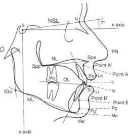
The cephalometric analysis was carried out by one author (CUJ) and included the reference points and lines shown in Figure 1 . The cephalogram was then
traced and the reference points digitized with the Dentofacial Planner (Dentofacial Software Inc., Toronto, Canada). The person tracing the cephalograms was blind to the degree of mandibular advancement and the dates of the post-operative radiographs. Conventional cephalometric variables as well as the co-ordinates of the reference points ( Table 1 ) were calculated by the computer program. The co-ordinate system had its origin at point s (sella), and its x -axis formed an angle of 7 degrees with the reference line NSL ( Figure 1 ). Overjet and overbite were calculated from the co-ordinates of the points: is (incision superior) and ii (incision inferior).
The systematic and accidental errors of the cephalometric analysis were evaluated by duplicate determinations of 11 cephalograms selected at random. The cephalograms were retraced and remeasured for a second time by the same author 2 weeks after the fi rst assessment. No systematic errors were found when the values were evaluated with a paired t -test. The accidental errors (si) were calculated with the formula si d n =
∑
2 2where d is the difference between the repeated measurements and n is the number of duplicate determinations ( Dahlberg, 1940 ). These errors are shown in Table 1 .
Most of the angular variables and co-ordinates of the skeletal reference points had accidental errors smaller than 1.0 degree or 1.0 mm, respectively. The exceptions were points tangent gonion (tGo, x -value) and soft tissue menton (Me ′ , x -value). tGo is a constructed point and thus more susceptible to tracing errors. Me ′ is located on a more rounded anatomical structure of the soft tissue chin which could have made location more diffi cult.
Statistical analysis
The effect of treatment, i.e. the differences between the variables and co-ordinates at T1 and T2, T4 and T5, T2 and T5, as well as T1 and T5 were tested with Wilcoxon’s matched-pair signed-ranks test. To increase the level of signifi cance, Bonferroni adjustments were carried out on the P value. The relationships between variables were analysed with the Spearman rank correlation coeffi cient.
Results
Table 2 shows the selected variables at T1 and T5. The mean changes, standard deviations, and ranges for the selected cephalometric parameters before surgery and during the subsequent observation periods are given in Tables 3 and 4 , and Figure 2 . Negative values imply a backward and positive values a forward movement of the
point in the horizontal plane. Negative values imply an upward and positive values a downward movement of the point in the vertical plane.
Due to Bonferroni adjustments ( P / n , n = 4), the values were * P ≤ 0.0125, ** P ≤ 0.0025, and *** P ≤ 0.00025. Skeletal changes
Horizontal. The mean advancement of the mandible immediately following surgery (T2 – T1) was 4.81 mm at point B, 5.33 mm at pogonion, and 4.14 mm at incision inferior.
At the long-term follow-up 12.7 years after surgery, the mean relapse (T5 – T2) at point B was with 2.42 mm ( P = 0.006) and at pogonion 3.21 mm ( P = 0.002), representing a loss of 50 and 60 per cent for pogonion of the
surgical advancement. The mean relapse (T5 – T2) at incision inferior was 1.73 mm ( P = 0.008), i.e. 42 per cent of the initial advancement (T2 – T1).
There was considerable variation in the net effects at point B and pogonion ( Figure 2 ). Mandibular advancement at point B relapsed in 12 subjects; in fi ve the relapse was complete, while in an additional fi ve a relapse of 50 per cent or more occurred. Further anterior movement of the mandible (point B) was seen in two patients 12.7 years post-surgically.
The antero-posterior net effects for advancement of the mandible at T5 were for incision inferior, point B, and pogonion 58, 50, and 40 per cent, respectively.
Vertical. The post-surgical relapse (T5 – T2) of menton was not signifi cant ( P = 0.069). tGo showed an ( P = 0.004) upward movement of 2.44 mm in the post-operative relapse period (T5 – T2) and pogonion a downward movement of 2.23 mm ( P = 0.0004).
Soft tissue changes
Horizontal. The post-surgical relapse (T5 – T2) of labrale superior showed a backward movement of 2.89 mm ( P = 0.001). The fi nal result (T5 – T1) was 1.61 mm ( P = 0.015). Labrale inferior had a very signifi cant ( P = 0.001) backward movement of 4.23 mm in the post-surgical relapse period (T5 – T2). The fi nal result (T5 – T1) showed a non-signifi cant ( P = 0.026) increase of 1.33 mm (range – 2.4 to 5.2 mm). Both soft tissue point B (3.5 mm) and soft tissue pogonion (3.34 mm) moved backward ( P = 0.001).
Vertical. Labrale superior (T5 – T2) moved downward by 2.66 mm ( P = 0.002). Menton showed a downward movement of 1.09 mm ( P = 0.215) in the post-surgical relapse period. The change at labrale inferior was 55 per cent of the advancement of incision inferior at T5 and at T4, 70 per cent. The corresponding values for point B to point B ′ and pogonion to pogonion ′ were 94 and 119 per cent, respectively, at T5. At T4, the values for point B and pogonion were 95 and 96 per cent, respectively.
Table 1 Accidental errors (si) of the cephalometric analysis.
Variables Si (°) Reference point (skeletal) Si (mm) Reference point (soft tissue) Si (mm) x y x y SNA 0.57 N 0.56 0.08 PN 0.38 0.47 SNB 0.34 Point-A 0.83 0.52 Point ′ -A 0.47 1.44 ANB 0.57 Point-B 0.66 0.96 Labrale superior 0.62 0.74 NL/NSL 0.82 Incision superior 0.57 0.52 Labrale inferior 0.47 0.70 ML/NSL 0.45 Incision inferior 0.52 0.30 Stomion superior 0.86 0.55 ML/NL 0.89 Pogonion 0.84 0.82 Stomion inferior 0.71 0.62 N – Spa 0.58 Menton 0.72 0.42 Point ′ -B 0.71 0.74
Spa – Me 0.54 tGonion 1.09 0.32 Pogonion ′ 0.86 0.85
N – Me 0.37 Spa 0.69 0.59 Menton ′ 2.09 0.75
S – tGo 0.30 Spp 0.63 0.48
Figure 1 Reference points and lines used in the cephalometric analysis.
S, sella; NSL, nasion – sella-line; N, nasion; x , horizontal reference plane; NL, nasal line; ILs, upper incisal line; Spp, spina nasalis posterior; Spa, spina nasalis anterior; PN, pronasion; point A ′ ; Mo, molar; OL, occlusal line; Ss, stomion superius; Ls, labrale superius; point A; Ii, incision inferior; Si, stomion inferior; Li, labrale inferior; Is, incision superior; tGo, tangent gonion; ML, mandibular line; point B; point B ′ ; Pg, pogonion; Pg ′ , soft tissue pogonion; Me, menton; Me ′ , soft tissue menton; and y , vertical reference plane.
Correlations
No signifi cant correlations for gender and age of the patients were found at any time point. Overjet (T2 – T1) correlated highly signifi cantly ( P = 0.001, R = 0.755) with relapse (T5 – T2) at point B ( x -value) and very signifi cantly ( P = 0.001, R = 0.908) with relapse at pogonion ( x -value). The amount of the mandibular advancement (T2 – T1) at point B ( x -value) was signifi cantly correlated ( P = 0.027, R = – 0.550) with the relapse (T5 – T2) at point B ( x -value).
Discussion
The larger number of females in this study group signifi es that mostly females seek treatment. Such distribution of gender has also been found in other investigations ( Kirkpatrick et al. , 1987; Caskey et al. , 1989; Moenning et al. , 1990; Watzke et al. , 1990; Mommaerts, 1991 ).
The mean antero-posterior advancement at point B was 4.81 mm (range 1.2 – 6.7 mm) and at pogonion 5.33 mm (range 1.7 – 7.5 mm). Several other studies have found the same amount of mandibular advancement ( Thomas et al. , 1986; Van Sickels et al. , 1986; Rubens et al. , 1988; Gassman et al. , 1990; Douma et al. , 1991; Abeloos et al. , 1993; Blomqvist et al. , 1997; Kallela et al. , 1998 ). At T5, the relapse at point B was 2.42 mm or 50 per cent of the initial surgical advancement (T2 – T1), and at pogonion 3.21 mm or 60 per cent of the initial surgical advancement.
Numerous studies have been published on short- and long-time post-surgical relapse after BSSO and RIF. However, there is a lack of research examining relapse after 10 or more years.
Mobarak et al. (2001b) examined 61 patients after BSSO for mandibular advancement 3 years post-operatively. In high-angle subjects, the relapse at pogonion was 36 per cent, while in low-angle subjects, it was only 27.6 per cent. The relapse in the low-angle group was mostly seen during
the two fi rst months post-surgery, while the high-angle group demonstrated a more continuous relapse. Hilbe and Puelacher (1994) reported a relapse of 12.9 per cent in 24 patients after 3.5 years according to the Wits analysis.
The patients in the present study showed a large variation in their post-operative response to BSSO. At T5, the mandible moved forwards in two patients, while in others, a smaller or greater relapse occurred. An additional advancement has also been repeated by others ( Van Sickels et al. , 1986, 1988; Caskey et al. , 1989; Krekmanov et al. , 1989; Moenning et al. , 1990 ).
It is considered that the surgery itself, the surgical methods used as well as remaining growth, and remodelling processes of the face play an important role for post-operative relapse or even further advancement of the mandible. Although the same surgical technique was used, having more than one surgeon can introduce a further degree of variability in the stability of the soft and hard tissues.
The initial growth of the patient’s face and continuous remodelling processes may lead to an advantageous or disadvantageous change of the position of the mandible after BSSO. Behrents (1985a,b) examined 113 untreated subjects from 17 to 80 years of age and showed that point B moved downward in both genders. Males presented anterior and downward rotation of the mandible, while females demonstrated a clockwise rotation (posterior and downward). However, point B and pogonion in females were likely to be stable in its sagittal position.
The fi ndings of that author indicate that there could be an improvement of the profi le in male advancement patients with age, but in females neither an improvement nor a worsening. The mean age of the patients at T5 in this investigation was 34.2 years for females, 33.6 years for males, and combined 34.1 years.
SNA angle was constant between T2 and T5, while SNB showed, for the same period, a highly signifi cant decrease
Table 2 Values for the selected cephalometric variables at T1 (before surgery) and T5 (12.7 years after surgery).
T1 T5
Mean SD Range Mean SD Range
SNA (°) 79.18 3.25 73.4 to 86.2 80.16 3.62 74.9 to 89.1 SNB (°) 75.54 3.00 68.7 to 80.5 76.71 3.23 70.8 to 82.1 ANB (°) 3.65 2.60 − 1.6 to 8.5 3.46 3.38 − 2.3 to 10.4 NSL/NL (°) 7.29 3.23 1.0 to 14.2 7.85 3.29 16.3 to 1.9 NSL/ML (°) 33.52 6.58 22.7 to 44.0 33.78 6.87 22.1 to 46.0 NL/ML (°) 26.23 6.59 15.7 to 37.2 25.93 6.89 14.9 to 35.8 Gonion angle (°) 121.90 5.64 113.8 to 137.0 124.03 5.19 116.0 to 132.3 Anterior face height (N – Me, mm) 120.14 5.38 111.3 to 130.8 121.05 5.90 111.1 to 130.0 Upper face height (N – Spa, mm) 52.34 3.21 46.0 to 57.0 52.44 3.24 45.9 to 57.3 Lower face height (Spa – Me, mm) 70.68 5.11 60.6 to 79.3 71.12 5.35 62.7 to 79.2 Posterior face height (S – tGo, mm) 80.06 6.55 69.6 to 91.7 79.37 7.37 68.6 to 93.8 Overjet (mm) 7.03 2.15 3.7 to 11.7 4.13 1.76 1.2 to 7.6 Overbite (mm) 2.84 1.68 0.6 to 6.2 3.64 1.89 0.5 to 6.4
T
able 3
Changes in the variables and co-ordinates of the mandibular incisors as a result of sur
gery . V ariable or co-ordinate T2 – T1 T5 – T4 T5 – T2 T5 – T1 Mean SD Range Mean SD Range Mean SD Range Mean SD Range Horizontal [ x -value (mm)] Point A 0.14 ns 1.15 − 2.0 to 3.1 1.23 * 1.25 − 1.3 to 3.3 0.99 ns 1.43 − 0.9 to 3.9 1.13 ns 1.62 − 1.7 to 4.6 Point B 4.81 *** 1.51 1.2 to 6.7 − 0.78 ns 1.92 − 3.9 to 2.7 − 2.42 * 2.62 − 6.4 to 4.7 2.39 ** 1.85 − 0.8 to 5.9 Pogonion 5.33 *** 1.53 1.7 to 7.5 − 1.48 * 1.91 − 4.6 to 2.2 − 3.21 ** 2.67 − 7.5 to 1.6 2.12 * 2.19 − 2.2 to 6.3 tGonion 4.52 ** 2.38 − 1.0 to 2.5 − 1.04 ns 1.91 − 4.5 to 1.6 − 3.04 ** 2.03 − 6.7 to 0.7 1.48 * 1.79 − 2.7 to 4.1 Incision superior − 0.20 ns 0.67 − 1.2 to 1.3 − 0.08 ns 1.55 − 2.3 to 2.7 − 0.28 ns 2.55 − 4.7 to 4.9 − 0.48 ns 2.45 − 4.1 to 4.9 Incision inferior 4.14 *** 2.19 0.7 to 8.9 − 0.99 ns 1.75 − 4.3 to 1.4 − 1.73 * 2.07 − 6.7 to 1.3 2.42 ** 1.89 − 1.4 to 6.2 V ertical [ y -value (mm)] Point B 0.38 ns 1.87 − 3.8 to 3.4 − 0.32 ns 3.10 − 8.3 to 4.4 − 0.39 ns 2.17 − 3.7 to 4.6 − 0.01 ns 2.69 − 4.3 to 5.9 Pogonion 0.31 ns 1.96 − 2.6 to 3.6 1.74 ns 2.27 − 2.6 to 6.1 2.23 * 2.29 − 1.3 to 5.8 2.54 * 3.00 − 2.8 to 8.5 Menton 0.80 * 0.96 − 1.0 to 2.5 0.69 ns 1.39 − 2.1 to 2.6 0.75 ns 1.73 − 2.2 to 5.2 1.55 * 1.76 − 1.8 to 6.2 tGonion 1.96 * 2.49 − 3.2 to 4.9 0.24 ns 1.96 − 3.0 to 2.8 − 2.44 * 2.86 − 8.0 to 3.4 − 0.48 ns 3.09 − 5.3 to 4.1 Incision superior 0.26 ns 0.89 − 2.4 to 1.7 0.28 ns 1.28 − 2.3 to 2.3 0.91 ns 1.25 − 1.6 to 2.5 1.16 * 1.41 − 1.7 to 4.2 Incision inferior − 0.04 ns 1.16 − 2.1 to 2.6 − 0.02 ns 2.05 − 4.1 to 3.0 0.41 ns 2.16 − 3.1 to 4.0 0.37 ns 2.33 − 3.1 to 4.8
Angular (°) and linear measurements (mm)
SNA 0.32 ns 1.16 − 2.4 to 2.3 0.96 * 1.15 − 1.3 to 2.9 0.66 ns 1.17 − 1.1 to 2.8 0.99 ns 1.66 − 2.5 to 3.1 SNB 2.92 *** 0.99 0.8 to 4.3 − 0.79 * 0.82 − 1.8 to 1.2 − 1.75 *** 1.03 − 3.5 to – 0.1 1.17 ** 1.01 − 0.4 to 2.9 ANB − 2.6 ** 1.40 − 5.6 to 0.1 1.78 ** 1.07 − 0.7 to 3.9 2.41 ** 1.49 0.0 to 6.0 − 0.19 ns 1.8 − 3.3 to 3.6 NSL/NL − 0.14 ns 0.81 − 1.8 to 0.8 0.62 ns 1.54 − 2.8 to 3.8 0.70 ns 1.45 − 3.1 to 3.2 0.56 ns 1.58 − 3.0 to 2.8 NSL/ML − 1.19 ns 2.19 − 5.2 to 3.1 − 0.38 ns 1.50 − 2.7 to 2.9 1.45 ns 2.18 − 2.1 to 6.6 0.26 ns 3.17 − 5.2 to 5.4 NL/ML − 1.05 ns 2.34 − 6.0 to 3.0 − 1.01 ns 1.98 − 4.8 to 2.4 0.76 ns 2.84 − 5.3 to 7.3 − 0.29 ns 3.37 − 6.7 to 5.2 Gonion angle 3.43 ns 4.19 − 3.9 to 8.9 − 1.4 ns 2.52 − 5.7 to 5.7 − 1.3 ns 2.35 − 6.9 to 3.8 2.13 ns 3.99 − 4.7 to 8.0 Overjet − 4.34 *** 2.01 − 1.4 to – 8.3 0.91 ns 1.32 − 0.8 to 3.5 1.44 * 1.68 − 1.0 to 5.0 − 2.9 * 2.94 − 6.5 to 2.4 Overbite 0.29 ns 1.34 − 2.0 to 2.6 0.31 ns 1.81 − 2.9 to 3.2 0.5 ns 2.08 − 3.7 to 4.5 0.79 ns 2.12 − 3.6 to 5.1 Spa – Me − 0.61 ns 1.04 − 1.7 to 1.8 0.78 ns 1.26 − 1.6 to 2.7 1.05 ns 1.65 − 1.6 to 3.6 0.44 ns 2.15 − 3.1 to 3.9 N – Me − 0.03 ns 0.86 − 1.3 to 1.8 0.63 ns 1.44 − 2.1 to 2.6 0.93 ns 1.77 − 2.0 to 5.3 0.91 ns 1.93 − 2.3 to 5.6 S – tGo 1.39 ns 2.49 − 3.7 to 4.6 0.39 ns 1.77 − 2.5 to 2.8 − 2.08 * 2.67 − 7.5 to 3.2 − 0.69 ns 2.95 − 5.5 to 3.3 T1, before sur gery;
T2, 5 days after sur
gery;
T5, 12.7 years after sur
gery; SD, standard deviation.
Bonferroni adjustments ( P / n , n = 4): * P ≤ 0.0125; ** P ≤ 0.0025; *** P ≤ 0.00025; ns, not signi fi cant.
T
able 4
Changes of the co-ordinates of the lower lip and the soft tissues of the lower face.
V ariable or co-ordinate T2 – T1 T5 – T4 T5 – T2 T5 – T1 Mean SD Range Mean SD Range Mean SD Range Mean SD Range Horizontal [ x -value (mm)] Labrale superior 1.28 * 1.38 − 0.5 to 4.5 − 1.26 ns 1.83 − 4.0 to 1.7 − 2.89 ** 1.90 − 6.0 to 1.7 − 1.61 ns 2.05 − 5.5 to 2.0 Labrale inferior 5.56 *** 2.09 1.0 to 10.3 − 1.04 * 1.37 − 3.1 to 1.3 − 4.23 ** 2.09 − 8.3 to 1.1 1.33 ns 2.06 − 2.4 to 5.2 Point B ′ 5.68 *** 1.62 2.2 to 7.8 − 0.76 ns 1.95 − 4.5 to 2.2 − 3.50 ** 2.36 − 8.9 to 1.1 2.24 * 2.41 − 2.3 to 6.7 Pogonion ’ 5.86 *** 1.79 1.5 to 8.2 − 0.94 ns 2.25 − 4.5 to 3.2 − 3.34 ** 2.72 − 9.2 to 1.3 2.53 * 2.54 − 3.0 to 6.2 Menton ’ 6.43 *** 3.22 0.4 to 10.6 − 3.43 ns 6.05 − 18.1 to 8.8 − 6.30 ** 4.20 − 12.0 to 3.0 0.99 ns 4.71 − 5.8 to 9.7 V ertical [ y -value (mm)] Labrale superior − 1.14 ns 1.99 − 4.0 to 2.9 0.54 ns 1.49 − 2.1 to 3.6 2.66 ** 2.32 − 3.0 to 6.7 1.52 * 1.64 − 1.1 to 4.2 Stomion superior − 0.70 ns 1.86 − 4.5 to 3.6 0.76 ns 1.70 − 2.6 to 4.4 2.10 * 2.26 − 4.4 to 3.8 1.40 ns 1.93 − 2.1 to 5.9 Stomion inferior − 1.03 ns 1.65 − 4.0 to 1.9 0.47 ns 2.16 − 5.8 to 3.0 1.56 * 1.92 − 3.8 to 4.4 0.53 ns 2.17 − 4.5 to 2.9 Labrale inferior 0.20 ns 2.65 − 3.4 to 6.2 − 0.36 ns 1.88 − 4.6 to 2.0 − 1.04 ns 2.47 − 5.6 to 3.1 − 0.84 ns 2.22 − 5.1 to 2.2 Point B ′ 0.33 ns 2.23 3.3 to 4.7 0.34 ns 2.42 − 5.0 to 3.5 0.98 ns 2.77 − 4.4 to 4.7 1.31 ns 2.66 − 4.1 to 7.2 Pogonion ’ − 0.21 ns 2.55 − 4.8 to 4.9 1.41 ns 2.94 − 5.5 to 6.3 2.49 ns 3.37 − 3.6 to 9.0 2.28 ns 3.45 − 3.8 to 10.8 Menton ’ 1.39 * 1.67 − 2.1 to 4.8 1.60 ns 3.34 − 3.6 to 1 1.7 1.09 ns 3.16 − 3.9 to 9.3 2.48 ** 2.69 − 0.6 to 9.2 T1, before sur gery;
T2, 5 days after sur
gery;
T5, 12.7 years after sur
gery; SD, standard deviation.
Bonferroni adjustments ( P / n , n = 4): * P ≤ 0.0125;** P ≤ 0.0025;*** P ≤ 0.00025; ns, not signi fi cant.
of 1.75 degrees. The net effect (T5 – T1), nevertheless, was a signifi cant improvement of 1.17 degrees. Behrents (1985a,b) found no signifi cant change in SNB from 17 to 80 years of age.
At the level of the teeth, the post-surgical relapse (T5 – T2) was more stable than the sagittal relapse at point B of 50 per cent. Overjet increased signifi cantly by 1.44 mm in the period T5 – T2. The fi nal result (T5 – T1) was a signifi cant improvement in overjet by 2.9 mm. The mean value for overbite was 2.8 mm at T1 and at T5 there was a non-signifi cant increase of 0.79 mm. As a result of the development of overjet and overbite, there was dental compensation to improve the skeletal relapse ( Figures 2 ).
Among factors which contribute to post-surgical relapse are the amount of mandibular advancement, control of the proximal segment of the mandible, increase of the posterior face height, and occlusal stability ( Epker and Wessberg, 1982; Van Sickels et al. , 1988; Will and West, 1989; Gassman et al. , 1990; Thüer et al. , 1994 ).
The relatively large relapse of the advancement found in this study is unsatisfactory and most likely due to incomplete setting of the condyles in the fossae before fi xation of the proximal segments. The manipulation of these segments is diffi cult, especially in large advancements where the soft tissues become considerably stretched.
Figure 2 Surgical and net effects 12.7 years post-surgery of the advancement of point B (a) and the change in overjet (b) in individual patients.
The amount of mandibular advancement at point B correlated signifi cantly ( P = 0.027) with the amount of the post-surgical relapse. Neither gender nor age had an infl uence on post-surgical relapse.
Analysis of the data shows that there was always a mean sagittal decrease of 0.8 mm at point B from T2 to T3, T3 to T4, and T4 to T5. The major part of the relapse (33 per cent) took place shortly after surgery. Between 13.9 months and 12.7 years after surgery, no signifi cant antero-posterior change was seen.
Beside possible condylar distraction, another contributing factor for short-term relapse could be that no splint was used to improve the occlusion during surgery. This would allow better determination of condylar positioning after fi xation. The post-operative orthodontic occlusal settling may, as well, have contributed to the short-term relapse. Aggressive post-surgical levelling would tend to rotate the mandible open with a posterior movement of the chin.
Unfortunately, the patients included in this study were referred by many orthodontists. Nothing is known about the quality of the pre-surgical orthodontic treatment, a factor that may infl uence occlusal stability at the time of surgery and therefore the possibility of confi rming condylar positioning during the operation.
The long-term relapse (T5 – T4) in these patients was rather small. Schendel and Epker (1980) considered the contributing factors to long-term relapse to be a result of unbalanced forces in the stomatognathic system.
Beside skeletal stability for aesthetic appearance, the soft tissue profi le is of great signifi cance. The mean net effect of the labial fold (point B ′ , x -axis) was 94 per cent of the advancement at point B after 12.7 years. The mean net effect for pogonion ’ was 119 per cent of the advancement at pogonion, and of labrale inferior 55 per cent of the advancement at incision inferior.
The values for the mean net effect published in the literature for point B to point B ′ are between 86 and 119 per cent, for pogonion to pogonion ′ between 94 and 111 per cent, and for incision inferior to labrale inferior between 26 and 85 per cent ( Lines and Steinhauser, 1974; Quast et al. , 1983; Mommaerts and Marxer, 1987; Dermaut and De Smit, 1989; Hernandez-Orsini et al. , 1989; Ewing and Ross, 1992; Thüer et al. , 1994; Mobarak et al. , 2001a ). A possible explanation as to why labrale inferior follows incision inferior only by 55 per cent is because the coronal part of the upper incisors normally supports the lower lip, even though the lower incisors relapse sagittally.
The initial post-surgical advancement of the upper lip was due to post-operative oedema. Mobarak et al. (2001a) mentioned the same fi ndings. Labrale superior ( x -axis) showed a net and non-signifi cant decrease of 1.61 mm.
Behrents (1985a,b) found that as a result of the increase in the distance between sella and labrale superior in
adulthood, a loss of soft tissue tension occurs and labrale superior moves downward. He also described a forward and downward movement of pogonion ′ and menton ′ for both genders in adulthood. Males achieved a more prominent pogonion ′ , a less accentuated mental fold, a longer and more prominent lower lip, and a larger and more angular nose compared with females.
Forsberg (1979) carried out a longitudinal study of facial growth over a 10-year period in 49 subjects between 24 and 34 years of age. He found a forward movement of the nose and a retrusion of the lips. He pointed out that a close relationship between the changes of the soft tissue and underlying hard tissue could not be expected. The soft tissues are also subject to the infl uence of tension of the oral musculature and the amount of subcutaneous fat present at different ages.
Conclusions
This study evaluated the long-term effects on skeletal stability after BSSO for mandibular advancement with RIF.
The fi ndings suggest that the amount of mandibular surgical advancement was responsible for long-term relapse. Skeletal relapse 12.7 years after surgery was 50 per cent for point B and 60 per cent for pogonion, with 33 per cent of the total relapse occurring between 5 days and 13.9 months after surgery. This was probably in part due to the initial soft tissue profi le, the initial growth direction, and remodelling processes of the hard tissues.
Address for correspondence
Dr Christof Joss Faculté de médecine
Section de médecine dentaire Rue Barthélémy-Menn 19 CH-1205 Genève
Switzerland
E-mail: [email protected]
Acknowledgements
The authors would like to thank Michael Vock, Department of Statistics, University of B ern, for his kind help with the statistical analysis.
References
Abeloos J , de Clercq C , Neyt L 1993 Skeletal stability following miniplate fi xation after bilateral sagittal split osteotomy for mandibular advancement . Journal of Oral and Maxillofacial Surgery 51 : 366 – 369 Behrents R G 1985a Growth in the aging craniofacial skeleton . Monograph
No 17, Craniofacial Growth Series, Center for Human Growth and Development, University of Michigan . Ann Arbor
Behrents R G 1985b An atlas of growth in the aging craniofacial skeleton . Monograph No 18, Craniofacial Growth Series , Center for Human Growth and Development, University of Michigan. Ann Arbor
Blomqvist J E , Ahlborg G , Isaksson S , Svartz K 1997 A comparison of skeletal stability after mandibular advancement and use of two rigid internal fi xation techniques . Journal of Oral and Maxillofacial Surgery 55 : 568 – 574
Blomqvist J E , Isaksson S 1994 Skeletal stability after mandibular advancement: a comparison of two rigid internal fi xation techniques . Journal of Oral and Maxillofacial Surgery 52 : 1133 – 1137
Buckley M J , Tulloch J F C , White R P , Tucker M R 1989 Complications of orthognathic surgery: a comparison between wire fi xation and rigid internal fi xation . The International Journal of Adult Orthodontics and Orthognathic Surgery 4 : 69 – 74
Caskey R T , Turpin D L , Bloomquist D S 1989 Stability of mandibular lengthening using bicortical screw fi xation . American Journal of Orthodontics and Dentofacial Orthopedics 96 : 320 – 326
Dahlberg G 1940 Statistical methods for medical and biological students . Interscience Publications , New York
Dermaut L R , De Smit A A 1989 Effects of sagittal split advancement osteotomy on facial profi les . European Journal of Orthodontics 11 : 366 – 374
Douma E , Kuftinec M M , Moshiri F A 1991 A comparative study of stability after mandibular advancement surgery . American Journal of Orthodontics and Dentofacial Orthopedics 100 : 141 – 155
Ellis III E , Reynolds S , Carlson D S 1988 Stability of the mandible following advancement: a comparison of three postsurgical fi xation techniques . American Journal of Orthodontics and Dentofacial Orthopedics 94 : 38 – 49
Epker B N , Wessberg G A 1982 Mechanisms of early skeletal relapse following surgical advancement of the mandible . British Journal of Oral Surgery 20 : 175 – 182
Epker B N , Wylie G A 1986 Control of the condylar-proximal mandibular segments after sagittal split osteotomies to advance the mandible . Oral Surgery, Oral Medicine, and Oral Pathology 62 : 613 – 617
Ewing M , Ross B R 1992 Soft tissue response to mandibular advancement and genioplasty . American Journal of Orthodontics and Dentofacial Orthopedics 101 : 550 – 555
Forsberg C M 1979 Facial morphology and ageing: a longitudinal cephalometric investigation of young adults . European Journal of Orthodontics 1 : 15 – 23
Gassman C J , Van Sickels J E , Thrash W J 1990 Causes, location, and timing of relapse following rigid fi xation after mandibular advancement . Journal of Oral and Maxillofacial Surgery 48 : 450 – 454
Hernandez-Orsini R , Jacobson A , Sarver D M , Bartolucci A 1989 Short-term and long-term soft tissue profi le changes after mandibular advancement using rigid fi xation techniques . The International Journal of Adult Orthodontics and Orthognathic Surgery 4 : 209 – 218
Hilbe M , Puelacher W 1994 Die Stabilität nach sagittaler Unterkieferosteotomie zur Korrektur distaler und progener Bisslagen . Informationen aus der Kieferorthopädie 26 : 63 – 69
Kallela I , Laine P , Suuronen R , Iizuka T , Pirinen S , Lindqvist C 1998 Skeletal stability following mandibular advancement and rigid fi xation with polylactide biodegradable screws . International Journal of Oral and Maxillofacial Surgery 27 : 3 – 8
Kirkpatrick T B , Woods M G , Swift J Q , Markowitz N R 1987 Skeletal stability following mandibular advancement and rigid fi xation . Journal of Oral and Maxillofacial Surgery 45 : 572 – 576
Krekmanov L , Lilja J , Ringqvist M 1989 Sagittal split osteotomy of the mandible without postoperative intermaxillary fi xation. A clinical and cephalometric study . Swedish Dental Journal Supplement 61 : 1 – 18 Lines P A , Steinhauser E W 1974 Soft tissue changes in relationship to
movement of hard structures in orthognathic surgery: a preliminary report . Journal of Oral Surgery 32 : 891 – 896
Luhr H G , Kubein-Meesenburg D , Schwestka-Polly R 1991 Bedeutung und Technik der Kiefergelenkspositionierung bei der sagittalen Spaltung des Unterkiefers . Fortschritte der Kieferorthopädie 52 : 66 – 72
Mobarak K A , Espeland L , Krogstad O , Lyberg T 2001a Soft tissue profi le changes following mandibular advancement surgery: predictability and
long-term outcome . American Journal of Orthodontics and Dentofacial Orthopedics 119 : 353 – 367
Mobarak K A , Espeland L , Krogstad O , Lyberg T 2001b Mandibular advancement surgery in high-angle and low-angle Class II patients: different long-term skeletal responses . American Journal of Orthodontics and Dentofacial Orthopedics 119 : 368 – 381
Moenning J E , Bussard D A , Lapp T H , Garrison B T 1990 A comparison of relapse in bilateral sagittal split osteotomies for mandibular advancement: rigid internal fi xation (screws) versus inferior border wiring with anterior skeletal fi xation . The International Journal of Adult Orthodontics and Orthognathic Surgery 5 : 175 – 182
Mommaerts M Y 1991 Lag screw versus wire osteosynthesis in mandibular advancement . The International Journal of Adult Orthodontics and Orthognathic Surgery 6 : 153 – 160
Mommaerts M Y , Marxer H 1987 A cephalometric analysis of the long-term, soft tissue profi le changes which accompany the advancement of the mandible by sagittal split ramus osteotomies . Journal of Cranio-Maxillofacial Surgery 15 : 127 – 131
Obwegeser H 1957 The surgical correction of mandibular prognathism and retrognathia with consideration of genioplasty . Journal of Oral Surgery 10 : 677 – 689
Quast D C , Biggerstaff R H , Haley J V 1983 The short-term and long-term soft-tissue profi le changes accompanying mandibular advancement surgery . American Journal of Orthodontics 84 : 29 – 36
Raveh J , Vuillemin T , Lädrach K , Sutter F 1988 New techniques for reproduction of the condyle relation and reduction of complications after sagittal ramus split osteotomy of the mandible . Journal of Oral and Maxillofacial Surgery 46 : 751 – 757
Richter U , Richter F , Klein J , Michel C 1990 Das vertikale und sagittale Rezidiv nach kieferorthopädisch-kieferchirurgischer Korrektur distaler Bisslagen. Eine kephalometrische Langzeituntersuchung . Informationen aus der Kieferorthopädie 22 : 461 – 479
Rubens B C , Stoelinga P J W , Blijdorp P A , Schoenaers J H A , Politis C 1988 Skeletal stability following sagittal split osteotomy using monocortical miniplate internal fi xation . International Journal of Oral and Maxillofacial Surgery 17 : 371 – 376
Schendel S A , Epker B N 1980 Results after mandibular advancement surgery: an analysis of 87 cases . Journal of Oral Surgery 38 : 265 – 282 Thomas P M , Tucker M R , Prewitt J R , Proffi t W R 1986 Early skeletal and
dental changes following mandibular advancement and rigid internal fi xation . The International Journal of Adult Orthodontics and Orthognathic Surgery 1 : 171 – 178
Thüer U , Ingervall B , Vuillemin T 1994 Stability and effect on the soft tissue profi le of mandibular advancement with sagittal split osteotomy and rigid internal fi xation . The International Journal of Adult Orthodontics and Orthognathic Surgery 9 : 175 – 185
Trauner R , Obwegeser H 1955 Zur Operationstechnik bei der Progenie und anderen Unterkieferanomalien . Deutsche Zahn-, Mund- und Kieferheilkunde 23 : 1 – 26
Van Sickels J E , Larsen A J , Thrash W J 1986 Relapse after rigid fi xation of mandibular advancement . Journal of Oral and Maxillofacial Surgery 44 : 698 – 702
Van Sickels J E , Larsen A J , Thrash W J 1988 A retrospective study of relapse in rigidly fi xated sagittal split osteotomies: contributing factors . American Journal of Orthodontics and Dentofacial Orthopedics 93 : 413 – 418
Watzke I M , Turvey T A , Phillips C , Proffi t W R 1990 Stability of mandibular advancement after sagittal osteotomy with screw or wire fi xation: a comparative study . Journal of Oral and Maxillofacial Surgery 48 : 108 – 121
Watzke I M , Tucker M R , Turvey T A 1991 Lag screw versus position screw techniques for rigid internal fi xation of sagittal osteotomies: a comparison of stability . The International Journal of Adult Orthodontics and Orthognathic Surgery 6 : 19 – 27
Will L A , West R A 1989 Factors infl uencing the stability of the sagittal split osteotomy for mandibular advancement . Journal of Oral and Maxillofacial Surgery 47 : 813 – 818