Carcinogenesls vol.9 no.l pp.147-151, 1988
Formation and persistence of 6^-(2-hydroxyethyl)-2'-deoxyguanosine
in DNA of various rat tissues following a single dose of
N-nitroso-iV-(2-hydroxyethyl)urea. An immuno-slot-blot study
Barbara I.Ludeke and Paul Kleihues
Laboratory of Neuropathology, Institute of Pathology, University of Zurich, CH-8091 Zurich, Switzerland
Rabbit antibodies against 0
6-(2-hydroxyethyI)-2'-deoxyguan-osine (O
6-HEdG) were used to develop a highly sensitive
immuno-slot-blot assay for this promutagenic base which
enabled the quantitation of >3.6 /tmol O
6-HEdG/mol
deoxy-guanosine, corresponding to S: 5 fmol in a 3-/<g DNA sample.
This assay was used to study DNA hydroxyethylation by
N-nitroso-AK2-hydroxyethyI)urea (HENU) in adult male F344
rats. Initial amounts of O^-HEdG 2 h after a single i.v. dose
of 50 mg/kg were highest in kidney (81 /unol O'-HEdG/mol
deoxyguanosine), followed by lung and liver (67 and 55
/xmol/mol dG respectively). Formation of 6^-HEdG in
cerebral DNA was considerably lower (18 /unol </-HEdG/
mol deoxyguanosine), probably reflecting delayed crossing of
the blood-brain barrier by HENU due to its hydrophilicity.
The formation of O*-HEdG in liver and kidney was strictly
proportional to dose over a range of 5 - 5 0 mg HENU/kg.
Repair of O^HEdG was very rapid in liver (apparent
half-life, 12 h), and somewhat slower in kidney and lung
(approx-imate half-life, 40 h and 48 h respectively). In contrast, 62%
of the initial amount of O'-HEdG hi cerebral DNA was still
present after 7 days. Saturation of the hepatic
O'-alkyl-guanine-DNA alkyttransferase by pretreatment with
/V-nitroso-dimethylamine (20 mg/kg) almost completely inhibited the
removal of 0*-HEdG, indicating that O^-HEdG is
predomi-nantly repaired by this repair enzyme.
Introduction
During the past decade, chemical DNA modifications by
methylating and ethylating carcinogens have been extensively
in-vestigated (1,2). DNA hydroxyethylation has attracted much less
attention although it is known that a large number of diverse
chemicals may yield a hydroxyethylating species as ultimate
reac-tant. /V-nitroso-/v'-(2-hydroxyethyl)urea (HENU*) has been shown
to induce thymic lymphoma in mice at an incidence similar to
and a latency period shorter than its ethyl analogue (3), indicating
that hydroxyethylation constitutes a powerful initiating DNA
lesion. HENU is highly mutagenic in bacteria (4,5). Reports on
miscoding in vitro have not been published, but mispairing during
DNA replication would be expected to be similar to that caused
by ethylated bases (1,2).
Other carcinogenic nitrosoureas known to hydroxyethylate
DNA include A^-nitroso-A'-(2-hydroxyethyl)-A''-etnylurea (6) and
N-nitroso-AK2-hydroxye%l)-A''-(2-chloroethyl)urea (7). The
'Abbreviations: C^-HEdG, O^-hydroxyethyl)^ '-deoxyguanosine; HENU, N-nitroso-JV-(2-hydroxyethyl)urea; O^-MEdG, O6-methyl-2'-deoxyguanosine;
MNU, /V-nitroso-Af-methylurea; NDMA, /V-nitrosodimethylamine; PBS, phos-phate-buffered saline (140 mM NaCl, 2.7 mM KC1, 8.1 mM NajHPCv 1.5 mM KH2PO4, pH 7.2); PBS-HS, PBS containing an additional 160 mM
NaCl.
carcinogenicity of therapeutic haloethylnitrosoureas has been
at-tributed to their capacity for DNA hydroxyethylation (8).
How-ever, the relative extent of this reaction appears to be significantly
lower in vivo than in vitro and occurs through a mechanism not
yet fully understood (9,10). Nitrosamines which primarily
methylate (N-nitrosomethylethylamine) or ethylate
(N-nitroso-diethylamine) DNA have recently been shown to yield
hydroxy-ethylating intermediates by minor metabolic pathways (11). DNA
hydroxyethylation has also been proposed for several
environ-mental and industrial pollutants, including
N-nitrosodiethanol-amine, a contaminant of cosmetics, tobacco and synthetic cutting
oils, and N-nitrosomorpholine, an airborne carcinogen prevalent
in the rubber industry (10,12,13). The pioneering work of
Seger-back (14) has provided evidence that ethene and ethylene oxide,
chemicals with extensive industrial applications, also produce
hydroxyethylated bases in addition to other DNA lesions.
This widespread occurrence of hydroxyethylating agents has
prompted us to develop a sensitive assay system for
Cfi-{2-hydroxyethyl)-2'-deoxyguanosine ((/-HEdG) with potential
ap-plication to molecular dosimetry in humans exposed to these
agents either therapeutically or environmentally. The
immuno-slot-blot technique described by Nehls et al. (15) appeared to
be particularly useful since it requires very small amounts of
DNA.
Materials and methods
ChemicalsHENU was provided by Dr W.Lijinsky (NCI, Frederick Cancer Research Facility, Frederick, MD). O*-{2-hydroxyethyl)guanosine was a kind gift of Dr W.Lijin-sky and Eh- G.Eisenbrand (University of Kaiserslautern, FRG). /V-Nrtrosodimethyl-amine (NDMA) was obtained from Schuchardt, Munchen, FRG. Calf thymus DNA was purchased from Sigma Chemie, Deisenhofen, FRG. DNA-grade hydroxyapatite was from Boehringer-Mannheim (Schweiz) AG, Rotkreuz, Switzerland. Keyhole limpet hemocyanin was purchased from Calbiochem AG, Luzem, Switzerland. Goat anti-rabbit IgG horseradish peroxidase conjugates were obtained from Nordic Immunology, Tilburg, The Netherlands (Lot 6-686) and from Kirkegaard and Perry Laboratories, Gaithersburg, MD (Lot HL43-5). 4-Chloro-l-naphthol was from Bio-Rad Laboratories AG, Glattbrugg, Switzerland. Antisera
06-(2-Hydroxyethyl-2'-deoxyguanosine (O*-HEdG) and 06
-methyl-2'-deoxy-guanosine (O'-MEdG) were coupled to keyhole limpet hemocyanin (16). Rab-bits were immunized by i.m. injections of the respective nuclcoside conjugate (1 mg/animal), emulsified 1:1 with Frcund's complete adjuvant. Animals were boosted 8, 16 and 18 weeks later with the same amount of antigen in Frcund's incomplete adjuvant. Antisera were collected 2 weeks later.
Alkylaled DNA standards
Hydroxyethylated and methylated DNA standards were prepared by reacting calf thymus DNA (dissolved in 80 mM Tris, pH 8.0, 2.2 mg/ml) with HENU or A'-nitroso-Af-methylurea (MNU), at a final concentration of 5.5 mg/ml. The alkyla-tion reacalkyla-tion was allowed to proceed at 37°C for 30 min. The DNA was pre-cipitated with 2-ethoxyethanol and washed twice in absolute ethanol and ether. The concentration of C^-alkylguanines was determined by HPLC with fluor-escence detection as described earlier (17) except that 50 mM NH4H2PO4 (pH
2.0) containing 3% mcthanol was used to elutc O6-(2-hydroxyethyl)guanine.
Animal experiments
Young male Fischer 344 rats (Charles River Wiga GmbH, Sulzfeld, FRG; 120 g average body wt) received i.v. injections of HENU (dissolved in 3 mM sodium citrate buffer, pH 6.0) at doses ranging from 5 to 50 mg/kg body wt (see figure
B.I.Ludeke and P.Kkihues
legends). Two rats were injected for each set of conditions. In one experiment, the animals received an i.p. injection of 20 mg/kg NDMA, followed 3 h later by i.v. administration of 50 mg/kg HENU. Rats were killed by exsanguination under ether anesthesia and the organs pooled and quickly frozen in liquid nitrogen. DNA isolation
DNA was purified by phenol extraction and adsorption onto hydroxyapatite as previously described (18). The DNA was dissolved in 10 mM Tris, pH 7.8, con-taining 1 mM EDTA and quantitated by reaction with diphenylamine (19) using standard curves generated with unmodified calf thymus DNA.
Immuno-slol-blots
Immuno-slot-blots were carried out essentially as described by Nehls el at. (15). Briefly, DNA samples 9 ng in 300 id) were heat-denatured for 10 min in a boil-ing water bath, quickly chilled on ice, and mixed with an equal volume of 2 M ammonium acetate. Single-stranded DNA was then immobilized on nitrocellulose filters (BA 52, Schkicher and SchOll, Feldbach, Switzerland) using a Minifold II microfiltration apparatus (Schleicher and Schflll). Filters were presoaked in 1 M ammonium acetate. After application of 200 y\ containing 3 /ig DNA, the slots were rinsed with 200 /A 1 M ammonium acetate. The filters were then removed from the support, soaked in 0.75 M NaCl and 0.075 M trisodium citrate for 5 min, blow-dried and baked at 80°C for 2.5 h. After treatment for 30 min at 37°C with 0.5% casein in phosphate-buffered saline (PBS) to block non-specific protein-binding shes, the filters were incubated overnight at 4°C with the appro-priate rabbit antiserum diluted 1:4000 (anti-O6-MEdG) or 1:15 000 (anti-06
-HEdG) in the same casein buffer. The filters were subsequently washed three times in casein buffer, once in high saline PBS (PBS-HS), and once in PBS-HS containing 0.1% Triton X-100, allowing 10 min for each buffer change. The last two steps were repeated once. Following a final wash in PBS, the bound antibodies were reacted with goat anti-rabbit IgG horseradish pcroxidase jugate, diluted 1:1000 (Nordic) or 1:2500 (Kirkegaard and Perry) in PBS con-taining 0.5% casein. The incubation was carried out for 3 h at room temperature. The nitrocellulose strips were then washed as above. Enzymatic activity was visualized by incubation in a solution of 2 mg/ml 4-chloro-l-naphthol and 0.015% hydrogen peroxide in 20 mM Tris, pH 7.5 and 500 mM NaCl for 20 min at 35°C. The blots were subsequently rinsed extensively with distilled water and stored wet in the dark at 4°C.
Densitometry
Densitometric evaluation was performed using a Shimadzu Model CS-930 dual-wavelength thin-layer chromatogram scanner in the zig-zag mode at 590 nm. Peak heights were measured manually. Standard curves were constructed by plotting peak heights after subtraction of background binding to unmodified DNA in a double log plot. Curve fitting was carried out by quadratic regression analysis (Figure 1).
Results
A typical immuno-slot-blot calibration curve for C^-HEdG in
heat-denatured DNA is shown in Figure 1. The peak heights
obtained by densitometric evaluation of the colored blots are
plotted against the C^-HEdG content in the respective slots after
subtraction of the value for background binding to unmodified
DNA. As can be seen from the diagram, ^ 1.4 fmol (9^-HEdG
can still be detected in a total amount of 3 ng DNA. This
corre-sponds to 0.9 jtmol C^-HEdG/mol deoxyguanosine. However,
we set the lower limit for quantitative determination at 3.6 /imol
C^-HEdG/mol deoxyguanosine. Sensitivity of the
immuno-slot-blot technique for the determination of C^-MEdG was similar,
with a detection limit of at least 2.8 fmol C^-MEdG in 3 /tg
DNA, corresponding to 1.8 /tmol 6^-MEdG/mol
deoxy-guanosine. Anti-O^-HEdG serum (NPZ 146-2) showed
cross-reactivity with C^-MEdG (Figure 2); the difference in
concen-tration required for obtaining identical peak heights was
~30-fold. Conversely, anti-C^-MEdG serum (NPZ 193-1)
showed no reactivity with hydroxyethylated DNA (not shown).
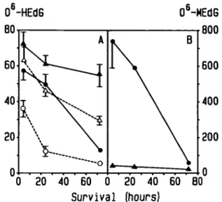
The persistence of C^-HEdG in DNA of various rat organs
following a single i.v. dose of 50 mg HENU/kg is shown in
Figure 3. Initial amounts of C^-HEdG were similar in DNA of
kidney and lung. Slightly lower levels of this modified base were
observed in liver DNA. However, the loss of C^-HEdG from
hepatic DNA was faster than from DNA of other organs, the
half-life being ~ 12 h. The rate of removal of this modified base
148
Peak height
1000
10
100 1000
0
6-HErJG (fmol/slot)
Fig. 1. Typical calibration curve for the determination of C^-HEdG. DNA (3 fig) containing C^-HEdG (1.3-328 fmol) was blotted onto nitrocellulose. After incubation with rabbit anti-CAHEdG serum, bound antibodies were reacted with a goat anti-rabbit IgG horseradish peroxidase conjugate. Enzymatic activity was measured via color development as described in the text. Peak height values are plotted against the respective amounts of C^-HEdG after subtraction of the value for non-specific binding to 3 /ig unmodified DNA.
Peak height (ram)
1000
100 1000 10000
0
6-alkyl-dG (fraol/slot)
Fig. 2. Reactivity of rabbit anti-O^HEdG serum with hydroxyethylated and methylated DNA. In a double logarithmic plot, the peak heights are plotted against the respective amounts of (/-HEdG ( A ) and C^-MEdG ( • ) applied to each slot. The dashed line represents the value for non-specific binding of the antiserum to nonalkylated DNA.from kidney and lung DNA was similar, with an apparent
half-life of 40 and 48 h respectively. In contrast, C^-HEdG
concen-trations in brain DNA was initially lower (35% of that in liver)
but persisted longer than in any other tissue investigated. The
7-day values, when expressed as a fraction of the initial (2-h)
C^-HEdG concentration, ranged from 4.7% (liver), 23.5%
(kidney), 25.7% (lung) to 62.4% (brain). The dose-dependent
formation of C^-HEdG was studied in liver and kidney. The
regression lines obtained (Figure 4) had slopes close to 1,
in-dicating that this promutagenic base is formed strictly
propor-tional to dose over the entire dose range investigated. The
regression equations as calculated by the least-squares method
Formation and persistence of O*-HEdG
0
6-HEdG
0
6-MEdG
40
80
120 160
Survival (hours)
Fig. 3. Persistence of O*-HEdG in DNA of various rat tissues following a
single i.v. dose of 50 mg/kg HENU and survival times ranging from 2 h to 7 days. Results are expressed as pmoUmol deoxyguanosine.
umol/mol
100
0.01
Dose (mraol/kg)
Fig. 4. Dose-dependent formation and concentration-dependent repair of
O^-HEdG in liver ( • ,O) and kidney (A,A) DNA. Rats were killed 4 h (closed symbols, solid lines) and 3 days (open symbols, dashed lines) after i single i.v. dose of HENU ranging from 0.039 to 0.38 mmol/kg,
corresponding to 5 to 50 mg/kg. Data represent mean ± SEM of 4 - 6 determinations.
were:
log C^-HEdG = 1.01 X log dose + 2.04 liver
log CP-HEdG = 1.01 X log dose + 2.14 kidney
In animals allowed to survive for 3 days after a single dose
of HENU, the amounts of C^-HEdG in kidney DNA were
generally lower and the slope of the regression line was steeper
than after 4 h, indicating that disproportionately more C^-HEdG
had been removed from DNA of animals exposed to lower doses
of HENU. The regression line was:
log C^-HEdG = 1.18 x log dose + 1.98 kidney
In liver DNA, most of the tf-HEdG initially formed had been
removed and a clear dose dependency was no longer evident after
3 days. However, at each of the doses administered, a small
200
0 20 40 60 0 20 40 60 BO
Survival (hours)
Fig. 5. (A) Persistence of O'-HEdG in liver and kidney DNA after
saturation of the C^-alkylguarune-DNA alkyltransferase with medG. Rats received i.p. injections of 20 mg/kg NDMA and, 3 h later, a chasing i.v. dose of 50 mg HENU per kg. After survival times of 4 h, 24 h and 3 days, Cfi-HEdG was determined by immuno-slot-blot analysis after correction for cross-reactivity of the anti-C^-HEdG serum whh Cfi-MEdG (closed symbols, solid lines). For comparison, me persistence of C^-HEdG without
pretreatment with NDMA is also shown (open symbols, dashed lines, data taken from Figure 3). Liver ( • , O ) ; kidney (A,A). The amounts of O^-alkylguanines are expressed as jimol/mol deoxyguanosine.
amount of C^-HEdG was found to persist ( ~ 3 —5 ftmol/mol
dG).
To test whether the removal of C^-HEdG competes with that
of other C^-alkylguanines, an additional experiment was carried
out in which animals were pretreated with 20 mg/kg of NDMA,
i.e. a dose known to produce saturating levels of C^-MEdG in
rat liver DNA. As shown in Figure 5, this almost completely
inhibited the repair of C^-HEdG in hepatic DNA during the first
24 h following HENU application. Although the amount of
C^-MEdG in kidney was ~ 19 times lower than in liver, this
still resulted in a significant inhibition of the repair of C^-HEdG
produced by a subsequent dose of HENU.
Discussion
The sensitivity of the immuno-slot-blot assay for C^-HEdG was
found to be similar to that achieved by Nehls et al. (15) for
C^-ethyldeoxyguanosine and (7*-ethyldeoxythymidine. These
authors employed murine monoclonal antibodies but we found
the use of rabbit antisera not to be disadvantageous. Significant
cross-reactivity was only observed for the reaction of the
anti-C^-HEdG serum with methylated DNA, whereas the anti-O
6-MEdG serum did not recognize C^-HEdG. Even the
simul-taneous presence of excess amounts of C^-MEdG in hepatic
DNA did not interfere with the quantitation of 10-fold lower levels
of C^-HEdG (Figure 5). In preliminary experiments we used an
alkaline phosphatase-conjugated second antibody for quantitative
evaluation. However, we found that the colored precipitate
resulting from the reaction of 4-chloro-l-naphthol with
horseradish peroxidase yielded a superior signal-to-noise ratio.
Since the intensity of the color reaction showed day-to-day
varia-tions, we found it mandatory to include a complete set of
stan-dards on each blot. In its present form, the assay enables the
quantitation of ^ 5 frnol ^-HEdG/S /tg DNA, corresponding
to 3.6 fimol C^-HEdG/mol deoxyguanosine. Bio-monitoring in
human individuals environmentally exposed to
hydroxyethylat-B.I.Ludeke and P.KIeihues
ing agents may require greater sensitivity and we are currently
investigating experimental modifications yielding a quantifiable
signal at lower levels of DNA alkylation.
To test this assay under in vivo conditions, we chose HENU
as a model compound since it yields hydroxyethyldiazonium
hydroxide as the only alkylating intermediate. As its
decomposi-tion is base catalyzed (20) and does not require enzymic
bio-activation, one would expect little organ variability in levels of
DNA alkylation, as has been shown previously for MNU (21)
and N-nitroso-N-ethylurea (22). Following a single i.v. injection
of 50 mg HENU/kg, the initial (2-h) concentrations of
&-HEdG were indeed similar in liver, kidney and lung. The extent
of C^-hydroxyethylation of guanine in liver and kidney was
determined as a function of dose. It was found that in both tissues
DNA hydroxyethylation was strictly proportional to amount of
HENU administered over the entire dose range (Figure 4). At
all doses the kidney values were 29% higher than those in liver.
This could be due to the fact that hydrophilic nitrosoureas such
as HENU are, to some extent, excreted via the urine and may
thus temporarily accumulate in the kidney. In contrast to its
methyl and ethyl analogues, HENU produced a low initial
ex-tent of DNA hydroxyethylation in cerebral DNA. This is
prob-ably due to its lower lipophilicity. The highly impermeable tight
junctions between the cells of the specialized capillary
endo-thelium of the central nervous system which form the
morpho-logical basis of the blood —brain barrier largely prevent low mol.
wt hydrophilic compounds from entering the brain parenchyma
by passive diffusion (23,24).
The persistence of C^-HEdG varied considerably among the
tissues investigated. The rate of removal was highest in liver.
Animals surviving for 7 days had C^-HEdG concentrations
amounting to 4.7% of those observed in animals sacrificed 2 h
after a single dose of 50 mg HENU/kg body wt. The steep decline
in C^-HEdG levels between 2 and 4 h (Figure 3) strongly
sug-gests that even the 2-h value (55 /tmol O^-HEdG/mol
deoxy-guanosine) does not truly reflect the initial extent of DNA
hydroxyethylation. Rapid removal of C^-HEdG from hepatic
DNA was also documented by the dramatic change in the
dose-response curve (Figure 4). Largely irrespective of the
in-itial dose, the 72-h values were reduced to a basal level of 3 - 6
/imol C^-HEdG/mol deoxyguanosine. This low level probably
represents persistence in non-parenchymal cells with a low repair
capacity (25,26).
The rate of loss of C^-HEdG from lung and kidney DNA was
somewhat lower than in liver, whereas in cerebral DNA there
was significantly less reduction in C^-HEdG concentrations.
These organ-specific differences closely parallel the activity of
the mammalian C^-alkylguanine-DNA alkyltransferase in
various rat tissues (27). Similar rates of repair were previously
observed for ^ - M E d G (28) and 0
6-ethyldeoxyguanosine (29).
C^-HEdG is known to be a substrate for C^-alkylguanine-DNA
alkyltransferase (30) but its rapid removal from rat liver DNA
is somewhat surprising since in vitro it is repaired at least 30
times more slowly than C^-MEdG and at least 10 times more
slowly than C^-ethyldeoxyguanosine (31). One reason for this
discrepancy could be that a glycosylase with high affinity for
C^-HEdG exists. It has been suggested that long-chain
C^-alkyl-guanines may be repaired by both the O^-alkylguanine-DNA
alkyltransferase (31) and glycosylase-mediated excision (32). To
test this hypothesis, we have carried out an additional
experi-ment based on the observation that the mammalian
C^-alkyl-guanine-DNA alkyltransferase stoichiometrically transfers the
alkyl group onto one of its own cysteine residues, thereby
becom-ing inactivated (33). When the available transferase molecules
have been consumed, de novo synthesis is required to restore
activity, a process which in vivo requires several days (34).
Pre-treatment with 20 mg NDMA/kg produced 750 /imol O
6-MEdG/mol deoxyguanosine in hepatic DNA, which is in close
agreement with the value of 36.7 y.mo\ C^-MEdG/mol
deoxy-guanosine/mg NDMA/kg body wt reported earlier (35).
Satura-tion of the hepatic alkyltransferase system in rats occurs at
concentrations above 180 /*mol C^-MEdG/mol deoxyguanosine
(35). One would, therefore, expect that alkyltransferase-mediated
removal of the C^-HEdG produced by a subsequent dose of
HENU would be significantly inhibited. This was indeed the case
(Figure 5A). During the first 24 h the repair of C^-HEdG in
liver was almost completely blocked. A similar, albeit less
mark-ed, effect was observed in rat kidney even though pretreatment
with NDMA produced C^-MEdG levels 19 times lower than in
liver. These findings strongly suggest that the repair of O
6-HEdG is indeed predominantly mediated by mammalian
Ch-alky lguanine-DN A Ch-alkyltransferase. This corroborates the
obser-vation that the mutagenicity of HENU in Escherichia coli does
not correlate with the capacity for excision repair (4). The
discrepancy between the rapid repair in vivo and the low rate
of removal by rat liver extracts in vitro is difficult to explain and
poses the question to which extent studies in cell-free systems
truly reflect in vivo repair capacity.
Although on the basis of chronic bioassay studies HENU must
be regarded as a powerful carcinogen in rats (6,7,36,37), mice
(3) and hamsters (38), its target specificity differs from that of
its methyl and ethyl analogs. Methyl- and ethylnitrosourea
pro-duce a high incidence of tumors of the central and peripheral
nervous system following a single perinatal exposure or multiple
doses in adult rats (39). In contrast, chronic administration of
HENU to F-344 rats led to the development of neural tumors in
< 15% of experimental animals (37). This reduced
neurocarcino-genicity may be due to the 3- to 4-fold lower extent of cerebral
DNA hydroxyethylation observed in the present study (Figure
3). However, due to the low rate of repair in the nervous system,
one would expect long-term accumulation of C^-HEdG to levels
higher than those in liver and other tissues (40,41). The low
in-cidence of neural tumors produced by HENU in rats may be due
to the presence of other target cell populations which undergo
malignant transformation more rapidly, e.g. lung and colon (38).
In conclusion, the immuno-slot-blot assay for C^-HEdG is
highly sensitive, does not require radiolabeled isotopes and allows
rapid and reliable quantitation of this promutagenic base under
in vivo conditions. It should be particularly useful in establishing
a molecular dosimetry during long-term exposure to
hydroxy-ethylating nitrosamines and therapeutic agents.
Acknowledgements
We thank Mrs I.CackeM for expert technical assistance. This work was supported by the Swiss National Fund and the Cancer League of the Canton of Zurich.
References
1. Saffhill.R., Margison.G.P. and O'Connor.P.J. (1985) Mechanisms of
car-cinogenesis induced by alkylating agents. Biochim. Biophys. Ada, 823, 111-145.
2. Singer.B. and Grunberger.D. (1983) Molecular Biology ofMiaagens and Car-cinogens. Plenum Press, New York.
3. Swenson.D.H., Frei J.V. and Lawley,P.D. (1979) Synthesis of 1-(2-hydroxy-ethyl)-l-nitrosourea and comparison of its carcinogeniciry with that of 1-ethyl-l-nitrosourea. J. Noll. Cancer Inst., 63, 1469-1473.
4. Kohda.K.H., Ninomiya.S., Washizu.K., Shiraki.K., Ebie.M. and Kawazoe.Y. (1987) Mutagenicity of a series of A'-alkyl-, /V-hydroxyalkyl-,
A'-haloalkyl-Formation and persistence of (/-HEdG
and /V-carboxyalkyl-A'-nitrosoureas in Escherichia coli tester strains: dependence on the uvrA DNA-repair system. Mulal. Res., 1T7, 219-228. 5. Lijinsky.W., Elespuru,R.K. and Andrews,A.W. (1987) Relative mutagenic and prophage-inducing effects of mono- and di-alkyl nitrosoiireas. Mutat. Res.,
178, 157-165.
6. Lijinsky.W., Singer.G.M. and Kovatch.R.M. (1985) Similar carcinogenic effects in rats of 1 -ethyl-1-nitroso-3-hydroxyethylurea and 1-hydroxyethyl-l-nitroso-3-ethylurea. Carcinogenesis, 6, 641-643.
7. Lijinsky,W., Kovatch.R.M. and Singer.S.S. (1986) Carcinogenesis in F-344 rats induced by nitrosohydroxyalkyl-chloroethylureas. /. Cancer Res. Clin, Oncol., 112,221-228.
8. Tong.W.P., Kirk.M.C. and Ludlum.D.B. (1981) Molecular pharmacology of the haloethyl nitrosoureas: formation of 6-hydroxyethylguanine in DNA treated with BCNU (A'^'-bis[2-cWoroethyl]-A'-nitrosourea). Biochem. Biophys. Res. Commun., 100, 351-357.
9. LownJ.W. and Chauhan.S.M.S. (1981) Mechanism of action of (2-halo-ethyl)-nitrosoureas on DNA. Isolation and reactions of postulated 2-{alkyl-imino)-3-nitrosooxazolidine intermediates in the decomposition of 1,3-bis-(2-chloroethyl)-, l-<2-chloroethyl)-3-cyclohexyl-, and l-<2-chlorocthyl)-3-(4'-trar£rrethylcyclohexyr)-l-nitrosourea. J. MetL Chenu, 24, 270-279. 10. Eisenbrand.G., Muller.N., Denkel.E. and Sterzel.W. (1986) DNA adducts
and DNA damage by antineoplastic and carcinogenic /V-nitrosocompounds. J. Cancer Res. Clin. Oncol., 112, 196-204.
11. von Hofe,E., Kleihues.P. and Keefer.L.K. (1986) Extent of DNA 2-hydroxyethylation by A'-nitrosomethylethylamine and JV-nitrosodiethylamine in vivo. Carcinogenesis, 7, 1335—1337.
12. Preussmann.R. and Stewart.B.W. (1984) JV-Nitroso carcinogens. In Searle.C.E. (ed.), Chemical Carcinogens, Vol. 2, ACS Monograph 182. American Chemical Society, Washington, DC, pp. 643-828.
13. Sabadie.N., Malaveille.C., Camus,A.-M. and Bartsch.H. (1980) Comparison of the hydroxy lation of benzo[a}pyrene with the metabolism of vinyl chloride, A'-nitrosomorpholine, and N-nitroso-W-methylpiperazine to mutagens by human and rat liver microsomal fractions. Cancer Res., 40, 119-126. 14. Segerback.D. (1983) Alkylation of DNA and hemoglobin in the mouse
follow-ing exposure to ethene and ethene oxide. Chenu-Biol. Interactions, 45, 139-151.
15. Nehls.P., AdamkiewiczJ. and Rajewsky.M.F. (1984) Immuno-slot-blot: a highly sensitive immunoassay for the quantitation of carcinogen-modified nucleosides in DNA. /. Cancer Res. Clin. Oncol, 108, 2 3 - 2 9 . 16. Muller.R. and Rajewsky,M.F. (1980) Immunological quantification by high
affinity antibodies of CP-ethyldeoxyguanosine in DNA exposed to A'-cthyl-A'-nitrosourea. Cancer Res., 40, 887-896.
17. von Hofe.E. and Kleihues.P. (1986) Comparative studies on hepatic DNA alkylation in rats by W-nitrosomethylethylamine and A'-nitrosodimethylamine plus A'-nitrosodiethylamine. J. Cancer Res. Clin. Oncol., 112, 205-209. 18. von Hofe.E., Grahmann.F., Keefer.L.K., Lijinsky.W., Nelson.V. and Kleihues.P. (1986) Methylation versus ethylation of DNA in target and non-target tissues of Fischer 344 rats treated with A'-nitrosomethylethylamine. Cancer Res., 46, 1038-1042.
19. Burton,K. (1956) A study of the conditions and mechanism of the diphenyl-amine reaction for the colorimetric estimation of deoxyribonucleic acid. Biochem, J., 62, 315-323.
20. Singer,S.S. (1984) Decomposition of A'-nitrosohydroxyalkylureas and N-nitrosooxazolidones in aqueous buffer. IARC Scient. Pub., 57, 371-375. 21. Kleihucs.P. and Magee.P.N. (1973) Alkylation of rat brain nucleic acids by A'-methyl-A'-nitrosourea and methyl methanesulfonate. J. Neurochem., 20, 595-606.
22. Goth.R. and Rajewsky.M.F. (1972) Ethylation of nucleic acids by ethylnitroso-urea-l-l4C in the fetal and adult rat. Cancer Res., 32, 1501-1505. 23. Brightman.M.W. and Reese.T.S. (1969) Junctions between intimately
ap-posed cell membranes in the vertebrate brain. J. Cell BioL, 40, 648—677. 24. Crone.C. (1963) The permeability of capillaries in various organs as deter-mined by use of the 'indicator diffusion' method. Acta Physiol. Scand., 58, 292-305.
25. LewisJ.G. and SwenbergJ.A. (1980) Differential repair of O*-methyl-guanine in DNA of rat hepatocytes and nonparcnchymal cells. Nature, 288, 185-187.
26. Menkveld.GJ., Van Der Laken.CJ., Hermsen.T., Kriek.E., Scherer,E. and Den Engelse.L. (1985) Immunohistochemical localization of (/-ethyldeoxy-guanosine and deoxyguanosin-8-yl-(acetyl)aminofluorene in liver sections of rats treated with diethylnitrosamine, ethylnitrosourea or A'-acetylammofluorcnc. Carcinogenesis, 6, 263—270.
27.Pegg,A.E., Dolan.M.E., Scicchitano.D. and Monmoto.K. (1985) Studies of the repair of (Aalkylguanine and C^-alkylthymine in DNA by alkyl-transferases from mammalian cells and bacteria. Environ, Health Perspect.,
62, 109-114.
28. Kleihues.P. and BuchelerJ. (1977) Long-term persistence of
0^-methyl-guanine in rat brain DNA. Nature, 269, 625—626.
29. Goth.R. and Rajewsky.M.F. (1974) Molecular and cellular mechanisms associated with pulse-carcinogcnesis in the rat nervous system by ethylnitroso-urea: ethylation of nucleic acids and elimination rates of ethylated bases from the DNA of different tissues. Z Krebsforsch., 82, 3 7 - 6 4 .
30. Pegg.A.E., Scicchitano.D. and Dolan.M.E. (1984) Comparison of the rates of repair of C^-alkylguanines in DNA by rat liver and bacterial O^-alkyl-guanine-DNA alkyltransferase. Cancer Res., 44, 3806-3811.
31. Morimoto.K., Dolan.M.E., Scicchitano.D. and Pegg.A.E. (1985) Repair of O^-propylguanine and C^-butylguanine in DNA by C^-alkylguanine-DNA alkyltransferases from rat liver and E.coli. Carcinogenesis, 6, 1027-1031. 32. BoyleJ.M., Margison.G.P. and Saffhill.R. (1986) Evidence for the excision repair of C^-n-butykieoxyguanosine in human cells. Carcinogenesis, 7, 1987-1990.
33. Pegg.A.E., Wiest.L., Foote.R.S., Mitra.S. and Perry.W. (1983) Purifica-tion and properties of O6-methylguanine-DNA transmethylase from rat liver.
J. BM. Chenu, 258, 2327-2333.
34. Kleihues.P. and Margison.G.P. (1976) Exhaustion and recovery of repair excision of C^-methylguanine from rat liver DNA. Nature, 259, 153 — 155. 35. Pegg.A.E. and Hui.G. (1978) Formation and subsequent removal of O6
-methylguanine from DNA in rat liver and kidney after small doses of di-methylnitrosamine. Biochem. J., 173, 739-748.
36. Iijinsky,W. and Reuber.M.D. (1983) Carcinogenicity of hydroxylated alkyl-nitrosoureas and of nhrosooxazolidones by mouse skin painting and by gavage in rats. Cancer Res., 43, 214-221.
37. Lijinsky.W. and Kovatch.R.M., (1987) Carcinogenesis by nitrosohydroxy-ethylurea and nhrosomethoxynitrosohydroxy-ethylurea in F344 rats. /. Cancer Res. Clin, Oncol., in press.
38. Lijinksy.W. (1984) Species differences in nitrosamine carcinogenesis. /. Cancer Res. Clin. Oncol., 108, 4 6 - 5 5 .
39. Kleihues.P., Lantos.P.L. and Magee.P.N. (1976) Chemical carcinogenesis in the nervous system. Int. Rev. Exp. PathoL, 15, 153—232.
40. Margison.G.P. and Kleihues.P. (1975) Chemical carcinogenesis in the ner-vous system. Preferential accumulation of C^-methylguanine in rat brain de-oxyribonucleic acid during repetitive administration of N-methyl-A'-nitrosourca. Biochem. J., 148, 521-525.
41. Cooper.H.K., Hauenstein.E., Kolar.G.F. and Kfcihues.P. (1978) DNA alkyla-tion and neuro-oncogenesis by 3,3-dimethyl-l-phenyltriazene. Acta Neuro-pathol, 43, 105-109.
Received on August 5, 1987; revised on October 8, 1987; accepted on October 15, 1987