N-linked oligosaccharide processing enzyme glucosidase II produces 1,5-anhydrofructose as a side product
8
0
0
Texte intégral
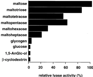
(2) K.Hirano et al.. Results N-Terminal sequence comparison of purified rat liver α-1,4-glucan lyase and glucosidase II The purification scheme is summarized in Table I. In the first DE 52 column chromatography step, the lyase activity was separated into two peaks. One was recovered in the flow through fraction and the other was eluted with 0.2–0.25 M sodium chloride. The ratio of these two activities was about 6:4. Since the unbound enzyme was not stable, only the bound protein was used for further purification. The final preparation showed two distinct bands on SDS–PAGE; the slower migrating band corresponded to 110 kDa and the other to 68 kDa. The sequence of seven N-terminal amino acid residues (V-D-R-S-N-F-K) of the 110 kDa protein was identical with the amino acid sequences of glucosidase II from other species such as mouse (accession number U92793), pig (U71273) and human (D42041). The N-terminal amino acid sequence of the 68 kDa degradation product (Y-L-Q-G-S-G-E-T-P-Q-T-D-V) showed identity with an internal known sequence of rat glucosidase II (340th and subsequent residues based on human sequence numbering). From these data, the 110 kDa protein exhibiting lyase activity is assumed to be glucosidase II and the 68 kDa protein, a proteolytically degraded product. Biochemical properties of purified α-1,4-glucan lyase The enzyme purified from the DE 52-retained fraction had a pI value of ∼4.5 determined by isoelectroric focusing and was also retained on a concanavalin A Sepharose column. This enzyme exhibited the highest activity in 20 mM sodium phosphate buffer at pH 7.5 between 40°C and 45°C. Lyase activity was greatly reduced in 20 mM Tris/HCl buffer (pH 7.0). The enzyme cleaved α-1,4 and α-1,3 glucosidic bonds, but not α-1,1, α-1,2, and α-1,6 bonds. When some maltosaccharides were used as substrates, stronger lyase activities were observed toward maltose and maltotriose, whereas the activity toward longer maltosaccharides decreased (Figure 1). These reactions also produced glucose at the same time (e.g., ∼90 µmol/min mg protein from maltose) and the amount was about 10,000-fold higher than that of 1,5-AnFru. 1,5-AnFru was not produced from glycogen or monosaccharides such as glucose and 1,5-AnGlc-ol. Fig. 1. Substrate specificity of purified α-1,4-glucan lyase. Partially purified α-1,4-glucan-lyase was used for the emzymatic measurements with different substrates. The indicated substrates were added to a final concentration of 10 mg/ml, incubated for 2 h at 37°C and then the 1,5-AnFru amounts were determined. The results are expressed as percentage of the 1,5-AnFru amounts produced from maltose.. (Figure 1). Furthermore, no activity was observed with β-cyclodextrin as substrate (Figure 1). 1,5-AnFru production was also detectable when the artificial substrate p-nitrophenyl-α-Dglucopyranoside was used (not shown). The lyase activity could be blocked with 1-deoxynojirimycin (80% reduction at 1 mM) and castanospermine (90% reduction at 1 mM), which are known to be glucosidase inhibitors. Furthermore, p-chloromercuribenzoic acid, which inhibits α-1,4-glucan lyase activity in fungi (Yu et al., 1997), at 2 mM completely inhibited the purified rat liver α-1,4-glucan lyase. At the same time, the glucose accumulation caused by this enzyme was also blocked by these inhibitors at the same concentrations (not shown). On the other hand, 1 mM EDTA had no influence on the lyase activity. Altogether, these biochemical properties of lyase activity generally resemble those reported for glucosidase II (Trombetta et al., 1996).. Table I. Purification of α-1,4-glucan lyase from rat liver. Purification step I. Liver lysate. Total lyase activitya. Total proteinb. Specific activity. Recovery. Purification. mU. mg. mU/mg protein. %. fold. 253.8. 41072. 6.18 ×. 10–3. 100. 1. 17.80 × 10⁻³. 10.68. 447.1. 41.56 × 10⁻³. 7.32. 7.15. 13.98. 77.9. 17.90 × 10⁻². 5.51. 29.09. V. Phenyl Superose. 3.54. 1.2. 2.95. 1.39. 487.23. VI. Mono Q. 0.85. 0.1. 8.50. 0.34. 1386.55. II. 1st DE 52. 26.99. III. Octyl Sepharose CL-4B. 18.58. IV. 2nd DE 52. 1515. 2.95. unit was defined as the amount of enzyme that produced 1µM of 1,5-AnFru in 1 min at 37 °C in 20 mM phosphate buffer (pH 7.0) using maltose (10 mg/ml) as substrate. bProtein contents were measured by Bradford method (Bradford, 1976). aOne. 1284.
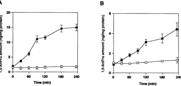
(3) Glucosidase II produces 1,5-anhydrofructose as side product. Trimming glucosidase II produces 1,5-anhydrofructose as a side product Next, we purified glucosidase II from rat liver microsomes using DEAE-Sepharose, concanavalin A Sepharose, Mono Q, Sephacryl S-300, and phenyl Superose column chromatography. Silver staining of the gel showed that the purified fraction contained two bands as usual: a major band at 110 kDa and a minor band at ∼90 kDa, both of which were immunoreactive for glucosidase II. Upon storage at 4°C, additional degradation bands occurred (Figure 2). The purified glucosidase II. produced 1,5-AnFru from maltose as substrate, but did not show lyase activity toward glycogen and glucose. Its lyase activity always involved high amounts of glucose production. In order to substantiate and independently confirm that glucosidase II exhibits additional lyase activity, cells deficient in glucosidase II were studied. Previously, we have demonstrated that the mouse lymphoma cell line PHAR2.7, which is deficient for glucosidase II activity (Reitman et al., 1982), also lacks mRNA for glucosidase II (Flura et al., 1997). As shown in figure 3, PHAR2.7 in contrast to the parental wild type line BW5147 lacked activity for α-1,4-glucan lyase. Likewise, the glucosidase II-deficient S. cerevisiae strain YG427 (Jakob et al., 1998) as compared with the wild type strain SS328 lacked activity for α-1,4-glucan lyase (Figure 3). These data demonstrate that glucosidase II is responsible for lyase activity. The lyase activities were detectable in these wild type cells when maltose was used as substrate, but not with glycogen or glucose as substrate. Furthermore, in CHO cells overexpressing pig liver glucosidase II (Flura et al., 1997), increasing glucosidase II activity levels were associated with increasing lyase activity (Figure 4). All these observations strongly indicate that glucosidase II produces minor amounts of 1,5-AnFru in addition to glucose. Discussion. Fig. 2. SDS–PAGE and Western blot analysis. Purified glucosidase II was separated in a 7.5% polyacrylamide gel; followed by silver staining (A), in parallel Western blot analysis using anti-pig glucosidase II antibodies was performed (B). As molecular standards, the following proteins were used; myosin (200 kDa), galactosidase (116 kDa), phosphorylase b (97 kDa), bovine serum albumin (67 kDa), ovalbumin (45 kDa).. Only a few studies have been reported for α-1,4-glucan lyase in mammalian organs (Kametani et al., 1996b), while several fungal and algal α-1,4-glucan lyase have been isolated and well characterized. A comparison of the amino acid sequence of α-1,4-glucan lyases isolated from fungi and red algae with other amino acid sequences from data bases showed a similarity to the glycoside hydrolase family 31 (Yu et al., 1999). In the present study, we demonstrate that the oligosaccharide trimming glucosidase II, a member of that hydrolase family, possesses the ability to produce 1,5-AnFru as a side product.. Fig. 3. Lyase activity measurement in mutant mouse lymphoma PHAR2.7 cells and in glucosidase II deficient yeast cells. Homogenates of these cells were incubated under the same condition as described in the Materials and methods section. Then, the production of 1,5-AnFru was measured. The protein amounts of the cell homogenates were determined by Bradford (Bradford, 1976). Each experiment was performed at least three times. Mouse lymphoma cell lines (A); BW5147 cells (solid circles) and PHAR2.7 cells (open circles), yeast cell homogenates (B); wild type (solid squares) and ∆-gls2 yeast strains (open squares).. 1285.
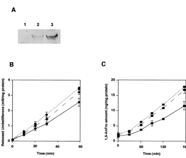
(4) K.Hirano et al.. Fig. 4. Enzymatic activities of CHO cells transfected with pig liver glucosidase II. Western blot analysis of pig liver glucosidase II overexpressed in CHO cells was performed as described in the text. Lane 1, untransfected CHO-K1; lane 2, CHO-gls2 clone 1; lane 3, CHO-gls2 clone 2. (A). Two different enzymatic activities of these cell extracts were measured as described in the text. Extracts of CHO-K1 (solid line), CHO-gls2 clone 1 (dotted line) and CHO-gls2 clone 2 (broken line) were measured for glucosidase II activity (B) and lyase activity (C).. In our attempts to purify α-1,4-glucan lyase, we observed distinct lyase activities in rat liver homogenate. After DEAE Cellulose column chromatography, two lyase activities were found and in the present study, we have focussed on the enzyme activity bound to the column. Further studies will aim at the characterization of the enzyme activity which was not retained onto DEAE cellulose. While the DEAE-bound protein was reactive with concanavalin A, the unbound protein was unreactive (unpublished observations). From these data we concluded that the two distinct lyase activities correspond to different enzymes and the occurrence of 1,5-AnFru in mammalian cells (Kametani et al., 1996b; Suzuki et al., 1996) may be attributed to different enzymes. The seven N-terminal amino acid residues of the purified 110 kDa protein exhibiting lyase activity were identical with the amino acid residues 33–40 in glucosidase II from human, pig, and mouse. In accordance, the N-terminal amino acid sequence of purified rat and pig liver glucosidase II (Trombetta et al., 1996; Flura et al., 1997; Hentges and Bause, 1997) started at the same position suggesting that cleavage occurs at this specific site. The amino acid sequence of the 68 kDa protein was identical with an internal sequence of pig liver glucosidase II starting from 1286. residue 340, indicating that the 68 kDa is a degradation product which occurred during the purification. These data show that the purified protein exhibiting lyase activity is attributed to glucosidase II. Glucosidase II removes the two glucose residues from the (Glc)2(Man)9(GlcNAc)2 oligosaccharide (Kornfeld and Kornfeld, 1985; Moremen et al., 1994), and in liver it is a resident protein of the endoplasmic reticulum (Lucocq et al., 1986). It is a ubiquitous enzyme and has been purified from various organs of different species (Burns and Touster, 1982; Brada and Dubach, 1984; Hino and Rothman, 1985; Trombetta et al., 1996; Hentges and Bause, 1997). We demonstrate here that the purified glucosidase II, which is a tetrameric glycoprotein consisting of 110 kDa subunits, is immunoreactive toward anti-pig liver glucosidase II and has a lyase activity using maltose as substrate. Furthermore, CHO cells overexpressing pig liver glucosidase II showed a 1.5- to 2-fold enhanced lyase activity compared to nontransfected cells. On the other hand, glucosidase II deficient yeast or mouse lymphoma cells showed no lyase activity. These two established glucosidase II knock out strains lack both immunological and catalytic activities of glucosidase II. Concerning mutant mouse lymphoma PHAR.
(5) Glucosidase II produces 1,5-anhydrofructose as side product. 2.7 cell, Northern blot, and Western blot analysis show the absence of mRNA encoding glucosidase II and expression of the protein, respectively (Flura et al., 1997). Construction of glucosidase II deficient yeast YG427 was performed by homologous recombination based on the sequence of pig liver glucosidase II (Jakob et al., 1998). Collectively, these data strongly indicate that glucosidase II possesses two different enzymatic activities whereby the lyase activity is the minor activity and is detectable in various species. We noticed that glucosidase II purified from rat liver, or from the homogenate of wild type yeast and BW5147 cells showed lyase activity only toward maltose but not toward glycogen. On the other hand, a crude hepatic preparation can produce 1,5-AnFru from glycogen (Kametani et al., 1996b). These data suggested that either some other not yet identified enzymes or amylases that can produce malto-oligosaccharides participate in this glycogenolytic pathway. The mechanism for the production of 1,5-AnFru by glucosidase II in mammals so far is unknown. Concerning the catalytic reactions of glucosidase II, several reports have been presented (Alonso et al., 1991). Brada and Dubach demonstrated that glucosidase II cleaves not only α-1,3-bonds but also α-1,4-bonds (Brada and Dubach, 1984). Based on the shared substrate and inhibitor specificity and the type of bond cleaved, one might speculate that two different enzymatic reactions, hydrolysis and elimination, take place at the same catalytic site forming a glucosyl-enzyme intermediate. The glucosyl moiety in the enzyme substrate complex may be released either hydrolytically or by elimination reaction primed by a different proton release mechanism (Yu et al., 1999). The idea of dual ways for glucosyl intermediate dissociation is reminiscent of the fact that several α-glucosidases catalyze hydration of glucal to 2-deoxyglucose. The initial step of this reaction to the enzymesubstrate complex may be the reversal of dissociation of the glycosyl-enzyme intermediate through elimination reaction (note that 1,5-AnFru is a tautomer of 2-hydroxyglucal). We failed to demonstrate the hydration of 1,5-AnFru to glucose by α-glucosidases. This seems reasonable because 1,5-AnFru is stabilized in its hydrated form (Kametani et al., 1996a) and seems to hardly undergo tautomeric isomerization to 2-hydroxyglucal in aqueous solution. The α-glucosidase-catalyzed hydration of glucal indicates that the enzyme has sufficient affinity to the unsaturated sugar. It is therefore anticipated that the structure of the 2,3-dihyropyran ring in glucal and 2-hydroxyglucal resemble the conformation of the backbone of the sixmembered ring of the glucosyl moiety in the transition state on the enzyme. Do all α-glucosidases produce 1,5-AnFru? Among those investigated, α-glucosidase II is the only glucosidase which produced 1,5-AnFru as a side product. Neither a recombinant α-glucosidase (maltase, EC 3.2.1.20) of E.coli origin nor γ-amylase from Aspergillus niger produced detectable amounts of 1,5-AnFru. Therefore, we tentatively conclude that only a few members of the glucoside hydrolase family produce 1,5-AnFru. The absence of 1,5-AnFru production in glucosidase II-deficient S.cerevisiae and PHAR2.7 cell also suggests that glucosidase II may be the only enzyme in these cells which is able to produce 1,5-AnFru. In comparison with the major glycogenolytic pathway, phosphorolysis, which is catalyzed by phosphorylase and provides every organ with a sufficient amount of glucose as the energy. source (Bollen et al., 1998), the activity of this anhydrosugar production is low in mammalian organs and is readily reduced to 1,5-AnGlc-ol. The physiological role of the production of 1,5-AnFru and 1,5-AnGlc-ol in mammalian cells is still unknown. Recently, it was demonstrated that 1,5-AnGlc-ol promotes glycogenolysis in E.coli (Shiga et al., 1999), and therefore one may expect that they have also some effects on glycogen metabolism in mammalian organs. Materials and methods Reagents and cells An anion exchanger DE 52 was purchased from Whatman International (Kent, UK), Octyl Sepharose CL-4B, concanavalin A Sepharose, Fast Flow Q Sepharose, Mono Q, Mono P, Superose 12, Sephacryl S-300, Phenyl Superose columns, Polybuffer 74 and silver staining kit were from Pharmacia Biotech Inc. (Uppsala, Sweden), glucose assay kit, nigerose, treharose, isomaltose, kojibiose were from Sigma (St. Louis, MO), PVDF membrane and protein molecular standards were from Bio-Rad Laboratories (Richmond, VA) and protease inhibitor cocktail tablets were from Roche Diagnostics (Rotkreuz, Switzerland). All other chemicals were obtained from Wako Pure Chemical Industries (Osaka, Japan). The mouse lymphoma cell line BW5147 (glucosidase IIpositive) and PHAR2.7 (glucosidase II-deficient) were kindly provided by Dr. I.Trowbridge (Salk Institute, San Diego, CA). Wild type CHO-K1 cells and CHO cells overexpressing pig liver glucosidase II (Flura et al., 1997) were cultured as described previously (Flura et al., 1997). BW5147 and PHAR2.7 cell lines were grown in Dulbecco’s modified Eagle’s medium containing 10% fetal bovine serum (FBS), penicillin (100 U/ml), and streptomycin (100 µg/ml) at 37°C and 10% CO2. CHO-K1 cells were grown in Ham’s F12 medium containing 10% FBS and CHO-K1 cells transfected with pig liver glucosidase II were grown in α–MEM containing 10% FBS and G418 (1 mg/ml) at 37°C and 5% CO2. The following yeast strains were used: SS328 as glucosidase II–positive wild type strain (Vijayraghavan et al., 1989) and YG427 as glucosidase II–deficient strain (Jakob et al., 1998). Purification of rat liver α-1,4-glucan lyase All steps were performed at 4°C. Frozen rat liver (200 g) were homogenized in 3 volumes of buffer A (20 mM sodium phosphate buffer, pH 7.0) containing 1.6 mM DTT. The cell debris, nuclei, mitochondria and lysosomes were removed by centrifugation at 38,000 × g for 20 min. The supernatant was acidified to pH 5.0 and left on ice for 30 min. Afterwards, the lysate was brought back to neutral pH and denatured proteins were removed. The supernatant was applied onto a DE 52 column (6 × 20 cm) equilibrated with buffer A. After washing with 2 volumes of buffer A, the proteins were eluted with a linear gradient of 0–0.5 M sodium chloride in buffer A. Two peaks of lyase activity, one in the flow through fractions and the other eluted with 0.2–0.25 M of sodium chloride, were obtained. The eluted fractions exhibiting lyase activity were concentrated by ultrafiltration (Amicon YM 30 membrane) and sodium sulfate was added to a final concentration of 1 M. Then the sample was loaded onto an Octyl Sepharose CL-4B column (5 × 15 cm) 1287.
(6) K.Hirano et al.. equilibrated with buffer A containing 1 M sodium sulfate. The column was washed with 2 volumes of this buffer and the proteins were eluted with buffer A. The eluted proteins were concentrated by ultrafiltration and rechromatographed on a DE 52 column (2.6 × 35 cm) using buffer A with the same sodium chloride gradient as described above. The fractions exhibiting lyase activity were concentrated and sodium sulfate was added to a final concentration of 1.5 M, then loaded onto Phenyl Superose column equilibrated with buffer A containing 1.5 M sodium sulfate. The column was washed with 2 volumes of this buffer and proteins were eluted by a decreasing linear salt gradient from 1.5 M to 0 M in buffer A. The peak fractions of phenyl Superose were diluted by adding 2 volumes of buffer B (20 mM sodium phosphate buffer, pH 7.8) and applied on a Mono Q column equilibrated with buffer B. Proteins were eluted with a linear gradient of 0–1 M sodium chloride in buffer B. The fractions exhibiting lyase activity were pooled and analyzed on a 6% SDS–polyacrylamide gel according to Laemmli (Laemmli, 1970). The gels were either silver stained or transferred to PVDF membrane (Towbin et al., 1979) for amino acid sequencing. Amino acid sequences were determined by using the protein sequencer system Procise Model 492 from PE Biosystems (Foster City, USA). Purification of rat liver glucosidase II Rat liver glucosidase II was purified as described previously (Brada and Dubach, 1984). Briefly, microsomes were isolated from 100 g of frozen rat liver and proteins were extracted by the addition of 1% of Triton X-100. Glucosidase II was further purified on Fast Flow Q Sepharose, concanavalin A Sepharose, Mono Q, Sephacryl S-300, and phenyl Superose column chromatography, successively. Measurement of enzymatic activity 1,5-AnFru activity was measured as described previously (Kametani et al., 1996a,b; Shiga et al., 1999). The reaction mixture (200 µl), which contained maltose (2 mg/ml) as substrate, was incubated at 37°C for various time periods. The reaction was stopped by the addition of 4 volumes of ethanol. As internal standard, 20 ng of 13C6-1,5-AnFru was added, and denatured proteins were removed by centrifugation. After drying the supernatant, 160 µl of water, 20 µl of 10% O-ethylhydroxylamine-HCl and 20 µl of 0.5 M Tris–HCl (pH 8.5) containing 1 mM EDTA were added to the sample and incubated at 80°C for 15 min. The resulting oximes were separated by a reverse-phase column (DIA-CHROMA ODS, 4.6 × 15 cm, Kakokishouji, Kawasaki) mounted on HPLC (LC-10, Shimadzu). The 1,5-AnFru ethyloxime was collected, dried, acetylated, and then injected onto GC-MS (Hewlett Packard HP 6890 GC System and 5973 Mass Selective Detector). The ion fragments of 1,5-AnFru (m/z = 169 and 211) and 13C6-1,5-AnFru (m/z = 175 and 217) were monitored. Glucosidase II activity was measured using 0.1 mM 4-methylumbelliferyl α-D-glucopyranoside as substrate (Brada and Dubach, 1984). The amount of released methylumbelliferone was measured using a fluorescence photometer (excitation 360 nm, emission 450 nm). The amount of glucose production was measured using a glucose assay kit. 1288. Isoelectric focusing The fractions exhibiting lyase activity were diluted with 3 volumes of 25 mM imidazole-HCl buffer (pH 7.4) and applied onto a Mono P column equilibrated with this buffer. The bound proteins were eluted with a linear gradient from pH 7.4 to 4.0 using polybuffer 74/HCl (pH 4.0) according to the manufacturer’s instructions. Western blot analysis using anti-glucosidase II antibody Purified glucosidase II or CHO cell homogenates were separated in a 7.5% SDS–polyacrylamide gel followed by transfer on nitrocellulose membrane using a semi-dry blotting apparatus (Towbin et al., 1979). The membrane was blocked with PBS containing 1% defatted milk powder and 0.05% Tween 20 for 1 h at room temperature and incubated with 1 µg/ml of anti-pig glucosidase II antibody overnight at 4°C, followed by alkaline phosphatase-conjugated goat anti-rabbit IgG antibodies. Color reaction was performed using nitro blue tetrazolium/BCIP as substrate. References Alonso,J.M., Santa,C.A. and Calvo,P. (1991) Glucosidase II from rat liver microsomes. Kinetic model for binding and hydrolysis. Biochem. J., 278, 721–727. Baute,M.A., Baute,R. and Deffieux,G. (1988) Fungal enzymic activity degrading 1,4-α-D-glucans to 1,5-D-anhydrofructose. Phytochemistry, 27, 3401–3403. Bojsen,K., Yu,S., Kragh,K.M. and Marcussen,J. (1999a) A group of α-1,4-glucan lyases and their genes from the red alga Gracilariopsis lemaneiformis: purification, cloning and heterologous expression. Biochim. Biophys. Acta, 1430, 396–402. Bojsen,K., Yu,S. and Marcussen,J. (1999b) A group of α-glucan lyase genes from the fungi Morchella costata, M.vulgaris and Peziza ostracoderma. Cloning, complete sequencing and heterologous expression. Plant Mol. Biol., 40, 445–454. Bollen,M., Keppens,S. and Stalmans,W. (1998) Specific features of glycogen metabolism in the liver. Biochem. J., 336, 19–31. Brada,D. and Dubach,U.C. (1984) Isolation of a homogeneous glucosidase II from pig kidney microsomes. Eur. J. Biochem., 141, 149–156. Bradford,M.M. (1976) A rapid and sensitive method for the quantitation of microgram quantities of protein utilizing the principle of protein-dye binding. Anal. Biochem., 72, 248–254. Burns,D.M. and Touster,O. (1982) Purification and characterization of glucosidase II, an endoplasmic reticulum hydrolase involved in glycoprotein biosynthesis. J. Biol. Chem., 257, 9990–10000. Flura,T., Brada,D., Ziak,M. and Roth,J. (1997) Expression of a cDNA encoding the glucose trimming enzyme glucosidase II in CHO cells and molecular characterization of the enzyme deficiency in a mutant mouse lymphoma cell line. Glycobiology, 7, 617–624. Hentges,A. and Bause,E. (1997) Affinity purification and characterization of glucosidase II from pig liver. Biol. Chem., 378, 1031–1038. Hino,Y. and Rothman,J.E. (1985) Glucosidase II, a glycoprotein of the endoplasmic reticulum membrane. Proteolytic cleavage into enzymatically active fragments. Biochemistry, 24, 800–805. Jakob,C.A., Burda,P., te Heesen,S., Aebi,M. and Roth,J. (1998) Genetic tailoring of N-linked oligosaccharides: the role of glucose residues in glycoprotein processing of Saccharomyces cerevisiae in vivo. Glycobiology, 8, 155–164. Kametani,S., Hashimoto,Y., Yamanouchi,T., Akanuma,Y. and Akanuma,H. (1987) Reduced renal reabsorption of 1,5-anhydro-D-glucitol in diabetic rats and mice. J. Biochem. (Tokyo), 102, 1599–1607. Kametani,S., Mizuno,H., Shiga,Y. and Akanuma,H. (1996a) NMR of allcarbon-13 sugars: an application in development of an analytical method for a novel natural sugar, 1,5-anhydrofructose. J. Biochem. (Tokyo), 119, 180–185. Kametani,S., Shiga,Y. and Akanuma,H. (1996b) Hepatic production of 1,5-anhydrofructose and 1,5-anhydroglucitol in rat by the third glycogenolytic pathway. Eur. J. Biochem., 242, 832–838..
(7) Glucosidase II produces 1,5-anhydrofructose as side product. Kornfeld,R. and Kornfeld,S. (1985) Assembly of asparagine-linked oligosaccharides. Annu. Rev. Biochem., 54, 631–664. Laemmli,U.K. (1970) Cleavage of structural proteins during the assembly of the head of bacteriophage T4. Nature, 227, 680–685. Lucocq,J.M., Brada,D. and Roth,J. (1986) Immunolocalization of the oligosaccharide trimming enzyme glucosidase II. J. Cell. Biol., 102, 2137–2146. Mizuno,H., Morita,M. and Akanuma,H. (1995) Phosphorylation of 1,5-anhydroD-glucitol in mammalian cells. J. Biochem. (Tokyo), 118, 411–417. Moremen,K.W., Trimble,R.B. and Herscovics,A. (1994) Glycosidases of the asparagine-linked oligosaccharide processing pathway. Glycobiology, 4, 113–125. Reitman,M.L., Trowbridge,I.S. and Kornfeld,S. (1982) A lectin-resistant mouse lymphoma cell line is deficient in glucosidase II, a glycoproteinprocessing enzyme. J. Biol. Chem., 257, 10357–10363. Sakuma,M., Kametani,S. and Akanuma,H. (1998) Purification and some properties of a hepatic NADPH-dependent reductase that specifically acts on 1,5-anhydro-D-fructose. J. Biochem. (Tokyo), 123, 189–193. Shiga,Y., Kametani,S., Mizuno,H. and Akanuma,H. (1996) Escherichia coli phosphorylates 1,5-Anhydroglucitol and releases 1,5-Anhydroglucitol 6-phosphate when glucose is absent in the medium. J. Biochem. (Tokyo), 119, 173–179. Shiga,Y., Kametani,S., Kadokura,T. and Akanuma,H. (1999) 1,5-Anhydroglucitol promotes glycogenolysis in Escherichia coli. J. Biochem. (Tokyo), 125, 166–172. Suzuki,M., Kametani,S., Uchida,K. and Akanuma,H. (1996) Production of 1,5-anhydroglucitol from 1,5-anhydrofructose in erythroleukemia cells. Eur. J. Biochem., 240, 23–9.. Towbin,H., Staehelin,T. and Gordon,J. (1979) Electrophoretic transfer of proteins from polyacrylamide gels to nitrocellulose sheets: procedure and some applications. Proc. Natl. Acad. Sci. USA, 76, 4350–4354. Trombetta,E.S., Simons,J.F. and Helenius,A. (1996) Endoplasmic reticulum glucosidase II is composed of a catalytic subunit, conserved from yeast to mammals and a tightly bound noncatalytic HDEL-containing subunit. J. Biol. Chem., 271, 27509–27516. Vijayraghavan,U., Company,M. and Abelson,J. (1989) Isolation and characterization of pre-mRNA splicing mutants of Saccharomyces cerevisiae. Genes Dev., 3, 1206–1216. Yamanouchi,T., Tachibana,Y., Akanuma,H., Minoda,S., Shinohara,T., Moromizato,H., Miyashita,H. and Akaoka,I. (1992) Origin and disposal of 1,5-anhydroglucitol, a major polyol in the human body. Am. J. Physiol., 263, E268-E273. Yu,S. and Pedersen,M. (1993) α-1,4-glucan lyase, a new class of starch/glycogendegrading enzyme. II. Subcellular localization and partial amino-acid sequence. Planta, 191, 137–142. Yu,S., Kenne,L. and Pedersen,M. (1993) α-1,4-glucan lyase, a new class of starch/glycogen degrading enzyme. I. Efficient purification and characterization from red seaweeds. Biochim. Biophys. Acta, 1156, 313–320. Yu,S., Ahmad,T., Kenne,L. and Pedersen,M. (1995) α-1,4-Glucan lyase, a new class of starch/glycogen degrading enzyme. III. Substrate specificity, mode of action and cleavage mechanism. Biochim. Biophys. Acta, 1244, 1–9. Yu,S., Christensen,T.M., Kragh,K.M., Bojsen,K. and Marcussen,J. (1997) Efficient purification, characterization and partial amino acid sequencing of two α-1,4-glucan lyases from fungi. Biochim. Biophys. Acta, 1339, 311–320. Yu,S., Bojsen,K., Svensson,B. and Marcussen,J. (1999) α-1,4-glucan lyases producing 1,5-anhydrofructose from starch and glycogen have sequence similarity to α-glucosidase. Biochim. Biophys. Acta, 1433, 1–15.. 1289.
(8) K.Hirano et al.. 1290.
(9)
Figure
Documents relatifs
Neurospora crassa, un champignon modèle pour les études de génétique, est capable de pousser avec de la cellulose comme seule source de carbone, ce que ne
(Indeed, for the leading terms xy, xz, yz, or xy, zx, yz, we can take the element zy N x, for the leading terms yx, xz, yz, or yx, xz, zy, the element zx N y, for the leading terms
contents of a consecutive block of main memor,y locations starting at initial address IA and ending at final. address FA, in the form commonly used for RECOMP
PURPOSE: To input a number to any location in memor,y, either in Fixed or Floating Form.. tA.).0 Fixed DeeiJDal to Floating Binary Inpzt.. PIlL SOl1RCi b1ltton
are being ent ered (before pushing tbe enter key). the dilplay and allow. In no case should any of the .tart or di8play button. be prelsed whil. a number is being
Although tt Is assumed that aU the precautions have been taken to check out this program thoroughly, no responsib:Jity is taken by the originator of this
.Furthermore, no responsibility is taken by Autonetlc~ Industrial Products for the correct reoroductions uf this program... Since all storages are upgraded
To gain further insights into the interactions mode of the most potent compound 3g in the active site of PPA and explain the related inhibitory effect in close relationship with
