European Journal of Oriho&Mki 16 (1994) 479-490 O 1994 European Orthodontic Society
Light maxillary expansion forces with the magnetic
expansion device. A preliminary investigation
M. AN Darendeliler, C. Strahm, and J. P. Joho
Department of Orthodontics and Pedodontics, University of Geneva, Switzerland
SUMMARY An active maxillary magnetic expansion device (MED) was developed to be used clinically. The aim was to show the effects of light and continuous forces producing less traumatic stimulation of maxillary sutural growth than a conventional rapid maxillary expan-sion device.
In this study, two different types of appliances were used on six patients between 7 years 4 months and 16 years 2 months: the first type was bonded, the other one used bands. For better appreciation, four implants were placed on each patient: two apically between central and lateral incisors, and two between second premolars and molars. A standardized radio-graphic technique was used to take occlusal radiographs, and postero-anterior and lateral headplates. The results varied according to the age and the appliance used. The skeletal effect with the banded MED was between 16 and 77 per cent, and for the bonded MED 0 and 25 per cent in comparison to the overall expansion.
It seems that 250-500 g of continuous magnetic forces can produce dental and skeletal movements in a light force expansion concept, but further studies with larger samples are needed to make firm conclusions.
Introduction
Since Angell (1860) described the use of the maxillary expansion appliance in 1860, several investigators have used different types of maxil-lary expanders with different force levels and durations (Haas, 1961; Krebs, 1964; Reitan, 1964; Stockfisch, 1969; Hicks, 1978; Howe, 1982). They concluded that this application could have orthodontic and orthopaedic influ-ences depending on age, growth potential, and sex of the patient, but individual changes (Krebs, 1958; Haas, 1961, 1965; Wertz, 1970; Hicks, 1978; Linder-Aronson and Lindgren, 1979; Timms, 1981; Mossaz-Joglson and Mossaz, 1989) are unpredictable.
Skeletal effects, dental effects, and stability after rapid (RME), semi-rapid and slow expan-sion (SME), as well as after maxillary expanexpan-sion with functional appliances and maxillary expan-sion combined with different surgical tech-niques, have been studied by several researchers. Nevertheless, palatal expansion is not yet fully understood and the different findings are contra-dictory (Chaconas and de Albay y Levy, 1977; Schwarz et ai, 1985; Warren et ai, 1987).
It is reported that, during rapid maxillary expansion, forces between 3 and 10 pounds are produced by single activation of jackscrew appliances, while multiple daily activations could result in cumulative loads of 20 pounds or more (Isaacson and Ingram, 1964; Zimring and Isaacson, 1965). It has been proved that the major resistance to maxillary expansion does not come from the mid-palatal suture, but from the other sutures of the maxilla (Isaacson and Ingram, 1964; Zimring and Isaacson, 1965; Bishara and Staley, 1987).
Under the influence of such heavy forces created by RME, root resorptions have been detected especially in anchor teeth which were extracted from humans and animals for subsequent histological examination (Dabbane, 1958; Rinderer, 1966; Starnbach et ai, 1966; Moss, 1968; Timms, 1968; Timms and Moss, 1971; Barber and Sims, 1981; Langford and Sims, 1982; Vardimon et ai, 1991). Thus, in adult patients, several authors suggest cortico-tomy with or without palatal osteocortico-tomy (Lines, 1975; Bell, 1976; Glassmann, 1984; Lehmann et ai, 1984; Alpern and Yurosko, 1987; Mossaz
et al., 1992) in order to avoid root resorption, to obtain skeletal effects and to ensure stability. Actually, there is a tendency to use more continuous and less heavy forces (Mew, 1977; Mossaz-Jofilson and Mossaz, 1989). Semi-rapid and slow expansions are found to be more within biological boundaries (Rinderer, 1966; Barber and Sims, 1981; Vardimon et al., 1991). Slow maxillary expanders such as the Quad-helix (Hicks, 1978) or Minne-expander (Mossaz-JoSlson and Mossaz, 1989) incorpor-ate a force system varying from several ounces up to approximately 2 pounds. Krebs (1958) found that almost 50 per cent of the total reaction with the RME was skeletal and Hicks (1978) observed 16-50 per cent skeletal reaction with the SME.
Over the last 15 years, magnets and magnetic fields have been introduced to orthodontics (Blechman, 1985; Darendeliler and Joho, 1992; Gianelly et al., 1988; Kawata et al., 1987) and dentofacial orthopaedics (Darendeliler and Joho, 1993; Vardimon et al; 1987, 1989). Static and electromagnetic fields have been investi-gated and found to be effective on the rate and velocity of tooth movement and on bone healing (Darendeliler et al., 1992; Stark and Sinclair, 1987). It is well known today that magnetic forces are able to produce orthodontic tooth movements (Darendeliler and Joho, 1992; Kawata et al., 1987). In addition, they open an entirely new field in functional orthopaedic con-cepts, such as liberty of functioning and func-tional adaptation (Darendeliler and Joho, 1993). The transversal effects of repulsive mag-nets on the maxilla have been studied by Vardimon et al., (1987) on animals and have been applied to patients in the Department of Orthodontics, University of Geneva.
This study examines the effects of light mag-netic forces for maxillary expansion on patients of different ages. The aim was to observe the dental and skeletal responses to light and con-tinuous forces in the presence of static magnetic fields and to examine if a less traumatic stimula-tion of maxillary sutural growth could be obtained.
Subjects and methods
An active maxillary magnetic expansion device, (MED), which applies forces between 250 and 500 g (0.55-1.1 pounds), was developed to be
used on children and adolescents. Basically, in the MED, the conventional expansion screw was replaced by two repelling Samarium-Cobalt magnets (Sm2Co17). They measured 4 x 5 x 16mm and were coated with vacuum-moulded plastic plus acrylic. The magnetic flux density induced was 1.6 Tesla (earth magnetic field, 0.05 milliTesla). The expansion force, pro-duced between magnets, depends greatly on the distance between them: in the present study, the maximum force after coating was 500 g, the air gap being 0 mm and the magnets therefore being in full contact.
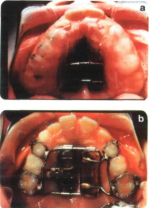
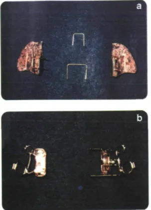
Two different types of MED appliance were used. A bonded MED (Fig. 1A) was placed in two patients while four patients received a banded MED (Fig. IB). Pins and tubes had to be placed in the right and left acrylic parts of the appliance in order to guide the separation during treatment (Fig. 2). In the bonded type, two heavy wires shaped like open staples held the two acrylic parts together during the bond-ing procedure (Figs 2A and 3A) and were removed afterwards (Fig. 1A). In the banded
MAGNETIC MAXILLARY EXPANSION 481
Figure 2 (A) The bonded MED before insertion. The 'U' form shaped heavy wires help to hold the acrylic parts together during bonding. (B) The banded MED before insertion. The expansion screw allows one of the magnets to move when activation is needed. A ligature holds the two parts together during cementing.
type, ligature wires were used to keep the two parts in contact during cementing of the appli-ance (Fig. 3B).
As the force decreases with the separation of the magnets, occasional activation is needed. In the banded type, the activation was made pos-sible by an expansion screw which moved one of the magnets towards the other until the gap between them was closed again (Figs IB, 2B, 3B and 3C). The screw was activated every 3 weeks. The repulsive force decreased from 500 g to approximately 250 g at the end of the third week (Fig. 4). This appliance is similar to a Minne-expander as far as the type of activation and the continuity of the force is concerned, but the force level with the Minne-expander is almost double (Mossaz-Jo€lson and Mossaz,
1989).
In the bonded type, the appliance was activ-ated once by adding smaller magnets on one
Figure 3 (A) The bonded MED in place. (B) The banded MED assembled before cementing. (C) The banded MED (case PL) OcclusaJ radiographs (a) day 1, (b) after 2 weeks, (c) at the end of the expansion period, and (d) at the end of the retention period.
side to fill the gap (Fig. 5C). The expansion was continued until 25 per cent overcorrection was obtained.
The MED was used on six patients aged between 7 years 4 months and 16 years 2 months. For a more accurate evaluation of the results, four implants were placed on each patient: one pair, right and left, apically between
M.A. DARENDELILER ET AL.
Figure 4 The repulsive force diagram of the banded MED.
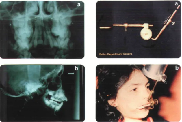
upper central and lateral incisors, and another pair (Fig. 6), again on the right and left sides between upper second premolars and first molars. Occlusal radiographs, lateral and postero-anterior headplates, study models, and intra-oral photographs were taken on the day the appliance was placed, at the end of active treatment, after 6 months of retention and after a post-retention period which varied from 1 to 2% years. Occlusal radiographs were taken with
a standardization device (Fig. 7). A conven-tional maxillary retainer (Hawley appliance) was placed the day the MED was removed and worn for the 6 months of retention. The trans-verse distance between both canine points and both first molar central fossae on the study models, and the transverse distance between both anterior implants and both posterior implants on the occlusal and postero-anterior headplates were measured to evaluate the over-all and skeletal expansion, and the relapse. Results
Clin ical findings
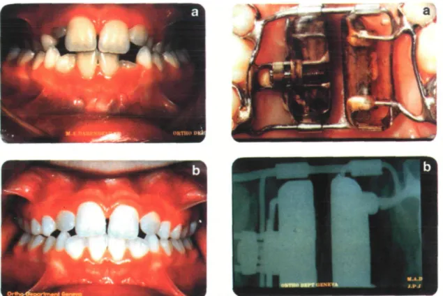
The results indicate that the dental expansion was successful in all six cases (Figs 8-10, Table 1). Both types of MED were well toler-ated by the patients, but oral hygiene was much more of a problem with the bonded appliance. All patients showed speech problems at the beginning of treatment which disappeared within 2 weeks.
In one patient wearing the bonded type (FS), the posterior pin slipped out of its guiding tube
Figure 5 The bonded MED (case FS) occlusal radiographs. (A) Day 1. (B) After 2 weeks. (C) By adding two small magnets on one side the intermagnet distance is reduced, the repelling force is increased which corresponds to an activation of the appliance (after 6 weeks). (D) At the end of the expansion period (note that the posterior pin slipped out from its guiding tube).
MAGNETIC MAXILLARY EXPANSION 483
Figure 6 (A and B) Postero-anterior and lateral radio-graphs showing the implants one pair, apically between upper central and lateral incisors, the other pair, between upper second premolars and first molars.
just before the last appointment and thus allowed more dental tipping (Fig. 5).
Removal of the bonded appliance took con-siderably more time than the banded one and the palatal mucosa was more erythematous and inflamed.. The well-known midline diastema which is usually observed in conventional RME Table 1 Cast measurements (overall expansion).
Figure 7 (A) Standardization device used for the occlusal radiographs. (B) In situ.
appliances was not observed in any of the six patients.
Cast analysis
The expansion gained and the post-retention changes in the anterior and posterior parts of the maxilla are described in Table 1. In all patients, the posterior width increased more
Patient RL* PS PL CM FS* DK Age 7.4 ».l 8 J 9.8 10.2 16.2 Treatment duration 6.5 months 5.5 months 4.0 months 8.0 months 5.5 months 4.0 months ANTERIOR Post-retention Expansion width change 6 6.5 4.2 2.2 4 3.6 +0.5 0 - 1 . 2 + 0.9 + 0.9 - 1 . 5 POSTERIOR Post-retention Expansion width change 6.6 7.2 7 83 ?.S 7.0 + 0.8 - 1 . 7 - 2 . 5 + 1.3 - 1 . 6 - 3 . 9 + : Increase in the transverse dimension
—: Decrease in the transverse dimension •Patients with the bonded MED.
484 M.A. DARENDELILER ET AL.
Figure 8 (A and B) Patient FS, age 10.2 years, before and after 22 weeks of appliance wear (the bonded MED).
Figure 9 (A and B) Patient CM, age 9.8 years, before and after 32 weeks of appliance wear (the banded MED).
than the anterior width. In the 16-year-old patient, post-retention dental relapse was 56 per cent after 17 months. The 8-year 6-month old boy who did not follow the instructions of full-time wear of the retainer also showed a post-retention relapse of 36 per cent. The other four patients showed less or no relapse after a 12-18-month post-retention period.
Radiographic findings
Occlusal radiograph and P-A headplates
In the four patients between 7 years 4 months and 9 years 8 months, an opening of the mid-palatal suture was seen on the occlusal radio-graph taken during the second week of expan-sion (Fig. 11). Anterior and posterior skeletal changes measured between the implants on the occlusal radiograph and P-A headplates were recorded three times and the mean values of the six measurements are reported in Table 2.
Almost no skeletal effects were detected in patients with bonded MED or in the 16-year-old patient (DK) with a banded MED. Skeletal
versus overall expansion varied from 0 to 25 per cent in cases with bonded MED and from 16 to 77 per cent in cases with banded MED, the 16-year-old patient excepted (DK) (Table 3).
Skeletal width changes after 1-2 years post-retention showed almost no skeletal relapse (Table 2).
Angular changes of the upper first molars. In
half of the cases, the expected buccal tipping of the first molars relapsed to a greater extent during retention and post-retention periods (Table 4). The amount of buccal tipping during expansion varied from 1.5 to 9.5 degrees except for one patient (FS), in which the tipping reached almost 20 degrees probably because the posterior pin had slipped out from its guiding tube.
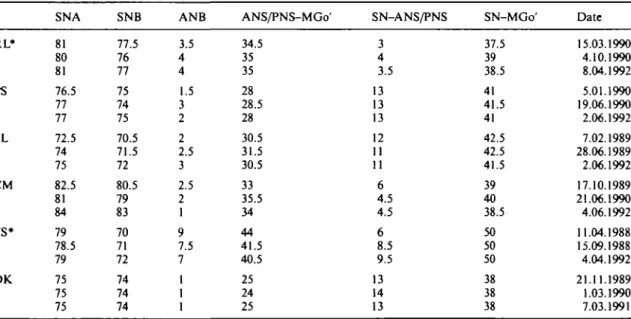
Lateral cephalograms. Table 5 indicates the cephalometric values measured during and after treatment: no skeletal changes were observed, with the exception of a slight opening of the
MAGNETIC MAXILLARY EXPANSION 4S1
Figure 10 (A and B) Patient PS, age 8.1 years, before and after 22 weeks of appliance wear (the banded MED).
mandibular plane angle during expansion which closed again during retention.
Discussion
Clinical researchers have used various force levels to expand the maxilla and have focused on the amount of skeletal effect obtained. It has
Figure 11 (A) A banded type of MED in place. (B) Opening of the mid-palatal suture is evident in the second week of expansion.
been reported that up to 20 pounds of force have been applied to the facial bones when using conventional RME appliances (Isaacson and Ingram, 1964; Zimring and Isaacson, 1965) and that approximately 50 per cent skeletal response has been obtained after 1-4 weeks (Timms, 1981). However, the optimal force Table 2 Measurements between left and right implants (skeletal expansion).
Patient RL* PS PL CM FS* DK. Age 7.4 8.1 8.6 9.8 10.2 16.2 Treatment duration 6.5 months 5.5 months 4.0 months 8.0 months 5.5 months 4.0 months Expansion 0.7 1.8 0.7 1.7 0.3 0.2 Anterior Post-retention width change + 0.1 - 0 . 6 + 0.3 + 0.2 + 0.4 + 0.1 Posterior Post-retention Expansion width change 1.7 3.0 1.7 4.1 0.7 0.5 + 0.5 + 1.3 + 0.1 + 0.7 + 1.8 +0.6 + : Increase in the transverse dimension
—: Decrease in the transverse dimension 'Patients with the bonded MED.
486
Table 3 Percentage of skeletal changes.
Patient RL* PS PL CM FS* DK Age 7.4 8.1 8.6 9.8 10.2 16.2 Skeletal (mm) 0.7 1.8 0.7 1.7 0.3 0.2 Anterior Overall (mm) 6.0 6.5 4.2 2.2 4.0 3.6 Skeletal (%) 11.7 27.6 16.6 77.2 7.5 5.5 Skeletal (mm) 1.7 3.0 1.7 4.1 0.7 0.5 Posterior Overall (mm) 6.6 7.2 7.0 8.3 7.6 7.0 Skeletal (%) 25.7 41.6 24.2 49.3 9.2 7.1 •Patients with the bonded MED.
Table 4 6/6 tipping (angulation between the upper molars and mid-sagittal plane measured on postero-anterior
headfilms). Patient RL* PS PL CM FS* DK Before treatment (°) 6/ 16 12 16 11 11 19 /6 10.5 11 12.5 21 9 18 After treatment 6/ 16 19 23 1 23 28.5 O /6 10.5 12.5 18.5 16.5 28.5 22.5 Tipping during expansion 6/ 0 + 7 + 7 - 1 0 + 12 + 9.5 O /6 0 + 1.5 + 6 - 4 . 5 + 19.5 +4.5 Post-retention period (' 6/ 18 13 16 5 13 21 ') /6 11 12.5 15 21.5 11.5 21 Tipping post-during retention (°) 6/ + 2 - 6 - 7 + 4 - 1 0 - 7 . 5 K> + 1 0 - 3 . 5 + 5 - 1 7 - 1 . 5 •Patients with the bonded MED.
Table 5 Sagittal and vertical changes during treatment and post-retention period.
SNA SNB ANB ANS/PNS-MGo' SN-ANS/PNS SN-MGo' Date
RL* PS PL CM FS* DK 81 80 81 76.5 77 77 72.5 74 75 82.5 81 84 79 78.5 79 75 75 75 77.5 76 77 75 74 75 70.5 71.5 72 80.5 79 83 70 71 72 74 74 74 3.5 4 4 1.5 3 2 2 2.5 3 2.5 2 1 9 7.5 7 1 1 1 34.5 35 35 28 28.5 28 30.5 31.5 30.5 33 35.5 34 44 41.5 40.5 25 24 25 3 4 3.5 13 13 13 12 11 11 6 4.5 4.5 6 8.5 9.5 13 14 13 37.5 39 38.5 41 41.5 41 42.5 42.5 41.5 39 40 38.5 50 50 50 38 38 38 15.03.1990 4.10.1990 8.04.1992 5.01.1990 19.06.1990 2.06.1992 7.02.1989 28.06.1989 2.06.1992 17.10.1989 21.06.1990 4.06.1992 11.04.1988 15.09.1988 4.04.1992 21.11.1989 1.03.1990 7.03.1991 •Patients with the bonded MED.
MAGNETIC MAXILLARY EXPANSION 487
levels for different ages have not yet been estab-lished. More recently, research results showed that slower maxillary expansion with the Minne-expander, applying forces up to 2 pounds, allowed 16-30 per cent skeletal expansion in 8-13 weeks in patients between 10 and 15 years old (Hicks, 1978), and 50 per cent skeletal response in patients between 8 and 12 years old (7-15 weeks) (Mossaz-JoSlson and Mossaz, 1989).
In the present study, using Light Maxillary Expansion Forces (LMEF) with the MED, generating forces between 250 and 500 g (banded MED), 16-77 per cent skeletal effect versus total expansion was obtained in patients between 8 years 1 month and 9 years 8 months (16-32 weeks). It is evident that, to obtain an orthopaedic expansion of the maxilla, the force must be sufficient to overcome the bio-elastic resistance of the periodontium, the alveolar bone and the sutural elements (Storey, 1955).
Orthopaedic effects on the maxillary complex are usually the result of a mechanical reposi-tioning followed by an adaptive growth of the facial sutures depending on age and growth potential of the patient. In the sagittal plane, forward or backward growth modifications of the maxilla are observed by using forces between 400 and 2000 g (Cleall et al., 1965; Storey, 1973). These forces influence the sutures of the naso-maxillary complex which are orientated in a similar, mostly sagittal, plane (Remmelink, 1988). Because these sutures are orientated in the sagittal plane and not in the vertical plane one would suppose that there is more resistance to the forces applied during transverse paedic correction than during sagittal ortho-paedic correction: this could be a reason in favour of the use of heavy forces to expand the maxilla. It is also well known that the major resistance to expansion is not the mid-palatal suture itself, but the other sutures of the maxilla (Bishara and Staley, 1987). The easiest technical way to apply transversal forces on the maxilla is the jackscrew and this is probably the reason for its large clinical use even though the forces applied are too heavy. Pathological effects such as root resorption, alveolar dehiscence and fen-estration due to overpowered appliances have been reported by several authors (Rinderer, 1966; Barber and Sims, 1981; Vardimon et al., 1991). In addition, mechanical separation at the mid-palatal suture can lead to an
unfavour-able healing response (Melsen, 1972). Melsen examined biopsy samples taken from the mid-palatal sutures of eight children aged between 8 and 13 years at various stages of RME and following expansion. She found numerous mic-rofractures at sites of bony interdigitations in the older patients of that sample. Her findings clearly showed that lighter forces, for example those delivered by the Minne-expander, should be used. However, we still do not know if any pathological effect exists with the Minne-expander. Since an orthopaedic effect can be obtained with an expansion force of 250-500 g as shown in the present study, side effects of heavy expansion might be eliminated.
With regard to the different force levels, the duration of the expansion varies: approximately 1 month with RME (Krebs, 1958), 2-4 months with SME (Minne-expander) (Mossaz-Joe'lson and Mossaz, 1989) and 4-8 months with LME (using MED).
Isaacson and Ingram (1964), and Zimring and Isaacson (1965) suggested that a more physiological expansion of the maxillary com-plex without the accumulation of a large resid-ual load could be obtained using slower rates of expansion. Storey (1973) mentioned, follow-ing a study on rats and rabbits, that in animals subjected to slow maxillary expansion, sutural integrity was maintained and that the relapse potential was lower than in animals treated with RME. Ohshiama (1972) reported that there was less dental tipping with SME than with RME in monkeys. The rate of mid-palatal suture separation with slow expansion systems appar-ently leads to a more physiological response by the sutural elements than the disruptive nature of RME (Skieller, 1964). The enhanced main-tenance of tissue integrity in slowly expanded sutural elements has been associated with greater stability and less relapse potential during reorganization of the maxillary complex (Skieller, 1964; Hicks, 1978; Timms, 1981; Bell, 1982).
With a slow expansion rate and light forces, the age and growth potential of the patient become more important. Mossaz-JoSlson and Mossaz (1989) found more skeletal effects (50 per cent) in patients aged 8-12 years than Hicks (1978) who used the same amount of force on patients aged 10-15 years and found 16-30 per cent skeletal effect. Thus, the LMEF, should be more effective if applied early.
Another aspect of the present study was the use of Static Magnetic Fields (SMF). Until now, very few histological findings concerning the effects of magnetic fields on facial bones, sutures and condyles have been published. Gerling et al. (1985) found an enlarged prolifer-ative layer in guinea-pigs' condyles in the pres-ence of pulsed electromagnetic fields (PEMF), but did not observe any increase in length of the mandible after a 10- and 30-day experi-mental period. In a previous study, on a sample of guinea-pigs, accelerated bone healing was found in the presence of SMF and PEMF after mandibular osteotomy. It is also mentioned that these magnetic fields could increase the rate of orthodontic tooth movement (Stark and Sinclair, 1987). Some authors (Cleall et al., 1965; Stark and Sinclair, 1987; Darendeliler et al., 1992) suppose that magnetic fields like electrical currents (Davidovitch et al., 1980; Davidovitch, 1981) could change the membrane permeability and thus influence the exchange of cellular nucleotides, calcium uptake (Colaccicco and Pilla, 1983) and lead to a quicker appear-ance of osteoblasts and osteoclasts (Stark and Sinclair, 1987). It is not known if SMF have advantages or disadvantages in maxillary expansion and further histological evidence and clinical research is needed.
Conclusion
With such a large number of parameters and such a small sample, conclusions are only hypo-thetical, but it seems that 250-500 g of continu-ous magnetic force can produce dental and skeletal movements in a light force expansion concept. Further studies orientated towards the following three goals must be carried out:
1. The use of larger samples in order to study the efficiency of LMEF versus RME and SME, in particular versus the Minne-expander.
2. Histological research is needed to evaluate the effect of LME on root resorption and surrounding tissues.
3. The use of neodymium magnets which are more powerful than SmCo magnets. The same amount of force could be obtained with a smaller and thus less bulky MED.
Address for correspondence Dr M. AH Darendeliler
Division d'Orthodontie et de Pedodontie Ecole de Medecine Dentaire
19, rue Barth.-Menn 1211 Geneva 4 Switzerland Acknowledgements
We express our thanks to Mrs Fiona McLennan Med. Dent., as well as to Mrs Aysin Darendeliler Med. Dent, for their help in preparing this publication.
References
Alpern M C, Yurosko J J 1987 Rapid palatal expansion in adults with and without surgery. Angle Orthodontist 57: 245-263
Angell E C 1860 Treatment of irregularities of the perman-ent or adult teeth. Dperman-ental Cosmos 1: 540-544, 599-601 Barber A F, Sims M R 1981 Rapid maxillary expansion
and external root resorption in man: a scanning electron microscope study. American Journal of Orthodontics 79: 630-652
Bell R A 1982 A review of maxillary expansion in relation to the rate of expansion and patient's age. American Journal of Orthodontics 81: 32-37
Bishara S E, Staley R N 1987 Maxillary expansion: Clinical implications. American Journal of Orthodontics and Dentofacial Orthopedics 91: 3-14
Blechman M A 1985 Magnetic force systems in orthodont-ics. Clinical results of a pilot study. American Journal of Orthodontics 87: 201-210
Chaconas S J, de Albay y Levy J A 1977 Orthopedic and orthodontic applications of the quad-helix appliance. American Journal of Orthodontics 72: 422-428 Cleall J F, Bayne D, Posen J, Subtelny J 1965 Expansion
of the mid-palatal suture in the monkey. Angle Orthodontist 35: 23-35
Colaccicco G, Pilla A A 1983 Electromagnetic modulation of biological processes: chemical, physical and biological correlations in the Ca-uptake by embrional chick tibia in vitro. Bioelectrochemistry Bioenergetics 10: 119-131 Dabbane E F 1958 A cephalometric and histologic study
of the effect of orthodontic expansion of the midpalatal suture of the cat. American Journal of Orthodontics 44: 187-219
Darendeliler M A, Joho J P 1992 Class II bimaxillary protrusion treated with magnetic forces. Journal of Clinical Orthodontics 16: 361-368
Darendeliler M A, Joho J P 1993 Magnetic activator device for correction of Class II/l malocclusions. American Journal of Orthodontics and Dentofacial Orthopedics 103: 223-239
Darendeliler M A, Sinclair P M, Kusy R P 1992 Effects of static magnetic fields and pulsed electromagnetic fields
MAGNETIC MAXILLARY EXPANSION 489
on orthodontic tooth movement. Journal of Dental Research 71: 251 (Abstract)
Davidovitch Z. 1981 Tissue reaction and bone turnover in orthodontic treatment. In: Barrer H G (ed.) Orthodontic—The State of Art. University of Pennsylvania Press, Philadelphia, pp. 297-313
Davidovitch Z, Finkelson M D, Steigman S, Shanfeld J, Montgomery P, Korostoff E 1980 Electrical currents, bone remodelling and orthodontic tooth movement. I. The effect of electrical currents on the periodontal cyclic nucleotides. American Journal of Orthodontics 77: 14-32 Gerling J A, Sinclair P M, Roa R L 1985 The effects of pulsating electromagnetic fields on condylar growth in guinea pigs. American Journal of Orthodontics 87: 211-223
Gianelly A A, Vaistas A S, Thomas W M, Berger D G 1988 Distalization of molars with repelling magnets—a case report. Journal of Clinical Orthodontics 22: 40-44 Glassmann A S 1984 Conservative surgical orthodontic
adult rapid palatal expansion: sixteen cases. American Journal of Orthodontics 86: 207-213
Haas A J 1961 Rapid expansion of the maxillary dental arch and nasal cavity by opening the midpalatal suture. Angle Orthodontist 31; 73-90
Haas A J 1965 The treatment of maxillary deficiency by opening the midpalatal suture. Angle Orthodontist 35: 200-217
Hicks E P 1978 Slow maxillary expansion: a clinical study of the skeletal vs dental response to low magnitude force. American Journal of Orthodontics 73: 121-141 Howe R P 1982 Palatal expansion using a bonded appliance.
American Journal of Orthodontics 82: 464-468 Isaacson R J, Ingram A H 1964 Forces produced by rapid
maxillary expansion II. Forces present during treatment. Angle Orthodontist 34: 261-270
Kawata T, Katsuhiko H, Kohji S 1987 A new orthodontic force system of magnetic brackets. American Journal of Orthodontics and Dentofacial Orthopedics 92: 241-248 Kennedy J W, Bell W H 1976 Osteotomy as an adjunct to
rapid maxillary expansion. American Journal of Orthodontics 70: 123-137
Krebs A 1958 Expansion of the midpalatal suture studied by means of metallic implants. Transactions of the European Orthodontic Society pp. 163-171
Krebs A 1964 Rapid expansion of midpalatal suture by fixed appliance. An implant study over a 7 year period. Transactions of the European Orthodontic Society pp. 141-142
Langford S R, Sims M R 1982 Root surface resorption, repair, and periodontal attachment following rapid maxil-lary expansion in man. American Journal of Orthodontics 81: 108-115
Lehmann J A, Haas A J, Haas D G 1984 Surgical ortho-dontic correction of transverse maxillary deficiency: A simplified approach. Journal of Plastic and Recon-structive Surgery 73: 62-68
Linder-Aronson S, Lindgren J 1979 The skeletal and dental effects of rapid maxillary expansion. British Journal of Orthodontics 6: 25-29
Lines P A 1975 Adult rapid maxillary expansion with corticotomy. Angle Orthodontics 67: 44-56
Melsen B 1972 A histological study of the influence of sutural morphology and skeletal maturation on rapid maxillary expansion in children. Transactions of the European Orthodontic Society pp. 499-507
Mew J R C 1977 Semi-rapid maxillary expansion. British Dental Journal 143: 301-306
Moss J P 1968 Rapid expansion of maxillary arch. Journal of Practical Orthodontics 2: 168-171, 216-223
Mossaz C F, Byloff F K, Richter M 1992 Unilateral and bilateral corticotomies for correction of maxillary trans-verse discrepancies. European Journal of Orthodontics 14: 110-116
Mossaz-Jofilson K, Mossaz C F 1989 Slow maxillary expan-sion: a comparison between banded and bonded appli-ances. European Journal of Orthodontics 11: 67-76 Ohshiama O 1972 Effect of lateral expansion force on the
maxillary suture in cynomolgus monkey. Journal of the Osaka Dental University 6: 11-50
Reitan K 1964 Effects of force magnitude and direction of tooth movement on different alveolar bone types. Angle Orthodontist 34: 244-255
Remmelink H-J 1988 Orientation of maxillary sutural sur-faces. European Journal of Orthodontics 10: 223-226 Rinderer L 1966 The effects of expansion on the palatal
suture. Transactions of the European Orthodontic Society pp. 365-377
Schwarz G M, Trash W J, Byrd L D, Jacobs J D 1985 Tomographic assessment of nasal septum changes follow-ing surgical orthodontic rapid maxillary expansion. American Journal of Orthodontics 87: 39-45
Skieller V 1964 Expansion of the mid-palatal suture by removable plates, analysed by the implant method. Transactions of the European Orthodontic Society pp. 143-157
Stark T M, Sinclair P M 1987 Effects of pulsed electromag-netic fields on orthodontic tooth movement. American Journal of Orthodontics and Dentofacial Orthodontics 91: 91-104
Starnbach H, Bayne D, Cleall J, Subtelny J 1966 Facioskeletal and dental changes resulting from rapid maxillary expansion. Angle Orthodontist 36: 152-164 Stockfisch H 1969 Rapid expansion of the maxilla—Success
and relapse. Transactions of the European Orthodontic Society pp. 469-481
Storey E 1955 Bone changes associated with tooth move-ment: a histological study of the effect of force in the rabbit, guinea pig and rat. Australian Dental Journal 59: 147
Storey E 1973 Tissue response to the movement of bones. American Journal of Orthodontics 64: 229-247 Timms D J 1968 An occlusal analysis of lateral maxillary
expansion with mid-palatal suture opening. Dental Practitioner 18: 435-440
Timms D J, Moss J P 1971 An histological investigation into the effects of rapid maxillary expansion on the teeth and their supporting tissues. Transactions of the European Orthodontic Society pp. 263-271
Timms J T 1981 Rapid maxillary expansion. Quintessence Books, Berlin
490
Vardimon A D, Graber T M, Voss L R, Verrusio E 1987 Magnetic versus mechanical expansion with different forces. Thresholds and points of force application. American Journal of Orthodontics and Dentofacial Orthopedics 92: 455-466
Vardimon A D, Stutzmann J J, Graber T M, Voss L R, Petrovic A G 1989 Functional orthopedic magnetic appli-ance (FOMA) II - modus operandi. American Journal of Orthodontics and Dentofacial Orthopedics 95: 371-387 Vardimon A D, Graber T M, Voss L, Lenke J 1991 Determinants controlling iatrogenic external root resorp-tion and repair during and after palatal expansion. Angle Orthodontist 61: 113-122
Warren D W, Hershey H G, Turvey T A, Hinton V A, Hairfield W M 1987 The nasal airway following maxillary expansion. American Journal of Orthodontics and Dentofacial Orthopedics 91: 111-118
Wertz R A 1970 Skeletal and dental changes accompanying rapid midpalatal suture opening. American Journal of Orthodontics 58: 41-66
Zimring J F, Isaacson R J 1965 Forces produced by rapid maxillary expansion III. Forces present during retention. Angle Orthodontist 35: 178-186