Aerosol Delivery of Functionalized Gold
Nanoparticles Target and Activate Dendritic
Cells in a 3D Lung Cellular Model
Kleanthis Fytianos,
†Savvina Chortarea,
†Laura Rodriguez-Lorenzo,
†Fabian Blank,
§Christophe von Garnier,
§Alke Petri-Fink,
†,‡and Barbara Rothen-Rutishauser*
,††
Adolphe Merkle Institute and
‡Department of Chemistry, University of Fribourg, CH-1700 Fribourg, Switzerland
§Respiratory Medicine, Inselspital, University of Bern, 3012 Bern, Switzerland
*
S Supporting InformationABSTRACT:
Nanocarrier design combined with
pulmo-nary drug delivery holds great promise for the treatment of
respiratory tract disorders. In particular, targeting of
dendritic cells that are key immune cells to enhance or
suppress an immune response in the lung is a promising
approach for the treatment of allergic diseases.
Fluores-cently encoded poly(vinyl alcohol) (PVA)-coated gold
nanoparticles, functionalized with either negative
(−COO
−) or positive (
−NH
3+
) surface charges, were
functionalized with a DC-SIGN antibody on the particle
surface, enabling binding to a dendritic cell surface
receptor. A 3D coculture model consisting of epithelial
and immune cells (macrophages and dendritic cells) mimicking the human lung epithelial tissue barrier was employed to
assess the e
ffects of aerosolized AuNPs. PVA-NH
2AuNPs showed higher uptake compared to that of their
−COOH
counterparts, with the highest uptake recorded in macrophages, as shown by
flow cytometry. None of the AuNPs induced
cytotoxicity or necrosis or increased cytokine secretion, whereas only PVA-NH
2AuNPs induced higher apoptosis levels.
DC-SIGN AuNPs showed signi
ficantly increased uptake by monocyte-derived dendritic cells (MDDCs) with subsequent
activation compared to non-antibody-conjugated control AuNPs, independent of surface charge. Our results show that
DC-SIGN conjugation to the AuNPs enhanced MDDC targeting and activation in a complex 3D lung cell model. These
findings highlight the potential of immunoengineering approaches to the targeting and activation of immune cells in the
lung by nanocarriers.
KEYWORDS:
gold nanoparticles, dendritic cells, aerosol exposures, cellular uptake, immunomodulation
T
he human body has a number of portals of entry
available for drug administration, such as the lungs (via
inhalation), the gastrointestinal tract (via digestion),
the blood vessels (via intravenous injection), and the skin (via
transdermal application). The lung, with its huge internal
surface of ca. 150 m
2(i.e., alveoli and airways),
1provides a
promising, noninvasive portal of entry for aerosolized drugs. A
number of inhaled drugs are currently applied to treat airway
diseases, are in development, or have been considered for the
treatment of various lung diseases. In 2004, there were about 25
drugs licensed for use in treating pulmonary diseases via
inhalation, such as asthma, emphysema, chronic in
flammation,
chronic obstructive pulmonary disease (COPD), and lung
cancer.
2In addition to the large lung surface, areas of a dense network
of antigen-presenting cell populations, such as dendritic cells
(DCs) and macrophages, are present within the lung
parenchyma.
3DCs are among the most important immune
cells in the lung, orchestrating innate and adaptive immune
responses.
4Speci
fic targeting of DCs via receptor-mediated
mechanisms has previously been described as an innovative
approach to drug delivery.
5,6Dendritic cell-speci
fic intracellular
adhesion molecule 3 (ICAM-3)-grabbing non-integrin
(DC-SIGN) is a well-studied receptor that is found in abundance on
the surface of mature DCs. It has a high a
ffinity for mannose
and is involved in the enhancement of antigen uptake,
processing, and presentation to T-cells in the draining of
lymph nodes. Targeting DC-SIGN may lead to the
develop-ment of immunomodulatory applications such as a change in
pro-in
flammatory cytokine secretion.
6http://doc.rero.ch
Published in "ACS Nano 11(1): 375–383, 2017"
which should be cited to refer to this work.
The development of nanomaterials (i.e., a material with one,
two, or three external dimensions in the nanoscale (<100 nm)
(ISO/TS, 2008)) for drug delivery, medical imaging, and
diagnostic purposes has led to the discovery of potential
e
ffective therapeutics. Depending on their size and properties,
inhaled nanomaterials may enter the lung and interact with the
respiratory wall.
7,8While larger materials are deposited in the
conducting airways, smaller ones reach the peripheral gas
exchange region and may pass the air
−blood tissue barrier to
enter the systemic circulation. The interaction with the lungs,
and particularly the bioavailability of nanomaterials, depends on
various nanomaterial characteristics, such as size,
hydro-phobicity, solubility, and enzymatic hydrolysis. Currently,
nanomaterial-based (i.e., iron oxide, liposomes, polymer
nanoparticles, emulsions, fullerenes, and carbon nanotubes)
drugs are employed in biomedical applications due to their
e
ffectiveness in modulating immune responses.
9,10Recently, our group has developed a library of homo- and
heterofunctionalized,
fluorescently labeled and polymer-coated
gold nanoparticles (AuNPs).
11Fluorescently labeled
nano-particles (NPs) may be utilized for detection with techniques
such as laser microscopy and
flow cytometry, and the signals
may be a useful tool for measurements such as cellular uptake
and marker expression.
12Furthermore, AuNPs possess
excep-tional optical properties (scattering and absorption from
localized surface plasmon resonances) and can be synthesized
by rapid and highly reproducible methods. In addition, they are
not cytotoxic at biomedically relevant concentrations. Using
this NP library, we have shown that surface modi
fication can
alter cellular uptake, with the PVA-NH
2and PVA-COOH
AuNPs being taken up by human blood monocyte-derived
dendritic cells (MDDCs) in vitro, while functional and
immunological properties were not impaired at concentrations
of 20
μg/mL.
13,14In this study, we have further explored the potential of
aerosolized PVA-COOH and PVA-NH
2AuNPs in terms of
their cellular uptake and functional and immunological
properties in an advanced in vitro coculture cell model
representing the human lung epithelial tissue barrier
15under
realistic exposure scenarios, that is, exposure of the AuNPs as
an aerosol over the lung cell surface. Furthermore, the ability of
DC-SIGN-conjugated AuNPs to speci
fically target MDDCs in
the coculture model was investigated.
RESULTS AND DISCUSSION
AuNP Synthesis and Characterization. AuNPs were
synthesized by the Turkevich method,
16coated with poly(vinyl
alcohol) (PVA) with either a positive (
−NH
3+) or a negative
(
−COO
−) charge and tagged with ATTO590
fluorescent
molecules), as described by Rodriguez-Lorenzo et al.
It has been reported that the use of adequate conjugation
strategies, in order to correctly orient the DC-SIGN antibody
(Ab), could strongly a
ffect the accessibility of antigen binding
sites.
17−19Therefore, the Ab conjugation was carried out by the
fixed orientation of the Ab on the NP surface. In the case of
PVA-COOH, the conjugation was achieved through ionic and
covalent interactions as described by Parolo et al.
46The
conjugation is based on the fact that when the pH of an Ab
solution is lower than its isoelectric point, the concentration of
positive charges (
−NH
3+group of Lys residues) in the
fragment-crystallizable region (Fc region, which is the tail
region of the Ab) of the Ab is high. Therefore, negatively
charged AuNPs (COO
−groups of PVA-COOH) most likely
interact through the Fc region of the Ab, and this oriented ionic
attraction is bound via carbodiimide chemistry, guaranteeing
that the antigen binding sites remain accessible. In contrast, the
conjugation strategy of PVA-NH
2AuNPs consists of the use of
the polysaccharidic region located in the Fc region of the Ab for
covalent binding with amino group of the PVA-NH
2polymer.
17
Bradford assay measurements indicated that the concentration
of Ab covalently bound to PVA-COOH was 2.7
± 2.0 μg, while
that bound to PVA-NH
2was 0.7
± 0.5 μg, indicating that the
conjugation was more e
fficient through peptide-bond formation
than via the polysaccharidic region. It should be noted,
however, that the number of ATTO590 molecules per AuNP
decreased after Ab conjugation in both cases, which is most
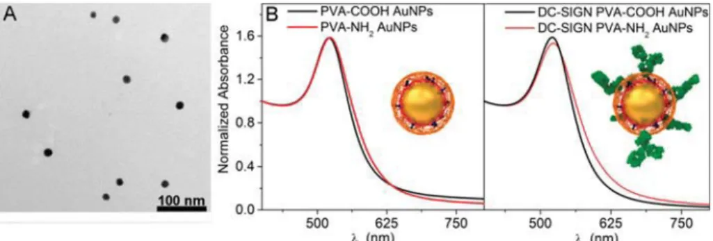
Figure 1. Characterization of AuNPs. TEM micrograph of 15 nm AuNPs (A) and UV−vis spectra (B) of the heterofunctionalized ATTO590 PVA-COOH and PVA-NH2AuNPs before and after DC-SIGN Ab conjugation. Inset: Schematic illustration of the designed AuNPs.
Table 1. Physicochemical Characterization of the Heterofunctionalized PVA-COOH and PVA-NH
2AuNPs
AuNP sample hydrodynamic diameter [nm] (PDI %)a ξ [mV] (SD) ATTO590 per AuNPb DC-SIGN [μg] (SD)cDC-SIGN per AuNP (SD)
PVA-COOH 66 (40) −13.6 (9.2) 720
PVA-NH2 111 (38) 10.6 (3.2) 350
DC-SIGN PVA-COOH 100 (42) −11.5 (8.8) 40 2.7 (2.0) 6 (4)
DC-SIGN PVA-NH2 175 (49) 8.6 (3.1) 28 0.7 (0.5) 2 (1)
aAt the 0.05 level, the difference between the population means is not significantly different.bFluorescence measured by means of Victor plate reader
with thefilter set to 560/615.cTotal protein estimation using Bradford assay.
likely due to desorption of a part of the polymer layer during
Ab conjugation.
Prior to employing the AuNPs in cell culture experiments, we
thoroughly characterized both PVA-COOH and PVA-NH
2AuNPs by assessing their colloidal stability using UV
−vis
spectroscopy, their hydrodynamic radii, and polydispersity
indices (PDI) using dynamic light scattering (DLS) and their
surface charges through
ζ-potential measurements of AuNP
suspensions in PBS. All AuNP types were stable and showed no
evidence of aggregation-induced red shifting or band
broad-ening in their UV
−vis spectra (
Figure 1
B).
Analysis by DLS showed that DC-SIGN AuNPs presented
hydrodynamic diameters higher than those of the unconjugated
homologues: 100 nm versus 66 nm for PVA-COOH and 175
nm versus 111 nm for PVA-NH
2(
Table 1
). This increase may
be attributed to the presence of Ab, which can produce an
increase up to 20 nm
20together with a swelling of polymer
layer due to the variation of electrostatic interaction, as
described in other studies.
21,22PVA-COOH AuNPs were
negatively charged with a
ζ-potential of −13.6 ± 9.2 mV, and
PVA-NH
2AuNPs were positively charged with a
ζ-potential of
10.6
± 3.2 mV (
Table 1
).
Interestingly, the surface charge remained negative (
−11.5 ±
8.6 mV) and positive (8.6
± 3.1 mV) after Ab conjugation,
which allowed a direct comparison of charged particles with
and without Ab on cellular uptake patterns and MDDC-speci
fic
targeting potential and immunomodulatory e
ffects.
AuNP suspensions (120
μg/mL) were prepared, and 1 mL
was nebulized using the air
−liquid interface cell exposure
(ALICE) system.
23The deposited values on the bottom of the
chamber were determined using a quartz crystal microbalance
(QCM) and were 3.44
± 0.36 and 4.34 ± 0.42 μg/cm
2for the
PVA-COOH and PVA-NH
2, respectively, and 4.34
± 0.42 and
6.17
± 0.19 μg/cm
2for the DC-SIGN AuNPs. TEM images
(
Figure S1
) showed that AuNPs were monodisperse and that
there was a uniform deposition after aerosolization.
Cellular Uptake. Four di
fferent AuNP types were used to
study the interaction with the individual cell types in the
coculture lung model. The particles were aerosolized over the
lung cell surface, and cultures were post-incubated for 24 h
before sample processing.
For
flow cytometric analysis, cells were digested by
trypsin-EDTA and labeled for speci
fic cell surface markers for
multicolor detection as previously described (Clift et al.,
manuscript under revision). Particle uptake was measured as
mean
fluorescence intensity (ΔMFI) of ATTO590 from the
EpCAM
+cell population (A549 cells), CD80
+(MDDCs), and
CD11a
+(monocyte-derived macrophages, MDMs) (
Figure 2
).
For all three cell types, a low uptake of PVA-COOH was
observed, whereas a signi
ficant increase in particle-positive
MDMs was seen when the cultures were exposed to PVA-NH
2AuNPs. When DC-SIGN PVA-COOH particles were used, a
signi
ficant increase in MDMbut also in MDDCwas
observed as well as observation of more DC-SIGN PVA-NH
2AuNPs inside MDDCs than non-Ab-labeled ones. For epithelial
cells, a generally low frequency of positive cells was observed
for all four particle types.
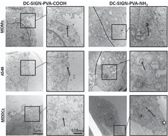
TEM was used to qualitatively con
firm the uptake of AuNPs,
and the NPs were detected in all three cell types of the
coculture (
Figures 3
and
S5
). AuNPs were found in all di
fferent
cell types either as single particles or as small agglomerates.
Positively charged DC-SIGN AuNPs showed higher uptake
levels in all cells compared to their negatively charged
counterparts. Generally, AuNPs were found in all cell types
either as single NPs, small or large agglomerates inside vesicles,
or in the cytosol.
In addition to TEM, laser scanning microscopy images were
obtained to show that the cell morphology and the epithelial
monolayer structure were not impaired upon exposure to any
of the AuNPs (
Figure S4
).
Cell Viability, Apoptosis, Cytotoxicity, and Cytokine
Secretion. In the next step, the cell viability and induction of
cell responses upon exposure to AuNPs was assessed. Only
PVA-NH
2(i.e., non-DC-SIGN-PVA-NH
2-functionalized)
AuNPs signi
ficantly induced late apoptosis compared to the
negative control (
Figure 4
). However, the PVA-NH
2polymer
alone, which was used as a control, also induced signi
ficant late
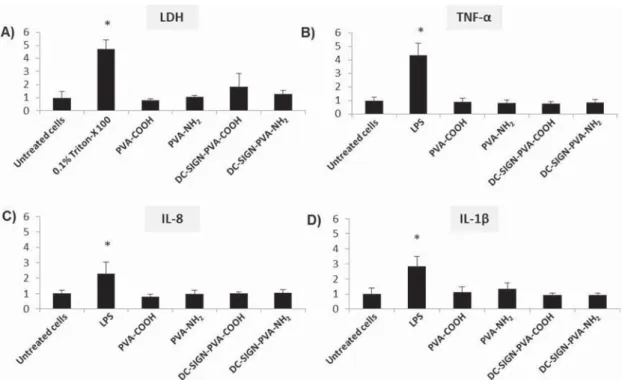
apoptosis levels. None of the AuNPs tested induced
cytotoxicity, as shown by lactate dehydrogenase (LDH).
Considering cytokine secretion levels, enzyme-linked
im-munosorbent assay (ELISA) measurements showed no increase
of release of various cytokines, such as IL-8, TNF-
α, and IL-1β
(
Figure 5
), implying that none of the AuNP types tested
interfere with the functional properties of the cocultures.
MDDC Surface Marker Expression. The surface marker
expression of MDDCs was investigated by
flow cytometry
analysis. DC-SIGN-conjugated AuNPs of both surface
functionalizations signi
ficantly upregulated MHC-II expression
compared to the negative control (
Figure 6
).
AuNPs without DC-SIGN functionalization did not alter
surface marker expression. In order to verify that the
upregulated MHC-II expression can be attributed to the
conjugated AuNPs and not to the antibody alone, DC-SIGN
antibody suspensions at concentration levels of 2.7
μg/mL (a
concentration level that corresponds to the AuNP suspensions)
were aerosolized as a control measurement, and no e
ffects were
observed on surface marker expression (data not shown).
Speci
fic Targeting Approach. Over the past few years, a
signi
ficant amount of research has been conducted on drug
delivery to the lungs by making use of NPs in aerosolized form,
Figure 2. Cellular uptake of AuNPs in the cocultures. The cells grown on the membrane insert were detached from the inset after exposure to the AuNPs for 24 h (insert) and analyzed by flow cytometry. The three different cell populations were identified by the distinct cell surface markers that were used to label the cells. The MFI of ATTO590 was calculated for the three different cell populations that expressed these markers. Results indicated that PVA-NH2AuNPs were predominantly taken up by the MDMs.
DC-SIGN-conjugated AuNPs showed an enhanced targeting of MDDCs. Each experiment was repeated three times, and cells from different cell cultures were obtained (n = 3, *p < 0.05).
as reviewed by Mu
̈ller and colleagues.
24Various types of NPs
have proven capable of speci
fic cell or organ targeting and
size-speci
fic delivery. The aim of the study was to target MDDCs in
vitro with aerosolized surface-modi
fied AuNPs. By achieving
this, an immune response might be triggered in vivo via
cross-talk of pulsed DCs and T-cells.
We recently showed that AuNPs coated with PVA-COOH
and PVA-NH
2were the NPs taken up by MDDCs at the
highest numbers in monoculture suspensions, in comparison to
AuNPs with polyethylene glycol surface modi
fications,
11,13compelling us to continue with more sophisticated experiments
with these two particle types. PVA is an FDA-approved
polymer that can improve colloidal stability, biocompatibility,
and cell interactions both in vivo and in vitro.
25−27In the
current study, we explore a more complex lung barrier model in
order to further investigate the NP
−cell interaction and employ
an air
−liquid exposure system to aerosolize AuNPs.
28The
tested AuNPs were aerosolized and shown to be uniformly
deposited in the cell culture inserts, while also remaining
monodisperse after aerosolization. The deposition of the
particles was quanti
fied by QCM, and we measured deposition
values ranging from 3 to 6
μg/cm
2(
Figure S1
).
In order to investigate which cell types are taken up in the
cocultures, we utilized a
flow cytometry protocol where the
insets from the cocultures were digested with trypsin/EDTA
and the cell suspensions were stained with speci
fic surface
markers for each cell type.
29The results indicated that
signi
ficantly more MDMs were measured with NPs for the
positively charged particles (i.e., PVA-NH
2AuNPs) compared
to negatively charged ones (i.e., PVA-COOH AuNPs). Similar
results have been reported by Bachler et al.
30We also detected
more NP-positive MDMs in comparison to epithelial cells and
MDDCs. A similar pattern has also been observed by Seydoux
and colleagues, wherein AuNPs with the same surface
modi
fication were mainly detected in the broncho-alveolar
lavage macrophages in vivo.
14We observed the presence of
intracellular PVA-COOH particles by TEM imaging, and
further research is ongoing to determine if the
fluorophore
Figure 3. TEM micrographs. TEM images of internalized DC-SIGN AuNPs in MDMs, A549 (apical side of the 3D cocultures), and MDDCs (basolateral side of the 3D cocultures). Left panel: DC-SIGN-PVA-COOH AuNPs. Right panel: DC-SIGN-PVA-NH2AuNPs. AuNPs were
found inside cellular vesicles 24 h after exposure using the ALICE system. The boxes (overview) and zoomed-in images show the particles (arrows) (samples with lead citrate and uranyl acetate staining).
Figure 4. Apoptosis and cell viability. An Annexin V (apoptosis)/PI (cell death) assay was performed in order to determine the percentage of healthy, early and late apoptotic, and dead cells. PVA-COOH AuNPs did not influence cell viability, and when cells were exposed to PVA-NH2 AuNPs, a higher frequency of late
apoptotic cells was recorded compared to the other treatments. Cells exposed to 2 mM captothecin for 24 h were used as a positive control for apoptosis, and cells exposed to−80 °C for 30 min were used as a positive control for cell death (n = 3, *p < 0.05).
properties of ATTO590 in the polymer undergo quenching in
an intracellular fashion.
Since our major aim was to establish a NP target and activate
dendritic cells in the lung tissue, we conjugated DC-SIGN Ab
onto the polymer surface of AuNPs. Both positively- and
negatively charged DC-SIGN AuNPs were taken up by MDMs.
In MDDCs, DC-SIGN-bound AuNP uptake was increased 4
−5
fold compared to naked AuNPs, indicating that speci
fic
targeting of DCs is feasible.
E
ffect of AuNPs on the Functioning of MDDCs. None of
the AuNPs tested had an e
ffect on cytotoxicity and
pro-in
flammatory cytokine secretion. The AuNPs did not elicit
signi
ficant LDH release, and the cytokine secretion levels were
similar to those in negative control cells. These
findings agree
with the literature, which suggests that aerosolized AuNPs do
not cause significant adverse effects, either in vitro or in vivo.
15,23We have recently shown that PVA-COOH and PVA-NH
2AuNPs do not interfere with cytotoxicity and cytokine secretion
in MDDC suspensions, at similar concentration levels and
exposure times.
13After PVA-NH
2AuNP exposure
correspond-ing to 3.44
± 0.36 μg/cm
2of deposited Au in the insets, the
cells showed increased late apoptotic levels compared to the
negative control. The apoptosis levels of the PVA-COOH
AuNP-treated cells were similar to those of the negative
control. In general, AuNPs can cause apoptosis only at very
high concentrations.
31,32In order to clarify if the increased late
apoptotic levels are due to the polymer itself, polymers without
AuNPs were aerosolized in the ALICE, and the results
indicated that the apoptosis pattern is observed due to the
PVA-NH
2polymer and not the AuNPs. Recently, we observed
that aerosolized (PEG-PVA)-NH
2AuNPs led to increased
apoptotic levels compared to the negative control after 3 days
of repeated exposure. This means that apoptosis might be
caused by the polymer coating (PVA-NH
2) rather than the gold
itself, and therefore, prolonged exposure to such biomedical
particles needs to be studied in more detail (data not shown).
Interestingly, we could show that when the polymer was
shielded by the DC-SIGN Ab, the induction of late apoptosis
decreased to control levels.
Considering immunological properties, the
immunopheno-typing marker expression remained unaltered in our study since
CD1c expression was not impaired after AuNP exposure. The
same applied for co-stimulatory markers such as CD40, CD80,
and CD86. Considering activation marker expression, CD83
expression also remained unaltered, while MHC-II expression
was signi
ficantly higher in MDDCs when the cocultures were
Figure 5. Cytotoxicity and cytokine secretion. Neither AuNP type elicited significant cytotoxicity levels, as indicated by LDH release (A). Inserts exposed to dH2O 0.9% NaCl were used as negative controls, and inserts exposed to 0.2% Triton-X 100 (under submerged conditions)
were used as positive controls. AuNP suspensions without cells were used as interference controls. At the given concentration levels (20μg/ mL), no interference was measured. TNF-α (B), IL-8 (C), and IL-1β (D) secretion levels were at similar levels as the negative control (inserts exposed to H2O), as measured by ELISA. Eachy axis is normalized to the respective negative control value. For all the cytokines tested, inserts
exposed to 1μg/mL LPS (under submerged conditions) were used as positive controls (n = 3, *p < 0.05).
Figure 6. MDDC marker expression. The following MDDC surface markers were examined: CD1c, CD40, CD80, CD83, CD86, and MHC-II. AuNPs without DC-SIGN functionalization had no effect on the MDDC marker expression. In contrast, DC-SIGN-conjugated AuNPs significantly upregulated MHC-II expression (n = 3, *p < 0.05).
exposed to DC-SIGN AuNPs compared to the negative
control. This result highlights the immunomodulatory
proper-ties of the DC-SIGN-conjugated AuNPs. We have previously
shown that MHC-II expression was not impaired after
PVA-COOH and PVA-NH
2AuNP exposure in suspension
experi-ments using MDDM monocultures. Blank et al.
33observed
increased MHC-II expression when MDDCs were exposed to
superparamagnetic iron oxide NPs. MHC-II molecules are
found in abundance on the surface of mature DCs. Increased
MHC-II levels indicate that MDDCs have reached a maturation
state. This might enable them to migrate to lymphoid tissues
and interact with the T-cell receptor (TcR) of CD4
+T-cells,
thus in
fluencing antigen presentation to T-cells and initiating
immune responses.
34−37Complementarily, Seydoux et al.
14have shown that cationic AuNPs that have been intranasally
instilled can enhance antigen-speci
fic CD4
+T-cell proliferation
in the lung draining lymph nodes.
There is evidence in the literature for the utilization of
DC-SIGN AuNPs in biomedical applications. Arnaiz et al.
38synthesized 1.8 nm gold glyconanoparticles, some of which
were internalized by immature DCs by both DC-SIGN- and
non-DC-SIGN-mediated pathways, while the other part was
colocalized with SIGN receptors. This resulted in
DC-SIGN receptor blocking by these AuNPs and thus also
inhibition of HIV-1 infection via T-cell communication
cross-talk in vitro. Chiodo and colleagues
6engineered AuNPs
conjugated to galactofuranose, a sugar which binds to the
DC-SIGN receptor. These AuNPs induced pro-in
flammation of
MDDCs, as well as CD80, CD86, and HLA-DR upregulation.
In addition, Arosio et al.
39synthesized AuNPs functionalized
with
α-fucosylamide, a molecule which facilitated DC-SIGN
receptor targeting. These AuNPs were internalized by MDDCs
via the DC-SIGN receptor without triggering immune
responses, thus they can be utilized for delivery purposes.
Our results indicate that surface-modi
fied AuNPs have great
potential in the clinical
field because, apart from their functional
and immunomodulatory properties, they can also be cleared by
alveolar macrophages.
40Another explanation of the increased MHC-II expression
might be attributed to particle size. Cruz et al.
41demonstrated
that nanosized poly(lactic-co-glycolic acid) particles were able
to interact with DC-SIGN receptors of human DCs in vitro and
elicit a signi
ficant T-cell response compared to their microsized
counterparts. Manolova and co-workers
42also demonstrated
that the targeting e
fficacy (and thus immune response
inductions) caused by virus-like particles (30 nm) also depends
on the size of the particulate system, with smaller NPs being
more potent than their larger counterparts (200 nm).
DC-SIGN is a well-studied receptor that is involved both in CD4
+and CD8
+T-cell stimulation.
43Vaccines that target DCs show
considerable advantages and are considered a promising
strategy for the further advancement of immunotherapy, as
described by Kasternmu
̈ller et al.
43and Kreutz et al.
44In this
article, we investigate speci
fic targeting of MDDCs by
DC-SIGN AuNPs in a complex 3D lung barrier coculture model.
Sehgal et al., Cruz et al., and Arosio et al.
39,41,45tested DC
targeting via the DC-SIGN receptor with polymer
nano-particles in DC monocultures in vitro. E
fficient antigen uptake
by DCs was observed, which resulted in enhanced T-cell
targeting.
CONCLUSION
By applying an advanced 3D human lung model combined with
air
−liquid exposure of nanoparticles onto the cell surface and
subsequent cellular analysis, we have shown that PVA-NH
2AuNPs were taken up mainly by MDMs, a
finding which we
con
firmed in an animal study.
14When the particles were
additionally functionalized with a DC-SIGN Ab, the targeting
of MDDCs present at the basal side of a 3D lung cell model
was signi
ficantly enhanced. In addition, DC-SIGN AuNPs
induced upregulation of MHC-II levels in the MDDC
population, compared to the non-DC-SIGN-functionalized
AuNPs. These
findings demonstrate the effects of specific
targeting of MDDCs via enhanced DC-SIGN AuNP uptake.
Our results demonstrate that there is potential for the
development of pulmonary nanocarriers that can target lung
DCs and elicit immune responses with this NP-based system.
With our well-de
fined in vitro system, it is possible to identify
the optimal NP candidate(s) for further in vivo experiments to
test downstream immune reactions, such as activation of
T-cells, and also to determine if the particles are degraded,
eliminated, or transferred to secondary organs.
MATERIALS AND METHODS
Synthesis and Characterization of AuNPs. PVA Coating of AuNPs. Carbodiimide chemistry was used to conjugate the primary amines of PVA-NH2(M12, Mw= 240000, Erkol S.A., Spain) or the
hydroxyl groups of both PVA (Mowiol 3-85, Mw = 15000,
Calbiochem, EMD Bioscience, Inc. La Jolla, CA, USA) and PVA-COOH (KL506, Mw = 30000−50000, Kuraray Specialties Europe
GmbH, Hattersheim am Main, Germany) with the activated carboxyl groups of the ATTO590 NHS ester (Sigma-Aldrich, Buchs, Switzer-land), as previously described by Rodriguez-Lorenzo et al.11 AuNPs (15 nm in diameter, [Au] = 0.5 mM) were synthesized following the protocol of Turkevich.16 In order to synthesize the PVA-NH2 and
PVA-COOH AuNPs, PBS (1×, pH 7.4) solutions containing 8 mg of dye-conjugated PVA-NH2per 80 mg of PVA (40% ATTO590-PVA
and 60% Mowiol 3-85 PVA) and 40 mg of dye-conjugated COOH-PVA per 80 mg of COOH-PVA (40% ATTO590-COOH-PVA and 60% Mowiol 4-88 PVA, Mw = 30000), respectively, were first prepared. Then, the
respective polymer solution, which was previously sonicated for 1 h at 60 °C, was added dropwise at RT with shaking to 48 mL of synthesized AuNPs. The mixture was incubated in the dark at RT for 24 h to reach thermodynamic equilibrium. Labeled, polymer-coated NPs were centrifuged (10000g, 1 h) to remove excess polymer and redispersed in 40 mL of PBS (1×, pH 7.4). A second coating with unlabeled polymers (5 mg of NH2-PVA/50 mg Mowiol 3-85 for
PVA-NH2 AuNPs and 30 mg of COOH-PVA/60 mg Mowiol 4-88 for
COOH-PVA AuNPs) was then further applied using the same conditions. The samples were centrifuged at 10000g for 1 h and redispersed in 1 mL of PBS (1×, pH 7.4).
DC-SIGN Antibody Conjugation to COOH-PVA AuNPs. Thefixed orientation conjugation of the antibody to COOH-PVA AuNPs was carried out as previously reported by Parolo et al.46with modifications. A solution of 20μL of 10 mg/mL EDC (TCI Europe) was mixed with 30.8μL of sulfo-NHS (10 mg/mL) at room temperature for 10 min. Then, 53.8μL of a solution containing 3.7 mg/mL of EDC and 5.7 mg/mL of sulfo-NHS (Sigma-Aldrich) was mixed with 0.5 mg of PVA-COOH AuNPs, and the total volume was increased to 800μL with MES buffer (10 mM, pH 5). The resulting solution was incubated at 25°C for 1 h with shaking to activate the carboxylic groups. The resulting activated PVA-COOH AuNPs were mixed with 5μg of DC-SIGN antibody (Biolegend, cat. no. 343002) and left to react overnight at 4°C. After the incubation, 2 μL of a 40 mM solution of ethanolamine (Sigma-Aldrich) was added to the conjugated AuNPs, and these were incubated for 1 h to block the remaining activated −COOH group. The obtained conjugates were centrifuged at 10000g for 1 h. Finally, the resulting oriented DC-SIGN PVA-COOH AuNPs
were redispersed in PBS (1×, pH 7.4), reaching a concentration of 450 μg/mL.
DC-SIGN Antibody Conjugation to PVA-NH2AuNPs. Conjugation
via the polysaccharidic region of the Ab was carried out based on the procedure reported by Puertas et al.17A solution containing 10μg of DC-SIGN antibody in 120μL of PBS (1×, pH 7.4) was incubated with 20μL of a 0.01 M NaIO4aqueous solution for 3 h at 4°C in the
dark. Then, 5μL of glycerol was added to quench the reaction, and the mixture was left to react for 1 h. The Ab solution was incubated with 0.5 mg of PVA-NH2(1.4 mL of PBS) overnight at 4°C. The Schiff
bases formed from the amine groups of the AuNPs and the aldehyde groups of the oxidized Ab were reduced by incubation with 7.7μL of 10 mM sodium cyanoborohydride (NaNCBH3, Sigma-Aldrich) and
162.87μL of 0.2 M TRIS. The mixture was gently stirred at 25 °C for 1 h. The obtained conjugates were centrifuged at 10000g for 1 h. Finally, the resulting oriented-DC-SIGN PVA-NH2 AuNPs were
redispersed in PBS (1×, pH 7.4), reaching a concentration of 315 μg/ mL.
Particle Characterization Methods. UV−vis spectra of the samples were recorded at 37 °C using a Jasco V-670 spectropho-tometer (Jasco Europe S.R.L., Milano, Italy), using 10 mm path length quartz cuvettes. Micrographs of the AuNPs were taken with a Tecnai Spirit transmission electron microscope (FEI), operating at 120 kV. The images were recorded at a resolution of 2048 × 2048 pixels (Veleta CCD camera, Olympus). The hydrodynamic radius was measured using a PBS solution containing 50μg/mL AuNPs at 37 °C by DLS at 90° using a commercial goniometer instrument (3D LS spectrometer, LS Instruments AG, Switzerland). The surface charges of the AuNP samples were measured using 50μg/mL suspensions in PBS at 25 °C using a phase amplitude light scattering (PALS) ζ-potential analyzer (Brookhaven ZetaPALS). The Smoluchowski approximation was fitted to 20 cycles of electrophoretic mobility measurements, and 10 replicates were obtained for each sample to estimate the mean and the standard deviation (SD).
The Ab conjugation yield was determined using the Bradford protein concentration assay for both immobilization strategies. The AuNPs, after being conjugated with antibody, were centrifuged, and the supernatant was dialyzed against water to carry out the assay. A reference solution was prepared having exactly the initial Ab concentration under the same conditions. Therefore, the decrease in Ab concentration in the supernatant can be directly correlated with the amount of Ab conjugated on the AuNPs. The amount of ATTO590 on the AuNPs was determined byfluorescence using a PerkinElmer Victor microplate reader (560/615filter set). Ten microliter samples of the AuNPs were dissolved with 10μL of 40 mM KCN (Sigma-Aldrich) to avoid any interference during the fluorescence measure-ments. All measurements were carried out in triplicate, and standard errors were never greater than 5%. Finally, all AuNP types were tested for endotoxin contamination with the Pierce LAL chromogenic endotoxin quantification kit (cat. no. 88282, Thermo Scientific, Waltham, MA, USA) according to the manufacturer’s instructions, and all AuNP types were found to have a value of <0.5 EU/mL.
Exposure Experiments. Air−Liquid Interface Cell Exposure. The ALICE is a well-established system that delivers aerosols in a dose-controlled manner. It has been used to study the potentially hazardous effects of different types of nanomaterials23,47
and AuNPs.48 After nebulization of the AuNP suspensions onto the lung cell surface, cocultures were post-incubated for 24 h.47 dH2O containing 0.9%
NaCl was used as control, with a QCM deposition value of 0μg/cm2. The instrument’s lowest limit of detection corresponds to 0.09 μg/ cm2.
Triple-Cell Coculture Model. Cocultures were prepared as previously described.15,49 Briefly, adenocarcinoma human alveolar basal epithelial cells (A549 cells) were grown under submerged conditions for 5 days on cell culture inserts (surface area of 4.2 cm2,
pores with 3.0μm diameter, high pore density PET membranes for six-well plates (BD Biosciences, Basel, Switzerland; 353502) at a density of 1× 10⁶cells/mL).
Monocytes were isolated from human buffy coat from healthy volunteers (Blood Donation Service, Bern University Hospital, Bern,
Switzerland) by gradient centrifugation (Lymphoprep, Axis Scield, Oslo, Norway) as previously described by Blank et al.9and Fytianos et al.13
Monocytes were differentiated into MDDCs by adding 10 ng/mL granulocyte−monocyte−colony stimulating factor and 10 ng/mL IL-4. MDMs were differentiated by adding 10 ng/mL monocyte−colony stimulating factor. Twenty-four hours after the assembly of the cocultures, cells were put into the air−liquid interface (ALI). Cells were maintained in ALI conditions for 24 h in order to produce surfactant,48by removing the medium in the apical part and replacing the medium in the basolateral part with 1.2 mL of fresh medium.
Cellular Uptake of AuNPs. Flow Cytometry. The cellular uptake of aerosolized AuNPs within the individual cell types was measured as described in Clift et al. (manuscript under revision). Briefly, after exposure, inserts were treated with trypsin-EDTA (0.5 mL in the upper and 1 mL in the lower part of the inset). After 2 min, 100μL of fetal calf serum was added to each part of the inset to inhibit trypsin activity. Cells were collected, washed with PBS, counted, and transferred to flow cytometry tubes at a density of 106 cells/mL.
They were stained with epithelial cell adhesion molecule EpCAM-APC (Biolegend, cat. no. 324208), CD80-FITC (Biolegend, cat. no. 305206), CD40-Pacific Blue (Biolegend, cat. no. 334320), and CD11a-PE-Cy7 (Biolegend, cat. no. 301220) at 4 °C for 30 min. Cells were washed, andflow cytometric analysis was performed with a LSRFortessa instrument (BD Biosciences). All antibodies were diluted 1:50. All antibodies were purchased from Biolegend. For every measurement, 30000−50000 gated events were recorded. Figure S3
shows a detailed description of the gating strategy.
Transmission Electron Microscopy. In order to visualize the AuNPs after the nebulization process, TEM grids were integrated inside the exposure chamber and, after exposure, were left to dry in order to visually confirm uniform deposition of the AuNPs. Coculture samples were prepared as described by Chortarea et al.47Briefly, after the exposure and 24 h incubation, inserts were fixed with 2.5% glutaraldehyde in HEPES buffer, then postfixed with 1% osmium tetroxide. This was followed by dehydration via graded ethanol series as well as Epon embedding. Finally, ultrathin sections of 50−80 nm were cut from the Epon, and samples were mounted on TEM grids. Images were taken using a TECNAI Spirit transmission electron microscope (FEI) operating at 120 kV. The images were recorded at a resolution of 2048× 2048 pixels (Veleta CCD camera, Olympus).
Annexin V/Propidium Iodide Assay. Apoptosis (Annexin V) and cell viability (propidium iodide) were measured with the Annexin V Fluos kit (Roche Diagnostics, Zug Switzerland; 11-828-681-001). Measurements were taken using aflow cytometer, and 10000 gated events were recorded. For positive controls, cocultures exposed to dH2O 0.9% NaCl aerosol were digested and incubated with 2μM
camptothecin (Sigma-Aldrich, cat. no. C9911-100MG) for 30 min or were cooled to−80 °C for 30 min, in order to induce apoptosis and necrosis, respectively. For negative controls, cocultures exposed to dH2O 0.9% NaCl aerosol were digested and were not treated further.
The gating strategy is shown inFigure S4.
Cytotoxicity and Cytokine Secretion. Supernatants in the basal cell culture chamber from the cocultures were collected and LDH (Roche Diagnostics, Zug Switzerland; 11-644-793-001) and ELISA were utilized to measure cytotoxicity and pro-inflammatory cytokine secretion, respectively. TNF-α (R&D Systems, Abington, UK; DTA00C), 8 (R&D Systems, Abington, UK; D8000C), and IL-1β (R&D Systems, Abington, UK; SLB50) secretion was measured according to the manufacturer’s protocols. For negative controls, the supernatants from cocultures exposed to dH2O 0.9% NaCl aerosol
were collected. Then, 0.2% of Triton-X 100 and 1μg/mL LPS were added under submerged conditions in the apical part of the insets, and the collected supernatants were used as positive controls for cytotoxicity and cytokine secretion, respectively.
MDDC Marker Expression. In order to measure potential immunomodulatory effects of the AuNPs on MDDCs, a MDDC surface marker expression study was conducted. The sample procedure was the same as that described in theFlow Cytometrysection. Cells were stained with CD1c-APC-Cy7 (Biolegend, cat. no. 331520),
CD40-Pacific Blue (Biolegend, cat. no. 334320), CD80-FITC (Biolegend, cat. no. 305206), CD83-biotinylated streptavidin-PE-Cy5 (eBioscience, cat. nos. 13-0839-80 and 15-4317-82), CD86-APC (Biolegend, cat. no. 305412), and MHC-II-PE-Cy7 (eBioscience, cat. no. 25-9952-41). The applied gating strategy was the same as described in theFlow Cytometrysection.
Statistics. Results are presented as mean (n = 3; i.e., independent experiments) ± standard deviation. The Mann−Whitney U-test comparison for nonparametric data was utilized. The comparisons were performed using Microsoft Excel.
ASSOCIATED CONTENT
*
S Supporting InformationThe Supporting Information is available free of charge on the
ACS Publications website
at DOI:
10.1021/acsnano.6b06061
.
Figures S1
−S5 as described in the text (
)
AUTHOR INFORMATION
Corresponding Author
*E-mail:
barbara.rothen@unifr.ch
. Phone: +41 26 300 9502.
ORCID
Kleanthis Fytianos:
0000-0002-7082-9493Notes
The authors declare no competing
financial interest.
ACKNOWLEDGMENTS
This study was supported by grants of the Swiss National
Science Foundation (Grant Nos. 310030_159847/1 and
146249), the NRP 64 program
“Opportunities and Risks of
Nanomaterials
”, and the Adolphe Merkle Foundation.
REFERENCES
(1) Gehr, P.; Bachofen, M.; Weibel, E. R. The Normal Human Lung: Ultrastructure and Morphometric Estimation of Diffusion Capacity. Respir. Physiol. 1978, 32, 121−140.
(2) Patton, J. S.; Fishburn, C. S.; Weers, J. G. The Lungs as a Portal of Entry for Systemic Drug Delivery. Proc. Am. Thorac. Soc. 2004, 1, 338−344.
(3) Holt, P. G.; Stumbles, P. A. Characterization of Dendritic Cell Populations in the Respiratory Tract. J. Aerosol Med. 2000, 13, 361− 367.
(4) von Garnier, C.; Nicod, L. P. Immunology Taught by Lung Dendritic Cells. Swiss Med. Wkly. 2009, 139, 186−192.
(5) Carrillo-Conde, B.; Song, E. H.; Chavez-Santoscoy, A. Mannose-Functionalized “Pathogen-like” Polyanhydride Nanoparticles Target C-type Lectin Receptors on Dendritic Cells. Mol. Pharmaceutics 2011, 8, 1877−1886.
(6) Chiodo, F.; Marradi, M.; Park, J. Galactofuranose-coated Gold Nanoparticles elicit a Pro-inflammatory Response in Human Monocyte-derived Dendritic Cells and are recognized by DC-SIGN. ACS Chem. Biol. 2014, 9, 383−389.
(7) Heyder, J.; Gebhart, J.; Rudolf, G.; Schiller, C. F.; Stahlhofen, W. Deposition of Particles in the Human Respiratory-Tract in the Size Range 0.005−15-Mu-M. J. Aerosol Sci. 1986, 17, 811−825.
(8) Patton, J.; Byron, P. R. Inhaling Medicines: Delivering Drugs to the Body through the Lungs. Nat. Rev. Drug Discovery 2007, 6, 67−74. (9) Blank, F.; Gerber, P.; Rothen-Rutishauser, B.; Sakulkhu, U.; Salaklang, J.; De Peyer, K.; Gehr, P.; Nicod, L. P.; Hofmann, H.; Geiser, T.; Petri-Fink, A.; Von Garnier, C. Biomedical Nanoparticles Modulate Specific CD4+ T Cell Stimulation by Inhibition of Antigen Processing in Dendritic Cells. Nanotoxicology 2011, 5, 606.
(10) Smith, D. M.; Simon, J. K.; Baker, J. R. Applications of Nanotechnology for Immunology. Nat. Rev. Immunol. 2013, 13, 592− 605.
(11) Rodriguez-Lorenzo, L.; Fytianos, K.; Blank, F.; von Garnier, C.; Rothen-Rutishauser, B.; Petri-Fink, A. Fluorescence-Encoded Gold Nanoparticles: Library Design and Modulation of Cellular Uptake into Dendritic Cells. Small 2014, 10, 1341−1350.
(12) Vanhecke, D.; Rodriguez-Lorenzo, L.; Clift, M. J. D.; Blank, F.; Petri-Fink, A.; Rothen-Rutishauser, B. Quantification of Nanoparticles at the Single Cell Level - an Overview about State-of-the Art techniques and their Limitations. Nanomedicine (London, U. K.) 2014, 9, 1885−1900.
(13) Fytianos, K.; Rodriguez-Lorenzo, L.; Clift, M. J. D.; Vanhecke, D.; von Garnier, C.; Petri-Fink, A.; Rothen-Rutishauser, B. Uptake Efficiency of Surface Modified Gold Nanoparticles does not Correlate with Functional Changes and Cytokine Secretion in Human Dendritic Cells in Vitro. Nanomedicine (N. Y., NY, U. S.) 2015, 11, 633−644.
(14) Seydoux, E.; Rodriguez-Lorenzo, L.; Blom, R. A.; Stumbles, P. A.; Petri-Fink, A.; Rothen-Rutishauser, B. M.; Blank, F.; von Garnier, C. Pulmonary Delivery of Cationic Gold Nanoparticles Boost Antigen-Specific CD4+ T Cell Proliferation. Nanomedicine 2016, 12, 1815− 1826.
(15) Rothen-Rutishauser, B.; Kiama, S. G.; Gehr, P. A Three-Dimensional Cellular Model of the Human Respiratory Tract to Study the Interaction with Particles. Am. J. Respir. Cell Mol. Biol. 2005, 32, 281−289.
(16) Enustun, B. V.; Turkevich, J. Coagulation of Colloidal Gold. J. Am. Chem. Soc. 1963, 85, 3317−3328.
(17) Puertas, S.; Moros, M.; Fernandez-Pacheco, R.; Ibarra, M. R.; Grazu, V.; de la Fuente, J. M. Designing Novel Nano-Immunoassays: Antibody orientation versus Sensitivity. J. Phys. D: Appl. Phys. 2010, 43, 474012.
(18) Conde, J.; Dias, J. T.; Grazu, V.; Moros, M.; Baptista, P. V.; de la Fuente, J. M. Revisiting 30 years of Biofunctionalization and Surface Chemistry of Inorganic Nanoparticles for Nanomedicine. Front. Chem. 2014, 2, 00048.
(19) Marciello, M.; Filice, M.; Olea, D.; Velez, M.; Guisan, J. M.; Mateo, C. Useful Oriented Immobilization of Antibodies on Chimeric Magnetic Particles: Direct Correlation of Biomacromolecule Orienta-tion with Biological Activity by AFM Studies. Langmuir 2014, 30, 15022−15030.
(20) Lai, Y. H.; Koo, S.; Oh, S. H.; Driskell, E. A.; Driskell, J. D. Rapid Screening of Antibody−Antigen Binding using Dynamic Light Scattering (DLS) and Gold Nanoparticles. Anal. Methods 2015, 7, 7249.
(21) Khare, A. R.; Peppas, N. A. Swelling/Deswelling of Anionic Copolymer Gels. Biomaterials 1995, 16, 559−567.
(22) Skouri, R.; Schosseler, F.; Munch, J. P.; Candau, S. J. Swelling and Elastic Properties of Polyelectrolyte Gels. Macromolecules 1995, 28, 197−210.
(23) Lenz, A. G.; Karg, E.; Lentner, B.; Dittrich, V.; Brandenberger, C.; Rothen-Rutishauser, B. A Dose-Controlled System for Air−Liquid Interface Cell Exposure and Application to Zinc Oxide Nanoparticles. Part. Fibre Toxicol. 2009, 6, 32.
(24) Müller, L.; Lehmann, A. D.; Johnston, B. D.; Blank, F.; Wick, P.; Fink, A.; Rothen-Rutishauser, B. Inhalation Pathway as a Promising Portal of Entry: What Has To Be Considered in Designing New Nanomaterials for Biomedical Application? In Handbook of Nano-toxicology, Nanomedicine and Stem Cell Use in Toxicology, Sahu, S. C., Casciano, D. A., Eds.; John Wiley & Sons, Ltd.: New York, 2014.
(25) Hirsch, V.; Kinnear, C.; Moniatte, M.; Rothen-Rutishauser, B.; Clift, M. J. D.; Fink, A. Surface Charge of Polymer Coated SPIONs Influences the Serum Protein Adsorption, Colloidal Stability and subsequent Cell Interaction. Nanoscale 2013, 5, 3723−3732.
(26) Alconcel, S. N. S.; Baas, A. S.; Maynard, H. D. FDA-approved Poly(ethylene glycol)−Protein conjugate Drugs. Polym. Chem. 2011, 2, 1442.
(27) EFSA Opinion of the Scientific Panel on Food Additives, Flavourings, Processing Aids and Materials in Contact with Food on a request from the Commission related to the use of polyvinyl alcohol as a coating agent for food supplements, 2005; pp 1−5.
(28) Brandenberger, C.; Rothen-Rutishauser, B.; Muhlfeld, C.; Schmid, O.; Ferron, G. A.; Maier, K. L. Effects and Uptake of Gold Nanoparticles Deposited at the Air−Liquid Interface of a Human Epithelial Airway Model. Toxicol. Appl. Pharmacol. 2009, 242, 56−65. (29) Clift, M. J. D.; Fytianos, K.; Vanhecke, D.; Petri-Fink, A.; Rothen-Rutishauser, B. A Novel Technique To Determine the Cell Type Specific Response within an In Vitro Co-culture Model via Multi Color Flow Cytometry. Sci. Rep. 2016, submitted for publication.
(30) Bachler, G.; Losert, S.; Umehara, Y.; von Goetz, N.; Rodriguez-Lorenzo, L.; Petri-Fink, A.; Rothen-Rutishauser, B.; Hungerbuehler, K. Translocation of Gold Nanoparticles across the Lung Epithelial Tissue Barrier: Combining In Vitro and In Silico Methods to Substitute In Vivo Experiments. Part. Fibre Toxicol. 2015, 12, 00908.
(31) Selim, M. E.; Hendi, A. A. Gold Nanoparticles Induce Apoptosis in MCF-7 Human Breast Cancer Cells. Asian. Pac. J. Cancer. Prev. 2012, 13, 1617−1620.
(32) Lan, M. Y.; Hsu, Y. B.; Hsu, C. H.; Ho, C. Y.; Lin, J. C.; Lee, S. W. Induction of Apoptosis by High-Dose Gold Nanoparticles in Nasopharyngeal Carcinoma Cells. Auris, Nasus, Larynx 2013, 40, 563− 568.
(33) Blank, F.; Gerber, P.; Rothen-Rutishauser, B.; Sakulkhu, U.; Salaklang, J.; de Peyer, K. Biomedical Nanoparticles Modulate Specific CD4+ T Cell Stimulation by Inhibition of Antigen Processing in Dendritic Cells. Nanotoxicology 2011, 1, 606.
(34) ten Broeke, T.; Wubbolts, R.; Stoorvogel, W. MHC Class II Antigen Presentation by Dendritic Cells Regulated through Endo-somal Sorting. Cold Spring Harbor Perspect. Biol. 2013, 5, a016873.
(35) Villadangos, J. A.; Schnorrer, P.; Wilson, N. S. Control of MHC class II Antigen Presentation in Dendritic Cells: a Balance between Creative and Destructive Forces. Immunol. Rev. 2005, 207, 191−205. (36) Shin, J. S.; Ebersold, M.; Pypaert, M.; Delamarre, L.; Hartley, A.; Mellman, I. Surface Expression of MHC class II in Dendritic Cells is Controlled by Regulated Ubiquitination. Nature 2006, 444, 115−118. (37) Villadangos, J. A.; Cardoso, M.; Steptoe, R. J.; van Berkel, D.; Pooley, J.; Carbone, F. R.; Shortman, K. MHC Class II Expression Is Regulated in Dendritic Cells Independently of Invariant Chain Degradation. Immunity 2001, 14, 739−749.
(38) Arnaiz, B.; Martinez-Avila, O.; Falcon-Perez, J. M.; Penades, S. Cellular Uptake of Gold Nanoparticles Bearing HIV gp120 Oligomannosides. Bioconjugate Chem. 2012, 23, 814−825.
(39) Arosio, D.; Chiodo, F.; Reina, J. J.; Marelli, M.; Penades, S.; van Kooyk, Y.; Garcia-Vallejo, J. J.; Bernardi, A. Effective Targeting of DC-SIGN by α-Fucosylamide Functionalized Gold Nanoparticles. Bioconjugate Chem. 2014, 25, 2244−2251.
(40) Balasubramanian, S. K.; Poh, K. W.; Ong, C. N.; Kreyling, W. G.; Ong, W. Y.; Yu, L. E. The Effect of Primary Particle Size on Biodistribution of Inhaled Gold Nano-agglomerates. Biomaterials 2013, 34, 5439−5452.
(41) Cruz, L. J.; Tacken, P. J.; Fokkink, R.; Joosten, B.; Stuart, M. C.; Albericio, F.; Torensma, R.; Figdor, C. G. Targeted PLGA Nano- but not Microparticles Specifically Deliver Antigen to Human Dendritic Cells via DC-SIGN In Vitro. J. Controlled Release 2010, 144, 118−126. (42) Manolova, V.; Flace, A.; Bauer, M.; Schwarz, K.; Saudan, P.; Bachmann, M. F. Nanoparticles Target Distinct Dendritic Cell Populations according to their Size. Eur. J. Immunol. 2008, 38, 1404−1413.
(43) Kastenmüller, W.; Kastenmüller, K.; Kurts, C.; Seder, R. A. Dendritic Cell-Targeted Vaccines Hope or Hype? Nat. Rev. Immunol. 2014, 14, 705−711.
(44) Kreutz, M.; Tacken, P. J.; Figdor, C. G. Targeting Dendritic Cellswhy bother? Blood 2013, 121, 2836−2844.
(45) Sehgal, K.; Ragheb, R.; Fahmy, T. M.; Dhodapkar, M. V.; Dhodapkar, K. M. Activation via IL-15-Dependent DC (DC) Subsets Leads to Enhanced T Cell Targeting of Multiple Human Dendritic Cell Nanoparticle-Mediated Combinatorial Crosstalk. J. Immunol. 2014, 193, 2297−2305.
(46) Parolo, C.; de la Escosura-Muniz, A.; Polo, E.; Grazu, V.; de la Fuente, J. M.; Merkoci, A. Design, Preparation, and Evaluation of a Fixed-Orientation Antibody/Gold-Nanoparticle Conjugate as an
Immunosensing Label. ACS Appl. Mater. Interfaces 2013, 5, 10753− 10759.
(47) Chortarea, S.; Clift, M. J.; Vanhecke, D.; Endes, C.; Wick, P.; Petri-Fink, A.; Rothen-Rutishauser, B. Repeated Exposure to Carbon Nanotube-Based Aerosols does not affect the Functional Properties of a 3D Human Epithelial Airway Model. Nanotoxicology 2015, 9, 983.
(48) Brandenberger, C.; Muhlfeld, C.; Ali, Z.; Lenz, A. G.; Schmid, O.; Parak, W.; Gehr, P.; Rothen-Rutishauser, B. Quantitative Evaluation of Cellular Uptake and Trafficking of Plain and Polyethylene Glycol-Coated Gold Nanoparticles. Small 2010, 6, 1669−1678.
(49) Blank, F.; Rothen-Rutishauser, B.; Schurch, S.; Gehr, P. An Optimized In Vitro Model of the Respiratory Tract Wall to Study Particle Cell Interactions. J. Aerosol Med. 2006, 19, 392−405.


