doi: 10.1093/intimm/dxh043
BAFF production by antigen-presenting
cells provides T cell co-stimulation
Bertrand Huard
1, Lionel Arlettaz
2, Christine Ambrose
1, Vincent Kindler
4,
Davide Mauri
5, Eddy Roosnek
2, JuÈrg Tschopp
6, Pascal Schneider
6and
Lars E. French
11Department of Dermatology, University Medical Center, 1 rue Michel-Servet, 7211 Geneva 4, Switzerland 2Division of Immunology and Allergology, University Hospital, Geneva 7211, Switzerland
3Department of Gene Discovery, Biogen, Cambridge, MA 02148, USA 4Division of Hematology, University Hospital, Geneva 1211, Switzerland 5Apotech Corp., Epalinges 1066, Switzerland
6Institute of Biochemistry, Lausanne University, Epalinges 1066, Switzerland
Keywords: antigen-presenting cell, co-stimulation, T cell, tumor necrosis factor Abstract
The B cell-activating factor from the tumor necrosis factor family (BAFF) is an important regulator of B cell immunity. Recently, we demonstrated that recombinant BAFF also provides a co-stimulatory signal to T cells. Here, we studied expression of BAFF in peripheral blood leukocytes and correlated this expression with BAFF T cell co-stimulatory function. BAFF is produced by antigen-presenting cells (APC). Blood dendritic cells (DC) as well as DC differentiated in vitro from monocytes or CD34+stem cells express BAFF mRNA. Exposure to bacterial products further
up-regulates BAFF production in these cells. A low level of BAFF transcription, up-regulated upon TCR stimulation, was also detected in T cells. Functionally, blockade of endogenous BAFF produced by APC and, to a lesser extent, by T cells inhibits T cell activation. Altogether, this indicates that BAFF may regulate T cell immunity during APC±T cell interactions and as an autocrine factor once T cells have detached from the APC.
Introduction
The B cell-activating factor from the tumor necrosis factor (TNF) family, BAFF (also called BLyS, TALL-1, zTNF-4 and THANK), is a member of the TNF superfamily (1±3). BAFF regulates B cell immunity. At a resting state, recombinant BAFF induces the survival (4) and maturation of B cells (5). Upon activation, BAFF decreases activation-induced cell death in stimulated B cells (6). BAFF also stimulates T cells by delivering co-stimulation to TCR-dependent signals (7). The B cell immuno-stimulatory activity of BAFF has been further observed in vivo. Injection of recombinant BAFF into mice results in an enhancement of the pool of peripheral B cells (3) and humoral immune responses (6). Blockade of BAFF with a soluble form of one of its receptors inhibits production of speci®c antibodies (8,9) and development of autoimmune manifestations (9±11). The T cell immuno-stimulatory activity of BAFF has been also observed in vivo. Indeed, a soluble form of a BAFF receptor was found to inhibit T cell priming upon antigenic challenge, although an indirect role of B cells could not be excluded in this report (11).
Expression of BAFF was originally studied at the mRNA level by northern blot analysis. A predominant expression in lymphoid tissues (spleen, lymph nodes and bone marrow) and peripheral blood was shown (1,3,12). These studies were further re®ned, and abundant expression in myeloid cells and granulocyte colony stimulating factor-treated neutrophils was reported (13±15). In this study, we have further characterized the expression of BAFF at the mRNA and protein levels in antigen-presenting cells (APC) and T cells, and show that endogenous production of BAFF is able to mediate T cell co-stimulation.
Methods Cells
Peripheral blood mononuclear cells (PBMC) were obtained from healthy donors after Ficoll-Paque centrifugation. Cell subsets were puri®ed by immunomagnetic procedures
Correspondence to: B. Huard; E-mail: Bertrand.huard@medecine.unige.ch
(Dynal, Oslo, Norway). For T cells, we depleted PBMC with an CD19 (J4.119; Immunotech, Marseille, France), an anti-CD14 (RMO 52; Immunotech), an anti-MHC class II (IVA12; ATCC, Manassas, VA) and an anti-CD56 antibody (B159; PharMingen, San Diego, CA). For monocytes, we depleted PBMC with the anti-CD56, the anti-CD19 and an anti-CD2 antibody (XC3, kindly provided by Dr F. Triebel, Chatenay Malabry, France). NK cells and B cells were positively puri®ed from total PBMC with the anti-CD56 and the anti-CD19 antibody respectively. Puri®ed activated CD4+ and CD8+ T
cells were obtained by stimulating total PBMC with phytohe-magglutinin (PHA) for 4 days, and FACS-sorting the relevant subpopulation following staining with an CD4 and an anti-CD8 respectively. Purity of the different cellular preparations was 95% or more as assessed by immunostaining and ¯ow cytometry analysis. Dendritic cells (DC) were obtained from puri®ed monocytes cultured for 5±7 days with granulocyte macrophage colony stimulating factor (GM-CSF; 50 ng/ml) and IL-4 (100 U/ml) kindly provided by Dr C. Caux (Schering Plough, Dardilly, France). DC derived from CD34+stem cells
were generated as originally described (16). Brie¯y, CD34+
cells were puri®ed from cord blood using anti-CD34 M450 Dynabeads and Detach-a-Bead (Dynal). The cells were then ampli®ed using an hematopoietin cocktail composed of Flt3-L, thrombopoietin and stem cell factor for a minimum of 6 days, and differentiated in DC with GM-CSF and IL-4 for 5 days. Human T cell clones were generated from FACS-sorted CD3+
blood T cells stimulated with PHA and irradiated allogeneic feeder cells. To avoid the presence of feeder cells in the samples, they were used 2 weeks after the last in vitro re-stimulation. Blood DC were puri®ed according to O'Doherty et al. (17). PBMC were labeled with FITC-conjugated anti-CD3 (Dako, Zug, Switzerland), anti-CD14 (M5e2; PharMingen), anti-CD16 (3G8; PharMingen) and anti-CD19 (HIB19; PharMingen), phycoerythrin-conjugated anti-CD4 (RPA-T4; PharMingen), and Cy5-conjugated anti-MHC class II (G46-6; PharMingen). Cells harboring a high forward scatter and a low level of CD4 expression were ®rst gated. A second gating was performed on cells expressing MHC class II with an absence of expression of the lineage-speci®c markers CD3, CD14, CD16 and CD19 (gate R2). The puri®cation was performed by FACS (FACS Vantage SE; Becton Dickinson, Mountain View, CA).
Reagents
The rat mAb speci®c for huBAFF Buffy-2 and the mouse mAb speci®c for APRIL aprily-1 were obtained from Apotech (Epalinges, Switzerland). These mAb were produced against recombinant soluble BAFF (amino acids 83±285) and APRIL (amino acids 93±233) respectively. The rabbit polyclonal antiserum against BAFF (CT) was purchased from Prosci (Poway, CA). This polyclonal antiserum was produced against the peptide 254±269 of huBAFF. Soluble recombinant huTNF (amino acids 88±223), huBCMA (B cell maturation antigen)±Ig (amino acids 2±54), muBAFF-R±Ig (amino acids 2±70), huTACI (transmembrane activator and CAML interactor)±Ig (amino acids 2±118) and muFn14±Ig (amino acids 1±75) were obtained from Apotech. A soluble form of huCD40L was obtained by fusing its extracellular portion (amino acids 116±
261) to the adipocyte complement-related protein of 30 kDa (ACRP30, amino acids 18±111) as recently reported (18). Prior to cell stimulation assays, endotoxin levels of the puri®ed molecules were veri®ed to be <0.1 ng/mg of puri®ed proteins as assessed with the QCL-1000 kit according to the manu-facturer's instructions (Biowhittaker, Walkersville, MD). IFN-g was purchased form R & D Systems (Minneapolis, MN). Lipopolysaccharide (LPS) from Salmonella abortus-equi, lipoteichoic acid (LTA) and puri®ed huIgG1 were purchased from Sigma (St Louis, MO).
Gene expression analysis
Total RNA was prepared from cells using TRIzol (Gibco/BRL, Life Technology, Basel, Switzerland). Total RNA (25 ng) was reverse transcribed and ampli®ed using a One-Step RT-PCR kit (Qiagen, Basel, Switzerland). Two different primer pairs were used to detect BAFF mRNA. Primer pair A consisted of the forward primer 5¢-ggagaaggcaactccagtcagaac-3¢ and the reverse primer 5¢-caattcatccccaaagacatggac-3¢. Primer pair B consisted of 5¢-gctccaggagaaggcaactc-3¢ as forward primer and 5¢-cagcagtttcaatgcaccaa-3¢ as reverse primer. For APRIL, 5¢- tctcagttgccctctggttg-3¢ and 5¢- gagtctcctgcctt-ccttgg-3¢ were used as forward and reverse primers respect-ively. The control actin primers were forward 5-ttaacga-gaagctgtgctacgtc-3¢and reverse 5¢-atagtcctgcttgcttgctgatc-cac-3¢. Denaturation was performed at 94°C for 1 min, annealing at 55°C for 1 min and extension at 72°C for 1 min. Forty cycles were applied for non-quantitative studies. Ampli®ed mRNAs were visualized on agarose gels and ethidium bromide staining. To exclude ampli®cation of genomic DNA, all the primers used in this study spanned intronic sequences on the genomic DNA. Speci®city of the ampli®cations was checked by restriction analysis with three selected enzymes for each primer pair. For semi-quantitative analysis, the reaction was not performed to saturation by applying <40 cycles.
For blood DC, 2000 cells were FACS sorted in duplicate wells of 96 V-bottomed plates. Cells were lysed and cDNA was synthesized with the MMLV reverse transcriptase reaction kit (Gibco/BRL) in 0.12% Triton X-100 with 80 U/ml of RNAsin, 200 ng/ml of oligo-dT, 50 nM of dNTP and 3 mg of tRNA for 1 h at 37°C. A standard PCR was performed on the cDNA obtained. The purity of blood DC was veri®ed with the CD3e-speci®c primers aacataggcggtgatgaggatg-3¢ (forward) and 5¢-ctcctcgtgtcacaggct tg-3¢ (reverse) as well as CD14-speci®c primers agctcagaggttcggaagactta-3¢ (forward) and 5¢-atctccacctctactgcagacaca-3¢ (reverse).
For quantitative real-time PCR, total RNA was puri®ed from 105 cells using the RNeasy kit from Qiagen. cDNA were
synthesized with 60 U of reverse transcriptase and 40 ng of oligo-dT. The quantitative PCR was performed on the cDNA obtained with the LightCycler System (Roche Diagnostics, Rotkreuz, Switzerland) in microcapillary tubes in QuantiTect SYBR Green PCR kit solution (Qiagen). After a 10-min denaturation step at 95°C, 50 PCR cycles of 15 s at 95°C, 20 s at 55°C and 60 s at 72°C were performed. To con®rm the purity and speci®city of the reaction, a melting curve analysis was performed at the end of the PCR by slowly increasing (0.1°C/s) the temperature of the reaction from 65 to 95°C. The
RT-PCR products were also analyzed by gel electrophoresis. A standard curve was established with pure cDNA product. The results are expressed in arbitrary units according to each standard dilution curve where 106U is equivalent to ~15 pg of
puri®ed cDNA. Actin ampli®cation was used as an internal reference.
Cell lysis, immunoprecipitation, SDS±PAGE and western blotting
Lysates were prepared from cells with 1% Triton X-100 lysis buffer containing 10 mM sodium phosphate, 1 mM EDTA, 1 mM EGTA, 1 mM NaF, 150 mM NaCl, 10 mg/ml trypsin inhibitor, 20 mM iodoacetamide, 0.1% NaDOC and 1 mM PMSF. To remove nuclei, lysates were spun at 10,000 g for 5 min at 4°C. The lysates were run on SDS±PAGE gels, blotted, and subjected to immunodetection with the indicated anti-bodies and an ECL detection kit.
Cell supernatants were ®rst pre-cleared with Protein A± Sepharose and then subjected to immunoprecipitation with 1 mg BCMA±Ig and Protein A±Sepharose. Beads were washed twice in PBS and immunoprecipitated materials were eluted with 2.5 mM glycine, HCl, pH 2.5 for 10 min. Eluted materials were neutralized with Tris buffer, run on SDS±PAGE gels and analyzed by western blot.
T cell proliferation assay
A T cell activation assay was performed with plastic-bound anti-CD3 (OKT3; ATCC). Brie¯y, anti-CD3 was adsorbed to plastic for 4 h at 37°C in PBS. Wells were washed twice with PBS. Cells were added in RPMI 1640 supplemented with sodium pyruvate, glutamine, HEPES and 10% heat-inactivated FCS. Proliferation was assessed after 72 h by [3H]thymidine
incorporation for the last 18 h.
Results
BAFF is expressed in T cells and DC
We studied expression of BAFF in subsets of peripheral blood leukocytes at the mRNA level. By RT-PCR analysis, we detected BAFF mRNA in DC derived from monocytes and from CD34+cells, and in both of these DC precursors (Fig. 1A).
BAFF expression was also found in T cells, albeit at a lower level than in DC. BAFF mRNA was only marginally expressed in NK cells and undetectable in B cells (Fig. 1A). When T cell subsets were further analyzed, we found BAFF mRNA in PHA-stimulated CD4 helper as well as CD8 cytotoxic T cells (Fig. 1A). To ascertain the expression of BAFF in T cells, we assessed BAFF mRNA expression in long-term culture of T cell clones. Figure 1(B) shows that the three T cell clones tested expressed BAFF mRNA. In these samples, no message for CD14 was detected by RT-PCR analysis (data not shown), excluding the presence of contaminating monocytes in the sample analyzed. Expression of BAFF mRNA in DC was con®rmed with ex vivo DC puri®ed from peripheral blood. Circulating DC were puri®ed from blood cells harboring a high forward scatter and a low level of CD4 expression (gate R1, Fig. 1C), and expressing MHC class II with an absence of expression of the lineage-speci®c markers CD3, CD14, CD16 and CD19 (gate R2, Fig. 1C). RT-PCR analysis of these blood DC shows BAFF mRNA expression (Fig. 1C, right panel). In this blood DC preparation, no message for CD14 and CD3 was found (data not shown), excluding the presence of monocytes and T cells. The BAFF expression in T cells and DC shown here was obtained with a second pair of BAFF-speci®c primers (primer pair B; data not shown). Altogether, these
Fig. 1. BAFF gene expression in peripheral blood leukocytes. RNA from the indicated cell subsets were extracted and subjected to RT-PCR (40 cycles) with a BAFF-speci®c primer pair. (A) BAFF mRNA was detected in CD3+T cells, CD14+monocytes, DC-derived from
these monocytes, hematopoietin-treated CD34+ cells and
DC-derived from these CD34+cells. CD4+and CD8+ T cells activated
with PHA were also tested. Control ampli®cations (actin) are also shown. (B) BAFF mRNA was detected in T cell clones (TCC). (C) Circulating DC were puri®ed from blood cells harboring a high forward scatter and a low level of CD4 expression (R1). These cells were further selected based on the expression of MHC class II with an absence of expression of CD3, CD14, CD16 and CD19 (R2). BAFF and control GAPDH mRNA were detected in these blood DC by RT-PCR.
experiments show that BAFF mRNA is expressed in DC and their precursors, and at a lower level in T cells.
We next studied BAFF at the protein level. The BAFF protein contains a transmembrane domain and a cleavage site for furin-like proteases in its extracellular portion. We failed to detect surface expression of BAFF in T cells as well as in DC, despite the fact that the four different mAbs and the recombinant soluble receptors (TACI±Ig and BCMA±Ig) used in FACS analyses ef®ciently stained 293T cells trans-fected with BAFF cDNA (data not shown). Total stainings after cell permeabilization did not allow us to detect BAFF expres-sion by ¯ow cytometry (data not shown), indicating that the expression level of cell-associated BAFF is below the detec-tion limit of the reagents used with cyto¯uorimetric analysis. We next analyzed cell lysates by western blot. In these experiments, the mAb Buffy-2 was used to detect BAFF protein. The speci®city of Buffy-2 was ®rst demonstrated on 293T cells transiently transfected with BAFF. Buffy-2 recog-nized a triplet of bands at ~35 kDa, likely to correspond to various glycosylation isoforms of full-length BAFF (Fig. 2A, left). Mock-transfected 293T cells did not give any reactivity. With this method, endogenous full-length BAFF protein was observed in activated T cells and DC as a single 35-kDa band,
while no reactivity was observed with B cells (Fig. 2A, right). The size of full-length BAFF is in agreement with the predicted mol. wt of 35 kDa. This analysis indicates that BAFF protein is detected in T cells and DC. We next looked for BAFF secretion. For this purpose, BAFF was immunoprecipitated with BCMA± Ig from cell supernatants and analyzed by western blot with the CT anti-BAFF serum. The CT anti-BAFF antibody was not used previously on cell lysates due to the presence of a cross-reactive band at ~30 kDa observed with this antibody. BAFF immunoprecipitation with BCMA±Ig and detection with the CT anti-serum was ®rst validated with secreted proteins from transfected cell supernatants (Fig. 2B, left panel). Immunoprecipitation of 1 ml of medium conditioned for 2 days by 106DC allowed the resolution of a faint 18-kDa band
for BAFF (Fig. 2B, left). The size of secreted BAFF is in reasonable agreement with the predicted mol. wt of 18 kDa for cleaved BAFF. Under similar conditions, BAFF was not detected in supernatants of activated T cells. According to the faint band obtained with DC supernatant and the lower expression level of BAFF in T cells, the absence of detectable secreted BAFF in T cell supernatants is most likely due to a sensitivity problem in the assay used. The apparent size of 18 kDa for cleaved BAFF, both for the transfected and the endogenously expressed protein, indicates that the
glycosy-Fig. 2. DC secrete BAFF. (A) Lysates from 0.02 3 106transfected
293T cells (left panel) or 0.2 3 106leukocytes (right panel) were
analyzed by western blot. BAFF protein was detected with the Buffy-2 mAb. Right panel shows full-length BAFF (35 kDa) in activated T cells (anti-CD3 and anti-CD28 for 48 h) and DC. (B) Supernatants of transfected 293T cells (left panel) or the indicated leukocytes (right panel) were immunoprecipitated with BCMA±Ig and analyzed by western blot. BAFF protein was detected with the CT anti-BAFF antibody. Soluble BAFF recovered from DC supernatants migrated at 18 kDa.
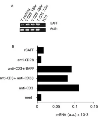
Fig. 3. BAFF expression is up-regulated following TCR±CD3 triggering. Puri®ed T cells were stimulated with coated anti-CD3 with or without co-stimulatory signals (anti-CD28 or BAFF) and total RNA subjected to RT-PCR analysis. (A) Semi-quantitative analysis after 35 cycles of standard RT-PCR. Actin transcription was monitored as a control. Similar results were obtained with T cells puri®ed from a second donor. (B) Total RNA was extracted after 18 h of stimulation and subjected to real-time PCR analysis of BAFF mRNA. Results are expressed in arbitrary units of mRNA. A similar result was obtained with T cells puri®ed from a second donor.
lation site in this domain is not used, consistent with previous observations (1).
BAFF is up-regulated following cellular activation
To further document BAFF expression in leukocytes, we studied the regulation of BAFF expression upon cellular activation. In T cells, semi-quantitative RT-PCR analysis indicated that BAFF mRNA appears up-regulated upon TCR±CD3 stimulation and that this up-regulated mRNA could be detected up to 3 days following stimulation (Fig. 3A). Further analyses with quantitative real-time PCR showed that T cell stimulation by an anti-CD3 antibody induced a 10-fold increase in BAFF mRNA (Fig. 3B). This increase was evident when RNA was extracted 18 h following stimulation, but was not yet observed after 4 h (data not shown). Addition of a co-stimulatory signal with an anti-CD28 antibody or with BAFF itself did not in¯uence the levels of BAFF mRNA (Fig. 3B), even when a suboptimal concentration of anti-CD3 antibody was used (data not shown). Co-stimulation signals in the absence of anti-CD3 did not modulate BAFF mRNA (Fig. 3B). Altogether, these data show that BAFF expression is markedly increased when T cells are stimulated through their TCR in a co-stimulation-independent manner.
In DC, we observed that exposure to the bacterial products, LPS or LTA, for 18 h resulted in a 3-fold up-regulation of BAFF mRNA (Fig. 4). On the contrary, other DC maturating agents belonging to the TNF family, such as TNF or CD40L, failed to up-regulate BAFF mRNA. In this experiment, IFN-g treatment also up-regulated BAFF mRNA in a similar range as previously reported (13). Similar up-regulation was also observed 4 h after LPS stimulation (data not shown). A comparison between the quantitative RT-PCR analyses performed on DC and T cells indicated that resting and activated T cells express only 0.3±1 and 3±10% respectively of the BAFF mRNA found in DC. These experiments show that BAFF is constitutively expressed in DC and that DC stimulation with bacterial products or IFN-g induces an up-regulation of BAFF expression.
Production of endogenous BAFF provides T cell co-stimulation
Since recombinant BAFF provides T cell co-stimulation, we tested whether blockade of endogenous BAFF could modu-late T cell activation. We ®rst performed an anti-CD3-driven T cell proliferation assay with puri®ed T cells. In this experiment, addition of a BAFF antagonist, TACI±Ig, resulted in a 2-fold inhibition of the T cell proliferation (Fig. 5A). In this anti-CD3 stimulation of puri®ed T cells, addition of an anti-CD28 increases the cellular proliferation, indicating the dependence for co-stimulation in this stimulation assay. The presence of contaminating APC that could have provided a source of BAFF is therefore unlikely in this assay. In a second experiment, we used total PBMC wherein circulating APC represents another source of BAFF. When these total PBMC were stimulated with the anti-CD3 antibody, addition of TACI±Ig at 50 mg/ml resulted in an almost complete inhibition of the T cell proliferation for 0.01 and 0.1 mg/ml of anti-CD3 (Fig. 5B). For 1 mg/ml of anti-CD3, TACI±Ig inhibition was bypassed. No inhibition was seen with the control huIgG1. Since huIgG1 may not be the best negative control, we also used in this experiment Fn14±Ig as an irrelevant TNF receptor fused to the constant region of huIgG1. No inhibition was observed in the presence of 50 mg/ml of Fn14±Ig (data not shown). In this assay using unseparated PBMC, the proliferation observed was mediated by T cells and was dependent on co-stimulation provided by APC. Indeed, B cell depletion (CD19+fraction)
had no effect and APC removal (CD14+ or MHC class II+
fraction) resulted in a strong inhibition (data not shown). TACI binds two ligands in the TNF superfamily, BAFF and APRIL (19,20). In addition to BAFF, monocytes and monocyte-derived DC expressed APRIL mRNA (Fig. 5C). However, the pattern of expression is dissimilar in other cell types, since APRIL mRNA is not expressed in T cells, resting or activated (Fig. 5C, higher panel). This APRIL pattern of expression was con®rmed at the protein level. Secreted APRIL was recovered upon immunoprecipitation of 5 ml of supernatant conditioned by monocyte or monocyte-derived DC at 0.2 3 106/ml for 4
days, but was absent from supernatant conditioned for 4 days by B or T cells at 1 3 106/ml (Fig. 5C, lower panel). In this
experiment, endogenous APRIL secreted from monocyte-derived DC resolved as a double band at ~22 kDa. A longer exposure time of the western blot also revealed a double band in the supernatant derived from monocytes (data not shown). The 22-kDa size is consistent with the predicted mol. wt of 16 kDa without glycosylation for secreted APRIL. The apparent double band could be due to different glycosylation patterns or to recovery in the immunoprecipitate of the two different APRIL isoforms a (accession no. NM 03808) and b (accession no. NM 172087) that differ by 16 internal amino acids. It is unlikely that the existence of two bands is due to the presence in the immunoprecipitate of the hybrid TWE-PRIL molecule generated by intergenic splicing between TWEAK and APRIL (21) or to heterodimeric BAFF/APRIL molecules (22), since the double band is also observed with 293T transfected with APRIL in the absence of TWEAK or BAFF (recovered from 1 ml supernatant conditioned for 4 days with transfected 293T cells at 0.1 3 106/ml) (Fig. 5C). Altogether, this analysis
demon-strates that APC express two TACI ligands, BAFF and APRIL.
Fig. 4. BAFF expression is up-regulated upon DC maturation with bacterial products. DC differentiated from monocytes were treated with LPS (20 ng/ml), LTA (10 mg/ml), TNF (10 ng/ml), soluble CD40L (100 ng/ml) or IFN-g (500 UI/ml) for 18 h. Total RNA was extracted and subjected to real-time RT-PCR analysis as in Fig. 3. A similar result was obtained with DC from a second donor.
On the contrary, T cells express only BAFF. In order to identify the TACI ligand produced by APC and involved in T cell co-stimulation, we used BAFF-R±Ig showing a strict speci®city for BAFF in the TNF family (23,24). BAFF-R±Ig blocked the T cell
proliferation obtained (Fig. 5D). The slight difference in the inhibition obtained here with BAFF-R±Ig and TACI±Ig is not conclusive, since we used the murine form of BAFF receptor and the human form of TACI. To formally demonstrate that
Fig. 5. Soluble TACI and BAFFR inhibit anti-CD3-driven T cell proliferation. (A) Puri®ed T cells (1 3 105) were activated with coated anti-CD3
at 1 mg/ml for 72 h. huTACI±Ig and huIgG1 (control Ig) were used at 50 mg/ml. Anti-CD28 was used at 100 ng/ml. The results are expressed as the mean thymidine incorporation from triplicate cultures 6 SD. Background proliferation in the absence of anti-CD3 was 500 6 50 c.p.m. (B) PBMC (1 3 105) were activated with increasing concentrations of coated anti-CD3. The proliferation obtained in the presence of medium
alone, huTACI±Ig (50 mg/ml) or huIgG1 (cIg, 50 mg/ml) is shown. Similar results were observed with PBMC from six independent donors. (C) RNA from the indicated cell subsets were extracted and subjected to RT-PCR (40 cycles) with an APRIL-speci®c primer pair. Upper panel shows APRIL mRNA expression in monocytes, DC derived from monocytes, and absence of expression in B and T cells. Control actin ampli®cation is also shown. Cell supernatants from the indicated cells were immunoprecipitated with BCMA±Ig and APRIL expression was studied in western blot analysis with the aprily-1 mAb. Soluble APRIL recovered from cell supernatants migrated as a double band of 22 kDa. (D) PBMC (1 3 105) were activated with coated anti-CD3 at 0.1 mg/ml. The proliferation obtained in medium alone or in the presence of
increasing concentration of muBAFF-R±Ig, huTACI±Ig or control Ig (huIgG1) is shown. Background proliferation in the absence of anti-CD3 was 5200 6 260 c.p.m. Similar results were obtained with three independent donors. (E) Puri®ed T cells (1 3 105) were activated with coated
anti-CD3 at 0.1 mg/ml in the presence of 1 3 104autologous puri®ed monocytes. TACI±Ig and control Ig (huIgG1) were used at 15 mg/ml.
circulating APC provide BAFF co-stimulation to T cells, we activated puri®ed T cells with coated anti-CD3 in the presence of puri®ed autologous monocytes. The presence of TACI±Ig resulted in an almost complete inhibition of the anti-CD3-driven proliferation, while no effect was seen with control Ig (Fig. 5E). Altogether, this demonstrates that BAFF produced by APC and, to a lesser extent, by T cells themselves provides co-stimulation to anti-CD3 stimulation of T cells.
Discussion
In the present study, we characterized the expression of the TNF ligand BAFF in subsets of human leukocytes and analyzed its T cell co-stimulatory activity. At the RNA level, we found expression of BAFF in APC. Indeed, ex vivo blood DC and DC derived from monocytes or CD34+ stem cells
express abundant levels of BAFF mRNA. We observed also BAFF expression in cells that are known to give rise to DC, CD34+ stem cells expanded in vitro with a hematopoietin
cocktail (25,26) and peripheral blood circulating monocytes (27,28). This indicates that BAFF expression is already initiated at the DC precursor stage. A low level of BAFF expression, up-regulated ~10-fold upon TCR±CD3 stimula-tion, was observed in T cells. The up-regulation of BAFF mRNA in T cells upon TCR±CD3 stimulation is consistent with the up-regulation previously observed with PHA stimulation (1). In DC, BAFF mRNA is highly expressed compared to T cells (100- to 300-fold more than in resting T cells), and BAFF expression is further up-regulated upon maturation with bacterial products such as LPS and LTA. Our results are in agreement with the expression of BAFF described in myeloid cells (1,2,13,14). On the contrary, BAFF expression in T cells is controversial to date (1,2,13). In this study, we detected BAFF message in T cell RNA with two independent sets of primers and BAFF protein in T cell lysates. The failure to detect BAFF gene expression in T cells by others is likely due to the lower expression level in these cells compared to myeloid cells.
Overexpression experiments have demonstrated that cleav-age of BAFF occurs at its furin recognition site (1,3). Our results strongly suggests that this site is also used in primary cells naturally expressing BAFF, such as DC. This is in agreement with the processing described at this furin site for transformed myeloid cells (13) and consistent with detection of BAFF in the sera of patients with autoimmune diseases (29,30). We did not detect BAFF secretion in T cells. This is likely due to the lower expression level of this molecule rather than an absence of BAFF cleavage in T cells. Indeed, biochemical studies with a furin synthetic substrate as described by Leitlein et al. (31) indicate the presence of functional furin(s) in cellular lysates from activated T cells (data not shown). These studies con®rm that DC produced and released BAFF protein.
We previously demonstrated that recombinant BAFF co-stimulates T cells (7). It was important to test whether endogenous expression of BAFF triggers this biological effect. In an anti-CD3 T cell stimulation assay using puri®ed T cells or unseparated PBMC, the BAFF antagonist TACI±Ig signi®cantly blocked T cell activation, especially when unseparated PBMC where used. This result is concordant with the inhibition obtained with TACI±Ig by Wang et al. in a similar assay with
murine splenocytes cells (11). Since we did not observe any detectable APRIL (the second TNF ligand binding to TACI) expression in T cells, this indicates that endogenous BAFF co-stimulates T cells in an autocrine fashion. Contrary to T cells, we observed that APRIL is co-expressed with BAFF in APC, consistent with previous reports (21,32,33). The inhibition observed in our experiments with soluble BAFF-R identi®es BAFF as the TACI ligand produced by APC and providing T cell co-stimulation. The almost complete inhibition obtained with BAFF-R±Ig in this experiment indicates that BAFF is mandatory for this co-stimulation pathway. We do not ®nd any evidence indicating a role for APRIL in this T cell activation assay.
To date, three receptors have been described for BAFF: BAFF receptor (or BR3) (23,24), BCMA and TACI (19,20,34). BAFF receptor and TACI have been previously reported expressed in T cells (23,35). The recently described pro-apoptotic signaling property described for TACI in B cells (36) is an argument against a T cell co-stimulatory activity for this receptor. BAFF receptor may therefore be the receptor involved in the T cell co-stimulation pathway described here. Additional experiments are needed to clarify this issue. The results presented here indicate that BAFF, in addition to its B cell stimulatory activity, also provides T cell co-stimulation. Its high expression in APC that can be up-regulated upon maturation indicates that BAFF may act preferentially when professional APC present an antigen to T cells. Its lower expression in T cells indicates that BAFF may also act in an autocrine fashion.
Acknowledgements
This work was supported by grants of the Swiss National Science Foundation, the Ligue Genevoise contre le Cancer, the Foundation pour la lutte contre le Cancer et pour des Recherches Medico-Biologiques and the Foundation Leenards. The authors thank B. Chappuis for access to real-time PCR technology. The valuable technical assistance of K. Grosdemange is greatly acknowledged
Abbreviations
BAFF B cell-activating factor from the TNF family BCMA B cell maturation antigen
GM-CSF granulocyte macrophage colony stimulating factor LTA lipoteichoic acid
LPS lipopolysaccharide
PBMC peripheral blood mononuclear cell PHA phytohemagglutinin
TACI transmembrane activator and CAML interactor TNF tumor necrosis factor
References
1 Schneider, P., MacKay, F., Steiner, V., Hofmann, K., Bodmer, J. L., Holler, N., Ambrose, C., Lawton, P., Bixler, S., Acha-Orbea, H., Valmori, D., Romero, P., Werner-Favre, C., Zubler, R. H., Browning, J. L. and Tschopp, J. 1999. BAFF, a novel ligand of the tumor necrosis factor family, stimulates B cell growth. J. Exp. Med. 189:1747.
2 Shu, H. B., Hu, W. H. and Johnson, H. 1999. TALL-1 is a novel member of the TNF family that is down-regulated by mitogens. J. Leukoc. Biol. 65:680.
3 Moore, P. A., Belvedere, O., Orr, A., Pieri, K., LaFleur, D. W., Feng, P., Soppet, D., Charters, M., Gentz, R., Parmelee, D., Li, Y.,
Galperina, O., Giri, J., Roschke, V., Nardelli, B., Carrell, J., Sosnovtseva, S., Green®eld, W., Ruben, S. M., Olsen, H. S., Fikes, J. and Hilbert, D. M. 1999. BLyS: member of the tumor necrosis factor family and B lymphocyte stimulator. Science 285:260.
4 Batten, M., Groom, J., Cachero, T. G., Qian, F., Schneider, P., Tschopp, J., Browning, J. L. and Mackay, F. 2000. BAFF mediates survival of peripheral immature B lymphocytes. J. Exp. Med. 192:1453.
5 Rolink, A. G., Tschopp, J., Schneider, P. and Melchers, F. 2002. BAFF is a survival and maturation factor for mouse B cells. Eur. J. Immunol. 32:2004.
6 Do, R. K., Hatada, E., Lee, H., Tourigny, M. R., Hilbert, D. and Chen-Kiang, S. 2000. Attenuation of apoptosis underlies B lymphocyte stimulator enhancement of humoral immune response. J. Exp. Med. 192:953.
7 Huard, B., Schneider, P., Mauri, D., Tschopp, J. and French, L. E. 2001. T cell co-stimulation by the TNF ligand BAFF. J. Immunol. 167:6225.
8 Yan, M., Marsters, S. A., Grewal, I. S., Wang, H., Ashkenazi, A. and Dixit, V. M. 2000. Identi®cation of a receptor for BLyS demonstrates a crucial role in humoral immunity. Nat. Immunol. 1:37.
9 Gross, J. A., Dillon, S. R., Mudri, S., Johnston, J., Littau, A., Roque, R., Rixon, M., Schou, O., Foley, K. P., Haugen, H., McMillen, S., Waggie, K., Schreckhise, R. W., Shoemaker, K., Vu, T., Moore, M., Grossman, A. and Clegg, C. H. 2001. TACI±Ig neutralizes molecules critical for B cell development and autoimmune disease. impaired B cell maturation in mice lacking BLyS. Immunity 15:289.
10 Gross, J. A., Johnston, J., Mudri, S., Enselman, R., Dillon, S. R., Madden, K., Xu, W., Parrish-Novak, J., Foster, D., Lofton-Day, C., Moore, M., Littau, A., Grossman, A., Haugen, H., Foley, K., Blumberg, H., Harrison, K., Kindsvogel, W. and Clegg, C. H. 2000. TACI and BCMA are receptors for a TNF homologue implicated in B-cell autoimmune disease. Nature 404:995.
11 Wang, H., Marsters, S. A., Baker, T., Chan, B., Lee, W. P., Fu, L., Tumas, D., Yan, M., Dixit, V. M., Ashkenazi, A. and Grewal, I. S. 2001. TACI±ligand interactions are required for T cell activation and collagen-induced arthritis in mice. Nat. Immunol. 2:632. 12 Mukhopadhyay, A., Ni, J., Zhai, Y., Yu, G. L. and Aggarwal, B. B.
1999. Identi®cation and characterization of a novel cytokine, THANK, a TNF homologue that activates apoptosis, nuclear factor-kappaB, and c-Jun NH2-terminal kinase. J. Biol. Chem.
274:15978.
13 Nardelli, B., Belvedere, O., Roschke, V., Moore, P. A., Olsen, H. S., Migone, T. S., Sosnovtseva, S., Carrell, J. A., Feng, P., Giri, J. G. and Hilbert, D. M. 2001. Synthesis and release of B-lymphocyte stimulator from myeloid cells. Blood 97:198.
14 Craxton, A., Magaletti, D., Ryan, E. J. and Clark, E. A. 2003. Macrophage- and dendritic cell-dependent regulation of human B-cell proliferation requires the TNF family ligand BAFF. Blood 101:4464.
15 Scapini, P., Nardelli, B., Nadali, G., Calzetti, F., Pizzolo, G., Montecucco, C., Cassatella, M. A. and Blystad, A. K. 2003. G-CSF-stimulated neutrophils are a prominent source of functional BLyS CD34+cell enrichment depletes atypical CD30+cells from
PBPC grafts in patients with HD. J. Exp. Med. 197:297.
16 Arrighi, J. F., Hauser, C., Chapuis, B., Zubler, R. H. and Kindler, V. 1999. Long-term culture of human CD34+progenitors with
FLT3-ligand, thrombopoietin, and stem cell factor induces extensive ampli®cation of a CD34±CD14±and a CD34±CD14+dendritic cell
precursor. Blood 93:2244.
17 O`Doherty, U., Steinman, R. M., Peng, M., Cameron, P. U., Gezelter, S., Kopeloff, I., Swiggard, W. J., Pope, M. and Bhardwaj, N. 1993. Dendritic cells freshly isolated from human blood express CD4 and mature into typical immuno-stimulatory dendritic cells after culture in monocyte-conditioned medium. J. Exp. Med. 178:1067.
18 Holler, N., Tardivel, A., Kovacsovics-Bankowski, M., Hertig, S., Gaide, O., Martinon, F., Tinel, A., Deperthes, D., Calderara, S., Schulthess, T., Engel, J., Schneider, P. and Tschopp, J. 2003. Two adjacent trimeric Fas ligands are required for Fas signaling
and formation of a death-inducing signaling complex. Mol. Cell. Biol. 23:1428.
19 Yu, G., Boone, T., Delaney, J., Hawkins, N., Kelley, M., Ramakrishnan, M., McCabe, S., Qiu, W. R., Kornuc, M., Xia, X. Z., Guo, J., Stolina, M., Boyle, W. J., Sarosi, I., Hsu, H., Senaldi, G. and Theill, L. E. 2000. APRIL and TALL-I and receptors BCMA and TACI: system for regulating humoral immunity. Nat. Immunol. 1:252.
20 Marsters, S. A., Yan, M., Pitti, R. M., Haas, P. E., Dixit, V. M. and Ashkenazi, A. 2000. Interaction of the TNF homologues BLyS and APRIL with the TNF receptor homologues BCMA and TACI. Curr. Biol. 10:785.
21 Pradet-Balade, B., Medema, J. P., Lopez-Fraga, M., Lozano, J. C., Kolfschoten, G. M., Picard, A., Martinez, A. C., Garcia-Sanz, J. A. and Hahne, M. 2002. An endogenous hybrid mRNA encodes TWE-PRIL, a functional cell surface TWEAK±APRIL fusion protein. EMBO J. 21:5711.
22 Roschke, V., Sosnovtseva, S., Ward, C. D., Hong, J. S., Smith, R., Albert, V., Stohl, W., Baker, K. P., Ullrich, S., Nardelli, B., Hilbert, D. M. and Migone, T. S. 2002. BLyS and APRIL form biologically active heterotrimers that are expressed in patients with systemic immune-based rheumatic diseases. J. Immunol. 169:4314. 23 Yan, M., Brady, J. R., Chan, B., Lee, W. P., Hsu, B., Harless, S.,
Cancro, M., Grewal, I. S. and Dixit, V. M. 2001. Identi®cation of a novel receptor for B lymphocyte stimulator that is mutated in a mouse strain with severe B cell de®ciency. Curr. Biol. 11:1547. 24 Thompson, J. S., Bixler, S. A., Qian, F., Vora, K., Scott, M. L.,
Cachero, T. G., Hession, C., Schneider, P., Sizing, I. D., Mullen, C., Strauch, K., Zafari, M., Benjamin, C. D., Tschopp, J., Browning, J. L. and Ambrose, C. 2001. BAFF-R, a newly identi®ed TNF receptor that speci®cally interacts with BAFF. Science 293:2108. 25 Caux, C., Vanbervliet, B., Massacrier, C., Dezutter-Dambuyant, C., de Saint-Vis, B., Jacquet, C., Yoneda, K., Imamura, S., Schmitt, D. and Banchereau, J. 1996. CD34+hematopoietic progenitors from
human cord blood differentiate along two independent dendritic cell pathways in response to GM-CSF + TNF alpha. J. Exp. Med. 184:695.
26 Salmon, P., Arrighi, J. F., Piguet, V., Chapuis, B., Zubler, R. H., Trono, D. and Kindler, V. 2001. Transduction of CD34+cells with
lentiviral vectors enables the production of large quantities of transgene-expressing immature and mature dendritic cells. J. Gene Med. 3:311.
27 Larregina, A. T., Morelli, A. E., Spencer, L. A., Logar, A. J., Watkins, S. C., Thomson, A. W. and Falo, L. D., Jr. 2001. Dermal-resident CD14+ cells differentiate into Langerhans cells. Nat.
Immunol. 2:1151.
28 Randolph, G. J., Beaulieu, S., Lebecque, S., Steinman, R. M. and Muller, W. A. 1998. Differentiation of monocytes into dendritic cells in a model of transendothelial traf®cking. Science 282:480. 29 Groom, J., Kalled, S. L., Cutler, A. H., Olson, C., Woodcock, S. A.,
Schneider, P., Tschopp, J., Cachero, T. G., Batten, M., Wheway, J., Mauri, D., Cavill, D., Gordon, T. P., Mackay, C. R. and Mackay, F. 2002. Association of BAFF/BLyS overexpression and altered B cell differentiation with SjoÈgren's syndrome. J. Clin. Invest. 109:59.
30 Cheema, G. S., Roschke, V., Hilbert, D. M. and Stohl, W. 2001. Elevated serum B lymphocyte stimulator levels in patients with systemic immune-based rheumatic diseases. Arthritis Rheum. 44:1313.
31 Leitlein, J., Aulwurm, S., Waltereit, R., Naumann, U., Wagenknecht, B., Garten, W., Weller, M. and Platten, M. 2001. Processing of immunosuppressive pro-TGF-beta 1,2 by human glioblastoma cells involves cytoplasmic and secreted furin-like proteases. J. Immunol. 166:7238.
32 Novak, A. J., Bram, R. J., Kay, N. E. and Jelinek, D. F. 2002. Aberrant expression of B-lymphocyte stimulator by B chronic lymphocytic leukemia cells: a mechanism for survival. Blood 100:2973.
33 Litinskiy, M. B., Nardelli, B., Hilbert, D. M., He, B., Schaffer, A., Casali, P. and Cerutti A. 2002. DC induce CD40-independent immunoglobulin class switching through BLyS and APRIL. Nat. Immunol. 3: 822.
Trabach, L., Hertig, S., Holler, N., Qian, F., Mullen, C., Strauch, K., Browning, J. L., Ambrose, C. and Tschopp, J. 2000. A soluble form of B cell maturation antigen, a receptor for the tumor necrosis factor family member APRIL, inhibits tumor cell growth. J. Exp. Med. 192:1677.
35 von Bulow, G. U. and Bram, R. J. 1997. NF-AT activation induced
by a CAML-interacting member of the tumor necrosis factor receptor superfamily. Science 278:138.
36 Seshasayee, D., Valdez, P., Yan, M., Dixit, V. M., Tumas, D. and Grewal, I. S. 2003. Loss of TACI causes fatal lymphoproliferation and autoimmunity, establishing TACI as an inhibitory BLyS receptor. Immunity 18:279.