Surgery for acute type a aortic dissection: comparison of techniques
q
Urs NiederhaÈuser
a,*, Hannes RuÈdiger
a, Andreas KuÈnzli
a, Burkhardt Seifert
b, JuÈrg Schmidli
a,
Paul Vogt
a, Marko Turina
aaClinic for Cardiovascular Surgery, University Hospital Zurich and City Hospital Triemli, Zurich, Switzerland bInstitute for Biostatistics, University Hospital Zurich and City Hospital Triemli, Zurich, Switzerland
Received 7 September 1999; received in revised form 7 March 2000; accepted 6 June 2000
Abstract
Objective: In order to determine the optimal surgical strategy for acute ascending aortic dissection, the graft inclusion technique was compared with the open resection technique. Methods: Between 1985 and 1995 a consecutive series of 193 patients (77% male, mean age 58 years) had emergency surgery during a mean interval of 13.2 h after onset of symptoms. Graft replacement of the ascending aorta was performed in all patients (supracoronary graft 143=193 74%, aortic root replacement 50=193 26%, aortic valve replacement 73=193 38%, arch replacement 44=193 20%) The open resection technique was applied in 93 patients and the inclusion technique in 100 patients with a Cabrol-shunt in 26%. Preoperative risk factors were equally distributed between groups (inclusion technique vs. open technique): left ventricular ejection fraction , 45% (13 vs. 2%, not signi®cant (n.s.)), neurological de®cit (31 vs. 25%; n.s.), systolic blood pressure , 90 mmHg (20 vs. 15%, n.s.) pericardial tamponade (25 vs. 9%, n.s.), renal failure (6 vs. 4%; n.s.). Results: The overall early mortality was 24%. Following graft inclusion it was 31% compared with 16% in the open technique group (P 0:0154). Postoperative complications (graft inclusion vs. open technique): myocardial infarction (9 vs. 12%, n.s.), low cardiac output (40 vs. 32%, n.s.), reexplora-tion for hemorrhage (23 vs. 25%, n.s.). Survival at 8 years was signi®cantly increased in the open technique group (P 0:0300). Pseudoa-neurysm formation occurred in 3% of patients and only after graft inclusion. Freedom from reoperation was 80% at 8 years and did not differ between groups. Graft inclusion was an independent signi®cant predictor of early (P 0:0069; relative risk 2:3673) and late mortality (P 0:0119; relative risk 2:0981). Conclusions: Surgery of acute ascending aortic dissection still carries a considerable early mortality whereas the late outcome is satisfactory. The open resection technique is the method of choice showing superior early and late results and avoiding pseudoaneurysm formation. The inclusion technique may be indicated in situations with increased risk of bleeding. A consequent decompression of the perigraft-space could reduce the rate of pseudoaneurysms. q 2000 Elsevier Science B.V. All rights reserved.
Keywords: Aortic dissection; Ascending aorta; Graft replacement; Surgical technique
1. Introduction
Acute ascending aortic dissection carries a high early mortality mainly caused by proximal aortic rupture with fatal pericardial tamponade. As treatment modality of choice undelayed surgery has evolved [1±5]. In the acute phase of the disease the aortic tissue, however, is friable because of the dissection process and the underlying aortic pathology making surgery technically dif®cult. Organ ische-mia caused by malperfusion of dissected aortic branches and
coagulation disorders add to the high early risk. The graft inclusion technique, published by Bentall and De Bono [6] for aortic root replacement, was used at a time when bleed-ing was a major problem. With the development of new surgical materials and techniques (perfusion techniques, organ protection, tissue glue, zero-porosity grafts) an open graft-implantation technique [7] could be adopted. It allowed the resection of pathologic aorta and a more anatomic graft interposition.
It was the aim of this study to determine the early and late results of surgery in acute ascending aortic dissection. Special interest was focused on the comparison of graft inclusion with the open resection technique.
2. Material and methods
Between 1985 and 1995 emergency surgery for acute type
1010-7940/00/$ - see front matter q 2000 Elsevier Science B.V. All rights reserved. PII: S1010-7940(00)00513-3
www.elsevier.com/locate/ejcts
qPresented at the 13th Annual Meeting of the European Association for
Cardio-thoracic Surgery, Glasgow, Scotland, UK, September 5±8, 1999. * Corresponding author. Clinic for Cardiovascular Surgery, Hospital Beau-Site, SchaÈnzlihalde 11, CH-3000 Bern 25, Switzerland. Tel.: 141-31-318-7766 (of®ce); 141-31-335-3333 (hospital); fax: 141-31-318-7768.
E-mail address: [email protected] (U. NiederhaÈuser).
A aortic dissection (Stanford classi®cation [8]) was performed in 193 patients at the University Hospital and the City Hospital Triemli, Zurich, Switzerland. In all patients the operation was performed during the ®rst 24 h after onset of dissection symp-toms with a mean interval of 13 ^ 7 h. Clinical data were obtained by retrospective review of hospital records. Post-operative follow-up data contain periodical cardiological reports and questionnaires. The mean follow-up in early survi-vors was 51.0 ^ 32.0 months.
Demographic, preoperative and intraoperative data are listed in Table 1.
Previous cardiac surgery was performed in 15 of 193 patients (7.7%): aortic valve replacement (AVR) in eight patients (3.9%); coronary artery bypass graft (CABG) in four patients (1.9%); atrial septal defect, arch replacement and mitral valve replacement in one patient each (0.5%). 2.1. Operation
A standard median sternotomy was performed and total cardiopulmonary bypass was instituted by cannulation of the femoral artery and the right atrium. The application of cold blood cardioplegia with high potassium content was antegrade in 106 patients and retrograde in 87 patients using a transatrial cannulation of the sinus venosus.
Graft inclusion was performed according to the technique described by Bentall [6] for aortic root replacement. The ascending aorta was not resected and was incised longitud-inally. The tubular graft was anastomosed inside and into
the true lumen of the aorta. In cases of severe destruction the dissected intimal layer of the aortic wall was `endarterecto-mized' and inclusion was performed only with the outer layer of the false channel. For aortic root replacement the coronary ostia were anastomosed to the graft in a side-to-side fashion [6]. Finally, wrapping of the graft was performed using the remnants of the aortic wall. In 26 patients of the inclusion group (26%) the perigraft space was decompressed using a Cabrol shunt [9] to the right atrium.
With the open technique the replaced and diseased aortic segment was resected and a graft was interposed using an end-to-end anastomosis. Gelatine±resorcinol±glutaralde-hyde/formaldehyde glue (Trigon GmbH, MoÈnchenglad-bach, Germany) [10] was used to seal the dissected aortic wall. With this technique aortic root reconstruction and valve competence could be achieved [11] and the aortic wall could be reinforced at the level of the graft anasto-moses.
During the ®rst half of the study period (1985±1990), 50 patients were operated on using the inclusion technique and 20 patients using the open technique (P 0:00004).
In the inclusion group mean extracorporeal circulation (ECC) time was 137.5 ^ 72.2 min, aortic cross-clamp time was 70.5 ^ 8.5 min and circulatory arrest time was 17.7 ^ 8.7 min. In the open group the corresponding times were 131.5 ^ 71.1 min (P 0:507), 71.6 ^ 30.7 min (P 0:868) and 18.3 ^ 9.3 min (P 0:003). Hypothermic circulatory arrest was used for interventions
Table 1
Demographic, preoperative and intraoperative dataa
Parameters Inclusion technique % Open technique % P
No. of patients 100 48.3 93 44.9 0.554
Age (years) 58 ^ 11 57 ^ 11 0.155
Male gender 72 72.0 76 81.7 0.111
Pericardial tamponade 25 25.0 8 8.8 0.003
Renal failure 6 6.0 4 4.3 0.595
Temporary neurologic de®cit 28 28.0 21 22.6 0.387
Persisting neurologic de®cit 3 3.0 2 2.2 0.711
AI severe 13/76 pat. 17.1 7/74 patients 9.4 0.169
LVEF ,30% 4/53 pat. 7.5 1/58 patients 1.7 0.140
Previous cardiac surgery 5 5 9 9.7 0.2107
Pulse de®cit at one localization 19 19 16 17.2 0.7463
Dissected
Asc. aorta 19 19.0 16 17.2 0.746
Asc. aorta 1 arch 15 15.0 17 18.3 0.540
Asc./desc. aorta 1 arch 66 66.0 59 63.4 0.710
Coronary artery 13 13.0 11 11.8 0.805
Site of intimal tear
Asc. aorta 73 73 77 82.8 0.1022
Arcus aortae 5 5 6 6.5 0.6638
Distal aorta 9 9 9 9.7 0.8715
Retrograde cardioplegia 31 31 59 63.4 ,0.001
Use of tissue glue 22 22 41 44.1 0.001
Use of aprotinine 41 41 71 77.2 ,0.001
a AI, aortic insuf®ciency; asc., ascending; desc., descending; LVEF, left ventricular ejection fraction. Echocardiographic data on left ventricular ejection
at the level of the aortic arch and for construction of an open distal anastomosis. It was applied in 44 patients of the inclu-sion group (44%) and in 63 patients (68%) of the open group (P 0:0009). During rewarming antegrade reperfusion was instituted if there was dissected aorta distal to the graft.
A segment of the ascending aorta was replaced in all patients with a tubular Dacron graft. A supracoronary graft was implanted in 143 of 193 patients (74%). In the inclusion group 66 patients (66%) had supracoronary graft implantation compared with 77 patients (83%) in the open group (P 0:00779). Aortic root replacement with a composite graft was performed in 50 of 193 patients, aortic valve replacement in 73 of 193 patients (38%) and arch replacement in 44 of 193 patients (20%) with a hemiarch procedure in 40 patients.
A simultaneous coronary revascularization procedure was performed in 10 patients (10%) with graft inclusion and in 21 patients (23%) with open graft implantation (P 0:017).
2.2. Statistical analyses
The Statistica software package (Stat Soft, Inc., 1993) and SPSS (SPSS Inc., Chicago, IL) were used for statistical analysis. Continuous variables were summarized as mean ^ standard deviation. Survival and event-free probabilities ^ standard error were calculated by actuarial analyses [12]. Differences between survival curves were estimated using the log rank test. Predictors for mortality and reoperation were determined by univariate and multivariate analysis. In univariate analysis discrete variables were analyzed by the chi-squared or Fisher's exact test. Continuous variables were analyzed by the Mann±Whitney test. Statistical signif-icance was associated with a P level of less than 0.05. Selected variables were entered into multivariate analysis by a stepwise logistic regression or by Cox proportional hazard regression to determine independent predictors. 3. Results
Early mortality in all 193 patients was 23.8% (46/193). Following graft inclusion early mortality was 31.0% (31/ 100) compared with 16.1% (15/93) after the open technique (P 0:0154). During the ®rst half of the study period (1985±1990), early mortality was 24.4 vs. 23.4% in the second half (P 0:8644). In the open technique group early mortality was lower during the ®rst half (10.7 vs. 32%, P 0:03985) and during the second half (18.5 vs. 30.9%, P 0:13596) of the study period. Univariate signif-icant predictors for early death were: increasing age (P 0:0032), pericardial tamponade (P 0:0001), preo-perative (P 0:0525) and postopreo-perative (P 0:0001) neurologic de®cit, inclusion technique (P 0:0154), dura-tion of ECC (P , 0:0001) and perioperative myocardial infarction. Independent signi®cant risk factors for early mortality were: the inclusion technique (P 0:0069,
rela-tive risk 2.3673) and postoperarela-tive neurologic de®cit (P 0:0001, relative risk 14.3525).
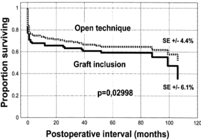
Following open graft implantation, survival was 77.3 ^ 3.0% after 30 days, 72.5, ^ 3.2% after 1 year, 64.5 ^ 3.7% after 5 years and 61.8 ^ 4.4% after 8 years. For graft inclusion the corresponding ®gures were 70.8% ^ 4.6% after 1 month, 65.6% ^ 4.8% after 1 year, 59.1 ^ 5.1% after 5 years and 55.1 ^ 6.1% after 8 years (P 0:02998) (Fig. 1). Complications in the late follow-up did not differ signi®cantly between grofollow-ups and are depicted in Table 2. Independent signi®cant risk factors for late mortality were: age, (P 0:0010, relative risk 1.0465); dissection of the coronary ostia (P 0:0615, rela-tive risk 2.1672); previous cardiac surgery (P 0:0517, relative risk 2.3141) and inclusion technique (P 0:0119, relative risk 2.0981).
During the follow-up period a total of 21 patients had to be reoperated after a mean interval of 26.2 months. In 13 of 21 patients the reintervention took place at the level of the proximal aorta. The following reoperations were performed (inclusion vs. open technique). Aortic valve replacement was done in three patients for an insuf®cient and primarily reconstructed valve (1.0 vs. 2.2% (P 0:9495)). For the same indication with additional aneurysm of the ascending aorta, valve replacement and supracoronary graft implanta-tion was performed in four patients (1.5 vs. 1.6% (P 0:5095)) and composite graft replacement of the aortic root in six patients (2.0 vs. 4.3% (P 0:6133)). Graft repla-cement of the thoracic and abdominal aorta for aneurys-matic dilatation was necessary in ®ve patients (3.0 vs. 2.2% (P 0:9345)). For ischemic complications vascular surgery on aortic branches had to be performed in three patients (3.0 vs. 0% (P 0:2708)).
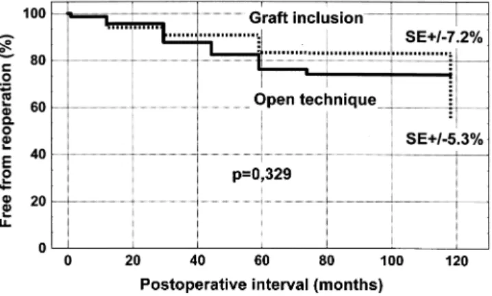
In three patients reoperation was indicated by a pseudoa-neurysm at the anastomotic suture lines. A pseudoapseudoa-neurysm only occurred after graft inclusion and never in the open technique group. In the open technique group, freedom from reoperation (including only early survivors) was 98.6 ^ 1.4% after 30 days, 95.6 ^ 2.0% after 1 year and
Fig. 1. Actuarial survival comparing the open resection technique with graft inclusion.
76.1 ^ 7.2% after 5 years. In the graft inclusion group the corresponding ®gures were 98.6 ^ 1.4% after 30 days, 94.0 ^ 2.9% after 1 year and 83.3 ^ 5.3% after 5 years (P 0:3293). (Fig. 2).
Late complications are listed in Table 2. At the end of the follow-up the mean functional NYHA (New York Heart Association) class was 1.73 ^ 0.72 after graft inclusion and 1.95 ^ 0.82 in the open technique group (P 0:1501). 4. Discussion
In acute dissection of the ascending aorta, undelayed surgery is the treatment method of choice. In the acute phase of the disease the surgical intervention is technically dif®cult and demanding. Despite diagnostic and therapeutic improvements over the last decades mortality remained considerably high and was 22.6% in our present series. Similar mortality rates between 21 and 26% are documented by other authors [13±16]. In our experience mortality did not change substantially over the years and Fann from the Stanford group [15] even noted a slight increase in mortality
over the last decade. We can only speculate about the reason for this tendency and we think that faster and accurate echo-cardiographic diagnosis, prompt patient referral and more ef®cient preoperative treatment has allowed more high-risk patients to come to operation.
The inclusion technique was described by Bentall and de Bono [6] for aortic root replacement with a composite graft. It was introduced at a time when bleeding problems and leaking graft material was a major surgical concern. Complete graft inclusion with the remaining aorta was a successful method to control bleeding. With the accumula-tion of blood in the perigraft space, however, anastomotic suture lines could get under tension. This is thought to be a possible mechanism for the development of pseudoaneur-ysms at these localizations. With the development of new techniques and materials (zero porosity grafts, tissue glue, te¯on felt as buttress for anastomotic sutures, aprotinin) an open technique for graft implantation was possible with resection of the replaced, pathologic aortic segment. This technique allowed a better exposition and visibility of perti-nent anatomic structures, a more anatomic reconstruction and improved surgical access for the control of hemostasis.
Table 2
Complications during the early and late postoperative period
Complications Inclusion technique % Open technique % P
Early
Perioperative myocardial infarction 9 9 11 11.8 0.520
Low cardiac output 40 40 29 31.5 0.202
Re-exploration (hemorrhage) 23 23 23 25.0 0.746 Neurology transient 8 8 13 14.0 0.165 Neurology permanent 10 10 19 20.4 0.036 Renal failure 8 8 8 8.8 0.880 Intestinal ischemia 2 2 1 1.1 0.527 Late
Myocardial infarction 1/52 patients 2 1/57 patients 2 0.5164
Hemorrhage 0 0
Embolism 3/52 patients 6 1/57 patients 2 0.5462
Endocarditis 0 0
Stroke 1/52 patients 2 7/57 patients 12 0.5164
In the present study the open technique was applied in 93 patients and the inclusion technique in 100 patients. Graft inclusion had a signi®cantly increased (P 0:01549) early mortality of 31% compared with a 16.1% mortality of the open technique. This is also true for the early half of the study period whereas during the late period this difference was no longer signi®cant. Graft inclusion was, moreover, an independent signi®cant risk factor for early and late death with a more than twofold risk. Survival after open graft implantation was also signi®cantly better (P 0:02998). Due to the retrospective analysis of our data the two treat-ment groups were not comparable in all relevant preopera-tive ®ndings and examinations. Cardiac tamponade, a univariate risk factor for early mortality, was signi®cantly more frequent in the open technique group. Despite this negative ®nding early survival remained signi®cantly increased in this subgroup of patients. On the contrary, left ventricular dysfunction and severe valve regurgitation were more frequent in the inclusion technique group. Despite lacking statistical signi®cance they may have nega-tively in¯uenced the early results of this treatment group. In a published series of 348 patients with composite graft replacement of the aortic root, Svensson [17] also compared both implantation techniques. Acute aortic dissection was only present in 34 patients (9.8%) of his series, which showed equal early mortality rates of 9% but improved survival after 3 years for the open technique. The survival difference was not statistically signi®cant (81 vs. 79%, P 0:280).
In our series the rate of reexploration for hemorrhage was 24% and almost similar in both groups. Kouchoukos [7] has published a series of 168 patients with composite graft replacement of the aortic root. Acute aortic dissection was present only in 17 patients of this study. The open resection technique had a signi®cantly lower rate of rethoracotomies (2 vs. 13.3%, P 0:024). The rates of perioperative myocardial infarction and of low cardiac output were simi-lar in both technical groups, a ®nding published also by other authors [17]. Persistent postoperative neurologic de®-cits were signi®cantly increased in our open group (20 vs. 10%, P 0:036). In a single patient of this group the neuro-logical complication could be directly related to the open technique because of documented embolization of polymer-ized glue into a cerebral vessel [18]. This complication should be avoidable by surgical technical measures [19]. Transient neurologic de®cits showed similar frequencies in both groups. In the report of Svensson [17] neither tech-nique of graft implantation was in univariate or multivariate analysis a risk factor for postoperative neurologic disorders. In contrast, the Cabrol method [9] of coronary ostial implan-tation was followed by signi®cantly less neurologic compli-cations. This study contained only a small number of acute dissections (9.8%), which were not analyzed separately. Therefore the total risk for neurologic complications was substantially decreased.
During the follow-up period the rates of
thromboembo-lism, endocarditis, hemorrhage, angina pectoris and myocardial infarction were similar in both groups. The stroke rate was signi®cantly increased in the open technique group. At the end of the follow-up there was no signi®cant difference in the mean functional class (dyspnoe NYHA class).
In contrast to survival, the rate of late reoperations was not signi®cantly different in our two groups. Pseudoaneur-ysms occurred in only three patients with graft inclusion and were completely avoided in the group with the open resec-tion technique. In the study popularesec-tion of Kouchoukos [7] reoperation-free survival was signi®cantly better with the open technique of graft implantation. Following this tech-nique the rate of pseudoaneurysms was 2% compared with 8.6% after graft inclusion (P 0:049). This author there-fore advocates the open technique in accordance with Svensson [17] who also documented a signi®cantly decreased rate of reoperations following open graft implan-tation. The small number of pseudoaneurysms in our inclu-sion group may be explained by the fact that in case of excessive bleeding we consequently perform a Cabrol shunt [9] to the right atrium, which was the case in 26% of patients with graft inclusion. This shunt decompresses the perigraft space in order to avoid external graft compression or tension on anastomotic suture lines. In the series of Kouchoukos [7] such a decompression of the perigraft space was not performed in any patient with graft inclusion and in the series of Svensson [17] it was performed in only 5% of patients. Because of our superior early and late results we consider the open resection technique as the method of choice. In case of expected excessive bleeding (coagulopa-thy, reoperation), graft inclusion may be indicated. Decom-pression of the perigraft space must be performed with a liberal indication and consequent use of the Cabrol shunt technique.
Certain limitations of the present study have to be mentioned. The choice of technique for graft implantation (open/inclusion) was performed in a non-randomized fash-ion and was at the surgeon's discretfash-ion. In additfash-ion, materi-als and methods have changed considerably during the study period (see Table 1). Together with the retrospective evalua-tion of the clinical data, these factors are responsible for limitations. A randomized and prospective trial could provide more statistical and predictive power.
We conclude that surgery for acute ascending aortic dissection still has a considerable early mortality and morbidity whereas the late outcome is satisfactory. The open resection technique is the method of choice for graft insertion and showed superior early and late results. Open graft implantation allows a more anatomic reconstruction and has not negatively in¯uenced hemostasis. In the long-term follow-up pseudoaneurysms could be completely avoided. The inclusion technique may be indicated in cases of excessive bleeding (coagulopathy, reoperation). With a liberal use of the Cabrol shunt technique the rate of pseudoaneurysms could be markedly reduced.
References
[1] DeBakey ME, McCollum CH, Crawford ES, Morris GC, Howell J, Noon GP, Lawrie G. Dissection and dissecting aneurysms of the aorta: twenty-year follow-up of ®ve hundred twenty-seven patients treated surgically. Surgery 1982;92:1118±1134.
[2] Crawford ES, Svensson LG, Coselli JS, Sa® HJ, Hess KR. Aortic dissec-tion and dissecting aortic aneurysms. Ann Surg 1988;208:254±273. [3] Crawford ES, Svensson LG, Coselli JS, Sa® HJ, Hess KR. Surgical
treatment of aneurysm and/or dissection of the ascending aorta, trans-verse aortic arch, and ascending aorta and transtrans-verse aortic arch. Factors in¯uencing survival in 717 patients. J Thorac Cardiovasc Surg 1989;98:659±674.
[4] Masuda Y, Yamada Z, Morooka N. Prognosis of patients with medi-cally treated aortic dissections. Circulation 1991;84:III7±III13. [5] Pressler V, McNamara JJ. Thoracic aortic aneurysm: natural history
and treatment. J Thorac Cardiovasc Surg 1980;79:489±498. [6] Bentall H, De Bono A. A technique for complete replacement of the
ascending aorta. Thorax 1968;23:338±339.
[7] Kouchoukos NT, Wareing TH, Murphy SF, Perrillo JB. Sixteen-year experience with aortic root replacement. Results of 172 operations. Ann Surg 1991;214:308±320.
[8] Daily PO, Trueblood HW, Stinson EB. Management of acute aortic dissections. Ann Thorac Surg 1970;10:237±247.
[9] Cabrol C, Pavie A, Gandjbakhch I, Villemont JP, Guiraudon G, Laughlin L, Etievent Ph, Cham B. Complete replacement of the ascending aorta with reimplantation of the coronary arteries. New surgical approach. J Thorac Cardiovasc Surg 1981;81:309±315. [10] Bachet J, Gigou F, Laurian C, Bical O, Goudot B, Guilmet D. Four-year
clinical experience with the gelatine-resorcine-formol biological glue in acute aortic dissection. J Thorac Cardiovasc Surg 1982;83:212±217. [11] NiederhaÈuser U, KuÈnzli A, Seifert B, Schmidli J, Lachat M, ZuÈnd G,
Vogt P, Turina M. Conservative treatment of the aortic root in acute type a dissection. Eur J Cardio-thorac Surg 1999;15:557±563. [12] Kaplan EL, Meier P. Nonparametric estimation from incomplete
observations. J Am Stat Assoc 1958;53:457±481.
[13] Bachet J, Goudot B, Dreyfus GD, Brodaty D, Dubois C, Delentdecker P, Guilmet D. Surgery for acute type A aortic dissection: the Hopital Foch experience (1977±1998). Ann Thorac Surg 1999;67:2006±2009. [14] Crawford ES, Kirklin JW, Naftel DC, Svensson LG, Coselli JS, Sa® HJ. Surgery for acute dissection of ascending aorta. Should the arch be included? J Thorac Cardiovasc Surg 1992;104:46±59.
[15] Fann JI, Glower DD, Miller DC, Yun KL, Rankin JS, White WD, Smith LR, Wolfe WG, Shumway NE. Preservation of aortic valve in type A aortic dissection complicated by aortic regurgitation. J Thorac Cardiovasc Surg 1991;102:62±75.
[16] Heinemann M, Laas J, Jurmann M, Karck M, Borst HG. Surgery extended into the aortic arch in acute type A dissection. Indications, techniques and results. Circulation 1991;84:III25.
[17] Svensson LG, Crawford ES, Hess KR, Coselli JS, Sa® HJ. Composite valve graft replacement of the proximal aorta: comparison of techni-ques in 348 patients. Ann Thorac Surg 1992;54:427±439.
[18] Carrel T, Maurer M, Tkebuchava T, Niederhauser U, Schneider J, Turina MI. Embolization of biologic glue during repair of aortic dissection. Ann Thorac Surg 1995;60:1118±1120.
[19] NiederhaÈuser U, Kaplan Z, KuÈnzli A, Genoni M, ZuÈnd G, Lachat M, Vogt PR, Turina MI. Disadvantages of local repair in acute type A aortic dissection. Ann Thorac Surg 1998;66:1592±1599.
Appendix A. Conference discussion
Dr G. Luciani (Verona, Italy): I have a question. It seems to me that if the
inclusion technique were to expose the patient to an incremental risk, this would be in the long term; however, you have shown that these patients do not have an increased risk of reoperation in the distance, and the difference in late mortality seems to be rather small compared with the great difference in early mortality. So my question to you is, are you looking at the same group of patients and at the same time interval, or were these patients operated during two different time periods? So is there a learning curve involved with the operation that may explain the difference in early mortality?
Dr NiederhaÈuser: It was a retrospective and non-randomized study, and the patients were not equally distributed over the time study period. The technique of operation was at the surgeon's discretion. In the second half of the study period the open technique was applied more often. In another trial about acute dissection, including a study period of 12 years, we have seen an increasing number of older patients with more comorbidity and more risk factors. All this may contribute to an inhomogeneous patient popula-tion making comparison between technical groups dif®cult. As you said, there was not a very impressive difference in late mortality but it was statistically signi®cant.
Dr R. Bonser (Birmingham, UK): There is one statement that you made in the conclusions, that I think we should draw issue with, and that is the use of the inclusion technique in the patient at high risk of bleeding. All acute aortic dissections, most probably, have a signi®cant coagulopathy. A lot of patients have hyper®brinogenemia, elevated D-dimers, as a consequence of the dissection process itself. And in fact, you demonstrated that there was more tamponade in the inclusion group, and so I'm not sure how you derived the conclusion that the inclusion technique may be better when there is a high risk of bleeding.
Dr NiederhaÈuser: The inclusion technique combined with a Cabrol shunt to the right atrium is indicated in patients who have coagulation disorders in addition to acute aortic dissection. These disorders may include thrombo-lytic therapy for suspected myocardial infarction or any preexisting form of coagulopathies.
Dr T. Mesana (Marseille, France): I have a short comment and a short question. I think it's hard to talk about pseudoaneurysms without having a routine follow-up of patients, including MRI or whatsoever. So did you enforce this type of follow-up?
Dr NiederhaÈuser: Our patients are followed by a regular protocol includ-ing a CT scan at 3 months postoperatively and thereafter at annual intervals. With these measures we cannot detect all pseudoaneurysms but a high rate of them.
Dr Mesana: Because most of these pseudoaneurysms are really asymp-tomatic, they are dif®cult to assess without a routine follow-up, up to 3 or 5 years. Also, your last comment mentioned that Cabrol shunt may prevent pseudoaneurysm. I didn't see clearly the reason why, in function of your results, you assessed that Cabrol shunt could reduce the rate of pseudoa-neurysms. Is it easier to do Cabrol shunt with such fragile adventitia tissue sutured to the atrium? It may be different in the repair of aneurysms and aortic dissections.
Dr NiederhaÈuser: Could you please repeat you second question? Dr Mesana: How did you assess that the Cabrol shunt can reduce the rate of late aneurysm in your population of patients and at the same time you demonstrated that the inclusion technique provided more aneurysm?
Dr NiederhaÈuser: I think the Cabrol shunt has to be performed because we want to decompress the perigraft space in order to reduce tension at the suture lines, which may be responsible for a pseudoaneurysm formation.
Dr A. Biederman (Warsaw, Poland): I have two questions for you. Do you still use both techniques currently? Do you still operate on patients with inclusion and open technique or just open technique now? That is the ®rst question. And the second is, maybe I missed, but I don't remember, did you state how many of your patients were type I or type II dissection?
Dr NiederhaÈuser: The open resection technique is standard procedure but in patients with an additional risk of bleeding we perform graft inclu-sion. All patients in this study had acute type A dissection and about 15% had DeBakey Type II dissection.